e ects of dopamine and glutamate on synaptic … · nism in drug use disorders appears to be synap-...
TRANSCRIPT

Original PaperS62
Qi Z et al. Eff ects of Dopamine and Glutamate on Synaptic Plasticity … Pharmacopsychiatry 2011; 44 (Suppl. 1): S62 – S75
Bibliography DOI http://dx.doi.org/ 10.1055/s-0031-1273707 Pharmacopsychiatry 2011; 44 (Suppl. 1): S62 – S75 © Georg Thieme Verlag KG Stuttgart · New York ISSN 0176-3679
Correspondence E. O. Voit, PhD 313 Ferst Drive Department of Biomedical Engineering GA 30332-0535 Atlanta USA Tel.: + 1 / 404 / 385 5057 Fax: + 1 / 404 / 894 4243 [email protected]
Eff ects of Dopamine and Glutamate on Synaptic Plasticity: A Computational Modeling Approach for Drug Abuse as Comorbidity in Mood Disorders
the orbitofrontal cortex that exhibits reduced behavioral inhibition, and the amygdala that is related to elevated impulsive behavior ( ● ▶ Fig. 1a ). In addition, pharmacological studies have shown that several transmitter systems are involved in the symptomatology of depressive disorders. Defi cits in norepinephrinergic, serotonergic and dopaminergic neurotransmission are well known [65] , a hyperfunction of the cholinergic system was proposed [24] , and lately also a GABA hypo-function hypothesis and a glutamate hyperfunc-tion hypothesis were discussed [30, 32, 60] . Although the etiology of MDD is not well under-stood, it has been suggested from a biochemical point of view that monoamines and the hypotha-lamic-pituitary-adrenal (HPA) axis in human brain are important contributors [33, 53] . Briefl y, the HPA axis responds to stress with the release
Introduction ▼ Mood disorder is mainly characterized by a dis-turbance in a patient ’ s mental well-being and includes major depressive disorder (MDD) and bipolar disorder. MDD is regarded as a leading cause of death world-wide with an estimated prevalence of 16 % in the United States [26, 39] . Neuropsychiatry already has identifi ed several macroanatomic brain structures and circuits by imaging studies and deep brain stimulation that are involved in the clinical phenomenology of mood disorders [3, 13, 35 – 38, 47] : the prefrontal cortex that is responsible for impaired cognitive operations, the nucleus accumbens that is related to loss of hedonic states, the hippocampus with its memory dysfunctions, the striatal complex that is involved in reduced psychomotor action,
Authors Z. Qi 1 , 2 , 3 , S. Kikuchi 1 , 3 , F. Tretter 4 , E. O. Voit 1 , 3
Affi liations 1 Department of Biomedical Engineering, Georgia Institute of Technology and Emory University Medical School, Atlanta, GA, USA
2 Center for Neurodegenerative Disease, Emory University School of Medicine, Atlanta, GA, USA 3 Integrative BioSystems Institute, Georgia Institute of Technology, Atlanta, GA, USA 4 Isar-Amper-Klinikum gemeinn ü tzige GmbH, Klinikum M ü nchen-Ost, Haar, Landkreis M ü nchen, Germany
Abstract ▼ Major depressive disorder (MDD) aff ects about 16 % of the general population and is a leading cause of death in the United States and around the world. Aggravating the situation is the fact that “ drug use disorders ” are highly comorbid in MDD patients, and vice versa . Drug use and MDD share a common component, the dopamine sys-tem, which is critical in many motivation and reward processes, as well as in the regulation of stress responses in MDD. A potentiating mecha-nism in drug use disorders appears to be synap-tic plasticity, which is regulated by dopamine transmission. In this article, we describe a com-putational model of the synaptic plasticity of GABAergic medium spiny neurons in the nucleus accumbens, which is critical in the reward sys-tem. The model accounts for eff ects of both dopamine and glutamate transmission. Model simulations show that GABAergic medium spiny neurons tend to respond to dopamine stimuli with synaptic potentiation and to glutamate
signals with synaptic depression. Concurrent dopamine and glutamate signals cause various types of synaptic plasticity, depending on input scenarios. Interestingly, the model shows that a single 0.5 mg / kg dose of amphetamine can cause synaptic potentiation for over 2 h, a phenomenon that makes synaptic plasticity of medium spiny neurons behave quasi as a bistable system. The model also identifi es mechanisms that could potentially be critical to correcting modifi ca-tions of synaptic plasticity caused by drugs in MDD patients. An example is the feedback loop between protein kinase A, phosphodiesterase, and the second messenger cAMP in the post-synapse. Since reward mechanisms activated by psychostimulants could be crucial in establishing addiction comorbidity in patients with MDD, this model might become an aid for identifying and targeting specifi c modules within the reward system and lead to a better understanding and potential treatment of comorbid drug use disor-ders in MDD.
Thi
s do
cum
ent w
as d
ownl
oade
d fo
r pe
rson
al u
se o
nly.
Una
utho
rized
dis
trib
utio
n is
str
ictly
pro
hibi
ted.

Original Paper S63
Qi Z et al. Eff ects of Dopamine and Glutamate on Synaptic Plasticity … Pharmacopsychiatry 2011; 44 (Suppl. 1): S62 – S75
of pro-infl ammatory cytokines. The stress response is regulated by the monoamine system, and disturbed monoamine transmis-sion therefore impairs the regulation of stress responses. Thus, functional disorders of monoamine transmission, the HPA axis, and stress together contribute to MDD [51] . Two types of monoamines, norepinephrine and serotonin, have been the primary subject of investigation in the context of MDD. In comparison, dopamine (DA) has attracted less attention, although DA transmission is very important for the disorder as well. For instance, one of the 2 required symptoms of MDD in the criteria of the Diagnostic and Statistical Manual of Mental Disor-ders [1] is anhedonia with a prevalence of almost 40 % among MDD patients [46] . Anhedonia is the inability to experience enjoyment from activities that had been pleasurable before, and a reduction in DA transmission in the nucleus accumbens has been suggested as the cause [3, 38, 75 – 77] . Because DA plays an important role in motivation and reward, the reduction in DA transmission, and the correspondingly reduced reward, can directly contribute to comorbidity between drug use and MDD [42] . Indeed, this association has been reported for quite some while [9, 58, 59] : MDD is diagnosed in about 30 % of cocaine addicts, and about 10 % of MDD patients have drug use disorders [61, 73] . Diff erent causalities have been suggested for the interaction between drug use disorders and a depressive disorder. One hypothesis suggests that neuronal adaptation is caused by chronic drug use due to frequent over-stimulation of brain reward pathways, and that drug withdrawal can therefore lead to depressive symptoms such as anhedonia [10, 16] . Another possible causality (called the self-medication hypothesis ) is that use of psychostimulants by MDD patients is an intentional strat-egy for improving their mental states [27, 34] . Other suggested mechanisms purport that drug use causes stress which then induces MDD or that drug use disorders share specifi c underly-ing processes with MDD. Such mechanisms are far from clear and may be complicated. However, these disorders have the DA system in common. In addition to the critical roles of DA in moti-
vation and reward, as well as its contribution to anhedonia, indi-rect support for the involvement of DA comes from pharmacological observations. Reserpine depletes monoamine and can cause depressive symptoms in some patients. Iproniazid inhibits the degradation of monoamine by monoamine oxidase and improves depressive moods, while imipramine blocks the reuptake of monoamine and can have an antidepressant eff ect. While these observations are mainly associated with norepine-phrine and serotonin, DA also belongs to the class of monoam-ines and shares almost the same metabolic pathway with norepinephrine. In fact, DA is the precursor of norepinephrine. Thus, antidepressant drugs should be expected to act on the DA system, especially by interfering with DA reuptake. Taking these fi ndings into account, the DA system and its transmission might be of crucial importance for the comorbidity of drug use disor-ders in MDD [65] . Although drug use disorders have been studied for a long time, our understanding of its governing processes is still rather lim-ited. One underlying mechanism seems to be the synaptic plas-ticity of neuronal pathways that are involved in reward and learning, and a corresponding hypothesis states that addictive drug use is a form of “ pathological learning ” [22, 23, 25] . Synap-tic plasticity is the capability of a synapse to adjust its connec-tion strength by changing the amount of released neurotransmitters and / or modifying the effi cacy of its response to neurotransmitter stimulation [15] . The intensity of a response to neurotransmitters is determined by the density of postsynap-tic receptors and by receptor conductance. In this study, we focus on the DA system and the eff ects of DA transmission on synaptic plasticity, which is hypothesized as an underlying mechanism for addictive drug use. The nucleus accumbens (NAc) is selected as locus of interest because the mesolimbic DA pathway is critical to reward and addictive drug use [69] . NAc is located in the ventral striatum and receives inputs for basal ganglia, and it is a component of the important cortico-striato-pallido-thalamo-cortical loop. In addition to dopamine input, NAc also receives glutamate projections from the cortex that can aff ect eff ects of DA transmission on synaptic plasticity. The mechanisms for regulating synaptic plasticity involve both sides of a synapse, that is, the presynapse and the postsynapse. Metabolic processes in the presynapse determine the amount of released neurotransmitters through the control of enzymatic reactions and the recycling of neurotransmitter between diff erent compartments. On the postsynaptic side, the density and conductance of receptors are regulated by second messenger systems and activity profi les of kinases and phos-phatases. These regulatory mechanisms quantitatively and dynamically govern the resultant synaptic plasticity in a com-plex manner that exceeds the intuition of the human brain and necessitates support of a mathematical model. In the future, this model of synaptic plasticity in the comorbidity between drug use disorders and MDD will become even more complex when the roles of acetylcholine, serotonin and norepinephrine are merged with the present focus on dopamine.
Biochemical and Physiological Considerations ▼ Nerve cells in NAc are mainly medium spiny neurons that receive several neurotransmitters, including dopamine and glutamate. Dopamine binds to its D 1 and D 2 receptors on the postsynaptic membranes of NAc neurons and activates or inhibits a second
CortexPrefrontalcortex
Orbitofrontalcortex
Anteriorcingulum
Hippocampus
Amygdala
ventraltegmentum Substantia
nigra
Limbicsystem
Midbrain
Nucleusaccumbens
Striatum
Thalamus Sensorysystems
Motorsystems
Reward-associated
stimuli
Attentionplanninga
Valuation
Memory
Reward
Impulseinhibition
Impulsivebehavior
Fig. 1a Macroanatomic brain circuitry related to symptoms of depres-sion as a local malfunctioning of certain brain regions. The diagram shows the main focus of the computational modeling study, namely the nucleus accumbens and its associated projections from the ventral tegmental area and the prefrontal cortex (highlighted in blue). Dashed arrows: dopamine projections. Anterior cingulum = anterior cingulated cortex, ventral tegmentum = ventral tegmental area.
Thi
s do
cum
ent w
as d
ownl
oade
d fo
r pe
rson
al u
se o
nly.
Una
utho
rized
dis
trib
utio
n is
str
ictly
pro
hibi
ted.

Original PaperS64
Qi Z et al. Eff ects of Dopamine and Glutamate on Synaptic Plasticity … Pharmacopsychiatry 2011; 44 (Suppl. 1): S62 – S75
messenger system (the cAMP system) which regulates the activ-ity of protein kinase A (PKA) and the activity of protein phos-phatase-1 (PP1). Glutamate binds to its ionotropic receptors ( e. g. , AMPA and NMDA receptors) and regulates the calcium fl ux into the postsynapse. This calcium fl ux in turn regulates the activity of Ca 2 + / calmodulin-dependent protein kinase (CaMKII) and the activity of protein phosphatase 2A (PP2A). These kinases and phosphatases control phosphorylation and dephosphoryla-tion of AMPA receptors, and the density and conductance of these receptors are representations of the synaptic plasticity of GABAer-gic medium spiny neurons in the striatum ( ● ▶ Fig. 1b – c ). Thus, the fi rst component of the model addresses processes in the presynapse, which include neurotransmitter production, storage, recycling, and degradation. In the case of dopamine (DA), several processes and mechanisms are critical, namely: DA synthesis, catalyzed by the enzyme tyrosine hydroxylase (TH); the packaging of DA into vesicles through vesicular monoamine transporters (VMAT2); the degradation of DA by the enzyme monoamine oxidase (MAO); and the reuptake of DA by dopamine transporters (DAT). These processes are shown in ● ▶ Fig. 2a . The dynamics of glutamate is not modeled here. Instead, glutamate signals are simply represented by their main eff ect, which is the infl ux of calcium into the postsynapse. The second component of the model addresses the postsynapse ( ● ▶ Fig. 2b ), where processes crucially important to the trans-duction of neurotransmitter signals include: the loop of PKA-phosphodiesterase (PDE)-cAMP-PKA (I, red line); interactions between diff erent phosphorylation sites of DARPP-32 (the d opamine- and c A MP- r egulated p hospho p rotein with 32 kDa molecular weight); the positive feedback loop of PKA-PP2A-DARPP-32-PKA (II, green line); regulation of PKA and PP1 by dif-ferent forms of DARPP-32 phosphorylation; the interaction between PP1 and CaMKII (III, lavender lines); and the interaction between PKA and PP1 via binding of Inhibitor-1 (I1) or via regu-lation of DARPP-32 phosphorylation (IV, blue lines). The details and magnitudes of the contributions of these component pro-cesses to synaptic plasticity are complicated and not yet entirely clear. The third component of the model describes the phosphoryla-tion, dephosphorylation and traffi cking of AMPA receptors (AMPARs) in the postsynapse, which are regulated by the fi rst and second model components presented above ( ● ▶ Fig. 2c ). AMPARs can be inserted into the membrane and removed from it based on their phosphorylation states, which are controlled by kinases and phosphatases. A buff er of AMPARs, controlled through synthesis and degradation, acts as a supply and storage unit for cytosolic AMPARs and communicates with the pool of membrane-associated AMPARs. Controlled by these mecha-nisms, membrane-associated AMPARs modify the synaptic effi -cacy of medium spiny neurons.
Modeling Methods ▼ The model is set up with ordinary diff erential equations (ODE) of biochemical reactions and signal transduction processes that describe the 3 components and ultimately connect dopamine and glutamate signals to synaptic plasticity of GABAergic medium spiny neurons. It is partially based on earlier models from our lab as well as from other groups. Specifi cally, the com-ponents of the system model include sub-modules for dopamine metabolism in the presynapse [48, 49] , signal transduction in
the postsynapse [2, 14, 31, 50] , and traffi cking of AMPA receptors [8, 18, 41] ( ● ▶ Fig. 1b ). These modules were adapted and inte-grated into a single model that accounts for signals of dopamine and glutamate, their transduction in medium spiny neurons, and their control of AMPA receptors, which are used as indica-tors of synaptic plasticity of medium spiny neurons in the stria-tum. In the following sections, we review the 3 component modules: (1) neurotransmitter dynamics and release, along with presynaptic eff ects of amphetamine; (2) signal transduction; (3) traffi cking of
Dopamine
Kinases Phosphatases
Glutamate I
b
II
III
DARPP-32
AMPA Receptor
Synaptic Plasticity
Fig. 1b The proposed computational model is structured in three modules, which are: I. neurotransmitter dynamics and release; II. signal transduction; III. traffi cking of AMPA receptors. Each module is adapted from earlier work: a detailed model of dopamine metabolism similar to [48, 49] ; a composite model of signal transduction based on [2, 14, 31, 50] ; and a model of AMPAR dynamics adapted from [8, 18, 41] .
Dopaminec Glutamate
cAMP
Thr34 Thr75
PP2A
DARPP-32
AMPAR
Synaptic Plasticity
PP2B CDK5CaMKIIPDE
I1
CaMKII
PKAc
PP1
Ca2+
Fig. 1c Mechanisms within the postsynapse that integrate dopamine and glutamate signals from diff erent presynaptic cells and ultimately lead to synaptic plasticity in neurons of the nucleus accumbens. Specifi cally, dopamine and glutamate signals aff ect the activity of various kinases and phosphatases, which interact with each other and with DARPP-32. Activ-ity profi les of kinases and phosphatases regulate phosphorylation and dephosphorylation of AMPAR, whose density and conductance modify synaptic plasticity of GABAergic medium spiny neurons. Ovals attached to DARPP-32 and denoted as Thr 34 and Thr 75 indicate phosphate groups at two threonine residues; their numbers refer to positions in the rat sequence. Blunted lines represent inhibition signals. Green and red boxes are kinases and phosphatases, respectively.
Thi
s do
cum
ent w
as d
ownl
oade
d fo
r pe
rson
al u
se o
nly.
Una
utho
rized
dis
trib
utio
n is
str
ictly
pro
hibi
ted.

Original Paper S65
Qi Z et al. Eff ects of Dopamine and Glutamate on Synaptic Plasticity … Pharmacopsychiatry 2011; 44 (Suppl. 1): S62 – S75
AMPA receptors. Subsequently, we will discuss pertinent specifi cs of module integration and additional issues of model design.
Neurotransmitter dynamics and role of amphetamine : The neurotransmitter dopamine is synthesized from its precursor L-DOPA, which is produced from tyrosine ( ● ▶ Fig. 2a ). Most syn-thesized dopamine is packed into vesicles for storage and for later release into the synaptic cleft. Released dopamine can bind to its receptors on the postsynaptic membrane and transfer neu-ronal signals. As an alternative to receptor binding, dopamine transporter (DAT) can carry the released dopamine back into the presynaptic terminal for recycling. Within the terminal and the synaptic cleft, dopamine can be enzymatically converted into other metabolites. The dopamine signal leaving the presynapse is composed of a basal level and the stimulated release in response to electrical signals received by the presynaptic mem-brane. The psychostimulant amphetamine increases the release of dopamine from vesicles into the cytosol through VMAT2 and to the synaptic cleft via DAT [66] . At the same time, it inhibits the enzyme MAO, which degrades excess dopamine, and pro-motes synthesis of dopamine through activation of the enzyme tyrosine hydroxylase (TH) [66] . Glutamate is produced and degraded in diff erent neurons and glia cells. For simplicity, glutamate signals are represented here by their main eff ect, namely the infl ux of calcium into the postsynapse.
Signal transduction : In the presence of dopamine, the D 1 receptors are activated while the D 2 receptors are inhibited. Since the D 1 and D 2 receptors regulate cAMP antagonistically, the eff ects of D 2 receptors in response to dopamine stimulation can be represented indirectly and in fi rst approximation through
the function of D 1 . Because mechanistic details of the antagonis-tic action are not known quantitatively, and in order to keep our model as simple as feasible, we therefore include only dopamine D 1 receptors in our model. When dopamine binds to its postsy-naptic receptors of D 1 subtype, a G-protein based mechanism triggers a second-messenger cAMP system ( ● ▶ Fig. 2b ). The cAMP system in turn activates protein kinase A (PKA). PKA then phosphorylates DARPP-32 at a specifi c threonine residue (DARPP-32-Thr34, where 34 refers to the position in the rat sequence) and thereby converts it into a potent inhibitor of pro-tein phosphatase-1 (PP1). Glutamate binds to its own ionotropic receptors ( e. g. , AMPAR and NMDAR) and induces Ca 2 + fl ux into the cell. The elevation of Ca 2 + activates protein phosphatase 2B (PP2B), which dephos-phorylates DARPP-32 and reduces its inhibition of PP1. Mean-while, Ca 2 + infl ux activates phosphorylation of DARPP-32 by cyclin-dependent kinase 5 (CDK5) at another threonine residue (DARPP-32-Thr75), which inhibits the activity of PKA.
Traffi cking of AMPA receptors : In response to dopamine and glutamate signals and their eff ects on DARPP-32, the kinases and phosphatases aff ect the phosphorylation and dephosphorylation state of AMPARs and thus their membrane insertion or removal, which in turn modifi es the synaptic effi cacy of medium spiny
Tyrosinea+
–
TH
MAO
L-DOPA
Dopamine (DA)
VMAT2
DAT DOPAL
DOPAC
DOPACHVA
DA
Amphetamine
Fig. 2a Dopamine dynamics in the presynaptic terminal. Dopamine is synthesized from its precursor L-DOPA, which is produced from tyrosine. Most synthesized dopamine is packed into storage vesicles for later release into the synaptic cleft. Dopamine transporter (DAT) proteins can carry dopamine from the synaptic cleft back to the presynaptic terminal for recycling. In addition, dopamine can be enzymatically converted into other metabolites or diff use out of the cleft. The psychostimulant amphetamine increases release of dopamine from the vesicles into the cytosol through vesicular monoamine transporters (VMAT2), and to the synaptic cleft via DAT. At the same time, amphetamine inhibits the enzyme monoamine oxidase (MAO), which degrades excess dopamine, and promotes synthesis of dopamine through activation of the enzyme tyrosine hydroxylase (TH). These mechanisms to alter dopamine metabo-lism in the presynapse by amphetamine are highlighted as red arrows.
Synaptic Cleft
Dopamine
b
AMPARBuffer
AC5
cAMPPDE1
PDE4
CK2
PP2C CDK5
PP2A
PP2B
PP2Bi
III
III
I
II
IV
Ca2+
CK1
I1
PKAc
PP1 DARPP-32Thr34
DARPP-32Thr75
DARPP-32Ser102
DARPP-32Ser137
DARPP-32
CaMKII
Kinases Phosphatases
D1
Gα
Gαβγ
Glutamate
AMPA NMDA
+
+
+
+
+
+
+
+ +
+ +
++
+
+
++
+
+
+
+
+
+
+
Fig. 2b Signal transduction in the postsynapse. Dopamine binds to its D 1 receptors and triggers the second-messenger cAMP, which subse-quently activates protein kinase A (PKA). PKA phosphorylates DARPP-32 at a specifi c threonine residue and thereby converts it into a potent inhibitor of protein phosphatase-1 (PP1). In contrast, glutamate binds to its own receptors (AMPAR and NMDAR) and induces Ca 2 + fl ux into the cell. The elevation of Ca 2 + activates protein phosphatase 2B (PP2B), which dephosphorylates DARPP-32 and reduces its inhibition of PP1. Meanwhile, Ca 2 + influx activates phosphorylation of DARPP-32 by cyclin-depen-dent kinase 5 (CDK5) at another threonine residue, which inhibits the activity of PKA. Color lines and dotted lines represent potentially critical mechanisms for synaptic plasticity and are subsystems under perturba-tion investigations: the negative feedback loop PKA – PDE – cAMP – PKA (I, the red line) ; the positive feedback loop of PKA – PP2A – DARPP-32-Thr75 – PKA (II, the green line); the alternative pathways of glutamate – PP2B – PP1 vs. glutamate – CaMKII – PP1 (III, the lavender lines); and alternative pathways of PKA – DARPP-32-Thr34 – PP1 vs. the pathway PKA – I1 – PP1 (IV, the blue lines).
Thi
s do
cum
ent w
as d
ownl
oade
d fo
r pe
rson
al u
se o
nly.
Una
utho
rized
dis
trib
utio
n is
str
ictly
pro
hibi
ted.

Original PaperS66
Qi Z et al. Eff ects of Dopamine and Glutamate on Synaptic Plasticity … Pharmacopsychiatry 2011; 44 (Suppl. 1): S62 – S75
neurons ( ● ▶ Fig. 2c ). The kinase PKA can phosphorylate AMPAR at site Ser845, while another kinase CaMKII can phosphorylate site Ser831. Both sites can be dephosphorylated by the phosphatase PP1 and the protein phosphatase PP2A. The double phosphoryla-tion of AMPAR enables the binding of an anchor and the insertion of cytosolic AMPAR into the postsynaptic membrane. The unphos-phorylated form of AMPAR separates from the anchor and moves back into the cytosol. A buff er of AMPARs, which is controlled by their synthesis and degradation, acts as a supply and deposit of cytosolic AMPAR. Thus, AMPARs travel between membrane, cytosol, and the buff er in diff erent phosphorylation states.
Model equations : The integrative model for synaptic plastic-ity of GABAergic medium spiny neurons in the striatum is set up based on ODEs and the law of mass action. Specifi cally, all reac-tions are represented in the form of an enzymatic reaction (Eq. 1) or a simple binding reaction (Eq. 2):
S E SE P EK
KKf
b
C+ ⎯ →⎯ +
(Eq. 1)
A B ABK
Kf
b+
(Eq. 2)
Features regarding dopamine metabolism in the presynapse were taken directly from [48] and are not repeated here. The remaining reactions and their kinetic details are listed
in ● ▶ Table 1 – 3 in the appendix. Initial conditions for the diff er-ential equations are presented in ● ▶ Table 4 . Altogether, the inte-grated model consists of 121 ordinary diff erential equations. After typical diagnostics of stability and robustness ( e. g. , [71, 72] ), which showed that the model behaves properly, we simulated the responses of the system to various dopamine and glutamate signals and in the context of comorbid drug use in MDD. These simulations included diff erent scenarios of neurotransmitter depletion, low fre-quency stimulation (LFS), and high frequency stimulation (HFS). Dopamine and glutamate signals were considered separately as well as in combination. Subsequently, drug injection of the psycho-stimulant amphetamine was simulated. Finally, we perturbed mechanisms that have the potential of critically aff ecting synaptic plasticity and observed their eff ects on the performance of the sys-tem in response to various input signals.
Results ▼ Synaptic plasticity caused by dopamine and glutamate A necessary step between model construction and application is the testing and validation of the model against biological and clinical observations. In the current context, these observations consist primarily of electrophysiological data. Specifi cally, the following observations of changes in synaptic effi cacy in response to diff erent stimuli of dopamine and glutamate are important. 1. Corticostriatal HFS → Synaptic depression: Corticostriatal
high-frequency stimulation causes the release of glutamate and induces reduction of synaptic effi cacy of the GABAergic medium spiny neurons. This reduction is termed synaptic depression [6, 7, 74] .
2. Corticostriatal HFS / substantia nigra HFS → Synaptic poten-tiation: Simultaneous high-frequency stimulation of projec-tions from the cortex and the substantia nigra (or the ventral tegmental area), which release both glutamate and dopamine, results in an elevation of the synaptic effi cacy of GABAergic medium spiny neurons. This phenomenon is known as syn-aptic potentiation [55, 57, 74] .
3. Corticostriatal HFS / DA depletion → No change in effi cacy or synaptic depression: When rat brain slices are stimulated through corticostriatal high-frequency stimulation and simultaneously depleted of striatal dopamine by toxic 6-hydroxydopamine, the change in synaptic effi cacy is either undetectable or tends toward synaptic depression of the GABAergic medium spiny neurons [57, 68] .
4. Substantia nigra HFS → Synaptic potentiation: High-frequency stimulation of the substantia nigra (or the ventral tegmental area) causes the release of dopamine and induces elevated syn-aptic effi cacy in GABAergic medium spiny neurons [55] .
5. Corticostriatal HFS / AMPT → Synaptic depression: Reduc-tion of dopamine release in the striatum through pretreat-ment with α -methyl paratyrosine (AMPT) does not block the synaptic depression induced by corticostriatal HFS [57] .
6. Corticostriatal HFS / substantia nigra LFS → Synaptic poten-tiation: Low-frequency stimulation of the substantia nigra (or the ventral tegmental area) blocks synaptic depression from corticostriatal HFS and induces a short period of synap-tic potentiation [56] .
We simulated all these diff erent signal scenarios and compared simulated results with electrophysiological observations. In these simulations, the basal levels of dopamine and calcium cation were set to 10 nM and 50 nM, respectively. The stimulated
PKAc
PKA
PP1
PP1
PP2A
PP2A PP2A
AMPARBuffer
Anchor +Anchor
PP2APP1
PP1
CaMKII
CaMKII
Fig. 2c Traffi cking of AMPA receptors (AMPARs) in the postsynapse. In response to dopamine and glutamate signals that reach DARPP-32, kinases and phosphatases in the postsynapse aff ect membrane insertion and removal of AMPARs, which modify the synaptic effi cacy of medium spiny neurons. The kinase PKA can phosphorylate AMPARs at site Ser845, while another kinase CaMKII can phosphorylate site Ser831. Both sites can be dephosphorylated by phosphatases PP1 and PP2A. The double phosphorylation of cytosolic AMPARs enables the insertion of AMPARs into the membrane. Unphosphorylated AMPARs can move back to cytosol. In addition, an AMPAR buff er supplies or stores cytosolic AMPARs as needed. Thus, AMPARs move between membrane, cytosol, and the storage buff er.Abbreviations: 3-4-dihydroxy-phenylacetaldehyde (DOPAL), homovanil-late (HVA), 3,4-dihydroxyphenylacetate (DOPAC), cyclic AMP (cAMP), NMDA receptor (NMDAR), AMPA receptor (AMPAR), protein phosphatase 2A (PP2A), dopamine- and cAMP-regulated phosphoprotein with 32 kDa molecular weight (DARPP-32), Ca 2 + /calmodulin-dependent protein kinase (CaMKII), cytosolic AMPAR (cAMPAR), membrane-associated AMPAR (mAMPAR), buff ered AMPAR (bulk_cAMPAR).
Thi
s do
cum
ent w
as d
ownl
oade
d fo
r pe
rson
al u
se o
nly.
Una
utho
rized
dis
trib
utio
n is
str
ictly
pro
hibi
ted.

Original Paper S67
Qi Z et al. Eff ects of Dopamine and Glutamate on Synaptic Plasticity … Pharmacopsychiatry 2011; 44 (Suppl. 1): S62 – S75
dopamine level maximally reached 2 μ M, while activated cal-cium cation maximally reached 5 μ M. Synaptic plasticity was represented by the ratio of the number of membrane AMPARs after and before a particular stimulus. In consideration of the conductance diff erence between diff erent phosphorylated forms of membrane AMPARs, we used a formula to calibrate the com-putation of synaptic plasticity. In this formula, single phosphor-ylation raises the basal conductance by 50 % , while double phosphorylation is assumed to result in twofold basal conductance. Overall, the results of the model simulations demonstrated good consistency with electrophysiological observations ( ● ▶ Fig. 3 ). The model simulations identifi ed 2 interesting phenomena of potential importance. First, they showed that changes in synap-tic plasticity are mostly of short duration, with a typical time frame of about 10 min. This result is consistent with the obser-vation that synaptic potentiation caused by substantia nigra (or the ventral tegmental area) stimulation mostly lasts for 10 – 15 min [56] . The second interesting result is a temporary synap-tic depression, which precedes synaptic potentiation in cases of concurrent dopamine and glutamate signals. We are not aware of clinical observations of this eff ect.
Synaptic plasticity caused by amphetamine and comorbidity with MDD To study the eff ect of the psychostimulant amphetamine on syn-aptic plasticity of GABAergic medium spiny neurons, we incor-porated the mechanisms triggered by amphetamine into the model of dopamine metabolism and compared its output with experimental observations [78] . A comparison of results showed that the model produced dynamic responses of extracellular dopamine very similar to those measured by the brain dialysis (results not shown). Accounting for the eff ects of amphetamine in the model, we also simulated eff ects of diff erent amounts of amphetamine on syn-aptic plasticity of GABAergic medium spiny neurons, with dos-ages varying from 0.1 mg / kg to 5 mg / kg, a range that corresponds to street use and medical treatments. The eff ective period of 0.5 mg / kg amphetamine turned out to be longer than 2 h, thus
requiring an increased time window for the simulations. As the results show, a single injection of 0.5 mg / kg amphetamine can potentiate synaptic effi cacy to a level that corresponds to about 3 times the basal level ( ● ▶ Fig. 4a, b ). The system behaves quasi in a bistable way, and the synaptic potentiation lasts for over 2 h. As there is suspected comorbidity between drug use disorders and MDD, we also studied the eff ect of the psychostimulant amphetamine on synaptic plasticity within an MDD-impaired DA system. Since antidepressants block the reuptake of monoam-ines, we simulated the opposite of this process in order to refl ect MDD, namely an increased (doubled) activation of DAT. We then challenged the impaired system with a single injection of 0.5 mg / kg amphetamine ( ● ▶ Fig. 4c , d ). The results show that activation of DA reuptake impairs DA transmission and causes a reduced reward of amphetamine in MDD. Furthermore, the impair-ment of DA transmission leads to a reduced synaptic potentiation eff ect of the psychostimulant. Another intervention causing reduced reward is the application of dopamine D 1 antagonists (e. g., SCH 39166), which in a model simulation causes an alteration of amphetamine eff ects on synaptic plasticity (data not shown).
Eff ects of various mechanisms on synaptic plasticity In an additional set of simulations of a diff erent type, we per-turbed mechanisms in the system that we expected to be poten-tially important to synaptic plasticity of GABAergic medium spiny neurons. These prescreened mechanisms included: the negative feedback loop PKA – PDE – cAMP – PKA (I, ● ▶ Fig. 2b ); the positive feedback loop of PKA – PP2A – DARPP-32-Thr75 – PKA (II, ● ▶ Fig. 2b ); diff erences in responses of the pathway of glutamate – PP2B – PP1 vs. glutamate – CaMKII – PP1 (III, ● ▶ Fig. 2b ); and diff erences in responses of the pathway PKA – DARPP-32-Thr34 – PP1 vs. the PKA – I1 – PP1 (IV, ● ▶ Fig. 2b ). Besides these mechanisms in the postsynapse, we tested proc-esses in the presynapse that were identifi ed as particularly criti-cal in the context of psychostimulants. Specifi cally, it has been suggested that amphetamine aff ects the synthesis, storage, recy-cling, and degradation of dopamine (red arrows in ● ▶ Fig. 2a ). These processes are primarily related to the function of VMAT2,
Glu HFS50
0
Syna
ptic
Pla
stic
ity [%
]
–500 500
t [s]
1 000
DA HFS50
0
Syna
ptic
Pla
stic
ity [%
]
–500 500
t [s]
1 000
Glu HFS / AMPT50
0
Syna
ptic
Pla
stic
ity [%
]
–500 500
t [s]
1 000
Glu HFS / DA LFS50
0
Syna
ptic
Pla
stic
ity [%
]
–500 500
t [s]
1 000
Glu HFS / DA HFS50
0
Syna
ptic
Pla
stic
ity [%
]
–500 500
t [s]
1 000
Glu HFS / DA depletion50
0
Syna
ptic
Pla
stic
ity [%
]
–500 500
t [s]
1 000
Fig. 3 Typical synaptic plasticity of GABAergic medium spiny neurons in response to various stimuli to the corticostriatal projections and the substantia nigra (or the ventral tegmental area). Time is given in units of seconds, while synaptic plasticity is expressed as the ratio of the number of membrane-associated AMPARs after and before a stimulus (blue lines), and as ratio of conductance of membrane-associated AMPARs (green lines).
Thi
s do
cum
ent w
as d
ownl
oade
d fo
r pe
rson
al u
se o
nly.
Una
utho
rized
dis
trib
utio
n is
str
ictly
pro
hibi
ted.

Original PaperS68
Qi Z et al. Eff ects of Dopamine and Glutamate on Synaptic Plasticity … Pharmacopsychiatry 2011; 44 (Suppl. 1): S62 – S75
DAT, MAO-B, and catechol O-methyltransferase (COMT). There-fore, we perturbed these components before applying ampheta-mine and studied their eff ects on synaptic plasticity. Perturbations of the involved mechanisms consisted of 10-fold activation and inhibition, which were implemented per multiplication of the rel-evant rate constants by 10 or 0.1, respectively.
(I) PKA – PDE – cAMP – PKA : This negative feedback loop is in eff ect because PKA phosphorylates PDE, both PDE and its phospho-rylated form convert cAMP into AMP, and cAMP activates PKA ( ● ▶ Fig. 5 a ). Simulations show that perturbations of this negative loop have a negligible infl uence on glutamate signals. However, this loop is critical to dopamine signals. Inhibition of this module eliminates the eff ects of dopamine on synaptic plasticity. Under this inhibition, even substantia nigra HFS (or the ventral tegmen-tal area HFS) cannot induce synaptic potentiation. For concurrent
signals of corticostriatal HFS / substantia nigra LFS, inhibition of this pathway causes synaptic depression instead of synaptic potentiation when there is no perturbation or an activation of the PKA – PDE – cAMP – PKA loop is applied ( ● ▶ Fig. 5 b ).
(II) PKA – PP2A – DARPP-32-Thr75 – PKA : PKA activates PP2A, which is the phosphatase responsible for removal of the phos-phate at threonine residue 75 of DARPP-32. Since DARPP-32 phosphorylated at Thr75 inhibits PKA, these processes together form a positive feedback loop ( ● ▶ Fig. 6 a ). The simulation results show that inhibition of this positive feedback loop can counter-act the synaptic depression eff ect of glutamate signals ( ● ▶ Fig. 6 b ). For dopamine signals, however, activation of this loop enhances synaptic potentiation. In response to concurrent dopamine and glutamate signals, inhibition of this loop – rather than its activation – counteracts the synaptic depression eff ect of glutamate signals.
2 500a
2 000
1 500
1 000
Extr
acel
lula
r Dop
amin
e [%
]
500
00 1 000 2 000 3 000 4 000
t [s]5 000 6 000 7 000 8 000
0.1 mg/kg0.5 mg/kg2 mg/kg5 mg/kg
Fig. 4a Eff ect of amphetamine on the synaptic plasticity of GABAergic medium spiny neurons. Experimental measurements (connected symbols) of released dopamine following single injections of diff erent doses of amphetamine, namely 0.1 mg / kg, 0.5 mg / kg, 2.0 mg / kg, and 5.0 mg / kg.
700c
0.5 mg/kg amphetamine0.5 mg/kg amphetamine andreuptake activation600
500
400
300
200
Extr
acel
lula
r Dop
amin
e [%
]
100
00 1 000 2 000 3 000 4 000
t [s]5 000 6 000 7 000 8 000
Fig. 4c Eff ect of amphetamine on the synaptic plasticity of GABAergic medium spiny neurons. Activation of DA reuptake, as it is presumably found in MDD, impairs DA transmission and causes a reduced reward for the consumption of amphetamine. Green line: DA released by a single dose of 0.5 mg / kg amphetamine in a normal DA system. Red line: DA released by a single dose of 0.5 mg / kg amphetamine in a DA system with activation of DA reuptake.
250b 0.5 mg/kg Amphetamine
200
150
100
Syna
ptic
Pla
stic
ity [%
]
50
0–1 000 0 1 000 2 000 3 000 4 000
t [s]5 000 6 000 7 000 8 000
Fig. 4b Eff ect of amphetamine on the synaptic plasticity of GABAergic medium spiny neurons. Synaptic plasticity caused by a single dose of 0.5 mg / kg amphetamine. Blue line: ratio of membrane-associated AMPARs after and before the injection of amphetamine. Green line: ratio of conductance of membrane-associated AMPARs after and before the injection of amphetamine.
250d 0.5 mg/kg Amphetamine
200
150
100
50
Syna
ptic
Pla
stic
ity [%
]
00–1 000 1 000 2 000 3 000 4 000
t [s]5 000 6 000 7 000 8 000
Fig. 4d Eff ect of amphetamine on the synaptic plasticity of GABAergic medium spiny neurons. The impairment of DA transmission by reuptake activation in the case of MDD leads to a reduced synaptic potentiation eff ect of the psychostimulant amphetamine. Solid lines: ratios of the number of membrane-associated AMPARs after and before a stimulus. Dashed lines: ratios of conductance of membrane-associated AMPARs after and before a stimulus. Green lines represent the normal DA system. Red lines represent the DA system with MDD-induced activation of DA reuptake.
Thi
s do
cum
ent w
as d
ownl
oade
d fo
r pe
rson
al u
se o
nly.
Una
utho
rized
dis
trib
utio
n is
str
ictly
pro
hibi
ted.

Original Paper S69
Qi Z et al. Eff ects of Dopamine and Glutamate on Synaptic Plasticity … Pharmacopsychiatry 2011; 44 (Suppl. 1): S62 – S75
Quantitatively, this positive feedback loop contributes more signifi -cantly to the eff ects of dopamine rather than glutamate.
(III) Glutamate – PP2B – PP1 vs. Glutamate – CaMKII – PP1 : Glutamate activates phosphatase PP2B, which removes phosphate at Thr34 residue of DARPP-32. Because this dephospho-rylation releases DARPP-32 inhibition of PP1, glutamate activates PP1 through this pathway. However, the autophosphorylation of the kinase CaMKII is also activated by glutamate and then inhibits PP1. Therefore, the eff ect of glutamate on synaptic plasticity can vary, depending on the relative magnitudes of its activation and its inhibition of PP1 ( ● ▶ Fig. 7a ). Inhibition of PP1 can be obtained through activation of the glutamate – CaMKII – PP1 pathway, which shows enhanced eff ects of both dopamine and glutamate signals. By contrast, inhibition of the glutamate – CaMKII – PP1 pathway diminishes the normal eff ects of both dopamine and glutamate sig-nals so that corticostriatal HFS induces synaptic potentiation instead of synaptic depression ( ● ▶ Fig. 7b ). In contrast to the glutamate – CaMKII – PP1 pathway, the glutamate – PP2B – PP1 pathway has a less signifi cant impact on synaptic plasticity.
(IV) PKA – DARPP-32-Thr34 – PP1 vs. PKA – I1 – PP1 : PKA indirectly inhibits PP1 through 2 pathways: one is PKA – DARPP-
32-Thr34 – PP1 and the other is PKA – I1 – PP1 ( ● ▶ Fig. 8a ). It is interesting to investigate their relative signifi cance for the regu-lation of synaptic plasticity. Our simulations show that PKA inhi-bition of PP1 through DARPP-32-Thr34 is more eff ective than inhibition through the PKA – I1 – PP1 pathway ( ● ▶ Fig. 8b ). Increased PKA inhibition of PP1 potentiates dopamine-induced synaptic plasticity, while reduced inhibition of PP1 by PKA diminishes dopamine-induced synaptic potentiation.
Perturbations aff ecting VMAT2, DAT, MAO-B, or COMT : A perturbation aff ecting each of these molecules was applied before a single application of 0.5 mg / kg amphetamine. Our simu-lations showed that the enzymes MAO-B and COMT are less sig-nifi cant than the transporters VMAT2 and DAT with respect to amphetamine eff ects on synaptic plasticity. Activation of DAT diminishes synaptic potentiation caused by amphetamine, while DAT inhibition enhances it (results not shown).
PKAa
PDE
cAMP
Fig. 5a Eff ect of the negative feedback loop PKA – PDE – cAMP – PKA on synaptic plasticity of GABAergic medium spiny neurons. Functional diagram of the loop PKA – PDE – cAMP – PKA. Green arrow: activation; red arrow: inhibition.
50b Glu HFS / DA LFS
40
30
20
10
0
–10
–20
Syna
ptic
Pla
stic
ity [%
]
–30
–40
–500 200 400 600
t [s]800 1 000
Fig. 5b Eff ect of the negative feedback loop PKA – PDE – cAMP – PKA on synaptic plasticity of GABAergic medium spiny neurons. High-frequency stimulation of the corticostriatal projections (Glu HFS), together with high-frequency stimulation of the substantia nigra or the ventral tegmental area (DA HFS). Solid lines: ratios of membrane-associated AMPARs after and before a stimulus. Dashed lines: ratios of conductance of membrane-assoc-iated AMPAR after and before a stimulus. Blue and green lines represent inhibition of the loop. Magenta and red lines represent activation of the loop.
PKAa
DARPP-32-Thr75
PP2A
Fig. 6a Eff ect of the positive feedback loop PKA – PP2A – DARPP-32-Thr75 – PKA on the synaptic plasticity of GABAergic medium spiny neurons. Functional diagram of the positive feedback loop PKA – PP2A – DARPP-32-Thr75 – PKA. Green arrows: activation; red arrows: inhibition. The blue hexagon attached to DARPP-32 indicates the phosphate group at Thr75.
50b Glu HFS
40
30
20
10
0
–10
–20
Syna
ptic
Pla
stic
ity [%
]
–30
–40
–500 200 400 600
t [s]800 1 000
Fig. 6b Eff ect of the positive feedback loop PKA – PP2A – DARPP-32-Thr75 – PKA on the synaptic plasticity of GABAergic medium spiny neurons. High-frequency stimulation of the corticostriatal projections (Glu HFS). Solid lines: ratios of the number of membrane-associated AMPARs after and before a stimulus. Dashed lines: ratios of conductance of membrane-associated AMPARs after and before a stimulus. Blue and green lines represent inhibition of the loop. Magenta and red lines represent activation of the loop.
Thi
s do
cum
ent w
as d
ownl
oade
d fo
r pe
rson
al u
se o
nly.
Una
utho
rized
dis
trib
utio
n is
str
ictly
pro
hibi
ted.

Original PaperS70
Qi Z et al. Eff ects of Dopamine and Glutamate on Synaptic Plasticity … Pharmacopsychiatry 2011; 44 (Suppl. 1): S62 – S75
Conclusions and Discussion ▼ MDD patients have a strongly elevated risk of developing comor-bidity with drug use disorders. Aggravating the situation, the model presented here suggests that the reward from drug use decreases in MDD-impaired DA systems, which will likely lead to the consumption of even higher doses of psychostimulants. With-out treatment, the relapse rate among drug abusers within one year is as high as 90 % [29] . One reason for this unfortunate situa-tion may be an insuffi cient understanding of their underlying mechanisms. It has been suggested that synaptic plasticity might be one of such mechanisms. Therefore, it is important to charac-terize synaptic plasticity in detail. Specifi cally, it seems useful to screen and manipulate those mechanisms that are potentially critical to synaptic plasticity and to explore means of altering and correcting drug-caused modifi cations. Of course it is diffi cult to study alterations and manipulations of the reward system in a sys-tematic fashion in situ , and the reward system itself is too compli-
cated to use intuition as the sole means of explanation. By contrast, a computational model of the system, once constructed and vali-dated, is easily simulated, manipulated with precisely targeted interventions and optimized toward a desirable goal. In this paper, we present how such an analysis can be performed. Two molecular mechanisms are primarily utilized for synaptic plasticity: (1) modifi cation of existing proteins; and (2) regula-tion of gene transcription and translation into new protein [54, 62] . The second mechanism accounts for long-lasting eff ects, but requires a longer response time than the fi rst mechanism does, because gene expression is a much slower process than metabolic regulation and protein degradation. In this study, we focused on the short-term eff ects of 2 neuro-transmitters, dopamine and glutamate, and on alterations of their eff ects under drug abuse. Under normal conditions, dopamine and glutamate eff ects last for about 10 – 20 min, but this time period extends to over 2 h under the infl uence of
Glua
PP2B
PP1 CaMKII
Fig. 7a Regulation of PP1 by glutamate through CaMKII has a more signifi cant eff ect on the synaptic plasticity of GABAergic medium spiny neurons than regulation of PP1 through PP2B. Functional diagram of the regulation of PP1 by glutamate through PP2B and CaMKII. Green arrow: activation; red arrow: inhibition.
50b Glu HFS
40
30
20
10
0
–10
–20
Syna
ptic
Pla
stic
ity [%
]
–30
–40
–500 200 400 600
t [s]800 1 000
Fig. 7b Regulation of PP1 by glutamate through CaMKII has a more signifi cant eff ect on the synaptic plasticity of GABAergic medium spiny neurons than regulation of PP1 through PP2B. High-frequency stimula-tion of the substantia nigra or the ventral tegmental area (DA HFS). Solid lines: ratios of membrane-associated AMPARs after and before a stimulus. Dashed lines: ratios of conductance of membrane-associated AMPAR after and before a stimulus. Blue and green lines represent inhibition of the pathway Glutamate – CaMKII – PP1. Magenta and red lines represent activation of this pathway.
50b Glu HFS / DA LFS
40
30
20
10
0
–10
–20
Syna
ptic
Pla
stic
ity [%
]
–30
–40
–500 200 400 600
t [s]800 1 000
Fig. 8b Inhibition of PP1 by PKA through DARPP-32 has a more signifi cant eff ect on the synaptic plasticity of GABAergic medium spiny neurons than inhibition of PP1 through I1. High-frequency stimulation of the corticostriatal projections (Glu HFS), together with high-frequency stimulation of the substantia nigra or the ventral tegmental area (DA HFS). Solid lines: ratios of membrane-associated AMPARs after and before a stimulus. Dashed lines: ratios of conductance of membrane-associated AMPARs after and before a stimulus. Blue and green lines represent inhibi-tion of the pathway PKA – DARPP-32-Thr34 – PP1. Magenta and red lines represent activation of this pathway.
PKA
a
DARPP-32-Thr34
PP1
I1
Fig. 8a Inhibition of PP1 by PKA through DARPP-32 has a more signifi -cant eff ect on the synaptic plasticity of GABAergic medium spiny neurons than inhibition of PP1 through I1. Functional diagram of the inhibition of PP1 by PKA through DARPP-32-Thr34 and I1. Green arrow: activation; red arrow: inhibition.
Thi
s do
cum
ent w
as d
ownl
oade
d fo
r pe
rson
al u
se o
nly.
Una
utho
rized
dis
trib
utio
n is
str
ictly
pro
hibi
ted.
Original Paper S71
Qi Z et al. Eff ects of Dopamine and Glutamate on Synaptic Plasticity … Pharmacopsychiatry 2011; 44 (Suppl. 1): S62 – S75
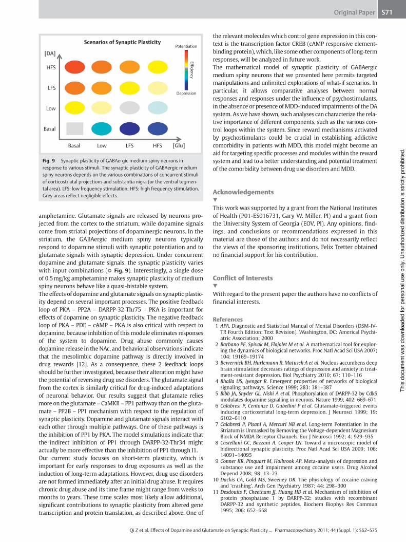
amphetamine. Glutamate signals are released by neurons pro-jected from the cortex to the striatum, while dopamine signals come from striatal projections of dopaminergic neurons. In the striatum, the GABAergic medium spiny neurons typically respond to dopamine stimuli with synaptic potentiation and to glutamate signals with synaptic depression. Under concurrent dopamine and glutamate signals, the synaptic plasticity varies with input combinations ( ● ▶ Fig. 9 ). Interestingly, a single dose of 0.5 mg / kg amphetamine makes synaptic plasticity of medium spiny neurons behave like a quasi-bistable system. The eff ects of dopamine and glutamate signals on synaptic plastic-ity depend on several important processes. The positive feedback loop of PKA – PP2A – DARPP-32-Thr75 – PKA is important for eff ects of dopamine on synaptic plasticity. The negative feedback loop of PKA – PDE – cAMP – PKA is also critical with respect to dopamine, because inhibition of this module eliminates responses of the system to dopamine. Drug abuse commonly causes dopamine release in the NAc, and behavioral observations indicate that the mesolimbic dopamine pathway is directly involved in drug rewards [12] . As a consequence, these 2 feedback loops should be further investigated, because their alteration might have the potential of reversing drug use disorders. The glutamate signal from the cortex is similarly critical for drug-induced adaptations of neuronal behavior. Our results suggest that glutamate relies more on the glutamate – CaMKII – PP1 pathway than on the gluta-mate – PP2B – PP1 mechanism with respect to the regulation of synaptic plasticity. Dopamine and glutamate signals interact with each other through multiple pathways. One of these pathways is the inhibition of PP1 by PKA. The model simulations indicate that the indirect inhibition of PP1 through DARPP-32-Thr34 might actually be more eff ective than the inhibition of PP1 through I1. Our current study focuses on short-term plasticity, which is important for early responses to drug exposures as well as the induction of long-term adaptations. However, drug use disorders are not formed immediately after an initial drug abuse. It requires chronic drug abuse and its time frame might range from weeks to months to years. These time scales most likely allow additional, signifi cant contributions to synaptic plasticity from altered gene transcription and protein translation, as described above. One of
the relevant molecules which control gene expression in this con-text is the transcription factor CREB (cAMP responsive element-binding protein), which, like some other components of long-term responses, will be analyzed in future work. The mathematical model of synaptic plasticity of GABAergic medium spiny neurons that we presented here permits targeted manipulations and unlimited explorations of what-if scenarios. In particular, it allows comparative analyses between normal responses and responses under the infl uence of psychostimulants, in the absence or presence of MDD-induced impairments of the DA system. As we have shown, such analyses can characterize the rela-tive importance of diff erent components, such as the various con-trol loops within the system. Since reward mechanisms activated by psychostimulants could be crucial in establishing addictive comorbidity in patients with MDD, this model might become an aid for targeting specifi c processes and modules within the reward system and lead to a better understanding and potential treatment of the comorbidity between drug use disorders and MDD.
Acknowledgements ▼ This work was supported by a grant from the National Institutes of Health (P01-ES016731, Gary W. Miller, PI) and a grant from the University System of Georgia (EOV, PI). Any opinions, fi nd-ings, and conclusions or recommendations expressed in this material are those of the authors and do not necessarily refl ect the views of the sponsoring institutions. Felix Tretter obtained no fi nancial support for his contribution.
Confl ict of Interests ▼ With regard to the present paper the authors have no confl icts of fi nancial interests.
References 1 APA . Diagnostic and Statistical Manual of Mental Disorders (DSM-IV-
TR Fourth Edition; Text Revision) . Washington, DC: Americal Psychi-atric Association ; 2000
2 Barbano PE , Spivak M , Flajolet M et al . A mathematical tool for explor-ing the dynamics of biological networks . Proc Natl Acad Sci USA 2007 ; 104 : 19169 – 19174
3 Bewernick BH , Hurlemann R , Matusch A et al . Nucleus accumbens deep brain stimulation decreases ratings of depression and anxiety in treat-ment-resistant depression . Biol Psychiatry 2010 ; 67 : 110 – 116
4 Bhalla US , Iyengar R . Emergent properties of networks of biological signaling pathways . Science 1999 ; 283 : 381 – 387
5 Bibb JA , Snyder GL , Nishi A et al . Phosphorylation of DARPP-32 by Cdk5 modulates dopamine signalling in neurons . Nature 1999 ; 402 : 669 – 671
6 Calabresi P , Centonze D , Gubellini P et al . Glutamate-triggered events inducing corticostriatal long-term depression . J Neurosci 1999 ; 19 : 6102 – 6110
7 Calabresi P , Pisani A , Mercuri NB et al . Long-term Potentiation in the Striatum is Unmasked by Removing the Voltage-dependent Magnesium Block of NMDA Receptor Channels . Eur J Neurosci 1992 ; 4 : 929 – 935
8 Castellani GC , Bazzani A , Cooper LN . Toward a microscopic model of bidirectional synaptic plasticity . Proc Natl Acad Sci USA 2009 ; 106 : 14091 – 14095
9 Conner KR , Pinquart M , Holbrook AP . Meta-analysis of depression and substance use and impairment among cocaine users . Drug Alcohol Depend 2008 ; 98 : 13 – 23
10 Dackis CA , Gold MS , Sweeney DR . The physiology of cocaine craving and ‘ crashing ’ . Arch Gen Psychiatry 1987 ; 44 : 298 – 300
11 Desdouits F , Cheetham JJ , Huang HB et al . Mechanism of inhibition of protein phosphatase 1 by DARPP-32: studies with recombinant DARPP-32 and synthetic peptides . Biochem Biophys Res Commun 1995 ; 206 : 652 – 658
Scenarios of Synaptic Plasticity
[DA]
[Glu]
HFS
Potentiation
Depression
Efficiency
LFS
Low
Basal
Basal Low LFS HFS
Fig. 9 Synaptic plasticity of GABAergic medium spiny neurons in response to various stimuli. The synaptic plasticity of GABAergic medium spiny neurons depends on the various combinations of concurrent stimuli of corticostriatal projections and substantia nigra (or the ventral tegmen-tal area). LFS: low frequency stimulation; HFS: high frequency stimulation. Grey areas refl ect negligible eff ects.
Thi
s do
cum
ent w
as d
ownl
oade
d fo
r pe
rson
al u
se o
nly.
Una
utho
rized
dis
trib
utio
n is
str
ictly
pro
hibi
ted.

Original PaperS72
Qi Z et al. Eff ects of Dopamine and Glutamate on Synaptic Plasticity … Pharmacopsychiatry 2011; 44 (Suppl. 1): S62 – S75
12 Di Chiara G , Imperato A . Drugs abused by humans preferentially increase synaptic dopamine concentrations in the mesolimbic system of freely moving rats . Proc Natl Acad Sci USA 1988 ; 85 : 5274 – 5278
13 Drevets WC , Price JL , Furey ML . Brain structural and functional abnor-malities in mood disorders: implications for neurocircuitry models of depression . Brain Struct Funct 2008 ; 213 : 93 – 118
14 Fernandez É , Schiappa R , Girault JA et al . DARPP-32 is a robust integrator of dopamine and glutamate signals . PLoS Comput Biol 2006 ; 2 : e176
15 Gaiarsa JL , Caillard O , Ben-Ari Y . Long-term plasticity at GABAergic and glycinergic synapses: mechanisms and functional signifi cance . Trends Neurosci 2002 ; 25 : 564 – 570
16 Gawin FH , Kleber HD . Abstinence symptomatology and psychiatric diagnosis in cocaine abusers. Clinical observations . Arch Gen Psychia-try 1986 ; 43 : 107 – 113
17 Girault JA , Hemmings HC Jr , Williams KR et al . Phosphorylation of DARPP-32, a dopamine- and cAMP-regulated phosphoprotein, by casein kinase II . J Biol Chem 1989 ; 264 : 21748 – 21759
18 Hayer A , Bhalla US . Molecular switches at the synapse emerge from receptor and kinase traffi c . PLoS Comput Biol 2005 ; 1 : 137 – 154
19 Hemmings HC Jr , Greengard P . DARPP-32, a dopamine- and adenosine 3 ′ :5 ′ -monophosphate-regulated phosphoprotein: regional, tissue, and phylogenetic distribution . J Neurosci 1986 ; 6 : 1469 – 1481
20 Hemmings HC Jr , Nairn AC , Greengard P . DARPP-32, a dopamine- and adenosine 3 ′ :5 ′ -monophosphate-regulated neuronal phosphoprotein. II. Comparison of the kinetics of phosphorylation of DARPP-32 and phosphatase inhibitor 1 . J Biol Chem 1984 ; 259 : 14491 – 14497
21 Hofmann F , Bechtel PJ , Krebs EG . Concentrations of cyclic AMP-depend-ent protein kinase subunits in various tissues . J Biol Chem 1977 ; 252 : 1441 – 1447
22 Hyman SE , Malenka RC . Addiction and the brain: the neurobiology of compulsion and its persistence . Nat Rev Neurosci 2001 ; 2 : 695 – 703
23 Hyman SE , Malenka RC , Nestler EJ . Neural mechanisms of addiction: the role of reward-related learning and memory . Annu Rev Neurosci 2006 ; 29 : 565 – 598
24 Janowsky DS , Overstreet DH . The role of acetylcholine mechanisms in mood disorders . In: Bloom FE, Kupfer DJ eds, Psychopharmacology: The fourth generation of progress . New York: Raven Press, Ltd ; 1995 , xlii+2002
25 Kauer JA . Learning mechanisms in addiction: synaptic plasticity in the ventral tegmental area as a result of exposure to drugs of abuse . Annu Rev Physiol 2004 ; 66 : 447 – 475
26 Kessler RC , Berglund P , Demler O et al . The epidemiology of major depressive disorder: results from the National Comorbidity Survey Replication (NCS-R) . Jama 2003 ; 289 : 3095 – 3105
27 Khantzian EJ . The self-medication hypothesis of addictive disorders: focus on heroin and cocaine dependence . Am J Psychiatry 1985 ; 142 : 1259 – 1264
28 Kim M , Huang T , Abel T et al . Temporal sensitivity of protein kinase a activation in late-phase long term potentiation . PLoS Comput Biol 2010 ; 6 : e1000691
29 Koob GF , Kenneth Lloyd G , Mason BJ . Development of pharmacothera-pies for drug addiction: a Rosetta stone approach . Nat Rev Drug Dis-cov 2009 ; 8 : 500 – 515
30 Krystal JH . N-methyl-D-aspartate glutamate receptor antagonists and the promise of rapid-acting antidepressants . Arch Gen Psychiatry 2010 ; 67 : 1110 – 1111
31 Lindskog M , Kim M , Wikstrom MA et al . Transient calcium and dopamine increase PKA activity and DARPP-32 phosphorylation . PLoS Comput Biol 2006 ; 2 : e119
32 Luscher B , Shen Q , Sahir N . The GABAergic defi cit hypothesis of major depressive disorder . Mol Psychiatry 2010
33 Maletic V , Robinson M , Oakes T et al . Neurobiology of depression: an integrated view of key fi ndings . International journal of clinical prac-tice 2007 ; 61 : 2030 – 2040
34 Markou A , Kosten TR , Koob GF . Neurobiological similarities in depres-sion and drug dependence: a self-medication hypothesis . Neuropsy-chopharmacology 1998 ; 18 : 135 – 174
35 Mayberg HS . Limbic-cortical dysregulation: a proposed model of depression . J Neuropsychiatry Clin Neurosci 1997 ; 9 : 471 – 481
36 Mayberg HS . Defi ning the neural circuitry of depression: Strategies toward treatment selection based on neuroimaging phenotypes . Psy-chiatric Annals 2006 ; 36 : 259 – 268
37 Mayberg HS . Targeted electrode-based modulation of neural circuits for depression . J Clin Invest 2009 ; 119 : 717 – 725
38 Monk CS , Klein RG , Telzer EH et al . Amygdala and nucleus accumbens activation to emotional facial expressions in children and adolescents at risk for major depression . Am J Psychiatry 2008 ; 165 : 90 – 98
39 Murray CJ , Lopez AD . Global mortality, disability, and the contribution of risk factors: Global Burden of Disease Study . Lancet 1997 ; 349 : 1436 – 1442
40 Nagase T , Murakami T , Nozaki H et al . Tissue and subcellular distribu-tions, and characterization of rat brain protein phosphatase 2A con-taining a 72-kDa delta/B ” subunit . J Biochem 1997 ; 122 : 178 – 187
41 Nakano T , Doi T , Yoshimoto J et al . A kinetic model of dopamine- and calcium-dependent striatal synaptic plasticity . PLoS Comput Biol 2010 ; 6 : e1000670
42 Nestler EJ , Carlezon WA Jr . The mesolimbic dopamine reward circuit in depression . Biol Psychiatry 2006 ; 59 : 1151 – 1159
43 Nishi A , Bibb JA , Snyder GL et al . Amplifi cation of dopaminergic signal-ing by a positive feedback loop . Proc Natl Acad Sci USA 2000 ; 97 : 12840 – 12845
44 Ouimet CC , da Cruz e Silva EF , Greengard P . The alpha and gamma 1 iso-forms of protein phosphatase 1 are highly and specifi cally concentrated in dendritic spines . Proc Natl Acad Sci USA 1995 ; 92 : 3396 – 3400
45 Patel S , Morris SA , Adkins CE et al . Ca2+-independent inhibition of inositol trisphosphate receptors by calmodulin: redistribution of cal-modulin as a possible means of regulating Ca2+ mobilization . Proc Natl Acad Sci USA 1997 ; 94 : 11627 – 11632
46 Pelizza L , Ferrari A . Anhedonia in schizophrenia and major depression: state or trait? Ann Gen Psychiatry 2009 ; 8 : 22
47 Price JL , Drevets WC . Neurocircuitry of mood disorders . Neuropsy-chopharmacology 2010 ; 35 : 192 – 216
48 Qi Z , Miller GW , Voit EO . Computational systems analysis of dopamine metabolism . PLoS One 2008 ; 3 : e2444
49 Qi Z , Miller GW , Voit EO . A mathematical model of presynaptic dopamine homeostasis: implications for schizophrenia . Pharmacops-ychiatry 2008 ; 41 (Suppl. 1) : S89 – S98
50 Qi Z , Miller GW , Voit EO . The internal state of medium spiny neurons varies in response to diff erent input signals . BMC Syst Biol 2010 ; 4 : 26
51 Raison CL , Capuron L , Miller AH . Cytokines sing the blues: infl ammation and the pathogenesis of depression . Trends Immunol 2006 ; 27 : 24 – 31
52 Rakhilin SV , Olson PA , Nishi A et al . A network of control mediated by regulator of calcium/calmodulin-dependent signaling . Science 2004 ; 306 : 698 – 701
53 Rao U , Hammen CL , Poland RE . Mechanisms underlying the comorbid-ity between depressive and addictive disorders in adolescents: inter-actions between stress and HPA activity . The American journal of psychiatry 2009 ; 166 : 361 – 369
54 Rayport SG , Kandel ER . Development of plastic mechanisms related to learning at identifi ed chemical synaptic connections in Aplysia . Neu-roscience 1986 ; 17 : 283 – 294
55 Reynolds JN , Hyland BI , Wickens JR . A cellular mechanism of reward-related learning . Nature 2001 ; 413 : 67 – 70
56 Reynolds JN , Wickens JR . Substantia nigra dopamine regulates synaptic plasticity and membrane potential fl uctuations in the rat neostriatum, in vivo . Neuroscience 2000 ; 99 : 199 – 203
57 Reynolds JN , Wickens JR . Dopamine-dependent plasticity of corticos-triatal synapses . Neural Netw 2002 ; 15 : 507 – 521
58 Ross HE , Glaser FB , Germanson T . The prevalence of psychiatric disor-ders in patients with alcohol and other drug problems . Arch Gen Psychiatry 1988 ; 45 : 1023 – 1031
59 Ross HE , Glasser FB , Stiasny S . Sex diff erences in the prevalence of psychiatric disorders in patients with alcohol and drug problems . Br J Addict 1988 ; 83 : 1179 – 1192
60 Sanacora G , Mason GF , Rothman DL et al . Reduced cortical gamma-aminobutyric acid levels in depressed patients determined by proton magnetic resonance spectroscopy . Arch Gen Psychiatry 1999 ; 56 : 1043 – 1047
61 Sanderson WC , Beck AT , Beck J . Syndrome comorbidity in patients with major depression or dysthymia: prevalence and temporal relation-ships . Am J Psychiatry 1990 ; 147 : 1025 – 1028
62 Shi SH , Hayashi Y , Petralia RS et al . Rapid spine delivery and redistri-bution of AMPA receptors after synaptic NMDA receptor activation . Science 1999 ; 284 : 1811 – 1816
63 Sim AT , Ratcliff e E , Mumby MC et al . Diff erential activities of protein phosphatase types 1 and 2A in cytosolic and particulate fractions from rat forebrain . J Neurochem 1994 ; 62 : 1552 – 1559
64 Snyder GL , Allen PB , Fienberg AA et al . Regulation of phosphorylation of the GluR1 AMPA receptor in the neostriatum by dopamine and psychostimulants in vivo . J Neurosci 2000 ; 20 : 4480 – 4488
65 Stahl SM . Stahl’s Essential Psychopharmacology: Neuroscientifi c Basis and Practical Applications . 3 rd edition ed: Cambridge University Press ; 2008 ; 1132
66 Sulzer D , Sonders MS , Poulsen NW et al . Mechanisms of neurotransmitter release by amphetamines: a review . Prog Neurobiol 2005 ; 75 : 406 – 433
67 Takahashi S , Ohshima T , Cho A et al . Increased activity of cyclin-dependent kinase 5 leads to attenuation of cocaine-mediated dopamine signaling . Proc Natl Acad Sci USA 2005 ; 102 : 1737 – 1742
Thi
s do
cum
ent w
as d
ownl
oade
d fo
r pe
rson
al u
se o
nly.
Una
utho
rized
dis
trib
utio
n is
str
ictly
pro
hibi
ted.

Original Paper S73
Qi Z et al. Eff ects of Dopamine and Glutamate on Synaptic Plasticity … Pharmacopsychiatry 2011; 44 (Suppl. 1): S62 – S75
68 Tang K , Low MJ , Grandy DK et al . Dopamine-dependent synaptic plas-ticity in striatum during in vivo development . Proc Natl Acad Sci USA 2001 ; 98 : 1255 – 1260
69 Tisch S , Silberstein P , Limousin-Dowsey P et al . The basal ganglia: anat-omy, physiology, and pharmacology . Psychiatr Clin North Am 2004 ; 27 : 757 – 799
70 Usui H , Inoue R , Tanabe O et al . Activation of protein phosphatase 2A by cAMP-dependent protein kinase-catalyzed phosphorylation of the 74-kDa B ” (delta) regulatory subunit in vitro and identifi cation of the phosphorylation sites . FEBS Lett 1998 ; 430 : 312 – 316
71 Voit EO . Computational analysis of biochemical systems: a practical guide for biochemists and molecular biologists . Cambridge, UK: Cam-bridge University Press ; 2000 , xii, 531 p.
72 Voit EO , Qi Z , Miller GW . Steps of modeling complex biological systems . Pharmacopsychiatry 2008 ; 41 (Suppl. 1) : S78 – S84
73 Weiss RD , Mirin SM , Griffi n ML . Methodological considerations in the diagnosis of coexisting psychiatric disorders in substance abusers . Br J Addict 1992 ; 87 : 179 – 187
74 Wickens JR , Begg AJ , Arbuthnott GW . Dopamine reverses the depression of rat corticostriatal synapses which normally follows high-frequency stimulation of cortex in vitro . Neuroscience 1996 ; 70 : 1 – 5
75 Willner P . Dopamine and depression: a review of recent evidence. I. Empirical studies . Brain Res 1983 ; 287 : 211 – 224
76 Willner P . Dopamine and depression: a review of recent evidence. II. Theoretical approaches . Brain Res 1983 ; 287 : 225 – 236
77 Willner P . Dopamine and depression: a review of recent evidence. III. The eff ects of antidepressant treatments . Brain Res 1983 ; 287 : 237 – 246
78 Zetterstrom T , Sharp T , Ungerstedt U . Further evaluation of the mech-anism by which amphetamine reduces striatal dopamine metabolism: a brain dialysis study . Eur J Pharmacol 1986 ; 132 : 1 – 9
Appendix ▼ Reactions, kinetics, and initial conditions in the mathematical model of signal transduction and traffi cking of AMPA receptors All reactions are represented in the form of an enzymatic reac-tion or a simple binding reaction, with K f denoting the rate con-
Table 1 Reactions and rate constants of signal transduction for DARPP-32 phosphorylation in dendrites of medium spiny neurons in the striatum (see legend of ● ▶ Fig. 2c for abbreviations).
Reaction K f (nM − 1 .s − 1 ) # K b (s − 1 ) K c (s − 1 ) Ref.
D1 + DA ↔ D1_DA 1.1E − 3 10.0 [31] D1_DA + G α β γ ↔ D1_DA_G α β γ 6.0E − 4 1.0E − 3 [31] D1 + G α β γ ↔ D1_G α β γ 6.0E − 5 3.0E − 4 [31] D1_G α β γ + DA ↔ D1_DA_G α β γ 3.3E − 3 10.0 [31] D1_DA_G α β γ → D1_DA + G α GTP + G β γ 20.0 a [31] G α GTP → G α GDP 10.0 a [31] G α GDP + G β γ → G α β γ 100.0 [31] G α GTP + AC5 ↔ G α GTP_AC5 3.9E − 2 50.0 [31] G α GTP_AC5 + ATP ↔ G α GTP_AC5_ATP 1.3E − 4 2.6E − 1 [31] G α GTP_AC5_ATP ↔ G α GTP_AC5 + cAMP 28.5 a 2.6E − 4 b [31] PKA + 2 cAMP ↔ PKA_cAMP 2 3.5E − 8 c 6.0E − 2 [41] PKA_cAMP 2 + 2 cAMP ↔ PKA_cAMP 4 2.7E − 5 c 0.28 [41] PKA_cAMP 4 ↔ 2 PKAc + PKAr 0.05 a 8.5E − 8 c [41] PDE1 + cAMP ↔ PDE1_cAMP → PDE1 + AMP 2.0E − 3 72.0 18.0 [31] PDE4 + cAMP ↔ PDE4_cAMP → PDE4 + AMP 2.0E − 3 72.0 18.0 [31] PKAc + PDE1 ↔ PKAc_PDE1 → PKAc + PDE1p 6.0E − 3 36.0 9.0 [4] PDE1p → PDE1 1.0E − 1 a [4] PKAc + PDE4 ↔ PKAc_PDE4 → PKAc + PDE4p 6.0E − 3 36.0 9.0 [4] PDE4p → PDE4 1.0E − 1 a [4] → Ca 2 + 1.0E + 2 e [4] Ca 2 + → 2.0 a [4] 2 Ca 2 + + PP2Bi ↔ PP2Bi_Ca 2 6.0E − 3 0.91 [4] 2 Ca 2 + + PP2Bi_Ca 2 ↔ PP2B 0.1 10.0 [4] AC5 + Ca 2 + ↔ AC5_Ca 1.0E − 3 0.9 [31] G α GTP + AC5_Ca ↔ G α GTP_AC5_Ca 1.9E − 2 25.0 [31] G α GTP_AC5_Ca + ATP ↔ G α GTP_AC5_Ca_ATP 6.0E − 5 1.3E − 1 [31] G α GTP_AC5_Ca_ATP ↔ G α GTP_AC5_Ca + cAMP 14.2 a 1.3E − 4 b [31] PP2A + 4 Ca 2 + ↔ PP2Ac 7.7E − 12 d 1.0E − 2 [31] PP2A + PKAc ↔ PP2A_PKAc → PP2Ap + PKAc 2.5E − 3 0.3 0.1 [70] PP2Ap → PP2A 4.0E − 3 a [31] CK1 → CK1p 1.0 a [14] PP2B + CK1p ↔ PP2B_CK1p → PP2B + CK1 3.0E − 2 24.0 6.0 [14] CDK5 + Ca 2 + ↔ CDK5c 3.0E − 3 1.0 PDE1p + cAMP ↔ PDE1p_cAMP → PDE1p + AMP 5.0E − 3 80.0 20.0 PDE4p + cAMP ↔ PDE4p_cAMP → PDE4p + AMP 5.0E − 3 80.0 20.0 a : Unit in s − 1 ; b : Unit in nM − 1 .s − 1 ; c : Unit in nM − 2 .s − 1 ; d : Unit in nM − 4 .s − 1 ; e : Unit in nM.s − 1 # : For a chemical reaction, K f is the rate constant for the forward process, K b is the rate constant for the backward process, while K c is the rate constant for the catalytic
step in a Michaelis-Menten kinetics
stant for the forward process, K b denoting the rate constant for the backward process, and K c denoting the rate constant for the catalytic step in a Michaelis-Menten kinetics.
Thi
s do
cum
ent w
as d
ownl
oade
d fo
r pe
rson
al u
se o
nly.
Una
utho
rized
dis
trib
utio
n is
str
ictly
pro
hibi
ted.

Original PaperS74
Qi Z et al. Eff ects of Dopamine and Glutamate on Synaptic Plasticity … Pharmacopsychiatry 2011; 44 (Suppl. 1): S62 – S75
Table 2 Reactions and rate constants of DARPP-32 phosphorylation in dendrites of medium spiny neurons in the striatum (see legend of ● ▶ Fig. 2c for abbreviations).
Reaction K f (nM − 1 .s − 1 ) # K b (s − 1 ) K c (s − 1 ) Ref.
D + PKAc ↔ D_PKAc → D34 + PKAc 2.7E − 3 8.0 2.0 [31] D34 + PP2B ↔ D34_PP2B → D + PP2B 1.0E − 2 2.0 0.5 [31] D + CDK5 ↔ D_CDK5 → D75 + CDK5 4.5E − 4 2.0 0.5 [31] D75 + PP2Ap ↔ D75_PP2Ap → D + PP2Ap 4.0E − 4 12.0 3.0 [31] D75 + PP2Ac ↔ D75_PP2Ac → D + PP2Ac 4.0E − 4 12.0 3.0 [31] D + CK2 ↔ D_CK2 → D102 + CK2 4.0E − 4 6.4 1.6 [17] D102 → D 1.6 a D + CK1 ↔ D_CK1 → D137 + CK1 4.4E − 3 12.0 3.0 [67] D137 + PP2C ↔ D137_PP2C → D + PP2C 7.5E − 3 12.0 3.0 [14] D34 + PP1 ↔ D34_PP1 1.0E − 2 1.0 [14] D34_PP1 + PP2B ↔ D34_PP1_PP2B → D + PP1 + PP2B 1.0E − 3 2.0 0.5 [14] D75 + PKAc ↔ D75_PKAc 4.6E − 3 2.4 [5] D + CDK5c ↔ D_CDK5c → D75 + CDK5c 1.8E − 3 4.0 1.0 2 Ca 2 + + CaM ↔ Ca 2 CaM 6.0E − 3 b 9.1 [31] 2 Ca 2 + + Ca 2 CaM ↔ Ca 4 CaM 0.1 b 1.0E + 3 [31] CaMKII + Ca 4 CaM ↔ CaMKII_Ca 4 CaM 0.01 0.8 [31] CaMKII_Ca 4 CaM → CaMKIIp + Ca 4 CaM 5.0E − 3 a [31] CaMKIIp + PP1 ↔ CaMKIIp_PP1 → CaMKII + PP1 1.0E − 4 1.4 0.35 [28, 31] PKAc + I1 ↔ PKAc_I1 → PKAc + I1p 1.4E − 3 5.6 1.4 [28, 31] PP1 + I1p ↔ PP1_I1p 1.0E − 3 5.0E − 3 PP2B + I1p ↔ PP2B_I1p → PP2B + I1 3.8E − 3 12.0 3.0 a : Unit in s − 1 ; b : Unit in nM − 1 .s − 1 ; c : Unit in nM − 2 .s − 1 ; d : Unit in nM − 4 .s − 1 ; e : Unit in nM.s − 1 # : For a chemical reaction, K f is the rate constant for the forward process, K b is the rate constant for the backward process, while K c is the rate constant for the catalytic
step in a Michaelis-Menten kinetics
Table 3 Reactions and rate constants of AMPAR traffi cking, AMPAR phosphorylation, and AMPAR dephosphorylation in the postsynapse of striatal projection neurons (see legend of ● ▶ Fig. 2c for abbreviations).
Reaction K f (nM − 1 .s − 1 ) K b (s − 1 ) K c (s − 1 ) Ref.
cAMPAR + PKAc ↔ cAMPAR_PKAc → cAMPAR_Ser845p + PKAc 2.5E − 3 4.0 1.0 [41] cAMPAR_Ser845p + PP1 ↔ cAMPAR_Ser845p_PP1 → cAMPAR + PP1 5.0E − 4 12.0 3.0 [64] cAMPAR_Ser845p + PP2Ap ↔ cAMPAR_Ser845p_PP2Ap → cAMPAR + PP2Ap 1.7E − 4 12.0 3.0 [64] cAMPAR_Ser845p + PP2Ac ↔ cAMPAR_Ser845p_PP2Ac → cAMPAR + PP2Ac 1.7E − 4 12.0 3.0 [64] cAMPAR_Ser845p + CaMKIIp ↔ cAMPAR_Ser845p_CaMKIIp → cAMPAR_Ser845p_Ser831p + CaMKIIp 1.0E − 4 2.0 0.5 [41] cAMPAR_Ser845p_Ser831p + PP1 ↔ cAMPAR_Ser845p_Ser831p_PP1 → cAMPAR_Ser845p + PP1 5.0E − 4 4.0 1.0 [64] cAMPAR_Ser845p_Ser831p + PP2Ap ↔ cAMPAR_Ser845p_Ser831p_PP2Ap → cAMPAR_Ser845p + PP2Ap 1.7E − 4 4.0 1.0 [64] cAMPAR_Ser845p_Ser831p + PP2Ac ↔ cAMPAR_Ser845p_Ser831p_PP2Ac → cAMPAR_Ser845p + PP2Ac 1.7E − 4 4.0 1.0 [64] mAMPAR + PKAc ↔ mAMPAR_PKAc → mAMPAR_Ser845p + PKAc 2.5E − 3 4.0 1.0 [41] mAMPAR_Ser845p + PP1 ↔ mAMPAR_Ser845p_PP1 → mAMPAR + PP1 5.0E − 4 0.8 0.2 [64] mAMPAR_Ser845p + CaMKIIp ↔ mAMPAR_Ser845p_CaMKIIp → mAMPAR_Ser845p_Ser831p + CaMKIIp 1.0E − 4 2.0 0.5 [41] mAMPAR_Ser845p_Ser831p + PP1 ↔ mAMPAR_Ser845p_Ser831p_PP1 → mAMPAR_Ser845p + PP1 5.0E − 4 4.0 1.0 [64] mAMPAR → cAMPAR + Anchor 0.8E − 3 a [18] cAMPAR_Ser845p_Ser831p + Anchor ↔ mAMPAR_Ser845p_Ser831p 1.0E − 5 0.1 [18] cAMPAR ↔ Bulk_cAMPAR 1 a 1.8E − 2 [18] cAMPAR_Ser845p → Bulk_cAMPAR 2.0E − 5 a [18] cAMPAR_Ser845p_Ser831p → Bulk_cAMPAR 2.0E − 5 a [18] a : Unit in s − 1 # : For a chemical reaction, K f is the rate constant for the forward process, K b is the rate constant for the backward process, while K c is the rate constant for the catalytic
step in a Michaelis-Menten kinetics
Thi
s do
cum
ent w
as d
ownl
oade
d fo
r pe
rson
al u
se o
nly.
Una
utho
rized
dis
trib
utio
n is
str
ictly
pro
hibi
ted.

Original Paper S75
Qi Z et al. Eff ects of Dopamine and Glutamate on Synaptic Plasticity … Pharmacopsychiatry 2011; 44 (Suppl. 1): S62 – S75
Table 4 Initial values for the DARPP-32 phosphorylation system in dendrites of medium spiny neurons in the striatum (see legend of ● ▶ Fig. 2c for abbreviations).
Molecule Concentration (nM) Reference
DA 10.0 [31] D1 5.0E + 2 [31] G α β γ 3.0E + 3 [31] AC5 2.5E + 3 [31] ATP 2.0E + 6 [31] PDE1 5.0E + 2 [31] PDE4 5.0E + 2 [31] DARPP-32 3.0E + 4 [19] PKA 6.6E + 3 [21] PP2Bi 4.0E + 3 [31] CDK5 1.2E + 3 [31] PP2A 8.0E + 2 [40, 63] CK1 2.0E + 3 PP2C 2.0E + 3 PP1 2.3E + 3 [11, 44] CK2 2.0E + 3 Anchor 11.56E + 3 [41] Bulk_cAMPAR 6.0E + 3 [41] CaM 1.0E + 4 [45, 52] CaMKII 2.0E + 4 [31] I1 1.0E + 3
Thi
s do
cum
ent w
as d
ownl
oade
d fo
r pe
rson
al u
se o
nly.
Una
utho
rized
dis
trib
utio
n is
str
ictly
pro
hibi
ted.