early- and late anthracycline-induced cardiac dysfunction
TRANSCRIPT
RESEARCH Open Access
Early- and late anthracycline-inducedcardiac dysfunction: echocardiographiccharacterization and response to heartfailure therapyJanine A. M. Kamphuis1*, Marijke Linschoten1, Maarten J. Cramer1, Pieter A. Doevendans1,2,3,Folkert W. Asselbergs1,4,5 and Arco J. Teske1
Abstract
Background: Anthracycline-induced cardiac dysfunction (ACD) is a notorious side effect of anticancer treatment. Ithas been described as a phenomenon of a continuous progressive decline of cardiac function, eventually leadingto dilated cardiomyopathy (DCM). This progressive nature suggests that patients with a delayed ACD diagnosishave greater compromise of cardiac function and more adverse remodeling, with a poor response to heart failure(HF) treatment. This study aimed to delineate the impact of a delayed ACD diagnosis on echocardiographiccharacteristics and response to HF treatment.
Methods and results: From the population of our cardio-oncology outpatient clinic, 92 ACD patients wereincluded in this study (age 51.6 ± 16.2 years, median cumulative anthracycline dose 329 [200–329] mg/m2), and amedian follow-up of 25.0 [9.6–37.2] months after ACD diagnosis. Median time to ACD diagnosis for patientsdiagnosed early (< 1 year) and late (> 1 year) was 4.0 vs. 47.7 months respectively. There were no echocardiographicdifferences between patients diagnosed early vs. late (LVEF 43.6 ± 4.9% vs. 43.0 ± 6.2% and iEDV 63.6 vs. 62.9 mL/m2). Eighty-three percent of patients presented with mild LV dysfunction and in 79% the LV was not dilated.Patients diagnosed early were more likely to have (partial) recovery of cardiac function upon HF treatment initiation(p = 0.015).
Conclusions: In the setting of a cardio-oncology outpatient clinic, patients with ACD presented with a hypokineticnon-dilated cardiomyopathy, rather than typical DCM. Timing of ACD diagnosis did not impact HF disease severity.However, in patients receiving an early diagnosis, cardiac function was more likely to recover upon HF treatment.
Keywords: Heart failure, Anthracyclines, Cardiac dysfunction, Cardiac effects of cancer treatment
© The Author(s). 2020 Open Access This article is licensed under a Creative Commons Attribution 4.0 International License,which permits use, sharing, adaptation, distribution and reproduction in any medium or format, as long as you giveappropriate credit to the original author(s) and the source, provide a link to the Creative Commons licence, and indicate ifchanges were made. The images or other third party material in this article are included in the article's Creative Commonslicence, unless indicated otherwise in a credit line to the material. If material is not included in the article's Creative Commonslicence and your intended use is not permitted by statutory regulation or exceeds the permitted use, you will need to obtainpermission directly from the copyright holder. To view a copy of this licence, visit http://creativecommons.org/licenses/by/4.0/.The Creative Commons Public Domain Dedication waiver (http://creativecommons.org/publicdomain/zero/1.0/) applies to thedata made available in this article, unless otherwise stated in a credit line to the data.
* Correspondence: [email protected] of Cardiology, Division of Heart and Lungs, University MedicalCenter Utrecht, University of Utrecht, E03.511, PO Box 85500, 3508 GAUtrecht, The NetherlandsFull list of author information is available at the end of the article
Kamphuis et al. Cardio-Oncology (2020) 6:23 https://doi.org/10.1186/s40959-020-00079-3
IntroductionAnthracyclines are potent antineoplastic drugs that consti-tute a cornerstone in the treatment of sarcomas, breastcancer and hematological malignancies. Shortly after theintroduction of these agents in the 1960’s, cardiac dys-function was discovered to be an important dose-limitingside effect [1]. However, despite dosage restrictions, theincidence of anthracycline-induced cardiac dysfunction(ACD) has been found to be 6% for overt heart failure andup to 18% for subclinical cardiac dysfunction [2]. Theprognosis of ACD is poor, with cardiovascular mortalityrates ranging from 9% at 5- and 24% at 10-years [3], up tomore dramatic outcomes of 60% at 2-years in patients thathave developed symptomatic HF [4].ACD has often been classified to occur either “early” or
“late”, with the first subtype developing within the 1st yearafter treatment and the latter more than 1 year afteranthracycline-containing therapy [5]. In recent years, thissubdivision has however been questioned, since a release oftroponins can be detected during- or early after anthracy-cline administration, indicating direct damage to the myo-cardium upon infusion [6, 7]. The initial damage, resultingin a declined pumping capacity, will not coincide with thedevelopment of clinical manifestations of HF in a majorityof patients. However, if serial echocardiographic assessmentis performed, almost all asymptomatic declines are detectedwithin the first year after anthracycline-containing chemo-therapy [8]. The delayed onset of symptomatic HF may berelated to the activation of compensatory mechanisms thatmodulate left ventricular (LV) function within a range, suchthat the functional capacity is preserved or only minimallydepressed [9]. These compensatory mechanisms includethe activation of neurohumoral systems and adaptivechanges within the myocardium commonly referred to asLV remodeling. However, prolonged activation of thesecompensatory mechanisms ultimately has detrimental ef-fects on the heart, leading to adverse LV remodeling withdilatation and wall thinning [10]. The onset of thesechanges typically marks the transition from asymptomatic-to symptomatic HF. Based on this theory, it can be postu-lated that over time, patients with ACD develop progressivesystolic dysfunction with dilated compartments, as is thecase in patients with familial DCM, long lasting left-sidedvalvular disease or hypertension. The aim of this consecu-tive cohort study was to (1) evaluate the impact of a delayeddiagnosis (> 1 year after anthracycline containing treatment)on echocardiographic characteristics and (2) assess the in-fluence of timing of diagnosis on HF treatment response.
MethodsStudy populationIn April 2015, a cardio-oncology clinic was launched atthe University Medical Center Utrecht, the Netherlands,
of which the protocol has been described in detail previ-ously [11].In short, the patient population mainly consists of pa-
tients with breast cancer or hematological malignancieswho are deemed to be at increased risk for ACD due totreatment- or patient-related factors. Furthermore, can-cer patients and survivors that have not received cardiacsurveillance in the past (e.g. received potentially cardio-toxic treatment prior to the initiation of the clinic in2015), are referred for screening of long-term cardiovas-cular complications at the discretion of the treating(hemato-)oncologist. One of the primary aims of thisclinic is to facilitate a timely diagnosis of cancer therapy-related cardiac dysfunction (CTRCD) by performing ser-ial echocardiographic assessments. When patients are di-agnosed with CTRCD, guideline-based HF therapy [12]is initiated if there are signs or symptoms of HF, orwhen the LV ejection fraction (LVEF) reaches < 45% re-gardless of the presence of cardiac complaints [11].All patients referred to the cardio-oncology clinic be-
tween April 2015 and February 2019 who were treatedwith anthracyclines were identified. Subsequently, we se-lected patients that showed impaired LV function onechocardiography or cardiac magnetic resonance (CMR).We excluded patients with LV dysfunction on multiple-gated acquisition (MUGA) scans, with echocardio-graphic images of insufficient quality, and patient thathad received treatment with the cardiotoxic monoclonalantibody trastuzumab. Time between the initiation ofanthracycline-containing therapy and ACD diagnosiswas used to divide patients in two groups, namely early-(< 1 year) and late (> 1 year) ACD diagnosis, as describedin the ESC Position Paper on Cancer Treatment andCardiovascular Toxicity [5]. The study was exemptedfrom formal approval by the Medical Ethics Committee.
Definition of ACDPatients were diagnosed with ACD if they met one ofthe following two criteria: (1) LVEF decline of ≥10 per-centage points below the lower limit of normal (LLN) (<53%) from baseline according to the European Associ-ation of Cardiovascular Imaging (EACVI) [13], or (2) anLVEF < 50% measured at > 1 time-point in case a base-line LVEF measurement prior to anthracycline treatmentwas not available. The diagnosis of ACD did not rely onthe presence of symptoms or signs of heart failure andtherefore, the selected population composes both pa-tients with clinical and subclinical ACD.Other possible causes of LV dysfunction were evalu-
ated to determine the likelihood of ACD. Based on thisassessment, patients were subdivided into three groupsnamely, ‘definite ACD’, ‘ACD with concomitant heartdisease’ and ‘possible ACD’. In patients with definiteACD, alternative causes of LV dysfunction were ruled
Kamphuis et al. Cardio-Oncology (2020) 6:23 Page 2 of 13
out or deemed highly unlikely. For patients that did notundergo ischemia detection, ischemic heart disease wasdetermined unlikely in the absence of chest pain, lowcardiovascular risk profile (< 2 risk factors), no coronaryartery disease (CAD) on computed tomography (CT) ofthe chest and lack of regional wall motion abnormalitieson echocardiography. Patients were diagnosed with ACDand concomitant heart disease if other causes affectedLV function, but these abnormalities were determined tobe insufficient to explain the degree of LV dysfunctionas a whole. Lastly, in patients with ‘possible ACD’, severevalvular heart disease, left bundle branch block (LBBB),sepsis-induced cardiac dysfunction and tachycardiomyo-pathy were ruled out. However, in these patients, thepresence of ischemic heart disease had not formally beenexcluded while there were signs of CAD on the chestCT. None of these patients had reported any chest com-plaints. The diagnosis of ACD was verified by two au-thors (JK and ML).
Oncological treatmentWe determined the timing of first anthracycline dose, aswell as the total cumulative dose (mg/m2) which was cal-culated to an equivalent doxorubicin dose [14]. More-over, data on mediastinal- or left-sided radiotherapy wascollected.
Echocardiographic analysisEchocardiographs were performed by trained techniciansat our cardio-oncology program. An extensive analysiswas performed on the echocardiogram on which ACDhad been diagnosed and on the most recent echocardio-gram. All measurements were analyzed by JK, and veri-fied by AT. Reference values and internationalechocardiography guidelines for echocardiographicexamination can be found in Supplemental Table 1. LVvolumes and -ejection fractions were preferably deter-mined on 3D echocardiographic images. If 3D imageswere not available, 2D biplane (modified Simpson’s) al-gorithm was performed on the 4- and 2-chamber apicalview. Global longitudinal strain (GLS) measurementswere performed with the vendor’s software packageusing the 4-, 3-, and 2-chamber apical view.
Follow-upPatients with a follow-up of ≥6 months, or patients withcomplete recovery of the LVEF within 6 months, wereincluded in the analysis of ACD reversibility. Reversibil-ity was based on the change between the nadir- and lastLVEF measurement and was classified according to theEACVI Expert Consensus [13]. In patients lacking abaseline LVEF measurements (n = 74), we deemed ACDto be 1) reversible in case there was an LVEF improve-ment of ≥10 percentage points to above the LLN, 2)
partially reversible if LVEF improved 5–10 percentagepoints to above the LLN, and 3) irreversible if the LVEFimproved < 10 percentage points from the nadir andremained below the LLN.
Statistical analysisContinuous data are expressed as means and standarddeviations (SD) or medians and interquartile ranges[IQR]. Categorical variables are expressed as numbers(percentages). Continuous data were compared using theindependent Student’s t-test or Mann-Whitney U. Cat-egorical data were tested using Chi-square or Fisherexact test as appropriate. Correlation was calculated witheither Pearson or Spearman correlation, where appropri-ate. Differences between > 2 groups were calculatedusing one-way analysis of variance (ANOVA) with Bon-ferroni post correction for multiple comparisons or theKruskal-Wallis test. A two-sided p-value < 0.05 was con-sidered significant. Statistical analyses were performedusing SPSS Statistics, version 25 (IBM Corp., Armonk,NY, USA).
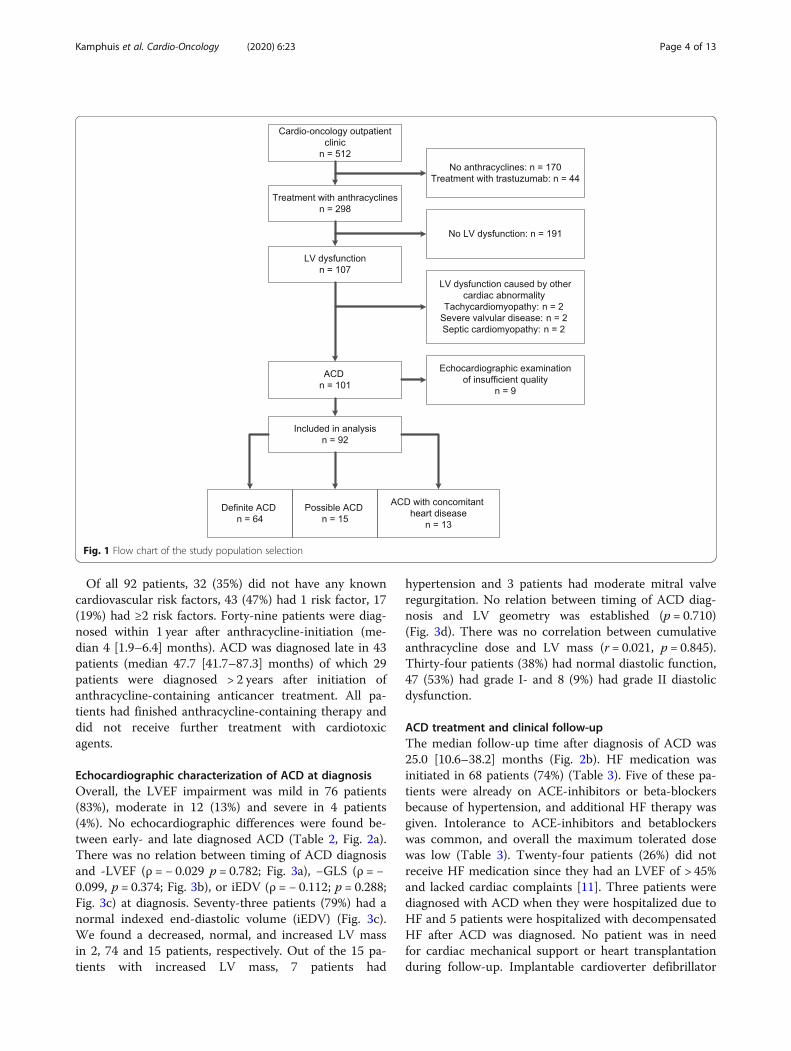
ResultsStudy populationBetween April 2015 to February 2019, a total of 512 pa-tients had been evaluated at the cardio-oncology out-patient clinic (Fig. 1). Anthracyclines were administeredin 342 patients, of which 44 patients were not eligiblefor this study due to concomitant treatment with trastu-zumab. Of the remaining 298 patients, 107 (35.9%) hadLV dysfunction. In six patients, the underlying cause forthe impaired LV dysfunction was deemed not related toanthracyclines. Additionally, nine patients had echocar-diographic images of insufficient quality to perform reli-able measurements. Thereby, a total of 92 patients wereincluded in this study. All patient characteristics at timeof ACD diagnosis are outlined in Table 1. Most patientswere referred for screening of cardiovascular toxicity(n = 78; 85%) and cardiac screening prior to stem celltransplantation was the main reason for referral (n = 56;61%). Cardiac complaints, including dyspnea, angina andpalpitations were the reason for referral in 14 patients(early n = 5; late n = 9).
Anthracycline-induced cardiac dysfunctionSixty-four patients (70%) were diagnosed with definiteACD, and thirteen patients (14%) had ACD with con-comitant heart disease(s), including LBBB (n = 3), CAD(n = 5), moderate mitral valve regurgitation (n = 7), non-compaction cardiomyopathy (n = 2) and hypertrophiccardiomyopathy (n = 1). In 15 patients (16%), CAD couldnot be excluded as additional investigations of coronaryartery status had not (yet) been performed. These pa-tients were classified as having possible ACD.
Kamphuis et al. Cardio-Oncology (2020) 6:23 Page 3 of 13
Of all 92 patients, 32 (35%) did not have any knowncardiovascular risk factors, 43 (47%) had 1 risk factor, 17(19%) had ≥2 risk factors. Forty-nine patients were diag-nosed within 1 year after anthracycline-initiation (me-dian 4 [1.9–6.4] months). ACD was diagnosed late in 43patients (median 47.7 [41.7–87.3] months) of which 29patients were diagnosed > 2 years after initiation ofanthracycline-containing anticancer treatment. All pa-tients had finished anthracycline-containing therapy anddid not receive further treatment with cardiotoxicagents.
Echocardiographic characterization of ACD at diagnosisOverall, the LVEF impairment was mild in 76 patients(83%), moderate in 12 (13%) and severe in 4 patients(4%). No echocardiographic differences were found be-tween early- and late diagnosed ACD (Table 2, Fig. 2a).There was no relation between timing of ACD diagnosisand -LVEF (ρ = − 0.029 p = 0.782; Fig. 3a), −GLS (ρ = −0.099, p = 0.374; Fig. 3b), or iEDV (ρ = − 0.112; p = 0.288;Fig. 3c) at diagnosis. Seventy-three patients (79%) had anormal indexed end-diastolic volume (iEDV) (Fig. 3c).We found a decreased, normal, and increased LV massin 2, 74 and 15 patients, respectively. Out of the 15 pa-tients with increased LV mass, 7 patients had
hypertension and 3 patients had moderate mitral valveregurgitation. No relation between timing of ACD diag-nosis and LV geometry was established (p = 0.710)(Fig. 3d). There was no correlation between cumulativeanthracycline dose and LV mass (r = 0.021, p = 0.845).Thirty-four patients (38%) had normal diastolic function,47 (53%) had grade I- and 8 (9%) had grade II diastolicdysfunction.
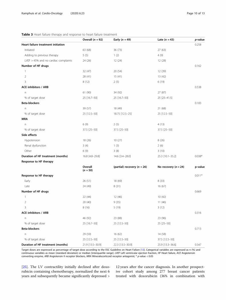
ACD treatment and clinical follow-upThe median follow-up time after diagnosis of ACD was25.0 [10.6–38.2] months (Fig. 2b). HF medication wasinitiated in 68 patients (74%) (Table 3). Five of these pa-tients were already on ACE-inhibitors or beta-blockersbecause of hypertension, and additional HF therapy wasgiven. Intolerance to ACE-inhibitors and betablockerswas common, and overall the maximum tolerated dosewas low (Table 3). Twenty-four patients (26%) did notreceive HF medication since they had an LVEF of > 45%and lacked cardiac complaints [11]. Three patients werediagnosed with ACD when they were hospitalized due toHF and 5 patients were hospitalized with decompensatedHF after ACD was diagnosed. No patient was in needfor cardiac mechanical support or heart transplantationduring follow-up. Implantable cardioverter defibrillator
Fig. 1 Flow chart of the study population selection
Kamphuis et al. Cardio-Oncology (2020) 6:23 Page 4 of 13

Table 1 Characteristics of study participants
Demographics Overall (n = 92) Early (n = 49) Late (n = 43) p-value
Male 68 (74) 38 (78) 30 (70)
Body Mass Index (kg/m2) 24.5 (4.5) 24.8 (4.5) 24.1 (4.5) 0.422
Body Surface Area (m2) 1.9 (0.2) 1.9 (0.2) 1.9 (0.2) 0.536
Age at start cancer therapy (years) 48.2 (18.1) 52.2 (15.8) 43.7 (19.2) 0.024
Age at diagnosis ACD (years) 51.6 (16.2) 52.4 (16.1) 50.8 (16.2) 0.646
Malignancy
Hematological 86 (94) 48 (98) 38 (88)
Acute leukemia 44 (51) 32 (67) 12 (32)
Non-Hodgkin’s lymphoma 6 (7) 1 (2) 5 (13)
Hodgkin’s lymphoma 26 (30) 8 (17) 18 (47)
Othera 10 (12) 7 (15) 3 (8)
Breast cancer 2 (2) 0 (0) 2 (5)
Other solid tumorsb 4 (4) 1 (2) 3 (7)
Cumulative anthracycline dose (mg/m2), [IQR] 329 [200–329] 329 [180–329] 308 [200–400] 0.114
Chest radiation 7 (8) 1 (2) 6 (14)
Functional class at diagnosis
NYHA class I + II 86 (94) 47 (96) 39 (91)
NYHA class III + IV 6 (6) 2 (4) 4 (9)
Electrocardiogram
Heart rhythm
Sinus rhythm 90 (98) 48 (98) 42 (98)
Atrial fibrillation 2 (2) 1 (2) 1 (2)
Heart axis
Normal 82 (89) 42 (86) 40 (93)
Left 8 (9) 5 (10) 3 (7)
Right 2 (2) 2 (4) 0 (0)
Ventricular conduction
Normal 86 (93) 45 (92) 41 (95)
Left bundle branch block 4 (4) 2 (4) 2 (5)
Right bundle branch block 2 (2) 2 (4) 0 (0)
Cardiovascular risk factors
Hypertension 19 (21) 7 (14) 12 (28)
Diabetes Mellitus 9 (10) 3 (6) 6 (14)
Hyperlipidemiac 12 (17) 5 (13) 7 (19)
Smoking status n = 80 n = 42 n = 38
Former smoker 31 (39) 24 (57) 7 (18)
Current smoker 13 (16) 6 (14) 7 (18)
Never smoked 36 (45) 12 (29) 24 (64)
Coronary / Peripheral Artery disease 13 (14) 8 (17) 5 (12)
Categorical variables are expressed as n (%) and continuous variables as mean (standard deviation) or median [interquartile range]; aOther hematologicalmalignancies included chronic myeloid leukemia (n = 4), multiple myeloma (n = 3), myelodysplastic syndrome (n = 2) and myelofibrosis (n = 1); bOther solid tumorsincluded sarcomas (n = 3) and Wilms tumor (n = 1); c n = 75 patients (38 early ACD; 37 late ACD); ACD Anthracycline-induced cardiac dysfunction
Kamphuis et al. Cardio-Oncology (2020) 6:23 Page 5 of 13
(ICD) implantation was performed in 4 patients, ofwhich 3 patients also received cardiac resynchronizationtherapy. One patient was successfully treated with aMitraClip because of severe, secondary mitral regurgita-tion, which developed after an initial decline in LV func-tion. In total, 38 patients died during follow-up. Onepatient died due to acute HF. Other deaths were pre-dominantly related to the underlying oncological disease(n = 30) or cancer treatment-related infections (n = 2). In5 patients, the cause of death was unknown.
ACD echocardiographic follow-up and reversibilityFollow-up echocardiographic examinations were avail-able in 67 patients, with a median follow-up time of 17.7[10.5–26.6] months between diagnosis and the last echo-cardiographic examination. Of the 53 patients who had a
normal iEDV at diagnosis, only 2 patients (3.7%) devel-oped an iEDV above the upper limit of normal (ULN)during follow-up. Thirteen out of 14 patients who pre-sented with a dilated LV showed normal iEDV values atfollow-up. There was no relation between follow-up dur-ation and the change in iEDV (ρ = − 0.079; p = 0.524).Sixty-seven patients could be analyzed for reversibility
of ACD. Of the 25 patients who were excluded from theanalysis, 20 patients died before cardiac follow-up couldbe performed. In 32 patients (48%) no recovery of LVfunction was observed (treatment in n = 24), 10 patients(15%) had partial recovery (treatment in n = 9), and 25patients (37%) showed complete recovery (treatment inn = 17). No differences were observed in GLS at diagno-sis between patients with- and without (partial) recovery((partial) recovery − 13.9 ± 3.0; no recovery − 14.0 ± 3.6;
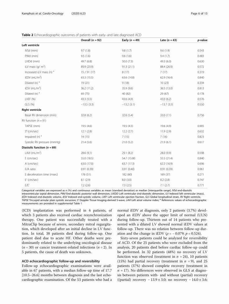
Table 2 Echocardiographic outcomes of patients with early- and late diagnosed ACD
Overall (n = 92) Early (n = 49) Late (n = 43) p-value
Left ventricle
IVSd (mm) 9.7 (1.8) 9.8 (1.7) 9.6 (1.9) 0.543
PWd (mm) 9.5 (1.6) 9.6 (1.6) 9.4 (1.7) 0.483
LVIDd (mm) 49.7 (6.8) 50.0 (7.3) 49.3 (6.3) 0.630
iLV mass (g/ m2) 89.9 (23.9) 91.3 (21.1) 88.4 (26.9) 0.572
Increased iLV mass (n) a 15 / 91 (17) 8 (17) 7 (17) 0.319
iEDV (mL/m2) 63.3 (15.5) 63.6 (14.8) 62.9 (16.4) 0.840
Dilated (n) a 19 (21) 9 (18) 10 (23) 0.334
iESV (mL/m2) 36.2 (11.2) 35.9 (9.6) 36.5 (13.0) 0.813
Dilated (n) a 69 (75) 40 (82) 29 (67) 0.178
LVEF (%) 43.3 (5.5) 43.6 (4.9) 43.0 (6.2) 0.576
GLS (%) −13.5 (3.3) −13.2 (3.1) −13.7 (3.5) 0.550
Right ventricle
Basal RV dimension (mm) 32.8 (6.2) 32.6 (5.4) 33.0 (7.1) 0.756
RV function (n = 91)
TAPSE (mm) 19.5 (4.6) 19.5 (4.3) 19.6 (4.9) 0.905
S′ (cm/sec) 12.1 (2.8) 12.2 (2.7) 11.9 (2.9) 0.632
Impaired (n) a 14 (15) 7 (15) 7 (16) 0.823
Systolic RV pressure (mmHg) 21.4 (5.6) 21.0 (5.2) 21.9 (6.1) 0.617
Diastolic function (n = 89)
LAVI (mL/m2) 28.6 (9.1) 29.1 (8.2) 28.0 (9.9) 0.598
E (cm/sec) 55.0 (18.5) 54.7 (15.80 55.5 (21.4) 0.840
A (cm/sec) 63.0 (17.0) 63.7 (17.3) 62.2 (16.9) 0.696
E/A ratio 0.91 (0.39) 0.91 (0.40) 0.91 (0.39) 0.961
E deceleration time (msec) 176 (51) 182 (60) 169 (37) 0.271
E’ (cm/sec) 8.1 (2.9) 8.0 (3.0) 8.2 (2.8) 0.747
E/E’ 7.2 (2.6) 7.3 (2.5) 7.1 (2.7) 0.771
Categorical variables are expressed as n (%) and continuous variables as mean (standard deviation) or median [interquartile range]. IVSd end-diastolicintraventricular septal dimension, PWd End-diastolic posterior wall dimension, LVEDD Left ventricular end-diastolic dimension, iLV Indexed left ventricular (mass),iEDV Indexed end-diastolic volume, iESV Indexed end-systolic volume, LVEF Left ventricular ejection fraction, GLS Global longitudinal strain, RV Right ventricle,TAPSE Tricuspid annular plain systolic excursion; S′ Doppler Tissue Imaging-derived S-wave, LAVI Left atrial volume index; a References values of echocardiographicmeasurements are provided in supplemental Table 1
Kamphuis et al. Cardio-Oncology (2020) 6:23 Page 6 of 13
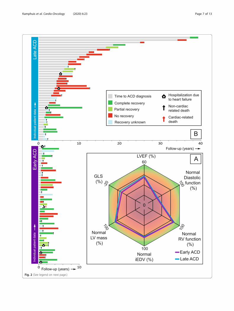
Fig. 2 (See legend on next page.)
Kamphuis et al. Cardio-Oncology (2020) 6:23 Page 7 of 13
p = 0.908). Patients with early ACD were more likely toshow (partial) recovery of LV function compared to lateACD patients, without significant differences in max-imum tolerated doses of HF medication (Table 3).
DiscussionThe aim of this consecutive cohort study was to evaluatethe echocardiographic phenotype of ACD, establish whatimpact a delayed diagnosis had on the degree of LV dys-function, LV dimensions and response to HF treatment.The key findings of this study are threefold: (1) the ma-jority of patients presented with mild LV dysfunctionwithout LV dilatation (2) the echocardiographic pheno-type was not different in patients diagnosed with earlyor late ACD and (3) patients with an early ACD diagno-sis and prompt initiation of HF treatment were morelikely to have a (partial) recovery of LV function, com-pared to patients with a late ACD diagnosis.In contrast to previous studies in which ACD is de-
scribed as a toxic cause of DCM [15], we found that LVdilatation was present in only one-fifth of cases at diag-nosis and only two patients developed LV dilatation dur-ing a median follow-up of 17.7 months. We believe thatthere are three major reasons for the absence of a DCM-like phenotype. Firstly, many patients received an earlydiagnosis of ACD due to serial echocardiographicscreening at a cardio-oncology clinic. In the past, thediagnosis of ACD was often established upon the devel-opment of symptomatic HF, and subclinical changes inLV function were not detected. In these patients, pro-longed activation of compensatory mechanisms, includ-ing the renin-angiotensin aldosterone system (RAAS),and subsequent LV remodeling, may have led to a morepronounced dilatation of the LV. However, in our study,dilatation was also not present in the majority of patientswith a late ACD diagnosis. The second possible reasonfor the absence of abnormal dimensions could be relatedto the reduction of cumulative anthracycline dose overthe last decades. While doxorubicin doses exceeding >500 mg/m2 were commonly administered in the past [1],the maximum cumulative dose of this agent is nowadaysrestricted to 450mg/m2 [16], with a median dose of 329[200–329] mg/m2 in this study (n = 9 receiving ≥450mg/m2). It is plausible that these dose restrictions haveresulted in an overall milder ACD phenotype. Thirdly, inpatients developing LV dysfunction with an LVEF < 45%,HF treatment was promptly initiated, aiming at
suppression of RAAS to prevent adverse LV remodeling.The early detection of ACD has shown to be beneficialin one study, where patients with early initiation of ther-apy were more likely to respond to pharmacologicaltreatment [17]. To our knowledge, our study is the firstto validate this time-dependent response to HF treat-ment. In conclusion, an earlier diagnosis, a restriction inthe maximum cumulative anthracycline dose and theinitiation of HF treatment might jointly have led to ahypokinetic non-dilated cardiomyopathy rather thanDCM [18]. Based upon the results of this study, moni-toring of LV function in patients at risk for ACD is rec-ommended to detect subclinical changes in LV functionas soon as possible and thereby allow for early initiationof HF treatment in case ACD develops.We did not find any echocardiographic differences in
dimension and function between patients diagnosedearly vs. late (Table 2). Furthermore, there were also nodiscrepancies in LV mass between the two groups. Todate, a number of imaging studies have evaluated thisparameter in ACD both by CMR [19–21] and echocardi-ography [22, 23]. Currently, CMR is considered as thegold standard for measurements of cardiac structure andvolumes [13]. With this technique, three studies found adecrease in LV mass [19–21], and an inverse correlationwith the anthracycline dose [20]. Furthermore, a lowerLV mass was predictive of cardiovascular death, appro-priate ICD therapy and HF hospitalization in a multi-variate model [20]. However, these findings arecontradictory to echocardiographic studies which foundan increase in LV mass [22, 23]. Armstrong et al. studiedadults who were treated with anthracyclines duringchildhood. They found a reduced LV mass in nearly halfof patients [24]. Comparison of CMR with echocardiog-raphy performed within 48 h, revealed that echocardiog-raphy overestimated LVEF and LV mass andunderestimates LV volumes. The absence of reduced LVmasses in our study population could therefore be re-lated to LV mass measurements with echocardiography.In contrast to our hypothesis, our data does not sup-
port the progressive nature of ACD regarding cardiac re-modeling. To date, longitudinal imaging studies inpatients with ACD are scarce, with a small number ofstudies performed in pediatric [25] and adult cancer pa-tients [22, 26]. Lipshultz et al. prospectively followed 115survivors of childhood acute leukemia with serial echo-cardiograms during a median follow-up of 11.8 years
(See figure on previous page.)Fig. 2 Echocardiographic characterization of early- and late diagnosed anthracycline-induced cardiac dysfunction and follow-up. a The radar chartshows the echocardiographic phenotype at diagnosis of early- and late ACD, which are both characterized by a mild hypokinetic, non-dilatedcardiomyopathy. LVEF and GLS are expressed as group means, LV mass, iEDV and RV function are expressed as % of patients with normaloutcomes and diastolic function is expressed as % of patients with diastolic dysfunction ≤ grade I; b Individual time periods of time to ACDdiagnosis and follow-up outcomes regarding i) hospitalization due to heart failure ii) recovery of LV function and iii) (non-)cardiac death
Kamphuis et al. Cardio-Oncology (2020) 6:23 Page 8 of 13
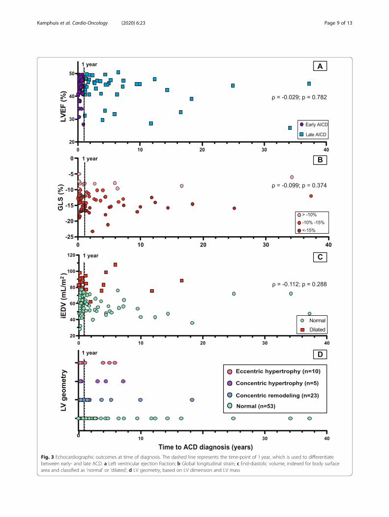
Fig. 3 Echocardiographic outcomes at time of diagnosis. The dashed line represents the time-point of 1 year, which is used to differentiatebetween early- and late ACD. a Left ventricular ejection fraction; b Global longitudinal strain; c End-diastolic volume, indexed for body surfacearea and classified as ‘normal’ or ‘dilated’; d LV geometry, based on LV dimension and LV mass
Kamphuis et al. Cardio-Oncology (2020) 6:23 Page 9 of 13
[25]. The LV contractility initially declined after doxo-rubicin containing chemotherapy, normalized the next 6years and subsequently became significantly depressed >
12 years after the cancer diagnosis. In another prospect-ive cohort study among 277 breast cancer patientstreated with doxorubicin (36% in combination with
Table 3 Heart failure therapy and response to heart failure treatment
Overall (n = 92) Early (n = 49) Late (n = 43) p-value
Heart failure treatment initiation 0.258
Initiated 63 (68) 36 (73) 27 (63)
Adding to previous therapy 5 (5) 1 (2) 4 (9)
LVEF > 45% and no cardiac complaints 24 (26) 12 (24) 12 (28)
Number of HF drugs 0.162
1 32 (47) 20 (54) 12 (39)
2 28 (41) 15 (41) 13 (42)
3 8 (12) 2 (5) 6 (19)
ACE inhibitors / ARB 0.538
n 61 (90) 34 (92) 27 (87)
% of target dose 25 [16.7–50] 25 [16.7–50] 25 [25–41.5]
Beta-blockers 0.183
n 39 (57) 18 (49) 21 (68)
% of target dose 25 [12.5–50] 18.75 [12.5–25] 25 [12.5–50]
MRA
n 6 (9) 2 (5) 4 (13)
% of target dose 37.5 [25–50] 37.5 [25–50] 37.5 [25–50]
Side effects
Hypotension 18 (26) 10 (27) 8 (26)
Renal dysfunction 3 (4) 1 (3) 2 (6)
Other 6 (9) 3 (8) 3 (10)
Duration of HF treatment (months) 16.8 [4.8–29.8] 14.6 [3.4–28.0] 25.3 [10.1–35.2] 0.038*
Response to HF therapy
Overall(n = 50)
(partial) recovery (n = 26) No recovery (n = 24) p-value
Response to HF therapy 0.011*
Early 26 (51) 18 (69) 8 (33)
Late 24 (49) 8 (31) 16 (67)
Number of HF drugs 0.669
1 22 (44) 12 (46) 10 (42)
2 20 (40) 9 (35) 11 (46)
3 8 (16) 5 (19) 3 (12)
ACE inhibitors / ARB 0.316
n 46 (92) 23 (88) 23 (96)
% of target dose 25 [16.7–50] 25 [12.5–50] 25 [25–50]
Beta-blockers 0.713
n 29 (59) 16 (62) 14 (58)
% of target dose 25 [12.5–50] 25 [12.5–50] 37.5 [12.5–50]
Duration of HF treatment (months) 21.9 [13.5–30.9] 22.3 [13.5–30.9] 25.9 [13.3–36.6] 0.547
Target doses are expressed as percentage of target dose according to the ESC Guideline on Heart Failure [12]. Categorical variables are expressed as n (%) andcontinuous variables as mean (standard deviation) or median [interquartile range]. LVEF Left ventricular ejection fraction, HF Heart failure, ACE Angiotensinconverting enzyme, ARB Angiotensin II receptor blockers, MRA Mineralocorticoid receptor antagonist; * p-value < 0.05
Kamphuis et al. Cardio-Oncology (2020) 6:23 Page 10 of 13

trastuzumab) the LVEF decline was not progressive duringa median follow-up of 2 years [22]. In a study by Joneset al. in 143 patients that were followed for 2 years, nonetransitioned to more advanced HF stages [26]. In ourstudy only 5.5% of patients progressed to symptomaticHF. Nevertheless, the presence of asymptomatic LV dys-function gives an increased risk of ultimately progressingto symptomatic HF. In a meta-analysis evaluating the riskin patients with systolic LV dysfunction due to various eti-ologies, the incidence of symptomatic HF was 8.4 (95% CI4.0–12.8) per 100 person-years, compared to 1.04 (95% CI0.0–2.2) per 100 person-years in the absence of LV dys-function, equaling a relative risk of 4.6 (95% CI 2.2–9.8)[27]. This meta-analysis illustrates the importance ofimplementing effective strategies in the pre-symptomaticstages to mitigate the progression rate to symptomaticHF. This progression might also be dependent on the de-velopment of other cardiac stressors, such as hyperten-sion, valvular disease, CAD, or the presence of pathogenicvariants in cardiomyopathy-associated genes [28, 29]. Inthe absence of these so called “second-hits”, it is possiblethat a considerable proportion of patients only developsmild LV dysfunction after anthracycline-containingchemotherapy, that remains stable for years. Larger obser-vational cohort studies, preferably with long-term follow-up can shed more light on the natural course of this spe-cific disease entity. Also, the outcomes of the TITAN-study, which compares intensive cardiac monitoring and-treatment to usual care, will be informative on the addedvalue of early identification of ACD [30].
LimitationsOur analysis was restricted to patients that visited thecardio-oncology outpatient clinic. Patients treated withpotentially cardiotoxic cancer therapy prior to its launchcurrently receive cardiological follow-up to screen forlong-term cardiovascular complications. It is possiblethat patients who developed a more severe ACD pheno-type presented at the emergency care unit earlier andwere never seen in an outpatient clinic setting and there-fore were not included in our study. Also, patientsdeemed to be at low risk for ACD do not receive cardiacscreening per protocol at our cardio-oncology service.This may overall lead to an underestimation of ACD inthe population of cancer patients treated withanthracyclines.Furthermore, the current study used patient informa-
tion collected during standard clinical care. Albeit thefollow-up of these patients is standardized to a great ex-tent [11], there still was heterogeneity in the data. Eventhough current guidelines recommend to perform car-diac screening prior to treatment [31], many patientswere referred after initiation of anthracycline-treatment,and therefore lacked cardiac baseline assessment. Subtle
changes in LV dimensions and –volumes within patientscould therefore have been overlooked. In addition, for pa-tients that did not undergo an echocardiography at base-line, pre-existent impaired cardiac function could havebeen misclassified as ACD. Also, additional testing forother causes of impaired LV function, such as CAD, hadnot (yet) been performed in a number of patients, leavinguncertainty of the diagnosis of ACD. Nevertheless, thisrepresents only a small subset of our cohort where, from aclinical point of view, ACD was very likely.Biomarkers, such as troponin or BNP, are not rou-
tinely performed at our cardio-oncology clinic and wereonly available in a limited number of patients. We there-fore were unable to include the outcomes of biomarkersin our analysis.
ConclusionsWe found that the ACD phenotype overall was mild anda majority of patients lacked cardiac complaints. In theabsence of serial echocardiographic assessment, it there-fore is plausible that the impaired cardiac functionwould have remained undetected. Since response to HFtreatment is time-dependent, detection of asymptomaticLV dysfunction is of great importance. When cardiaccompromise is detected, other cardiovascular risk factorscan be treated more aggressively, potentially decreasingthe risk of patients progressing to more advanced HFstages. Moreover, if a patient is planned to receive fur-ther cardiotoxic treatment, preventive actions can beconsidered [31]. The involvement of a cardiologist in amultidisciplinary setting at cardio-oncology clinics isvaluable to allow for the early detection of ACD andother adverse cardiovascular effects during cardiotoxictreatment [32].Importantly, future research within the field of
cardio-oncology should not only focus on the addedvalue of cardiac screening, but also possible draw-backs including medicalization and increased health-care costs [33]. Trials evaluating different follow-upstrategies, such as the TITAN-study [30], are requiredto achieve an optimal risk-benefit ratio. It is plausiblethat the optimal strategy varies per cancer type, asoften the prognosis of the malignancy is a dominantfactor. Longitudinal cohort studies establishing moreinsight into the natural course of ACD are herein ofpivotal importance.
Supplementary informationSupplementary information accompanies this paper at https://doi.org/10.1186/s40959-020-00079-3.
Additional file 1: Supplemental Table 1. Reference values ofechocardiographic measurements and international guidelines onechocardiographic examination [16].
Kamphuis et al. Cardio-Oncology (2020) 6:23 Page 11 of 13
AbbreviationsACD: Anthracycline-induced cardiac dysfunction; DCM: Dilatedcardiomyopathy; LV: Left ventricle; LVEF: Left ventricular ejection fraction;iEDV: Indexed end-diastolic volume; CTRCD: Cancer therapy-related cardiacdysfunction; CMR: Cardiac Magnetic resonance; MUGA: Multiple-gatedacquisition; ESC: European Society of Cardiology; LBBB: Left bundle branchblock; CAD: Coronary artery disease; CT: Computed tomography; BSA: Bodysurface area; GLS: Global longitudinal strain; LVIDd: Left ventricular internaldiameter at end-diastole; RWT: Relative wall thickness; TAPSE: Tricuspidannular plain systolic excursion; DTI: Doppler tissue imaging; EACVI: EuropeanAssociation of Cardiovascular Imaging; LLN: Lower limit of normal;IQR: Interquartile range; SD: Standard deviation; ANOVA: Analysis of variance;NYHA: New York Heart Association; RV: Right ventricle; ACE: Angiotensinconverting enzyme; ICD: Implantable cardiac defibrillator; ULN: Upper limit ofnormal; RAAS: Renin-angiotensin aldosterone system
AcknowledgementsNot applicable.
Authors’ contributionsJAMK, ML and AJT contributed to data collection, study design, data analysis,data interpretation, and writing of the manuscript. MJC, PAD and FWAcontributed to writing the manuscript. All authors critically reviewed themanuscript and provided important intellectual contributions. The finalmanuscript was approved by all authors.
FundingDr. Asselbergs has reported support from the University College LondonHospitals, National Institute for Health Research Biomedical Research Centre.Dr. Linschoten has reported support from the Alexandre Suerman Stipend ofthe University Medical Center (UMC) Utrecht. All other authors have reportedthat they have no relationships relevant to the contents of this paper todisclose.
Availability of data and materialsThe datasets generated and analysed during the current study are notpublicly available due to protecting participant confidentiality but areavailable from the corresponding author on reasonable request.
Ethics approval and consent to participateThe study was exempted from formal approval by the Medical EthicsCommittee (METC 19–304/C,Utrecht, the Netherlands).
Consent for publicationNot applicable.
Competing interestsNone declared.
Author details1Department of Cardiology, Division of Heart and Lungs, University MedicalCenter Utrecht, University of Utrecht, E03.511, PO Box 85500, 3508 GAUtrecht, The Netherlands. 2Netherlands Heart Institute, Utrecht, TheNetherlands. 3Central Military Hospital, Utrecht, The Netherlands. 4HealthData Research UK and Institute of Health Informatics, University CollegeLondon, London, UK. 5Institute of Cardiovascular Science, Faculty ofPopulation Health Sciences, University College London, London, UK.
Received: 2 August 2020 Accepted: 2 October 2020
References1. Von Hoff DD, Layard MW, Basa P, Davis HL, von Hoff AL, Rozencweig M,
et al. Risk factors for doxorubicin-induced congestive heart failure. AnnIntern Med. 1979;91:710–7. https://doi.org/10.7326/0003-4819-91-5-710.
2. Lotrionte M, Biondi-Zoccai G, Abbate A, Lanzetta G, D’Ascenzo F, Malavasi V,et al. Review and meta-analysis of incidence and clinical predictors ofanthracycline cardiotoxicity. Am J Cardiol. 2013;112:1980–4. https://doi.org/10.1016/j.amjcard.2013.08.026.
3. Fornaro A, Olivotto I, Rigacci L, Ciaccheri M, Tomberli B, Ferrantini C, et al.Comparison of long-term outcome in anthracycline-related versus
idiopathic dilated cardiomyopathy: a single Centre experience. Eur J HeartFail. 2018;20(5):898–906. https://doi.org/10.1002/ejhf.1049.
4. Felker GM, Thompson RE, Hare JM, Hruban RH, Clemetson DE, Howard DL,et al. Underlying causes and long-term survival in patients with initiallyunexplained cardiomyopathy. N Engl J Med. 2000;342(15):1077–84. https://doi.org/10.1056/NEJM200004133421502.
5. Zamorano JL, Lancellotti P, Rodriguez Muñoz D, Aboyans V, Asteggiano R,Galderisi M, et al. 2016 ESC position paper on cancer treatments andcardiovascular toxicity developed under the auspices of the ESC Committeefor practice guidelines: the task force for cancer treatments andcardiovascular toxicity of the European Society of Cardiology (ESC). EurHeart J. 2016;37(36):2768–801. https://doi.org/10.1093/eurheartj/ehw211.
6. Cardinale D, Sandri MT, Colombo A, Colombo N, Boeri M, Lamantia G, et al.Prognostic value of troponin I in cardiac risk stratification of cancer patientsundergoing high-dose chemotherapy. Circulation. 2004;109(22):2749–54.https://doi.org/10.1161/01.CIR.0000130926.51766.CC.
7. Auner HW, Tinchon C, Linkesch W, Tiran A, Quehenberger F, Link H, et al.Prolonged monitoring of troponin T for the detection of anthracyclinecardiotoxicity in adults with hematological malignancies. Ann Hematol.2003;82(4):218–22. https://doi.org/10.1007/s00277-003-0615-3.
8. Cardinale D, Colombo A, Bacchiani G, Tedeschi I, Meroni CA, Veglia F, et al.Early detection of Anthracycline Cardiotoxicity and improvement with heartfailure therapy. Circulation. 2015;131(22):1981–8. https://doi.org/10.1161/CIRCULATIONAHA.114.013777.
9. Mann DL, Bristow MR. Mechanisms and models in heart failure: thebiomechanical model and beyond. Circulation. 2005;111:2837–49. https://doi.org/10.1161/CIRCULATIONAHA.104.500546.
10. Mercurio V, Pirozzi F, Lazzarini E, et al. Models of heart failure based on thecardiotoxicity of anticancer drugs. J Card Fail. 2016;22(6):449–58. https://doi.org/10.1016/j.cardfail.2016.04.008.
11. Teske AJ, Linschoten M, Kamphuis JAM, Naaktgeboren WR, Leiner T, van derWall E, et al. Cardio-oncology: an overview on outpatient management andfuture developments. Neth Hear J. 2018;26(11):521–32. https://doi.org/10.1007/s12471-018-1148-7.
12. Ponikowski P, Voors AA, Anker SD, Bueno H, Cleland JG, Coats AJ, et al. 2016ESC guidelines for the diagnosis and treatment of acute and chronic heartfailure: the task force for the diagnosis and treatment of acute and chronicheart failure of the European Society of Cardiology (ESC). Developed withthe special contribution of the heart failure association (HFA) of the ESC. EurJ Heart Fail. 2016;18(8):891–975. https://doi.org/10.1093/eurheartj/ehw128.
13. Plana JC, Galderisi M, Barac A, Ewer MS, Ky B, Scherrer-Crosbie M, et al.Expert consensus for multimodality imaging evaluation of adult patientsduring and after Cancer therapy: a report from the American Society ofEchocardiography and the European Association of Cardiovascular Imaging.Eur Heart J Cardiovasc Imaging. 2014;15(10):1063–93. https://doi.org/10.1016/j.echo.2014.07.012.
14. Shankar SM, Marina N, Hudson MM, Hodgson DC, Adams J, Landier W, et al.Monitoring for cardiovascular disease in survivors of childhood Cancer:report from the cardiovascular disease task force of the Children’s oncologygroup. Pediatrics. 2008;121(2):e387–96. https://doi.org/10.1542/peds.2007-0575.
15. Japp AG, Gulati A, Cook SA, Cowie MR, Prasad SK. The diagnosis andevaluation of dilated cardiomyopathy. J Am Coll Cardiol. 2016;67(25):2996–3010. https://doi.org/10.1016/j.jacc.2016.03.590.
16. Ewer MS, Ewer SM. Cardiotoxicity of anticancer treatments. Nat Rev Cardiol.2015;12(9):547–58. https://doi.org/10.1038/nrcardio.2015.65.
17. Cardinale D, Colombo A, Lamantia G, Colombo N, Civelli M, De Giacomi G,et al. Anthracycline-induced cardiomyopathy: clinical relevance andresponse to pharmacologic therapy. J Am Coll Cardiol. 2010;55(3):213–20.https://doi.org/10.1016/j.jacc.2009.03.095.
18. Pinto YM, Elliott PM, Arbustini E, Adler Y, Anastasakis A, Böhm M, et al.Proposal for a revised definition of dilated cardiomyopathy, hypokineticnon-dilated cardiomyopathy, and its implications for clinical practice: aposition statement of the ESC working group on myocardial and pericardialdiseases. Eur Heart J. 2016;37(23):1850–8. https://doi.org/10.1093/eurheartj/ehv727.
19. Ferreira de Souza T, Quinaglia AC, Silva T, Osorio Costa F, Sha R, Neilan TG,Velloso L, et al. Anthracycline therapy is associated with Cardiomyocyteatrophy and preclinical manifestations of heart disease. JACC CardiovascImaging. 2018;11(8):1045–55. https://doi.org/10.1016/j.jcmg.2018.05.012.
20. Neilan TG, Coelho-Filho OR, Pena-Herrera D, Shah RV, Jerosch-Herold M,Francis SA, et al. Left ventricular mass in patients with a cardiomyopathy
Kamphuis et al. Cardio-Oncology (2020) 6:23 Page 12 of 13
after treatment with Anthracyclines. Am J Cardiol. 2012;110(11):1679–86.https://doi.org/10.1016/j.amjcard.2012.07.040.
21. Jordan JH, Castellino SM, Meléndez GC, Klepin HD, Ellis LR, Lamar Z, et al.Left ventricular mass change after Anthracycline chemotherapy. Circ HeartFail. 2018;11(7):e004560. https://doi.org/10.1161/CIRCHEARTFAILURE.117.004560.
22. Narayan HK, Finkelman B, French B, Plappert T, Hyman D, Smith AM, et al.Detailed echocardiographic Phenotyping in breast Cancer patients:associations with ejection fraction decline, recovery, and heart failuresymptoms over 3 years of follow-up. Circulation. 2017;135(15):1397–412.https://doi.org/10.1161/CIRCULATIONAHA.116.023463.
23. Tan TC, Bouras S, Sawaya H, Sebag IA, Cohen V, Picard MH, et al. Timetrends of left ventricular ejection fraction and myocardial deformationindices in a cohort of women with breast Cancer treated withAnthracyclines, Taxanes, and Trastuzumab. J Am Soc Echocardiogr. 2015;28(5):509–14. https://doi.org/10.1016/j.echo.2015.02.001.
24. Armstrong GT, Plana JC, Zhang N, Srivastava D, Green DM, Ness KK, et al.Screening adult survivors of childhood Cancer for cardiomyopathy:comparison of echocardiography and cardiac magnetic resonance imaging.J Clin Oncol. 2012;30(23):2876–84. https://doi.org/10.1200/JCO.2011.40.3584.
25. Lipshultz SE, Lipsitz SR, Sallan SE, Dalton VM, Mone SM, Gelber RD, et al.Chronic progressive cardiac dysfunction years after doxorubicin therapy forchildhood acute lymphoblastic leukemia. J Clin Oncol. 2005;23(12):2629–36.https://doi.org/10.1200/JCO.2005.12.121.
26. Jones DN, Jordan JH, Meléndez GC, Lamar Z, Thomas A, Kitzman DW, et al.Frequency of transition from stage a to stage B heart failure after initiatingpotentially Cardiotoxic chemotherapy. JACC Heart Fail. 2018;6(12):1023–32.https://doi.org/10.1016/j.jchf.2018.08.005.
27. Echouffo-Tcheugui JB, Ergou S, Butler J, Yancy CW, Fonarow GC. Assessingthe risk of progression from asymptomatic left ventricular dysfunction toovert heart failure: a systematic overview and meta-analysis. JACC Heart Fail.2016;4(4):237–48. https://doi.org/10.1016/j.jchf.2015.09.015.
28. Linschoten M, Teske AJ, Cramer MJ, van der Wall E, Asselbergs FW.Chemotherapy-related cardiac dysfunction: a systematic review of geneticvariants modulating individual risk. Circ Genom Precis Med. 2018;11(1):e001753. https://doi.org/10.1161/CIRCGEN.117.001753.
29. Garcia-Pavia P, Kim Y, Restrepo-Cordoba MA, Lunde IG, Wakimoto H, SmithAM, et al. Genetic variants associated with Cancer therapy-inducedcardiomyopathy. Circulation. 2019;140(1):31–41. https://doi.org/10.1161/CIRCULATIONAHA.118.037934.
30. Pituskin E, Haykowsky M, McNeely M, Mackey J, Chua N, Paterson I.Rationale and design of the multidisciplinary team IntervenTion in cArdio-oNcology study (TITAN). BMC Cancer. 2016;16:733.
31. Curigliano G, Lenihan D, Fradley M, Ganatra S, Barac A, Blaes A, et al.Management of cardiac disease in cancer patients throughout oncologicaltreatment: ESMO consensus recommendations. Ann Oncol. 2020;31(2):171–90. https://doi.org/10.1016/j.annonc.2019.10.023.
32. Ammon M, Arenja N, Leibundgut G, Buechel RR, Kuster GM, Kaufmann BA,et al. Cardiovascular management of cancer patients with chemotherapy-associated left ventricular systolic dysfunction in real-world clinical practice.J Card Fail. 2013;19(9):629–34. https://doi.org/10.1016/j.cardfail.2013.07.007.
33. Levis BE, Binkley PF, Shapiro CL. Cardiotoxic effects of anthracycline-basedtherapy: what is the evidence and what are the potential harms? LancetOncol. 2017;18(8):e445–56. https://doi.org/10.1016/S1470-2045(17)30535-1.
Publisher’s NoteSpringer Nature remains neutral with regard to jurisdictional claims inpublished maps and institutional affiliations.
Kamphuis et al. Cardio-Oncology (2020) 6:23 Page 13 of 13