ebola: a terrifying challenge - demystifying medicine · ebola: a terrifying challenge? or ......
TRANSCRIPT

Ebola: A Terrifying Challenge?
or
Demystifying Filoviruses!
Presented on January 27, 2015, by Jens H. Kuhn, MD, PhD, PhD, MS
Virology Lead, NIH/NIAID Integrated Research Facility at Fort Detrick (IRF-Frederick), Frederick,
MD

Filoviruses

Filoviruses

Definition of Viral Hemorrhagic Fevers

Medical category for severe clinical syndromes
Known for centuries
Vastly different case-fatality rates
Caused by unrelated agents
Transmitted by different means
Difficult differential diagnosis
Definition of Viral Hemorrhagic Fevers
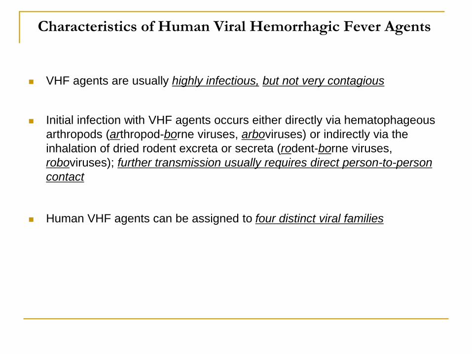
Characteristics of Human Viral Hemorrhagic Fever Agents
VHF agents are usually highly infectious, but not very contagious
Initial infection with VHF agents occurs either directly via hematophageous
arthropods (arthropod-borne viruses, arboviruses) or indirectly via the
inhalation of dried rodent excreta or secreta (rodent-borne viruses,
roboviruses); further transmission usually requires direct person-to-person
contact
Human VHF agents can be assigned to four distinct viral families
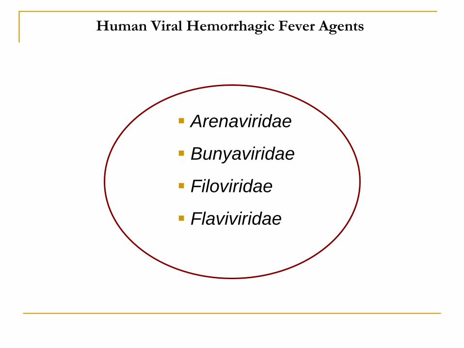
Human Viral Hemorrhagic Fever Agents
Arenaviridae
Bunyaviridae
Filoviridae
Flaviviridae
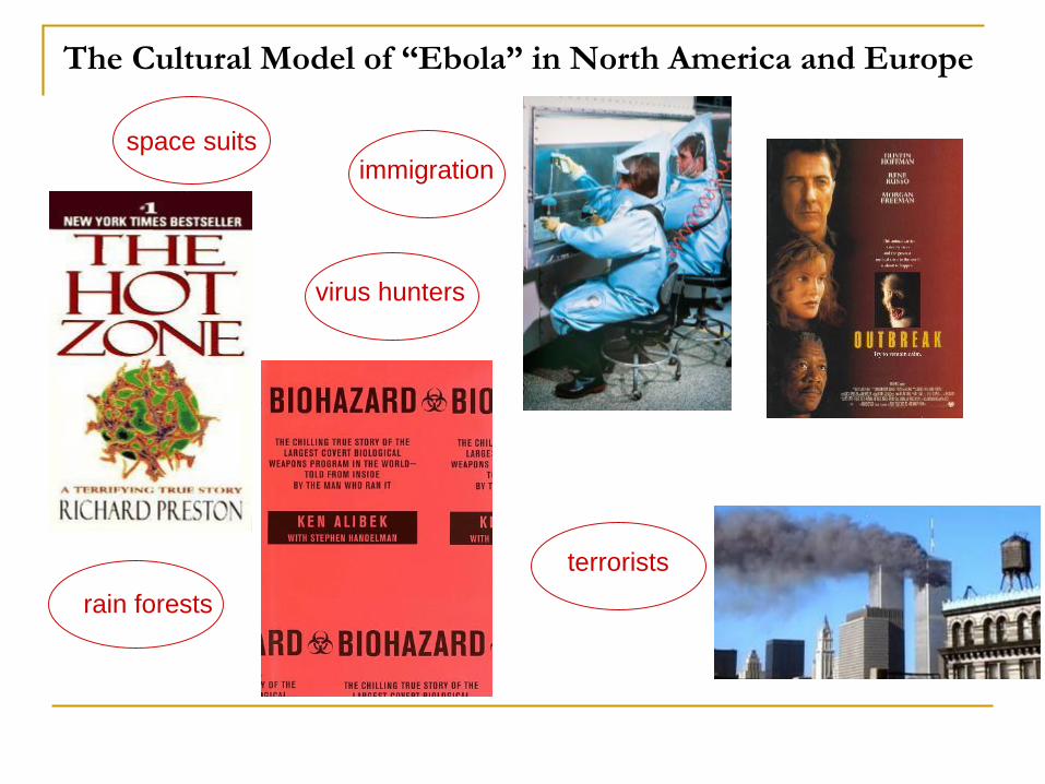
The Cultural Model of “Ebola” in North America and Europe
space suits
virus hunters
immigration
terrorists
rain forests
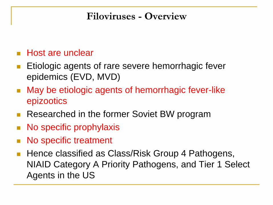
Host are unclear
Etiologic agents of rare severe hemorrhagic fever
epidemics (EVD, MVD)
May be etiologic agents of hemorrhagic fever-like
epizootics
Researched in the former Soviet BW program
No specific prophylaxis
No specific treatment
Hence classified as Class/Risk Group 4 Pathogens,
NIAID Category A Priority Pathogens, and Tier 1 Select
Agents in the US
Filoviruses - Overview


Filovirus TaxonomyYear ICTV Taxonomy
2011-present Order Mononegavirales
Family Filoviridae
Genus Marburgvirus
Species Marburg marburgvirus
Virus: Marburg virus (MARV)
Virus: Ravn virus (RAVV)
Genus Ebolavirus
Species Bundibugyo ebolavirus
Virus: Bundibugyo virus (BDBV)
Species Taï Forest ebolavirus
Virus: Taï Forest virus (TAFV)
Species Reston ebolavirus
Virus: Reston virus (RESTV)
Species Sudan ebolavirus
Virus: Sudan virus (SUDV)
Species Zaire ebolavirus
Virus: Ebola virus (EBOV)
Genus Cuevavirus
Species Lloviu cuevavirus
Virus: Lloviu virus (LLOV)
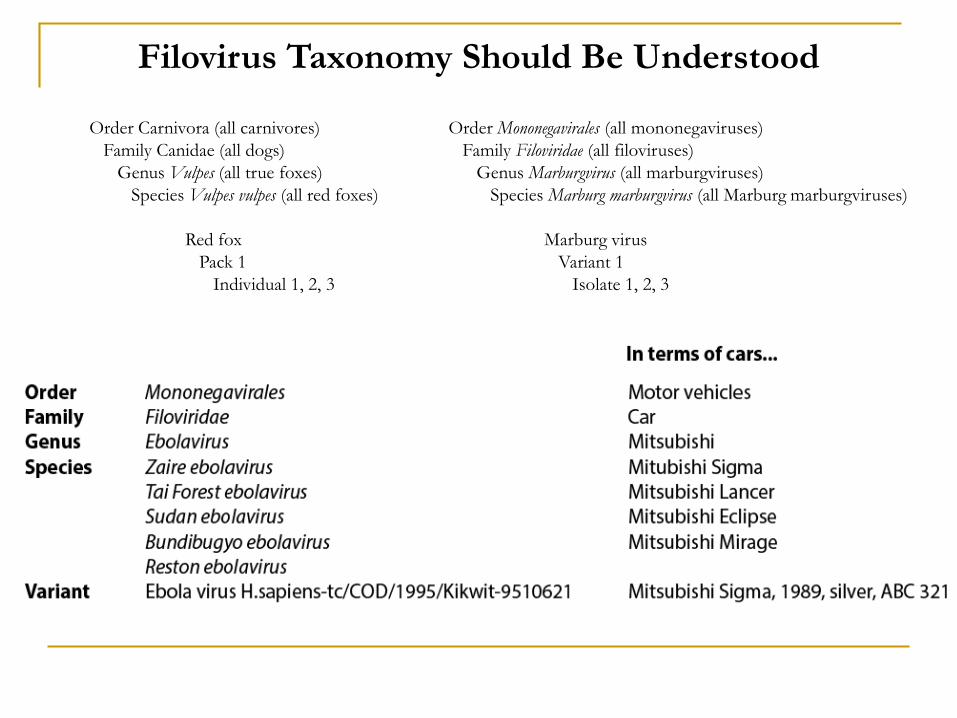
Order Carnivora (all carnivores)
Family Canidae (all dogs)
Genus Vulpes (all true foxes)
Species Vulpes vulpes (all red foxes)
Red fox
Pack 1
Individual 1, 2, 3
Order Mononegavirales (all mononegaviruses)
Family Filoviridae (all filoviruses)
Genus Marburgvirus (all marburgviruses)
Species Marburg marburgvirus (all Marburg marburgviruses)
Marburg virus
Variant 1
Isolate 1, 2, 3
Filovirus Taxonomy Should Be Understood
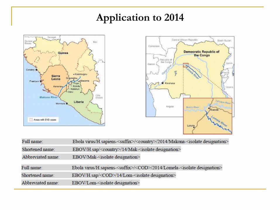
Application to 2014
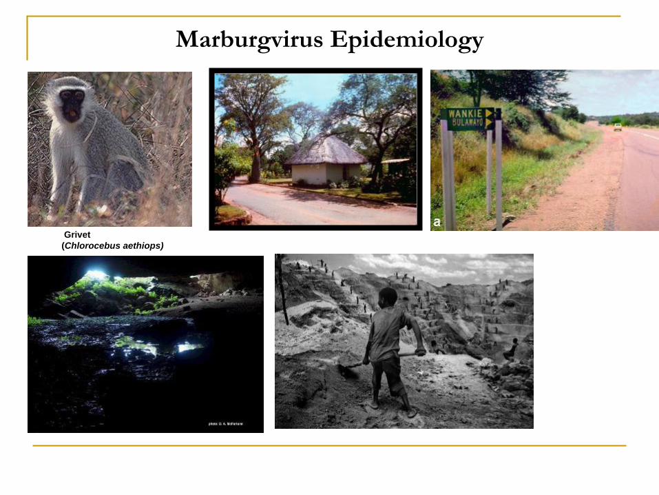
Marburgvirus Epidemiology
Grivet
(Chlorocebus aethiops)

Marburgvirus Epidemiology

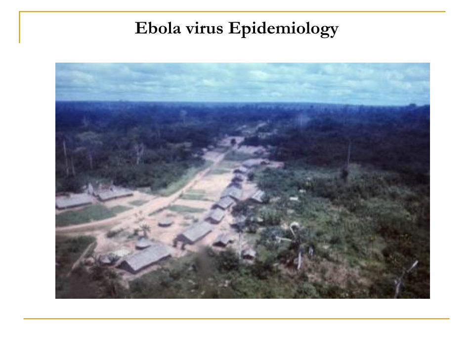
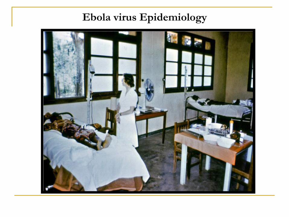
Ebola virus Epidemiology
Month Day Event
August 1976 26 First patient presents with fever in Yambuku;
receives chloroquine injection, fever resolves in four days
September 1976 1
16
23-25
30
Zaire Ebola hemorrhagic fever begins in first patient
Local clinician reviews 17 patients; reports unknown disease to Kinshasa
First medical team visit from Kinshasa; typhoid fever suspected; vaccination; evacuation of a Belgian
nun to capital
Hospital in Yambuku closed; 11 of 17 staff members dead
October 1976 2-6
3
8-12
13-14
14
18
19-27
30
Second medical team visit from Kinshasa; collection of specimens
Health zone quarantined by minister
Transmission occurring in Kinshasa hospital
Filovirus identified by electron microscopy in Belgium, UK, and USA
New virus (‘Ebola’) identified in USA
International commission formed
Survey team to Yambuku; reports active cases in eight villages
Airlift of surveillance teams to northeastern Zaire and to the border with neighboring Sudan
November 1976 2
5
16
Plasmapheresis program begins with convalescent patients
Last case dies
Surveillance, research and clinical care support arrives in Yambuku
December 1976 16 Emergency officially over
January 1977 28 Plasmapheresis program ends
Ebola virus Epidemiology

Ebola virus Epidemiology
Ebola virus Epidemiology

Ebola virus Epidemiology
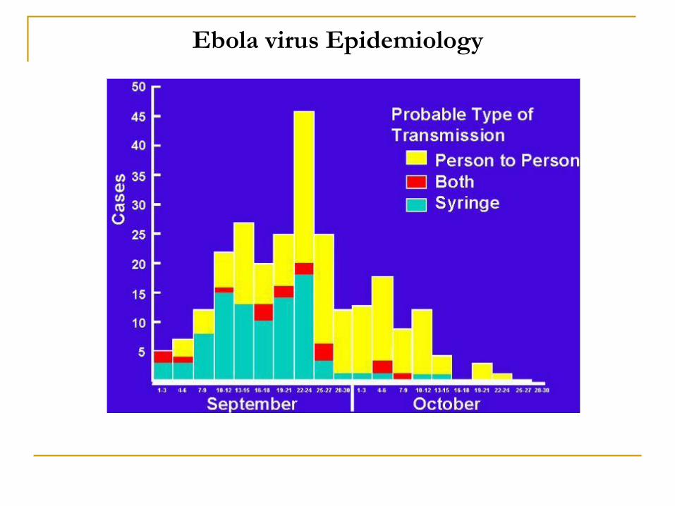
Ebola virus Epidemiology

Ebola virus Epidemiology

Ebola virus Epidemiology
Western lowland gorilla (Gorilla gorilla gorilla)Central chimpanzee (Pan troglodytes troglodytes)
Bush pig/red river hog (Potamochoerus porcus)
Blue duiker (Cephalophus monticola) Black-backed duiker (Cephalophus dorsalis)

Sudan virus Epidemiology
Olive baboon (Papio anubis)

Sudan virus Epidemiology

Sudan virus Epidemiology

Reston Virus Epidemiology
Crab-eating macaque (Macaca fascicularis)
Simian hemorrhagic fever virus (species Simian
hemorrhagic fever virus, genus Arterivirus, family
Arteriviridae)

Taï Forest Virus Epidemiology

Burial ceremonies play a significant role in the transmission
Filoviruses are transmitted by direct contact with blood, bodily secretions or tissues of infected animals or humans
- Viral titers often approach 107 pfu/ml
- The LD50 for rodents and nonhuman primates is thought to be 1-10 pfu
- 1 μl of blood (the equivalent of a needle stick) is thought to contain ≈1,000 LD50
Health-care workers often become infected while treating patients through close contact without the use of correct infection control precautions and adequate barrier-nursing procedures
Although highly infectious, filoviruses are not very contagious
Although animals are readily infected by the aerosol route, aerosol transmission has never been observed during natural outbreaks
Filovirus Transmission

Filovirus infections are characterized by the sudden onset of fever, intense weakness, myalgia, headache, and sore throat. This is often followed by vomiting, diarrhea, morbilliform rash, impaired kidney and liver function, and in some cases, hemorrhages. Laboratory findings show low counts of white blood cells and platelets as well as elevated liver enzymes and elevated D-dimers
Incubation period is 2 – 21 days
Currently limited supportive care is the only available treatment option
Clinical Presentation
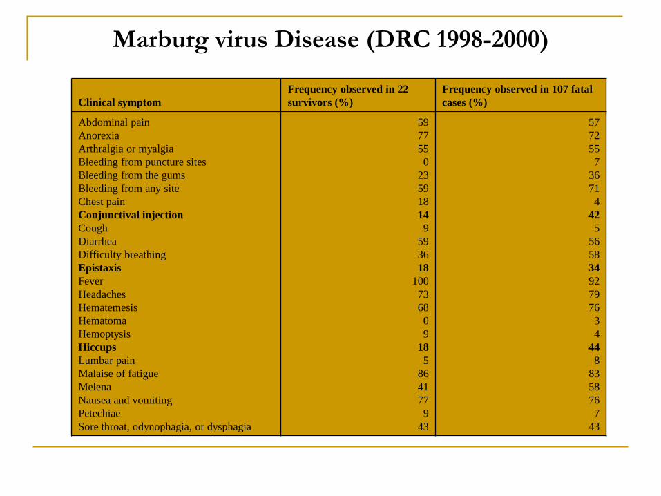
Marburg virus Disease (DRC 1998-2000)
Clinical symptom
Frequency observed in 22
survivors (%)
Frequency observed in 107 fatal
cases (%)
Abdominal pain
Anorexia
Arthralgia or myalgia
Bleeding from puncture sites
Bleeding from the gums
Bleeding from any site
Chest pain
Conjunctival injection
Cough
Diarrhea
Difficulty breathing
Epistaxis
Fever
Headaches
Hematemesis
Hematoma
Hemoptysis
Hiccups
Lumbar pain
Malaise of fatigue
Melena
Nausea and vomiting
Petechiae
Sore throat, odynophagia, or dysphagia
59
77
55
0
23
59
18
14
9
59
36
18
100
73
68
0
9
18
5
86
41
77
9
43
57
72
55
7
36
71
4
42
5
56
58
34
92
79
76
3
4
44
8
83
58
76
7
43
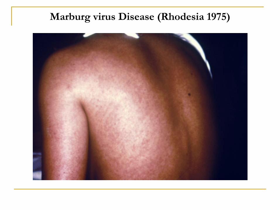
Marburg virus Disease (Rhodesia 1975)

Ebola Virus Disease (Zaire, 1995)Clinical symptom Frequency observed in 19 survivors (%) Frequency observed in 84 fatal cases (%)
Abdominal pain
Abortion
Anorexia
Anuria
Arthralgia or myalgia
Asthenia
Bleeding from puncture sites
Bleeding from the gums
Bloody stools
Chest pain
Conjunctival injection
Convulsions
Cough
Diarrhea
Dysesthesia
Epistaxis
Fever
Headaches
Hearing loss
Hematemesis
Hematoma
Hematuria
Hemoptysis
Hepatomegaly
Hiccups
Lumbar pain
Maculopapular rash
Melena
Nausea and vomiting
Petechiae
Sore throat, odynophagia, or dysphagia
Splenomegaly
Tachypnea
Tinnitus
68
5
47
0
79
95
5
0
5
5
47
0
26
84
5
0
95
74
11
0
0
16
11
5
5
26
16
16
68
0
58
5
0
11
62
2
43
7
50
85
8
15
7
10
42
2
7
86
0
2
93
52
5
13
2
7
0
2
17
12
14
8
73
8
56
2
31
1
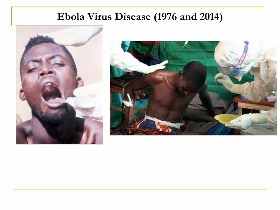
Ebola Virus Disease (1976 and 2014)
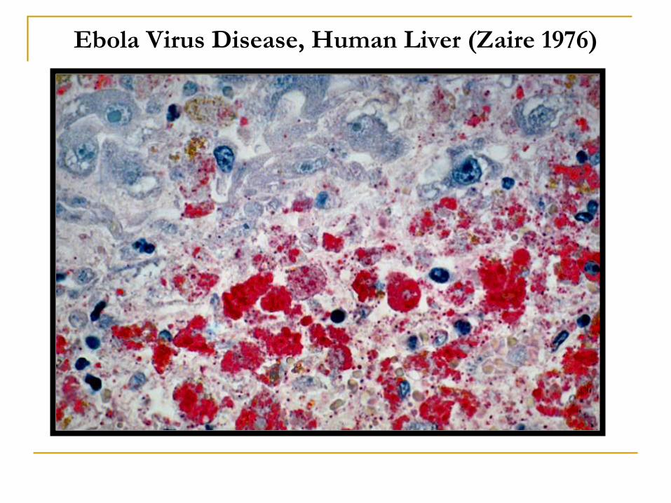
Ebola Virus Disease, Human Liver (Zaire 1976)

Ebola Virus Disease, Human Skin (Zaire 1976)

Filovirus Host Search
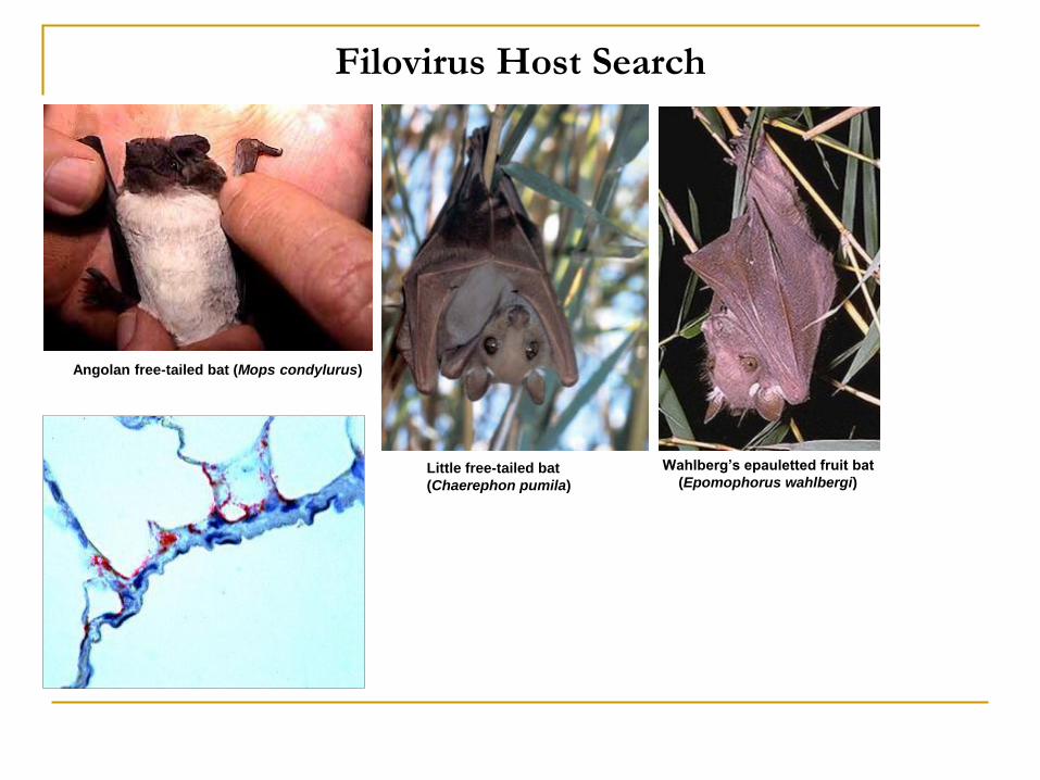
Filovirus Host Search
Angolan free-tailed bat (Mops condylurus)
Little free-tailed bat
(Chaerephon pumila)
Wahlberg’s epauletted fruit bat
(Epomophorus wahlbergi)

Filovirus Host Search
Social spider (Stegodyphus dumicola)
- Mus setulosus?
- Praomys sp.?
- Sylvisorex ollula?
- Aedes Stegomyia aegypti...?
African brown house snake (Lamprophis fuliginosus )
Common house mouse (Mus musculus)
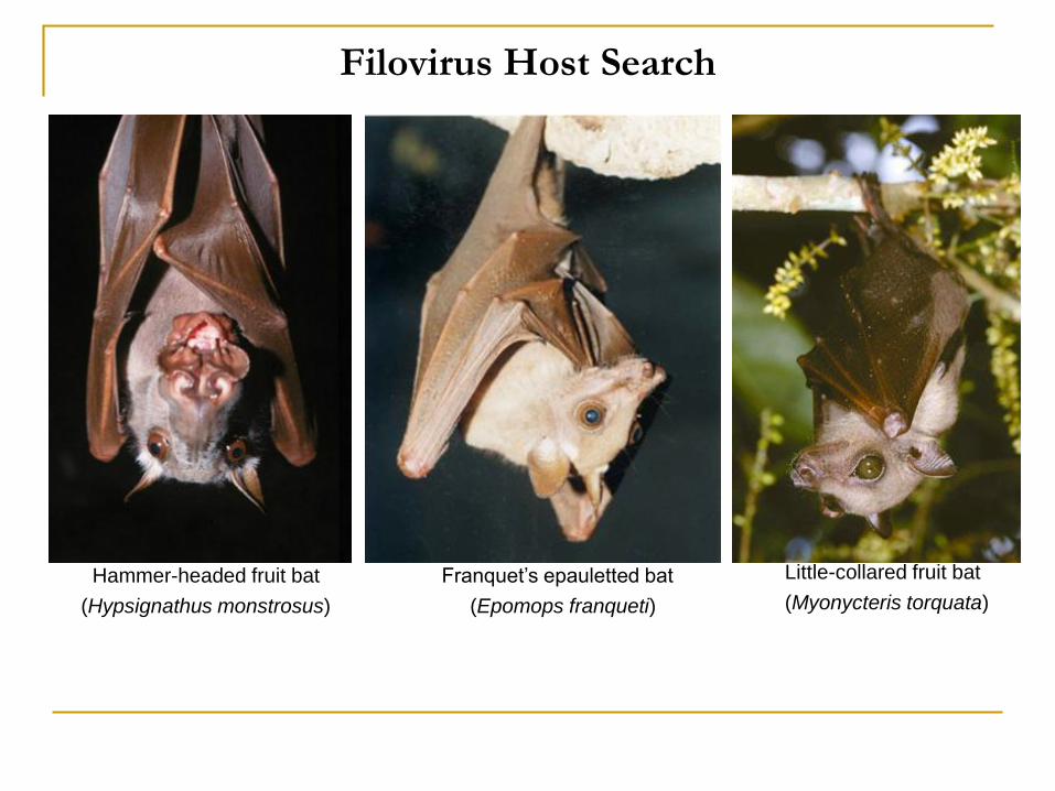
Filovirus Host Search
Hammer-headed fruit bat
(Hypsignathus monstrosus)
Franquet’s epauletted bat
(Epomops franqueti)
Little-collared fruit bat
(Myonycteris torquata)

Filovirus Host Search
Egyptian rousette (Rousettus aegyptiacus)

Filoviruses – Differences and Commonalities
Marburgviruses Ebolaviruses
Antigenic cross-reactivity with
members of the other genus
Minimal Minimal
Average particle length 665 nm 805 nm
Genome length 19.1 kb 18.9 kb
Gene overlaps One Several
Co-transcriptional GP mRNA editing No Yes
Protein profile Homologous sequences among all
isolates, clearly distinct from
ebolaviruses
Species-specific sequence
differences, clearly distinct from
marburgviruses
Case-fatality rate in humans ≥22-90% ≥25-90% (exceptions are Taï Forest
and Reston ebolaviruses at 0%)
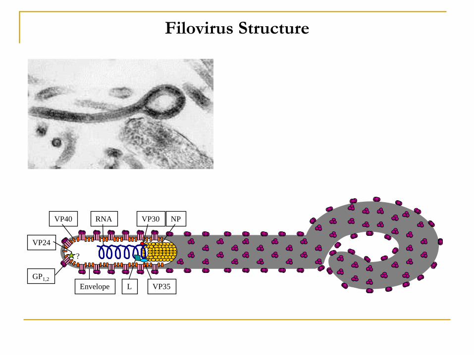
Filovirus Structure
RNA
L
NP
VP35
VP40 VP30
Envelope
VP24
GP1,2
?
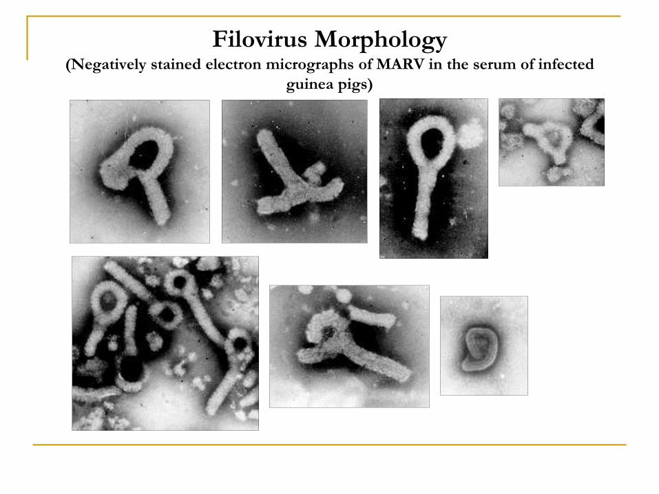
Filovirus Morphology(Negatively stained electron micrographs of MARV in the serum of infected
guinea pigs)
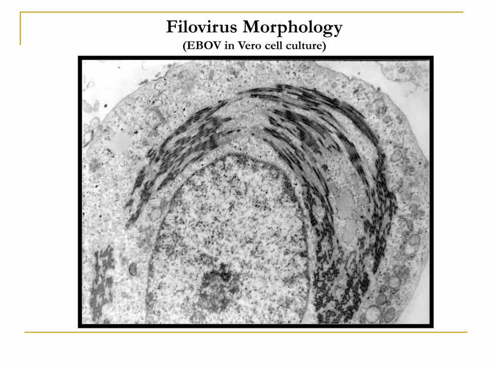
Filovirus Morphology(EBOV in Vero cell culture)

Filovirus Morphology

Filovirus Morphology
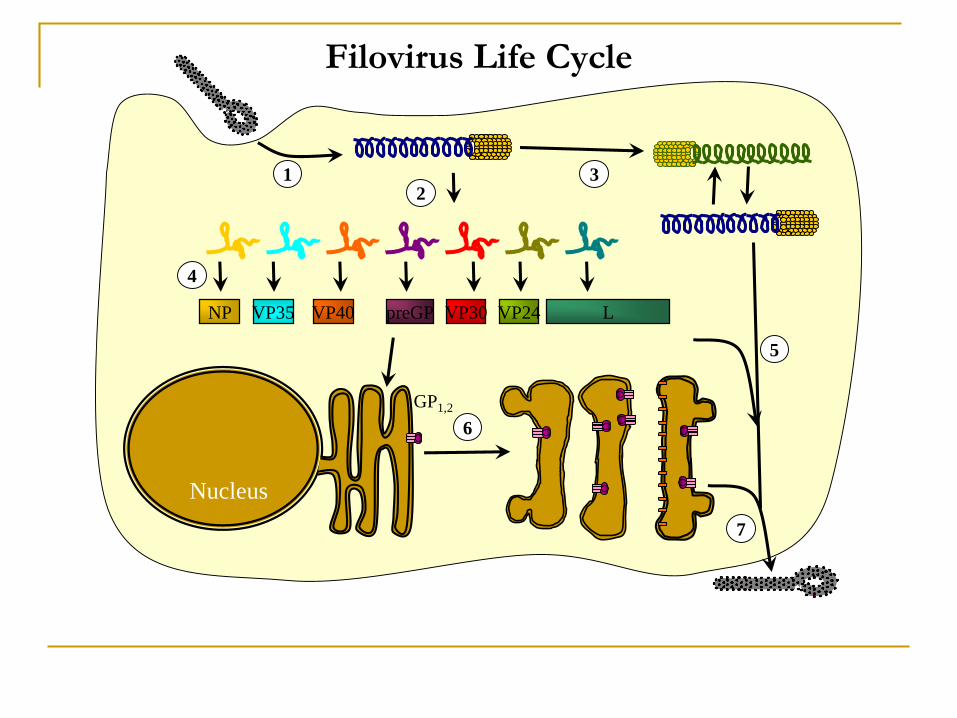
Filovirus Life Cycle
Nucleus
NP VP35 VP40 LVP24VP30
GP1,2
12
3
4
5
6
7
preGP
Animal Models of Filovirus Disease
Primate species subcutaneously infected with
1-10 LD50 EBOV-May
Mean time
to death
Hematological observations
Grivets (Chlorocebus aethiops) 7-8 days Fibrin depositions
Crab-eating macaques (Macaca fascicularis) 10-14 days Microcirculatory disturbances (capillary
stasis, erythrocyte aggregation), organs
engorged with blood, no hemorrhage, no
fibrin depositions
Hamadryas baboons (Papio hamadryas) 9-10 days Erythrocyte diapedesis
Rhesus monkeys (Macaca mulatta) 7-8 days Fibrin depositions, prominent hemorrhages
Animal Models of Filovirus Disease
Feature of Ebola virus
disease
Mice (adaptation
required)
Guinea pigs
(adaptation
required)
Nonhuman primates Humans
Disease duration to death
Virulence
Fever
Peak viremia
Hemorrhages
Maculopapular rash
DIC
Liver enzymes
Lymphopenia
Lymphocyte apoptosis
Thrombocytopenia
Cytokine response
4-7 days
High
No
≥7.5x10⁷-5.6x10¹¹ pfu/ml
Not profound
No
Not profound
Elevated
Controversial
?
Yes
Yes
6-12 days
High
Yes
≥105.2 pfu/ml
Rare
No
Conflicting data
Elevated
Yes
?
Yes
Yes
5-10 days
High
Yes
≥106-108 pfu/ml
Depending on species
Depending on species
Yes
Elevated
Yes
Yes
Yes
Yes
3-30 days
High
Yes
≥106.5 pfu/ml
Occasionally
Roughly 50% of the cases
Yes
Elevated
Yes
Yes
Yes
Yes
Filovirus Diagnosis
Diagnostic test
Detects Sample Material Advantage Disadvantage
Antigen capture ELISA Filoviral antigen Blood, serum, tissue Rapid, specific,
sensitive
Requires special equipment (ELISA
reader; γ-irradiation of samples or
handling of samples in BSL-4)
Electron microscopy (EM) Complete or partial
filovirus particles or
characteristic inclusion
bodies
Blood, serum, tissue Specific Insensitive, requires special equipment
(electron microscope)
IgG/IgM capture ELISA using native
or recombinant filoviral antigen
Antibodies to filoviral
antigen
Serum Rapid, specific,
sensitive
Requires special equipment (ELISA
reader and means to produce large
amounts of purified viral or
recombinant antigen)
Immunohistochemistry (IHC) Filoviral antigen Tissue (skin, liver) Fixed tissue can
be used
Requires time
In situ hybridization (ISH) Filoviral nucleic acids Tissue Fixed tissue can
be used
Requires special equipment
Indirect immunofluorescent assay
(IFA) using native or recombinant
filoviral antigen
Antibodies to filoviral
antigen
Serum Simple, safe Subjective interpretation, cross
reactions, insensitive
Reverse transcriptase-polymerase
chain reaction (RT-PCR)
Filoviral subgenomic
or genomic nucleic
acids
Blood, serum, tissue Rapid, sensitive Requires special equipment, possible
cross-contamination (false positives),
release of RT-PCR inhibitors from
tissue
Virus isolation in tissue culture or
animals
Filoviruses Blood, tissue Specific Requires maximum containment
laboratory and time
Western blot Antibodies to filoviral
antigen
Serum Viral protein-
specific
Difficult interpretation
Prevention/Treatment/Prophylaxis
Candidate vaccines (VSIV, adenovirus, fVLPs)
Candidate treatments (tissue factor inhibitors, antibody
cocktails, PMOs, siRNAs, small molecules)