ebv attachment stimulates fhos/fhod1 redistribution and co...
TRANSCRIPT

IntroductionHuman CD21 is a multifunctional cell surface glycoproteinthat is highly expressed on B-lymphocytes and folliculardendritic cells (FDCs), although it can be detected on manyadditional cell types. CD21 is the receptor for the C3dgfragment of complement (Iida et al., 1983), for CD23 (Aubryet al., 1994) and human CD21 and is also the major cellularattachment protein for Epstein-Barr virus (EBV) (Fingeroth etal., 1984). CD21 is composed of an extracellular domainconsisting of 15-16 short consensus repeat modules, ahydrophobic transmembrane, and a 34 amino acid cytoplasmicdomain (Fearon and Carroll, 2000; Moore et al., 1987). Thethree known ligands all bind within the two N-terminal repeats(Aubry et al., 1994; Fearon and Carroll, 2000), which form ahighly flexible domain as demonstrated by crystal structureanalysis (Prota et al., 2002).
At present, the role of human CD21 is best understood inthe context of the immune response. Many relevant studieshave been conducted in mice (Fearon and Carroll, 2000;Holers, 2000), where a related though clearly diverged protein(mCD21/CD35) serves the dual function of both a C3b
receptor (complement receptor type 1(CR1)/CD35) and a C3dreceptor (CR2/CD21), depending on the pattern of N-terminalsplicing. In man, two independent proteins, CD21 and CD35are synthesized; however, the intracellular domain of humanCD21 is most similar to that of mCD21/CD35 (Fingeroth,1990).
CD21 participates in regulation of antibody (Ab) productionthrough immune complex (C3d-Ab-Ag)-mediated modulationof B-cell receptor (BCR) signaling and internalization, Fcreceptor signaling and through retention of pathogenic antigens(including HIV) on FDCs for stimulation of B-cell memory(Cherukuri et al., 2001; Fearon and Carroll, 2000; Moir et al.,2000; Poe et al., 2001; Prodinger, 1999). Interaction of CD21with CD23 on B-cells is believed to additionally protect B-cellsfrom apoptosis (Bonnefoy et al., 1993). However, the functionof CD21 on most other cell types remains unknown.
The attachment of C3d-coated antigen to B-cells and FDCsphysically links CD21 to the BCR. Precisely how theseinteractions culminate in quantitative and qualitativealterations in antibody production is not fully understood. Onthe resting B-lymphocyte, noncovalent associations between
2709
CD21 is a multifunctional receptor for Epstein-Barr virus(EBV), for C3dg and for CD23. Upon engagement ofimmune complexes CD21 modulates immunoreceptorsignaling, linking innate and adaptive immune responses.The mechanisms enabling CD21 to independently relayinformation between the exterior and interior of the cell,however, remain unresolved. We show that forminhomologue overexpressed in spleen (FHOS/FHOD1) bindsthe cytoplasmic domain of human CD21 through its Cterminus. When expressed in cells, EGFP-FHOS localizesto the cytoplasm and accumulates with actin in membraneprotrusions. Plasma membrane aggregation, redistributionand co-localization of both proteins are stimulated whenEBV (ligand) binds CD21. Though widely expressed,FHOS RNA is most abundant in the littoral cell, a majorconstituent of the red pulp of human spleen believed to
function in antigen filtration. Formins are molecularscaffolds that nucleate actin by a pathway distinct fromArp2/3 complex, linking signal transduction to actinreorganization and gene transcription. Thus, ligandstimulation of FHOS-CD21 interaction may transmitsignals through promotion of cytoskeletal rearrangement.Moreover, formin recruitment to sites of actin assemblyinitiated by immunoreceptors could be a generalmechanism whereby co-receptors such as CD21 modulateintracellular signaling.
Supplemental data available online
Key words: Formin, CD21, Splenic littoral cell, Epstein-Barr virus,Actin cytoskeleton
Summary
EBV attachment stimulates FHOS/FHOD1redistribution and co-aggregation with CD21: formininteractions with the cytoplasmic domain of humanCD21Michael B. Gill 1,2,5,*, Jennifer Roecklein-Canfield 1,*, David R. Sage 1,2, Maria Zambela-Soediono 1,Nina Longtine 4,5, Marc Uknis 3 and Joyce D. Fingeroth 1,2,5,‡
1Divisions of Infectious Diseases, 2Experimental Medicine and 3Surgery, Beth Israel Deaconess Medical Center and 4Department of Pathology,Brigham and Women’s Hospital and 5Harvard Medical School, Boston, MA 02115, USA*These authors contributed equally to this work‡Author for correspondence (e-mail: [email protected])
Accepted 20 January 2004Journal of Cell Science 117, 2709-2720 Published by The Company of Biologists 2004doi:10.1242/jcs.01113
Research Article

2710
the extracellular domain of CD21 and other B-cell surfaceproteins, in particular CD19, that independently mediate B-cell signal transduction have been shown to play a keyrole (Fearon and Carroll, 2000; Poe et al., 2001). BothmCD21/CD35 and CD19 can co-localize with a retained BCRin cholesterol-rich microdomains (lipid rafts). This eventextends the duration and efficiency of intracellular signaling(Cherukuri et al., 2001), augments B-cell proliferation andmay logarithmically increase antibody production (Fearonand Carroll, 2000). There is still considerable debate aboutwhether human CD21 regulates ligand (C3d, EBV, CD23)internalization and/or contributes to transmission of anystimuli across the plasma membrane directly andindependently of CD19 (Bradbury et al., 1992; Carel et al.,1990; Martin et al., 1994; Tanner et al., 1987).
No direct role for the short conserved cytoplasmic domainof human CD21 in relation to CD21 biology has beenestablished. Recently, human CD46, a related complementreceptor, pathogen receptor and a TCR co-receptor, was shownto signal in vivo through its short cytoplasmic domains (cyt1and cyt2) (reviewed by Lee et al., 2002). Phosphorylation ofcyt2 was required to effectively anchor gonococcal pili tothe surface of epithelial cells. Furthermore, direct CD46stimulation of primary T-cells activated Vav whilecostimulation with CD3 enhanced Rac activation and inducedactin reorganization and morphological change (Zaffran et al.,2001). Crosslinking of human CD21 on normal B-lymphocytesby virus and by monoclonal antibodies (mAbs) has also beenobserved to result in rapid F actin assembly (Melamed et al.,1994) however, the relative roles of CD19 and associated B-cell proteins was not defined.
To investigate the function of the intracellular domain ofhuman CD21, we utilized a yeast two-hybrid assay to identifyproteins that directly interact with this region. We show that amember of the formin family, FHOS (formin homologueoverexpressed in spleen) (Westendorf et al., 1999) also knownas FHOD1 (Koka et al., 2003) interacts with CD21 inmammalian cells through its C terminus.
Formins are multidomain proteins conserved duringeukaryotic evolution that directly nucleate actin by amechanism independent of Arp2/3 complex (Evangelista et al.,2002; Kovar et al., 2003; Li and Higgs, 2003; Sagot et al.,2002b). They serve as molecular scaffolds, recruiting proteinsthat link cellular signal transduction pathways (Rho familyGTPases and src family tyrosine kinases) to actin-bindingproteins (profilins) and promote cytoskeletal reorganization(Tanaka, 2000; Zeller et al., 1999) and downstream genetranscription (Copeland and Treisman, 2002; Zeller et al.,1999). Formins are critical for cytokinesis, for theestablishment of cell polarity and for control ofmorphogenesis. FHOS, a human formin first identified in aspleen library has been shown to interact with the smallGTPase Rac1, to bind the actin binding protein profilin IIa andto activate the serum response transcription factor in vitro (Tojoet al., 2003; Westendorf, 2001). Recent studies indicate thatFHOS can interact with actin monomers and may play a rolein the regulation of cell motility and vesicle recycling as well(Koka et al., 2003; Tojo et al., 2003).
By means of fluorescence microscopy we document thatengagement of CD21 by extracellular ligand producescoordinate re-localization and clustering of FHOS and CD21
in epithelial cells lacking CD19. This receptor aggregation isgreatly diminished in the absence of the C-terminal aminoacids of FHOS. We propose a mechanism to explain theinteraction of these proteins in vivo, and suggest that anchorageand aggregation of cell surface proteins are an effector functionof certain formins. Unexpectedly, FHOS was abundant in thesplenic littoral cell, a major constituent of the red pulp ofhuman spleen that lines the splenic sinuses. Littoral cells arebelieved to play a key role in antigen filtration, though atpresent little is known about their biology.
Materials and MethodsConstruct design
Expression vectors: yeastcDNA encoding the cytoplasmic 34 amino acids (aa) of CD21(GenBank accession no. P20023) was amplified using Taqpolymerase, Advantage Taq-2 (Clontech) with the forward and reverseprimers [5′ GCGGAATTCTCAAAACACAGAGAACGCAAT 3 ′(EcoRI) and 5′ GCGCTGCAGGCTGGCTGGGTTGTATGG 3′(PstI)]. cDNA encoding the cytoplasmic 43 aa of CD21 (last 34 + 9aa of proximal transmembrane domain) was amplified by PCRusing the same reverse primer and the forward primer 5′GCGGAATTCTTGATTGTCATTACCTTA 3′ (EcoRI). Both PCRproducts were cloned into the pGBT.C vector (Clontech), placing theinsert CD21 sequence in frame with a Gal4 binding domain. Theresulting target plasmids were verified by sequencing and namedpGBT.C-CD21-CT and pGBT.C-CD21-TMCT respectively.
BacterialTo generate a glutathione S-transferase (GST)-FHOS CT fusionprotein, a 600 bp C-terminal fragment, including nucleotides thatencoded the last 199 aa and the stop codon of HeLa FHOS wasamplified by PCR using the gene-specific primers: 5′-GCG-CTCGAGCAGGCGGCCCGT-3′ (XhoI) and 5′-GCGCTCGAGT-CACACCTCCAGGC-3′ (XhoI). The PCR product was cloned in-frame into the expression plasmid, pGEX4T-1 (Pharmacia), andsequenced (both strands), generating pGEX4T-1-FHOS-CT.
MammalianTo synthesize pEGFP-FHOS (full length, 1-1164) and pEGFP-FHOS∆CT (C terminus deletion, 1-965), FHOS cDNA was amplifiedby PCR and cloned into pEGFPC2 (Clontech) using unique restrictionsites as follows: FHOS full length primers [MG182 5′GGGGTACCGCATGGCGGGCGGGGAAGACC 3′ (KpnI) andMG113 5′ CGGGATCCTCACACCTCCAGGCCAGGACC 3′(BamHI)] and FHOS∆CT primers [MG 182 and MG196 5′CGCGGATCCTCACGGGGTG TAGCCCAGG 3′ (BamHI)]. Togenerate pBABE-CD21, CD21 cDNA was cloned into the multiplecloning site of pBABE (Morgenstern and Land, 1990), an eukaryoticexpression vector.
CellsLines
Raji, Burkitt’s lymphoma; Nalm6, B-cell leukemia (a gift from ArnoldFreedman, Dana-Farber Cancer Institute); 293T, embryonic kidneyline transformed with adenovirus fragments and bearing SV40-Tantigen [a gift from Hava Avraham, Beth Israel Deaconess MedicalCenter (BIDMC)]; JY, B lymphoblastoid line (a gift from JackStrominger, Harvard University); HeLa, cervical carcinoma and themurine fibroblast line, 3T3 were utilized in these studies. Cells wereobtained from the American Type Tissue Collection except asindicated.
Journal of Cell Science 117 (13)

2711Formin interaction with CD21
Primary cellsB- and T-lymphocytes and monocyte/macrophages wereindependently isolated from freshly obtained normal human spleenusing a Rosette-Sep Cell purification kit (Stem Cell Technologies).Discarded splenic tissue was obtained in accordance with the policiesof the Institutional Review Board at BIDMC. The resultant cellpreparations were examined by flow cytometry to confirm the purityof the individual cell populations using phycoerythrin (PE)-labeledmAbs to human IgG, CD19, CD3, CD14 and CD16 (Sigma-Aldrich).Purified dendritic cells were a generous gift from Dr David Avigan,BIDMC, and FDCs were a generous gift from Dr Arnold Freedman.
Yeast two-hybrid screenA HeLa cDNA library, pGAD10-HeLa cDNA Matchmaker(Clontech), was screened with the pGBT.C-CD21.CT target plasmid.Briefly, plasmid DNA prepared from a single library amplification andpGBT.C-CD21.CT were co-transformed into the yeast strain, Y190(Clontech) by the lithium acetate method. Approximately 106
independent clones were screened. Transformants were selected onthe basis of their ability to activate both the GAL1::HIS3 andGAL1::lacZ reporter genes using standard procedures for yeastmanipulations (Chien et al., 1991).
Yeast plasmid DNA was isolated and sequenced. To confirm thespecificity of the candidate interactions, isolated plasmids wereindependently re-transformed into Y190 with either pGBT.C-CD21.CT or three unrelated target plasmids, encoding lamin (Bartelet al., 1996), bacteriophage T7 gene 2 (Bartel et al., 1996), or theintracellular domain of the Coxsackie and adenovirus receptor, CAR(Bergelson et al., 1997). Candidates that activated both reporter genes(as above) when co-transformed with pGBT.C-CD21.CT wereconsidered true positives.
Cloning of full length HeLA FHOS cDNAThe full-length FHOS HeLa cDNA sequence was amplified from aMarathon-ReadyTM human HeLa cDNA library (Clontech) using theFHOS-specific forward and reverse primers (5′ TGAGCCGGCC-GCAGAGCC 3′ and 5′ CGGGATCCTCACACCTCCAGGCCAG-GACC 3′) respectively. The Hela cDNA clone obtained wassequenced on both strands and deposited in GenBank under accessionno. AY192154.
Demonstration of FHOS-CD21 interactionIn vitro
Initially GST-FHOS.CT, GST-EBV.TK (thymidine kinase) and GSTalone were expressed and purified from BL21 carrying pGEX4T1-FHOS.CT, pGEX6P2-EBV.TK (Gustafson et al., 1998) and pGEX4T-1 vectors, respectively, using a glutathione-Sepharose 4B column(Amersham), and assessed for purity. Cell lysates were prepared fromboth Raji (CD21+) and Nalm6 (CD21-) cell lines. 107 cells were lysedin 0.5 ml of 1% NP-40 buffer supplemented with 1 mM of each ofthe following protease inhibitors: leupeptin, pepstatin, PMSF andaprotinin. The lysates were centrifuged at 800 g at 4°C for 10 minutesto remove cell debris and then incubated with 3 µg of purified (a)GST-FHOS.CT, (b) GST, or (c) GST-EBV.TK, overnight (o/n) at 4°C.For co-purification 0.1 ml of pre-washed glutathione-Sepharose 4Bbeads was added to each sample (a-c) and further incubated o/n at4°C. Beads were then washed three times in 0.1% NP-40/1×TPBS(0.1% NP-40 in PBS and 0.05% Tween 20) and then once in PBScontaining 0.5 M LiCl to remove non-specific proteins binding to theglutathione beads. The beads were boiled in sample buffer, separatedby 7% SDS-PAGE and transferred to nitrocellulose (Osmonics) byelectroblot. The nitrocellulose filter was pre-blocked in 1×TPBS 5%powdered milk (Carnation) o/n, incubated with rabbit anti-CD21serum (Prota et al., 2002) (diluted 1:250 in 1×TPBS/5% milk) for 2
hours at room temperature (RT), washed in 1×TPBS, incubated withgoat anti-rabbit-HRP secondary Ab (1:3000 dilution in 1×TPBS/5% milk) for 20 minutes at RT, re-washed and developed withchemiluminescent substrate, ECL (Amersham).
In vivoAn EGFP-modified version of the mammalian MATCHMAKER two-hybrid assay (Clontech, Catalog no. K1602-1) was used to confirm invivo FHOS-CD21 interaction. To increase the sensitivity of thereporter activity the chloramphenicol acetyl transferase (CAT)reporter gene in pG5CAT was replaced with EGFP (Clontech).Briefly, the CAT open reading frame was removed from pG5CAT byPCR with Pfu polymerase (Stratagene) using the forward and reverseprimers, 5′ GAAGATCTTTTAGCTTCCTTAGCTCC 3′ (BglII ) and5′ ATAAGAAT GCGGCCGCTTGCCCTTAAACGCCTGGTGC 3′(NotI), respectively. The PCR product (pG5∆CAT) was ligated withthe EGFP gene isolated from pEGFPN2 by BglII and NotI digestion.The resulting plasmid was named pG5∆CAT-EGFP.
To generate bait and target vectors containing either FHOS.CT orCD21.CT both genes were amplified, introducing unique (EcoRI)restriction sites for subcloning. The primers used were as follows: 5′CGGAATTCCAGGCGGCCCGTGAAGTGCGCATCATGC 3′ and 5′CGGAATTCTCACACCTCCAGGCCAGGACC 3′ (for FHOS.CT)and 5′ CGGAATTCAAACACAGAGAACGC 3′ and 5′ CG-GAATTCTCAGCTGGCTGGGTTGTATGG 3′ (for CD21.CT). Togenerate bait vectors, FHOS.CT (966-1164) and CD21.CT (1000-1033) were fused with the GAL4 DNA binding domain generatingthe clones pM-FHOS.CT and pM-CD21.CT, respectively. Togenerate target vectors FHOS.CT (966-1164) and CD21.CT (1000-1033) were fused to the VP16 activation domain, generating theclones pVP16-FHOS.CT and pVP16-CD21.CT, respectively. 293Tcells were co-transfected using Lipofectamine 2000™ (Invitrogen)with 0.5 µg of pG5∆CAT-EGFP plus each of the followingcombination of plasmids: (1) pM3-VP16 (+ control) (2) pM-53 +pVP16-T (+ control) (3) pM-53 + pVP16-CP (– control) (3) pM-FHOS.CT + pVP16-CD21.CT and (4) pM-CD21.CT + pVP16-FHOS.CT. Additionally, the bait and target vectors were co-transfected with an irrelevant corresponding positive control vector(pM-53 or pVP16-T). Twenty-four hours later cells were fixed andanalyzed by fluorescence microscopy.
Fluorescence microscopyFor indirect immunofluorescence cells were plated into 12-well traysat 2×10⁵ cells/well. Co-localization studies were performed with cells(293T, HeLa) that were transfected with 1 µg of pEGFP-FHOS,pEGFP-FHOS∆CT, or pBABE-CD21 alone or in combination asstated. At selected time points after transfection cells were fixed andstained as outlined below and imaged using the Eclipse E600fluorescence microscope (Nikon) at either 40× or 100× magnificationemploying Spot Advance for image capture and processing(Diagnostic Instruments). Twenty-four hours after transfection cellnuclei were stained with Hoechst 33342 (Molecular Probes) and fixedwith 4% paraformaldehyde in 0.1 M phosphate buffer (PB) (0.2 MNaH2PO4.H2O, 0.2 M NaHPO4, pH 7.2) for 30 minutes at RT. Fixedcells were permeabilized with 0.1% Triton X-100/PB/3% BSA for 15minutes at RT and extensively washed with PB. For actin staining,fixed cells were incubated with TRITC-labeled phalloidin (Sigma-Aldrich) in 1% BSA/PB (1:20,000 dilution) for 1 hours at RT. For α-tubulin staining fixed cells were incubated with anti-α-tubulin mAb(clone B-5-5-1-2, Sigma-Aldrich) in 1% BSA/PB at a 1:200 dilutionfor 1 hour at RT, washed and then visualized by staining with goatF(ab′)2 anti-mouse IgG R-PE (Biosource) in 1% BSA/BP at 1:200dilution for 1 hour at RT. All cells were mounted usingAquaPoly/mount (Polysciences). For CD21 staining, fixed cells(above) were incubated with anti-CD21 mAb HB-5 (Tedder et al.,

2712
1984) in 1% BSA/PB (10 µg/ml) for 1 hour at RT and visualized asdescribed for tubulin.
For ligand challenge experiments, prior to fixation, cells wereincubated with either concentrated EBV, prepared and purified asdescribed previously (Prota et al., 2002), concentrated Kaposi’ssarcoma-associated herpesvirus (Dezube et al., 2002) or withpolyclonal rabbit anti-CD21 SCR1-SCR2 antisera (Prota et al., 2002)diluted 1:100 in PB. This antiserum does not block binding of mAbHB-5, which recognizes SCR4 of CD21 (Carel et al., 1990). Pre-immune serum was used as a control. Cells were washed three timesin PB, fixed and developed for CD21 localization as above; UPC10(IgG2a) was the isotype matched control for HB-5.
RT-PCRTotal RNA was prepared from cells using a RNeasy Mini kit (Qiagen).RT-PCR was performed using a GeneAmp kit (Roche) with 1 µg oftotal RNA prepared from relevant cells, using FHOS (MG112 andMG113) and GAPDH-specific primers (MG121 5′ GCCATGAG-GTCCACCACCCTGT 3′ and MG122 5′ CTACTGGCGCTGCCA-AGGCTGT 3′).
In situ hybridizationNon-radioactive in situ hybridization was performed as describedpreviously (Berger and Hediger, 2001) using a digoxigenin-labeledcRNA probe containing 3496 nucleotides (ATG – STOP codon) ofFHOS cDNA, which had been first cloned into the pGEMT vector(Promega) to facilitate preparation of an antisense probe. Controlsections were incubated in an identical concentration of the senseFHOS probe.
ResultsThe cytoplasmic domain of CD21 interacts with theC-terminus of FHOSTo identify candidate proteins that interact with thecytoplasmic domain of human CD21 a modified version ofField’s yeast two-hybrid assay (Fields and Song, 1989) wasutilized (see Materials and Methods). A vector, CD21.CT(target) expressing the cytoplasmic 34 aa of human CD21 wassynthesized and then co-transformed into yeast with aHeLa cDNA vector library (bait). A positively identifiedtransformant was sequenced and shown to contain an 858 bpinsert predicting an open reading frame of 199 aa (966-1164)followed by a stop codon and 3′ untranslated sequence. Thecomplete DNA sequence encoding a 1164 aa protein wassubsequently cloned and found to be identical to FHOS(Westendorf et al., 1999). The HeLa cDNA fragment identifiedencodes the carboxyl terminus 199 aa of FHOS. This is avariable region located C-terminal to the highly conservedformin homology domains (FH1/FH2) that may confer uniqueinteraction(s) to different formin family members (Evangelistaet al., 2003; Wallar and Alberts, 2003).
Following identification of the HeLa FHOS fragment, thefull length HeLa cDNA clone was amplified by PCR (GenBankaccession no. AY192154) using a HeLa cDNA library as atemplate. Comparison of splenic and HeLa derived-sequencesrevealed 13 nucleotide alterations that resulted in 9predominantly conservative aa changes (E264D, E359D,S387T, D633E, V634L, T700S, R689Q, E745G, G751E,D849E) all located upstream of the C-terminal CD21interactive domain. As expected, HeLa-derived FHOScontained the predicted domains that are conserved among
diverse formin family members (Wallar and Alberts, 2003;Westendorf, 2001; Westendorf et al., 1999;) including the FHdomains, two coil-coiled domains, a collagen-like domain anda putative diaphanous autoregulatory domain (DAD)-likedomain (Wallar and Alberts, 2003). The latter is proposed tolink the N terminus with the C terminus in an intramolecularinhibitory interaction.
To verify the specificity of the CD21-FHOS interaction adirect yeast two-hybrid assay was performed. The bait vectorcontaining the C terminus of FHOS was co-transformed withCD21.CT (the original target vector) or with CD21-TMCT, arelated vector which contains nine amino acids from theadjacent transmembrane of CD21, together with three controltarget vectors expressing unrelated proteins. As shown in Fig.1a, activation of the HIS3 (and LacZ, data not shown) reportergene only occurred in co-transformants that expressedCD21.CT (or CD21-TMCT) together with the C terminus ofFHOS (Fig. 1a). These findings confirm that FHOS-CD21interaction is specific and highly reproducible in yeast.
CD21 expressed in human cells interacts with FHOSTo determine whether FHOS could interact with endogenousCD21 that is expressed on the plasma membrane of human B-lymphocytes a bacterial vector linking GST with the C-terminal 199 aa residues of FHOS was generated to enableexpression and purification of a GST-FHOS fusion protein.GST-FHOS was then immobilized on glutathione-Sepharoseand used to precipitate CD21 from freshly prepared cellmembrane lysates originating from B-cell lines. After elution,GST-FHOS and its bound ligand(s) were analyzed byimmunoblot using both an anti-GST Ab to confirm the elutionof the GST-FHOS fusion protein and using an anti-CD21 Abto confirm the presence of CD21 as a FHOS-interacting proteinon the column. When cell lysates prepared from CD21+ Rajicells were used (Fig. 1b, lane 4), CD21 could be readilydetected in the fraction eluted from GST-FHOS. No proteinwas detected when lysates from the CD21-Nalm6 line wasidentically analyzed (Fig. 1b, lane 6). When GST-FHOS wasreplaced with GST alone or with GST-TK (irrelevant control)as the immobilized ligand, neither of the control ligands boundCD21 (Fig. 1b, lanes 2 and 3). As shown in Fig. 1b, lane 1, thesize of CD21 detected in Raji cell lysates was the same as thatof CD21 co-precipitated by FHOS. These results demonstratethat FHOS interacts with endogenously expressed CD21 fromcell membrane lysates of human B-cells confirming that theFHOS-CD21 interaction first identified in yeast occurs in vitro.
FHOS interacts with CD21 in vivoTo demonstrate the in vivo interaction between FHOS andCD21 a modified version of the mammalian two-hybrid system(Clontech) was employed in which the reporter vector enzymeCAT was replaced with the fluorescent protein EGFP and cellswere analyzed by fluorescence microscopy to visualize EGFP(see Materials and Methods). First the fluorescent two-hybridassay was validated using the bait (pM) and target (pVP16)control vectors provided by the manufacturer (positive control:pM3-VP16 and pM-53+pVP16-T and negative control: pM-53+pVP16-CP) as shown in Fig. 1c, top. Both bait and targetvectors were then generated that contained either FHOS.CT (C
Journal of Cell Science 117 (13)

2713Formin interaction with CD21
terminus, 966-1164) or CD21.CT (cytoplasmic domain, 1000-1033), i.e. pM-FHOS.CT, pM-CD21.CT, pVP16-FHOS.CTand pVP16-CD21.CT to enable confirmation of proteininteraction in reciprocal assays. First, the recombinant vectors
were individually transfected into 293T cells together with themodified reporter vector, pG5∆CAT-EGFP, to ensure that noauto-activation of the reporter EGFP gene occurred (notshown). Next both combinations of the bait and the target
Fig. 1.The intracellular domain of CD21 interacts withFHOS. (a) in yeast. A GAL4 activation domain plasmidencoding the C-terminal amino acids of FHOS wasserially diluted (10–1-10–4; UD, undiluted) and co-transformed into a yeast reporter strain with plasmidsencoding the GAL4 DNA binding domain fused to oneof the following: the 34 aa residues of the CD21cytoplasmic tail (CD21-CT), the 34 aa residues of theCD21 cytoplasmic domain plus nine residues of the transmembrane domain (CD21-TMCT), lamin, the 105 aa residues of the CAR cytoplasmicdomain (CAR-CT), or bacteriophage T7 gene 2 (T7/gp2; see Materials and Methods). Co-transformants were selected on minimal medium.Growth (i.e. activation of the HIS3 reporter) indicates a positive interaction. +C (positive control) represents co-transformation of plasmidsencoding SNF1 and SNF4, proteins that interact in the two-hybrid assay. (b) In vitro. Purified fusion proteins expressing the C terminus ofFHOS, GST-FHOS, GST alone or GST-TK (irrelevant protein) were incubated with cell lysates prepared from the B-cell lines Raji (CD21+;lanes 2-4) and Nalm6 (CD21–; lanes 5 and 6). Proteins interacting with FHOS were co-purified on glutathione beads, separated by SDS-PAGEand detected by immunoblot using a monospecific rabbit anti-CD21 antiserum. A total protein lysate from Raji cells (lane 1) was analyzed as aCD21 positive control. (c) In vivo. 293T cells were transfected with pG5∆CAT-EGFP (reporter) plus the desired combination of bait (pM-X)and target (pVP16-X) vectors as indicated. X denotes the fusion protein of interest. Twenty-four hours after transfection, cells were identifiedwith Hoechst (nuclear stain), fixed, and analyzed by fluorescence microscopy. Fluorescent EGFP+ cells indicates a positive interaction betweenbait and target fusion proteins. Top panel (method verification): EGFP induction was assessed using positive control vectors (pM53-VP16 andpM-53 + pVP16-T) compared with negative control vectors (pM-53 + pVP16-CP, control protein; Clontech). Middle panel: EGFP inductionfollowing co-transfection of recombinant vectors encoding the cytoplasmic domain of CD21, CD21.CT together with the C terminus of FHOS,FHOS.CT. Right and left panels show reciprocal experiments. Bottom panel (control), EGFP induction following transfection of CD21.CT orof FHOS.CT together with cognate two-hybrid vectors encoding irrelevant proteins (T antigen or p53).

2714
vector (i.e. pM-FHOS.CT/pVP16-CD21.CT and pM-CD21.CT/pVP16-FHOS.CT) were co-transfected into 293Tcells along with pG5∆CAT-EGFP. As seen in Fig. 1c middle,each combination of the bait and target proteins was able tointeract in 293T cells inducing expression of the fluorescentreporter. For additional controls both the bait and target vectorswere transfected with a corresponding control plasmidprovided by the manufacturer (e.g. pM-CD21.CT + pVP16-T)(Fig. 1c, bottom) to ensure absence of nonspecific EGFPinduction. These results demonstrate that in mammalian cells,the C terminus of FHOS is able to specifically interact with thecytoplasmic domain of human CD21.
FHOS distribution and co-localization with actinNext, a series of experiments were undertaken to analyze thecell biology of the FHOS-CD21 interaction. First a eukaryoticexpression vector encoding full length FHOS fused at the N-terminus to the enhanced green fluorescent protein (EGFP),generating pEGFP-FHOS, was utilized to assess the cellulardistribution of FHOS in live cells. pEGFP-FHOS wastransfected into 293T cells and 24 hours later the cells wereimaged by fluorescence microscopy. As shown in Fig. 2a,panels 1 and 2, FHOS localized to the cytoplasm of the cell,where it was observed surrounding the nuclear membrane,though it was excluded from the nucleus itself as demonstratedby Hoechst staining (Fig. 2a, panels 5 and 8). FHOSaccumulated at the cell periphery (Fig. 2a) and at the bordersbetween two cells (Fig. 2b). However, FHOS was mostapparent close to the leading edge where it was prominentlyobserved in filopodia, microspikes (Fig. 2a, panel 2) and insmall lamellipodia (Fig. 2a, panels 3 and 6). Fig. 2a, panel 6further demonstrates FHOS accumulation along one side of the
nucleus as well as at the tips of elongating protrusions. Thesefindings are consistent with the integral role of formins in theregulation of actin organization (Pelham and Chang, 2002;Tolliday et al., 2002). Coordinate staining to detect both actin(TRITC-phalloidin) (Fig. 2a, panel 4) and microtubules (α-tubulin, anti-mouse PE) (Fig. 2a, panel 7) further showed thatFHOS specifically co-localizes with actin at the cell peripheryand at the borders between cells (Fig. 2a panel 5 and Fig. 2b,panels 1-7), but does not co-localize with tubulin (Fig. 2a,panel 8). Remarkably, as shown in Fig. 2a, panel 3,overexpressed FHOS formed actin cables (inset), as has beenreported for yeast formins (Feierbach and Chang, 2001; Sagotet al., 2002a). In the perinuclear region, the association ofFHOS with actin was less prominent than at other locations(Fig. 2b, panels 1-3), as has been previously noted (Koka et al.,2003).
Aggregation and co-localization of CD21 and FHOS isdynamically enhanced in the presence of a cross-linkingligandBased on the known functions of both CD21 and FHOS wehypothesized that the interaction of both proteins would beinfluenced by environmental and/or intracellular cues thatoccur only in vivo. To investigate the mechanics of thisinteraction in cells that lack CD19, 293T cells were initiallyco-transfected with both pEGFP-FHOS and pBABE-CD21 (aeukaryotic expression vector that encodes full length humanCD21) in order to highly express the respective proteins (Fig.3a). Twenty-four hours later the cells were analyzed with eithera mAb directed to human CD21 (anti-CD21, HB-5) (Fig. 3a,panel 5) or with an isotype-matched control mAb, UPC10 (Fig.3a, panel 2) or with second Ab alone, the latter to document
Journal of Cell Science 117 (13)
Fig. 2. FHOS associates with the actincytoskeletion. (a) FHOS co-localizes withactin in the cytoplasm and in protrusions.Transfected 293T cells expressing fulllength FHOS fused to EGFP, FHOS(green, panels 1-3, 6) were co-stained witheither TRITC-phalloidin (panel 4; red) todetect actin or with anti α-tubulin/goatF(ab’)2 anti-mouse Ig R-PE (panel 7; red).Merged images (panel 3 with 4 and panel 6with 7) are shown in panels 5 and 8,respectively. Hoechst 33342 (blue) wasused to detect nuclei in panels 5 and 8. (b)FHOS accumulates with actin at the borderbetween cells and without actin in aperinuclear patch. Transfected 293T cellsexpressing EGFP-FHOS (green; panels1,4-5) were co-stained with TRITC-phalloidin (panels 2 and 6; red) andmerged images (panel 1 with 2 and panel 5with 6) are shown in panels 3 and 7,respectively. Nuclei are stained blue as ina. All cells were fixed, stained and imagedusing an Eclipse E600 fluorescencemicroscope and processed using SpotAdvance technology.
2715Formin interaction with CD21
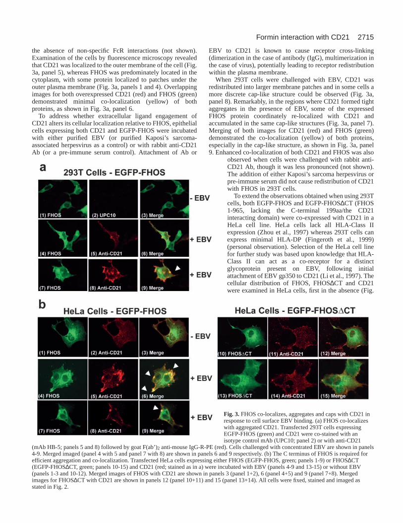
the absence of non-specific FcR interactions (not shown).Examination of the cells by fluorescence microscopy revealedthat CD21 was localized to the outer membrane of the cell (Fig.3a, panel 5), whereas FHOS was predominately located in thecytoplasm, with some protein localized to patches under theouter plasma membrane (Fig. 3a, panels 1 and 4). Overlappingimages for both overexpressed CD21 (red) and FHOS (green)demonstrated minimal co-localization (yellow) of bothproteins, as shown in Fig. 3a, panel 6.
To address whether extracellular ligand engagement ofCD21 alters its cellular localization relative to FHOS, epithelialcells expressing both CD21 and EGFP-FHOS were incubatedwith either purified EBV (or purified Kaposi’s sarcoma-associated herpesvirus as a control) or with rabbit anti-CD21Ab (or a pre-immune serum control). Attachment of Ab or
EBV to CD21 is known to cause receptor cross-linking(dimerization in the case of antibody (IgG), multimerization inthe case of virus), potentially leading to receptor redistributionwithin the plasma membrane.
When 293T cells were challenged with EBV, CD21 wasredistributed into larger membrane patches and in some cells amore discrete cap-like structure could be observed (Fig. 3a,panel 8). Remarkably, in the regions where CD21 formed tightaggregates in the presence of EBV, some of the expressedFHOS protein coordinately re-localized with CD21 andaccumulated in the same cap-like structures (Fig. 3a, panel 7).Merging of both images for CD21 (red) and FHOS (green)demonstrated the co-localization (yellow) of both proteins,especially in the cap-like structure, as shown in Fig. 3a, panel9. Enhanced co-localization of both CD21 and FHOS was also
observed when cells were challenged with rabbit anti-CD21 Ab, though it was less pronounced (not shown).The addition of either Kaposi’s sarcoma herpesvirus orpre-immune serum did not cause redistribution of CD21with FHOS in 293T cells.
To extend the observations obtained when using 293Tcells, both EGFP-FHOS and EGFP-FHOS∆CT (FHOS1-965, lacking the C-terminal 199aa/the CD21interacting domain) were co-expressed with CD21 in aHeLa cell line. HeLa cells lack all HLA-Class IIexpression (Zhou et al., 1997) whereas 293T cells canexpress minimal HLA-DP (Fingeroth et al., 1999)(personal observation). Selection of the HeLa cell linefor further study was based upon knowledge that HLA-Class II can act as a co-receptor for a distinctglycoprotein present on EBV, following initialattachment of EBV gp350 to CD21 (Li et al., 1997). Thecellular distribution of FHOS, FHOS∆CT and CD21were examined in HeLa cells, first in the absence (Fig.
Fig. 3.FHOS co-localizes, aggregates and caps with CD21 inresponse to cell surface EBV binding. (a) FHOS co-localizeswith aggregated CD21. Transfected 293T cells expressingEGFP-FHOS (green) and CD21 were co-stained with anisotype control mAb (UPC10; panel 2) or with anti-CD21
(mAb HB-5; panels 5 and 8) followed by goat F(ab’)2 anti-mouse IgG-R-PE (red). Cells challenged with concentrated EBV are shown in panels4-9. Merged imaged (panel 4 with 5 and panel 7 with 8) are shown in panels 6 and 9 respectively. (b) The C terminus of FHOS is required forefficient aggregation and co-localization. Transfected HeLa cells expressing either FHOS (EGFP-FHOS, green; panels 1-9) or FHOS∆CT(EGFP-FHOS∆CT, green; panels 10-15) and CD21 (red; stained as in a) were incubated with EBV (panels 4-9 and 13-15) or without EBV(panels 1-3 and 10-12). Merged images of FHOS with CD21 are shown in panels 3 (panel 1+2), 6 (panel 4+5) and 9 (panel 7+8). Mergedimages for FHOS∆CT with CD21 are shown in panels 12 (panel 10+11) and 15 (panel 13+14). All cells were fixed, stained and imaged asstated in Fig. 2.

2716
3b, panels 1-3 and 10-12) and then in the presence (Fig. 3b,panels 4-9 and 13-15) of EBV.
In the absence of EBV, there was limited co-localization ofFHOS and CD21 in HeLa cells (Fig. 3b, panels 1-3), identicalto that observed in 293T cells. In the presence of EBV, CD21re-localized and could be observed in concentrated aggregatesas shown in Fig. 3b, panel 5, and even seen in dense cap-likestructures (Fig. 3b panel 8). As expected, a portion of FHOScoordinately re-localized with CD21 into concentratedaggregates and cap-like structures as shown in Fig. 3b, panels4 and 7 (compared to Fig. 3b, panels 5 and 8). Merging of theCD21 (red) and FHOS (green) images demonstrated that bothFHOS and CD21 had specifically reorganized and aggregated,and accumulated at the same regions in the cell in response toEBV (Fig. 3b, panels 6 and also 9). Notably, in the absence ofits CD21 interacting domain, FHOS∆CT failed to significantlyco-localize with CD21 even in the presence of EBV (Fig. 3b,panels 13-15). FHOS∆CT did not appear to re-localize FHOSor cause significant stress fiber formation (Fig. 3b, panels 10and 12 compared with 1 and 3).
FHOS RNA is highly expressed in skeletal muscle,hematopoietic tissue, lung and heart, but is completelyabsent from brainExamination of an adult human tissue blot revealed an ~4 kbmessage, as previously reported (Westendorf et al., 1999), thatwas present in spleen, heart, lung, skeletal muscle, kidney, butnot in brain (Fig. S1a, http://jcs.biologists.org/supplemental).Expression of FHOS mRNA in other tissues was analyzed bya Multiple Tissue Expression Array (MTE). The rank orderof FHOS expression in these tissues was as follows:hematopoietic tissues including spleen, lymph nodes, bonemarrow, peripheral blood lymphocytes, fetal liver > adult liverand, to a lesser extent, fetal and adult muscle, lung and heart> kidney and colon. However, FHOS mRNA was not detectedin either whole adult or fetal brain tissue (Fig. S1b, http://jcs.biologists.org/supplemental). Furthermore, hybridizationanalysis of a MTE Array (Clontech) that contained spinal cordtissue as well as neural tissue obtained from 20 anatomicsubdivisions of the brain failed to detect full length FHOSmRNA (not shown). These findings establish that although
FHOS is expressed in many tissues, including tissues that doand do not express CD21, FHOS expression is not ubiquitousand is absent in the central nervous system. Of note, a related,but distinct human formin cDNA that maps to chromosome 18(FHOS maps to 16), in GenBank as KIAA1695: hypotheticalprotein FLJ22297 (accession no. BAB21786) was obtainedfrom brain.
FHOS is highly expressed in splenic littoral cellsBased on the observation that FHOS interacts with thecytoplasmic domain of CD21, we hypothesized that splenic‘overexpression’ of FHOS as described by Westendorf(Westendorf et al., 1999) would localize to B-lymphocytes andFDCs where CD21 is expressed. To identify the splenic celltype that abundantly expresses FHOS we purified cellpopulations from the hematopoietic constituents of splenicwhite pulp (B-cells, T-cells, macrophages, dendritic cells andFDCs), using both splenic and peripheral blood sources of therespective lineages to prepare total RNA. Surprisingly, whenanalyzed semi-quantitatively by RT-PCR (Fig. 4a), theexpression of FHOS mRNA in each cell population appearedapproximately equivalent suggesting that abundant expressionof FHOS RNA in spleen might result from cumulativeexpression of the transcript in white pulp. To test thishypothesis RNA in situ hybridization was performed on humansplenic tissue using full-length FHOS cDNA as a probe.Surprisingly FHOS mRNA was most highly transcribed in thered pulp, specifically in a population of sinus lining cellsknown as littoral cells (Fig. 4b). Littoral cells express cellsurface markers indicative of a mixed endothelial andhistiocytic lineage, although they also express the T-cell markerCD8 (Buckley, 1991). Characterization of this ubiquitous redpulp cell type (~30% of cellular constituents) has in fact beenquite limited (Arber et al., 1997; Hirasawa and Tokuhiro,1970). To confirm that the FHOS-expressing cells were littoralcells, an adjacent section was fixed in formalin and analyzedby immunoperoxidase staining. This revealed expression ofrelevant antigens including von Willebrands factor and CD8 onmorphologically identical cells (not shown). These studiesdemonstrate that FHOS is highly expressed in a specialized celltype that lines the vascular channels of the red pulp of human
Journal of Cell Science 117 (13)
Fig. 4. FHOS is abundantly expressed in splenic littoral cells. (a) FHOS expression in primary hematopoietic cells. RT-PCR of total RNA wasperformed using internal FHOS-specific primers (Materials and Methods) and demonstrates the ubiquitous expression of FHOS RNA in distinctprimary hematopoietic cells (B cells, T cells, monocytes, dendritic cells, FDCs) and certain cell lines (HeLa, JY). Negative control, murine 3T3cells. (b) FHOS expression in spleen. Normal frozen human spleen was sectioned and analyzed by in situ hybridization. The antisense paneldemonstrates high expression of FHOS mRNA in splenic littoral cells, whereas no signal can be detected in the sense panel (control).

2717Formin interaction with CD21
and is believed to be important for antigen internalization andclearance.
DiscussionFormins are a family of actin binding proteins that areconserved throughout eukaryotic evolution (Wasserman, 1998;Zeller et al., 1999). They function in dynamic remodeling ofthe cytoskeleton thereby regulating cell polarity and migration,cytokinesis, trafficking of vesicles, signaling to the nucleusand cell survival (Tanaka, 2000; Wallar and Alberts, 2003;Wasserman, 1998; Zeller et al., 1999). In higher eukaryotes,these functions form the basis for control of embryonicdevelopment and organ formation. Nine mammalian forminshave thus far been identified (Li and Higgs, 2003). In vivo,mutation of the C terminus of certain of these proteins has beenassociated with limb deformity (mouse formin1), renal aplasia(mouse formin1), deafness (human diaphanous1) and ovarianfailure (human diaphanous2, mouse formin2) (reviewed byLeader et al., 2002).
Until recently formins were believed to function solely asmolecular scaffolds providing a platform for crosstalk betweensignal transduction effectors (Rho family GTPases, src familykinases, wnt pathway proteins, others), actin binding proteins(profilin and profilactin binding proteins) and mediators oftranscriptional regulation (activators of the serum-responsetranscription factor). Several critical studies now show that thehighly conserved FH2 domain from yeast formins (Kovar etal., 2003; Pring et al., 2003; Pruyne et al., 2002; Sagot et al.,2002b) and from a mammalian formin, mDia1 (Li and Higgs,2003) can directly nucleate the barbed ends of actin filamentsleading to actin cable formation. This process is independentof assembly of branched actin filaments mediated by Arp2/3complex and provides evidence for a separate cellular pathwayof actin nucleation and dynamic control of cytoskeletalreorganization.
We show that FHOS, a human formin recently implicatedin the regulation of cell motility (Koka et al., 2003) andvesicle trafficking [i.e. of IRAP/GLUT4 (insulin-responsiveaminopeptidase/glucose transporter isoform type 4) bearingvesicles] (Tojo et al., 2003) binds to the short cytoplasmicdomain of human CD21. The FHOS-CD21 interaction, whichwas originally identified using a yeast two-hybrid assay, wasconfirmed in vitro using a stringent GST-FHOS pull downassay to precipitate B-lymphocyte CD21 and was furtherverified in vivo using an EGFP-modified version of amammalian two-hybrid assay. Interaction with CD21 occursvia the C-terminal amino acid residues of FHOS (aa 966-1164)in a region that encompasses a DAD-like domain and has beenproposed to bind its own N terminus in an intramolecularinhibitory interaction (Westendorf, 2001). In the case ofdiaphanous-related formins this interaction is released uponbinding of phosphorylated Rho GTPases to an N-terminalGTPase binding domain. Recently this region of FHOS hasalso been reported to interact with IRAP (Tojo et al., 2003).The significance of this overlapping FHOS domain isunknown, however, interestingly a related sequence in the shortinteractive region of each protein partner, ESSxK (in IRAP)and DTSxK (in CD21) can be identified (though this motif isnot conserved in mCD21/mCD35). These findings suggest thathuman FHOS may participate in vesicle trafficking or more
speculatively may regulate exo- and endocytosis. We arecurrently using our modified EGFP-mammalian two-hybridtechnology to fine map the region(s) in both FHOS and CD21that enables their specific interaction in vivo.
In addition to the direct binding experiments, the functionalconsequences of FHOS-CD21 interaction was directlyvisualized by overexpression of the full-length proteins in twoepithelial lines. These cells lack CD19 (and also CD35;personal observation), a protein that complexes with theextracytoplasmic domains of CD21 on B-lymphocytes and isbelieved to mediate the B-cell effector functions linked toCD21 ligation and crosslinking (Fearon and Carroll, 2000; Poeet al., 2001). In the absence of a suitable antibody and tooptimize visualization of intracellular distribution, EGFPfusion proteins were generated for both full length FHOS andFHOS lacking the C-terminal CD21 interactive domain. Whenexpressed in cells both EGFP fusion proteins localized to thecytoplasm of the cell, as has been reported recently (Koka etal., 2003). EGFP-FHOS was present together with F-actin inleading edge structures where it could be clearly visualized inelongating lamellipodia as well as in filopodia, microspikes,regions of cell-cell contact, as well as at the contractile ring individing cells (not shown).
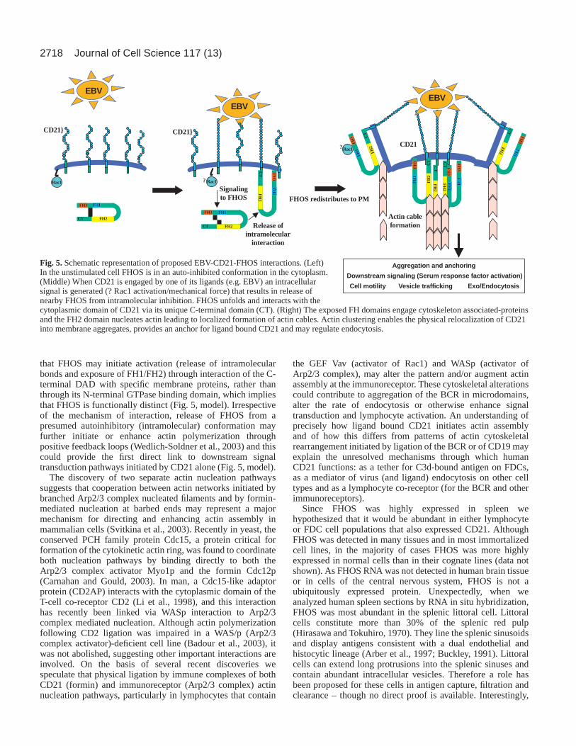
In cells that were co-transfected with both CD21 and FHOS,FHOS was predominately localized in the cytoplasm and CD21was uniformly expressed along the cell surface, thus co-localization under these conditions was limited. However,when challenged with either EBV or polyclonal antibodiesraised to the extracellular portion of CD21, both FHOS andCD21 coordinately re-localized forming aggregates and cap-like structures along the plasma membrane. When C-terminaltruncated FHOS was utilized in these studies, even in thepresence of EBV, neither re-localization of FHOS norsignificant CD21 aggregation was observed. From the dataobtained in this study we propose that upon extracellular ligandengagement of CD21 in vivo, FHOS binds to the cytoplasmicdomain of CD21, in turn linking this membrane protein to theactin cytoskeleton. Formin attachment anchors the ligandbound receptor and provides physical control for CD21 re-localization in the form of aggregation and cap formation atthe cell surface (Fig. 5).
The precise mechanisms that cause FHOS to redistribute tothe cell membrane enabling it to interact with and co-aggregateCD21 are not known. FHOS recruitment may occur uponligand induced modulation of intracellular CD21 (Barel et al.,2003) or may result from ligand-stimulated application of amechanical force generating a physical and/or spatial cue(?GTPase activation, Rac1) that attracts FHOS proximate toCD21 (Riveline et al., 2001). Although the overall structuralorganization of FHOS is similar to that of diaphanous-relatedformins, important differences are suggested by theobservation that Rac1 can bind FHOS in the absence ofphosphorylation (and therefore activation). Furthermore, thelocation of the FHOS GTPase binding domain is alteredrelative to that of other formins and the results of transcriptionregulation studies suggest that Rac1 may function downstreamrather than upstream of FHOS (Westendorf, 2001). AlthoughFHOS contains a C-terminal DAD-like domain, the structurediffers from that of diaphanous-related formins (Wallar andAlberts, 2003). Together with current findings (unpublisheddata) (Tojo et al., 2003) these observations raise the possibility
2718
that FHOS may initiate activation (release of intramolecularbonds and exposure of FH1/FH2) through interaction of the C-terminal DAD with specific membrane proteins, rather thanthrough its N-terminal GTPase binding domain, which impliesthat FHOS is functionally distinct (Fig. 5, model). Irrespectiveof the mechanism of interaction, release of FHOS from apresumed autoinhibitory (intramolecular) conformation mayfurther initiate or enhance actin polymerization throughpositive feedback loops (Wedlich-Soldner et al., 2003) and thiscould provide the first direct link to downstream signaltransduction pathways initiated by CD21 alone (Fig. 5, model).
The discovery of two separate actin nucleation pathwayssuggests that cooperation between actin networks initiated bybranched Arp2/3 complex nucleated filaments and by formin-mediated nucleation at barbed ends may represent a majormechanism for directing and enhancing actin assembly inmammalian cells (Svitkina et al., 2003). Recently in yeast, theconserved PCH family protein Cdc15, a protein critical forformation of the cytokinetic actin ring, was found to coordinateboth nucleation pathways by binding directly to both theArp2/3 complex activator Myo1p and the formin Cdc12p(Carnahan and Gould, 2003). In man, a Cdc15-like adaptorprotein (CD2AP) interacts with the cytoplasmic domain of theT-cell co-receptor CD2 (Li et al., 1998), and this interactionhas recently been linked via WASp interaction to Arp2/3complex mediated nucleation. Although actin polymerizationfollowing CD2 ligation was impaired in a WAS/p (Arp2/3complex activator)-deficient cell line (Badour et al., 2003), itwas not abolished, suggesting other important interactions areinvolved. On the basis of several recent discoveries wespeculate that physical ligation by immune complexes of bothCD21 (formin) and immunoreceptor (Arp2/3 complex) actinnucleation pathways, particularly in lymphocytes that contain
the GEF Vav (activator of Rac1) and WASp (activator ofArp2/3 complex), may alter the pattern and/or augment actinassembly at the immunoreceptor. These cytoskeletal alterationscould contribute to aggregation of the BCR in microdomains,alter the rate of endocytosis or otherwise enhance signaltransduction and lymphocyte activation. An understanding ofprecisely how ligand bound CD21 initiates actin assemblyand of how this differs from patterns of actin cytoskeletalrearrangement initiated by ligation of the BCR or of CD19 mayexplain the unresolved mechanisms through which humanCD21 functions: as a tether for C3d-bound antigen on FDCs,as a mediator of virus (and ligand) endocytosis on other celltypes and as a lymphocyte co-receptor (for the BCR and otherimmunoreceptors).
Since FHOS was highly expressed in spleen wehypothesized that it would be abundant in either lymphocyteor FDC cell populations that also expressed CD21. AlthoughFHOS was detected in many tissues and in most immortalizedcell lines, in the majority of cases FHOS was more highlyexpressed in normal cells than in their cognate lines (data notshown). As FHOS RNA was not detected in human brain tissueor in cells of the central nervous system, FHOS is not aubiquitously expressed protein. Unexpectedly, when weanalyzed human spleen sections by RNA in situ hybridization,FHOS was most abundant in the splenic littoral cell. Littoralcells constitute more than 30% of the splenic red pulp(Hirasawa and Tokuhiro, 1970). They line the splenic sinusoidsand display antigens consistent with a dual endothelial andhistocytic lineage (Arber et al., 1997; Buckley, 1991). Littoralcells can extend long protrusions into the splenic sinuses andcontain abundant intracellular vesicles. Therefore a role hasbeen proposed for these cells in antigen capture, filtration andclearance – though no direct proof is available. Interestingly,
Journal of Cell Science 117 (13)
CT FH2
FH3 FH1
CD21}
EBV
Rac1rac1
Signalingto FHOS
CT FH2
FH3 FH1
CT
FH
2
FH
3F
H1
?
FHOS redistributes to PM
Rac1
EBV
CD21}
Release ofintramolecular
interaction
Actin cableformation
CD21
CT
Rac1?
CT
FH
2
FH
3F
H1
CT
FH
2
FH
3F
H1
CT
FH2
FH3
FH1
EBV
CT
FH
2
FH
3F
H1
CT
FH2
FH3
FH1
Aggregation and anchoring
Downstream signaling (Serum response factor activation)
Cell motility Vesicle trafficking Exo/Endocytosis
Fig. 5. Schematic representation of proposed EBV-CD21-FHOS interactions. (Left)In the unstimulated cell FHOS is in an auto-inhibited conformation in the cytoplasm.(Middle) When CD21 is engaged by one of its ligands (e.g. EBV) an intracellularsignal is generated (? Rac1 activation/mechanical force) that results in release ofnearby FHOS from intramolecular inhibition. FHOS unfolds and interacts with thecytoplasmic domain of CD21 via its unique C-terminal domain (CT). (Right) The exposed FH domains engage cytoskeleton associated-proteinsand the FH2 domain nucleates actin leading to localized formation of actin cables. Actin clustering enables the physical relocalization of CD21into membrane aggregates, provides an anchor for ligand bound CD21 and may regulate endocytosis.

2719Formin interaction with CD21
in the littoral cell angioma, a vascular tumor of red pulp thatoriginates from littoral cells, the expression of CD21 issignificantly upregulated (Arber et al., 1997).
Although implicated in organ malformation, thus far noabnormalities in splenic development have been linked tomammalian formins. FHOS maps proximate to the locus forthe CREBBP gene on chromosome 16 and deletions ofCREBBP are associated with Rubinstein-Taybi syndrome.Intriguingly, in a report about the association of some cases ofRubinstein-Taybi syndrome with polysplenia and hypoplasticheart it was suggested that a contiguous gene syndrome maysometimes occur (Bartsch et al., 1999).
In this study we identify FHOS as a novel CD21 interactingprotein, a formin that binds the cytoplasmic domain of CD21through its own carboxyl terminus, and itself re-localizes in thecell coordinately with CD21 at the plasma membrane uponspecific ligand engagement of CD21. FHOS may provideCD21 with an independent and critical link to the cytoskeletalreorganization and interior signaling pathways of the cell.
This work was supported by an EIA from the American HeartAssociation and by National Institute of Health grants R01 DE12186and K24 to J.D.F. We thank Rong Li, Evelyn Kurt-Jones and CoxTerhorst for helpful comments.
ReferencesArber, D. A., Strickler, J. G., Chen, Y. Y. and Weiss, L. M. (1997). Splenic
vascular tumors: a histologic, immunophenotypic, and virologic study. Am.J. Surg. Pathol. 21, 827-835.
Aubry, J. P., Pochon, S., Gauchat, J. F., Nueda-Marin, A., Holers, V. M.,Graber, P., Siegfried, C. and Bonnefoy, J. Y. (1994). CD23 interacts witha new functional extracytoplasmic domain involving N-linkedoligosaccharides on CD21. J. Immunol. 152, 5806-5813.
Badour, K., Zhang, J., Shi, F., McGavin, M. K., Rampersad, V., Hardy, L.A., Field, D. and Siminovitch, K. A. (2003). The Wiskott-Aldrichsyndrome protein acts downstream of CD2 and the CD2AP and PSTPIP1adaptors to promote formation of the immunological synapse. Immunity18,141-154.
Barel, M., Balbo, M., le Romancer, M. and Frade, R. (2003). Activation ofEpstein-Barr virus/C3d receptor (gp140, CR2, CD21) on human cell surfacetriggers pp60src and Akt-GSK3 activities upstream and downstream to PI3-kinase, respectively. Eur. J. Immunol. 33, 2557-2566.
Bartel, P. L., Roecklein, J. A., SenGupta, D. and Fields, S. (1996). A proteinlinkage map of Escherichia coli bacteriophage T7. Nat. Genet. 12, 72-77.
Bartsch, O., Wagner, A., Hinkel, G. K., Krebs, P., Stumm, M.,Schmalenberger, B., Bohm, S., Balci, S. and Majewski, F. (1999). FISHstudies in 45 patients with Rubinstein-Taybi syndrome: deletions associatedwith polysplenia, hypoplastic left heart and death in infancy. Eur. J. Hum.Genet. 7, 748-756.
Bergelson, J. M., Cunningham, J. A., Droguett, G., Kurt-Jones, E. A.,Krithivas, A., Hong, J. S., Horwitz, M. S., Crowell, R. L. and Finberg,R. W. (1997). Isolation of a common receptor for Coxsackie B viruses andadenoviruses 2 and 5. Science275, 1320-1323.
Berger, U. V. and Hediger, M. A. (2001). Differential distribution of theglutamate transporters GLT-1 and GLAST in tanycytes of the third ventricle.J. Comp. Neurol. 433, 101-114.
Bonnefoy, J. Y., Henchoz, S., Hardie, D., Holder, M. J. and Gordon, J.(1993). A subset of anti-CD21 antibodies promote the rescue of germinalcenter B cells from apoptosis. Eur. J. Immunol. 23, 969-972.
Bradbury, L. E., Kansas, G. S., Levy, S., Evans, R. L. and Tedder, T. F.(1992). The CD19/CD21 signal transducing complex of human Blymphocytes includes the target of antiproliferative antibody-1 and Leu-13molecules. J. Immunol. 149, 2841-2850.
Buckley, P. J. (1991). Phenotypic subpopulations of machrophages anddendritic cells in human spleen. Scanning Microsc. 5, 147-157.
Carel, J. C., Myones, B. L., Frazier, B. and Holers, V. M. (1990). Structuralrequirements for C3d,g/Epstein-Barr virus receptor (CR2/CD21) ligand
binding, internalization, and viral infection. J. Biol. Chem. 265, 12293-12299.
Carnahan, R. H. and Gould, K. L. (2003). The PCH family protein, Cdc15p,recruits two F-actin nucleation pathways to coordinate cytokinetic actin ringformation in Schizosaccharomyces pombe. J. Cell Biol. 162, 851-862.
Cherukuri, A., Cheng, P. C., Sohn, H. W. and Pierce, S. K. (2001). TheCD19/CD21 complex functions to prolong B cell antigen receptor signalingfrom lipid rafts. Immunity14, 169-179.
Chien, C. T., Bartel, P. L., Sternglanz, R. and Fields, S. (1991). The two-hybrid system: a method to identify and clone genes for proteins thatinteract with a protein of interest. Proc. Natl. Acad. Sci. USA88, 9578-9582.
Copeland, J. W. and Treisman, R. (2002). The diaphanous-related forminmDia1 controls serum response factor activity through its effects on actinpolymerization. Mol. Biol. Cell13, 4088-4099.
Dezube, B. J., Zambela, M., Sage, D. R., Wang, J. F. and Fingeroth, J. D.(2002). Characterization of Kaposi sarcoma-associated herpesvirus/humanherpesvirus-8 infection of human vascular endothelial cells: early events.Blood100, 888-896.
Evangelista, M., Pruyne, D., Amberg, D. C., Boone, C. and Bretscher, A.(2002). Formins direct Arp2/3-independent actin filament assembly topolarize cell growth in yeast. Nat. Cell Biol. 4, 260-269.
Evangelista, M., Zigmond, S. and Boone, C. (2003). Formins: signalingeffectors for assembly and polarization of actin filaments. J. Cell Sci. 116,2603-2611.
Fearon, D. T. and Carroll, M. C. (2000). Regulation of B lymphocyteresponses to foreign and self-antigens by the CD19/CD21 complex. Annu.Rev. Immunol. 18, 393-422.
Feierbach, B. and Chang, F. (2001). Roles of the fission yeast formin for3pin cell polarity, actin cable formation and symmetric cell division. Curr. Biol.11, 1656-1665.
Fields, S. and Song, O. (1989). A novel genetic system to detect protein-protein interactions. Nature340, 245-246.
Fingeroth, J. D. (1990). Comparative structure and evolution of murine CR2.The homolog of the human C3d/EBV receptor (CD21). J. Immunol. 144,3458-3467.
Fingeroth, J. D., Weis, J. J., Tedder, T. F., Strominger, J. L., Biro, P. A. andFearon, D. T. (1984). Epstein-Barr virus receptor of human B lymphocytesis the C3d receptor CR2. Proc. Natl. Acad. Sci. USA81, 4510-4514.
Fingeroth, J. D., Diamond, M. E., Sage, D. R., Hayman, J. and Yates, J.L. (1999). CD21-Dependent infection of an epithelial cell line, 293, byEpstein-Barr virus. J. Virol. 73, 2115-2125.
Gustafson, E. A., Chillemi, A. C., Sage, D. R. and Fingeroth, J. D. (1998).The Epstein-Barr virus thymidine kinase does not phosphorylate gancicloviror acyclovir and demonstrates a narrow substrate specificity compared tothe herpes simplex virus type 1 thymidine kinase. Antimicrob. AgentsChemother. 42, 2923-2931.
Hirasawa, Y. and Tokuhiro, H. (1970). Electron microscopic studies on thenormal human spleen: especially on the red pulp and the reticulo-endothelialcells. Blood35, 201-212.
Holers, V. M. (2000). Phenotypes of complement knockouts.Immunopharmacology49, 125-131.
Iida, K., Nadler, L. and Nussenzweig, V. (1983). Identification of themembrane receptor for the complement fragment C3d by means of amonoclonal antibody. J. Exp. Med. 158, 1021-1033.
Koka, S., Neudauer, C. L., Li, X., Lewis, R. E., McCarthy, J. B. andWestendorf, J. J. (2003). The formin-homology-domain-containing proteinFHOD1 enhances cell migration. J. Cell Sci. 116, 1745-1755.
Kovar, D. R., Kuhn, J. R., Tichy, A. L. and Pollard, T. D. (2003). The fissionyeast cytokinesis formin Cdc12p is a barbed end actin filament cappingprotein gated by profilin. J. Cell Biol. 161, 875-887.
Leader, B., Lim, H., Carabatsos, M. J., Harrington, A., Ecsedy, J.,Pellman, D., Maas, R. and Leder, P. (2002). Formin-2, polyploidy,hypofertility and positioning of the meiotic spindle in mouse oocytes. Nat.Cell Biol. 4, 921-928.
Lee, S. W., Bonnah, R. A., Higashi, D. L., Atkinson, J. P., Milgram, S. L.and So, M. (2002). CD46 is phosphorylated at tyrosine 354 upon infectionof epithelial cells by Neisseria gonorrhoeae. J. Cell Biol. 156, 951-957.
Li, F. and Higgs, H. N. (2003). The mouse formin mDia1 is a potent actinnucleation factor regulated by autoinhibition. Curr. Biol. 13, 1335-1340.
Li, Q., Spriggs, M. K., Kovats, S., Turk, S. M., Comeau, M. R., Nepom,B. and Hutt-Fletcher, L. M. (1997). Epstein-Barr virus uses HLA class IIas a cofactor for infection of B lymphocytes. J. Virol. 71, 4657-4662.
Li, J., Nishizawa, K., An, W., Hussey, R. E., Lialios, F. E., Salgia, R.,

2720
Sunder-Plassmann, R. and Reinherz, E. L. (1998). A cdc15-like adaptorprotein (CD2BP1) interacts with the CD2 cytoplasmic domain and regulatesCD2-triggered adhesion. EMBO J. 17, 7320-7336.
Martin, D. R., Marlowe, R. L. and Ahearn, J. M. (1994). Determination ofthe role for CD21 during Epstein-Barr virus infection of B-lymphoblastoidcells. J. Virol. 68, 4716-4726.
Melamed, I., Stein, L. and Roifman, C. M. (1994). Epstein-Barr virusinduces actin polymerization in human B cells. J. Immunol. 153, 1998-2003.
Moir, S., Malaspina, A., Li, Y., Chun, T. W., Lowe, T., Adelsberger, J.,Baseler, M., Ehler, L. A., Liu, S., Davey, R. T., Jr et al. (2000). B cellsof HIV-1-infected patients bind virions through CD21-complementinteractions and transmit infectious virus to activated T cells. J. Exp. Med.192, 637-646.
Moore, M. D., Cooper, N. R., Tack, B. F. and Nemerow, G. R. (1987).Molecular cloning of the cDNA encoding the Epstein-Barr virus/C3dreceptor (complement receptor type 2) of human B lymphocytes. Proc. Natl.Acad. Sci. USA84, 9194-9198.
Morgenstern, J. P. and Land, H. (1990). Advanced mammalian genetransfer: high titre retroviral vectors with multiple drug selection markersand a complementary helper-free packaging cell line. Nucleic Acids Res. 18,3587-3596.
Pelham, R. J. and Chang, F. (2002). Actin dynamics in the contractile ringduring cytokinesis in fission yeast. Nature419, 82-86.
Poe, J. C., Hasegawa, M. and Tedder, T. F. (2001). CD19, CD21, and CD22:multifaceted response regulators of B lymphocyte signal transduction. Int.Rev. Immunol. 20, 739-762.
Pring, M., Evangelista, M., Boone, C., Yang, C. and Zigmond, S. H. (2003).Mechanism of formin-induced nucleation of actin filaments. Biochemistry42, 486-496.
Prodinger, W. M. (1999). Complement receptor type two (CR2,CR21): atarget for influencing the humoral immune response and antigen-trapping.Immunol. Res. 20, 187-194.
Prota, A. E., Sage, D. R., Stehle, T. and Fingeroth, J. D. (2002). The crystalstructure of human CD21: implications for Epstein-Barr virus and C3dbinding. Proc. Natl. Acad. Sci. USA99, 10641-10646.
Pruyne, D., Evangelista, M., Yang, C., Bi, E., Zigmond, S., Bretscher, A.and Boone, C. (2002). Role of formins in actin assembly: nucleation andbarbed-end association. Science297, 612-615.
Riveline, D., Zamir, E., Balaban, N. Q., Schwarz, U. S., Ishizaki, T.,Narumiya, S., Kam, Z., Geiger, B. and Bershadsky, A. D. (2001). Focalcontacts as mechanosensors: externally applied local mechanical forceinduces growth of focal contacts by an mDia1-dependent and ROCK-independent mechanism. J. Cell Biol. 153, 1175-1186.
Sagot, I., Klee, S. K. and Pellman, D. (2002a). Yeast formins regulate cellpolarity by controlling the assembly of actin cables. Nat. Cell Biol. 4, 42-50.
Sagot, I., Rodal, A. A., Moseley, J., Goode, B. L. and Pellman, D. (2002b).
An actin nucleation mechanism mediated by Bni1 and profilin. Nat. CellBiol. 4, 626-631.
Svitkina, T. M., Bulanova, E. A., Chaga, O. Y., Vignjevic, D. M., Kojima,S., Vasiliev, J. M. and Borisy, G. G. (2003). Mechanism of filopodiainitiation by reorganization of a dendritic network. J. Cell Biol. 160, 409-421.
Tanaka, K. (2000). Formin family proteins in cytoskeletal control. Biochem.Biophys. Res. Commun. 267, 479-481.
Tanner, J., Weis, J., Fearon, D., Whang, Y. and Kieff, E. (1987). Epstein-Barr virus gp350/220 binding to the B lymphocyte C3d receptor mediatesadsorption, capping, and endocytosis. Cell 50, 203-213.
Tedder, T. F., Clement, L. T. and Cooper, M. D. (1984). Expression of C3dreceptors during human B cell differentiation: immunofluorescence analysiswith the HB-5 monoclonal antibody. J. Immunol. 133, 678-683.
Tojo, H., Kaieda, I., Hattori, H., Katayama, N., Yoshimura, K., Kakimoto,S., Fujisawa, Y., Presman, E., Brooks, C. C. and Pilch, P. F. (2003). Theformin family protein, formin homolog overexpressed in spleen, interactswith the insulin-responsive aminopeptidase and profilin IIa. Mol.Endocrinol. 17, 1216-1229.
Tolliday, N., VerPlank, L. and Li, R. (2002). Rho1 directs formin-mediatedactin ring assembly during budding yeast cytokinesis. Curr. Biol. 12, 1864-1870.
Wallar, B. J. and Alberts, A. S. (2003). The formins: active scaffolds thatremodel the cytoskeleton. Trends Cell Biol. 13, 435-446.
Wasserman, S. (1998). FH proteins as cytoskeletal organizers. Trends CellBiol. 8, 111-115.
Wedlich-Soldner, R., Altschuler, S., Wu, L. and Li, R. (2003). Spontaneouscell polarization through actomyosin-based delivery of the Cdc42 GTPase.Science299, 1231-1235.
Westendorf, J. J. (2001). The formin/diaphanous-related protein, FHOS,interacts with Rac1 and activates transcription from the serum responseelement. J. Biol. Chem. 276, 46453-46459.
Westendorf, J. J., Mernaugh, R. and Hiebert, S. W. (1999). Identificationand characterization of a protein containing formin homology (FH1/FH2)domains. Gene232, 173-182.
Zaffran, Y., Destaing, O., Roux, A., Ory, S., Nheu, T., Jurdic, P.,Rabourdin-Combe, C. and Astier, A. L. (2001). CD46/CD3 costimulationinduces morphological changes of human T cells and activation of Vav, Rac,and extracellular signal-regulated kinase mitogen-activated protein kinase.J. Immunol. 167, 6780-6785.
Zeller, R., Haramis, A. G., Zuniga, A., McGuigan, C., Dono, R., Davidson,G., Chabanis, S. and Gibson, T. (1999). Formin defines a large family ofmorphoregulatory genes and functions in establishment of the polarisingregion. Cell Tissue Res. 296, 85-93.
Zhou, H., Su, H. S., Zhang, X., Douhan, J., III and Glimcher, L. H. (1997).CIITA-dependent and -independent class II MHC expression revealed by adominant negative mutant. J. Immunol. 158, 4741-4749.
Journal of Cell Science 117 (13)