ecg1 elkhatib
TRANSCRIPT
By M.elkhatib CIN


SA node- 60-100bpm
AV node- 40-60bpm
Bundle branches- 20-40bpm


Regular - RR interval constant
Basically regular – premature atrial/ventricular ectopic
Regularly irregular - RR interval variable but
with a pattern
Irregularly irregular - RR interval variable with
no pattern

Sinus Rhythm
Sinus Brady
Sinus Tachy
Atrial Tachy
Atrial Flutter
Junctional Tachy
AVNRT/AVRT
VT
1 & 3 degree HB


Sinus arrhythmia
Atrial fibrillation
Wenkebach
Mobitz 2
VF

Horizontally
One small box - 0.04 s
One large box - 0.20 s
Vertically
One large box - 0.5 mV


Small boxes
Number of small boxes between RR or PP
1500 divide by number of small boxes
Example 15 small boxes between the peaks
of two RR - 1500/15 = HR 100bpm.

Number of large boxes between 2 RR/PP
intervals
Divide 300 by the number of large boxes
Example
5 large boxes between 2 RR complexes
300/5 =60bpm

Quick and dirty method great for an irregular
rhythm
Measure 6 second strip and x by ten.

Sinus Brady
Sino atrial arrest
Wenkebach
Mobitz type 2
CHB

Sinus Tachy
Atrial Tachy
A flutter
Atrial fibrillation
VT
VF
Junctional Tachy
Nodal re-entrant Tachy
Re-entrant Tachy

Does a P wave precede every QRS
Is it positive or negative in the correct leads
Do all the p waves look alike
What is the shape of the p wave
What is the ratio of p waves to QRS complexes
Normal p waves upright (except AVR) round with 1:1ratio

P Mitrale
A wide bifurcated P - wave
Left atrial enlargement
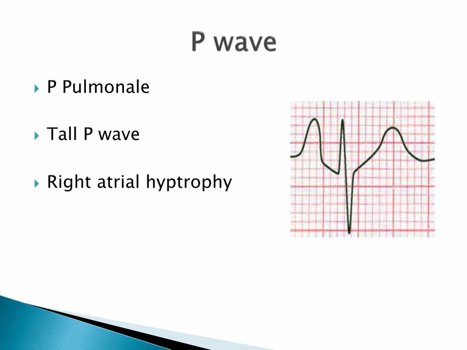
P Pulmonale
Tall P wave
Right atrial hyptrophy

AV junctional rhythm
Atrial fibrillation
Atrial flutter
Sino-atrial block

Dextrocardia
Incorrectly placed limb leads
Ectopic beats

Beginning of P wave to the beginning of QRS
Normal = 0.12 -0.20 secs
No more than 5 small squares

PR interval > 0.20 sec
secondary to drugs (digoxin/beta blockers)
Hypokaleamia
Congenital abnormalities (ASD, TGA)

First degree AV Block

<0.12 secs
Wolf Parkinson White syndrome (ventricular
muscle stimulated early)
Junctional rhythm
Dissociated beats (CHB)

Wenkebach
Wandering pacemaker
Premature atrial beats
AV dissociation

The length of time taken to depolarise the ventricles
<0.12 secs (3 small squares)
Q - first negative deflection
R- first positive deflection
S negative deflection after R
Any upward deflection after the R above the isoelectricline is classed as another R

QRS - 0.12 secs or greater
Common cause BBB
Abberant conduction
Think VT, VF, CHB

Dead or stunned myocardial tissue
Usually permanent
Can be reversed with early intervention
More than 1mm in depth
0.02 secs or greater in V1-V2
0.04 secs or more and greater than one-third of the R wave (although their is much dispute over this)

Reflects length of time from the beginning of
ventricular depolarisation to the end of repolarisation
QT interval corrected for heart rate variability
The ECG machine corrects this
The corrected figure is called the QTc
Should be no more than 440ms
Slight variation in peadiatrics depending on age
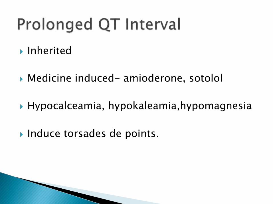
Inherited
Medicine induced- amioderone, sotolol
Hypocalceamia, hypokaleamia,hypomagnesia
Induce torsades de points.

Digoxin therapy
Hypercalacaemia

Represents recovery or repolarisation of the
ventricles
Measured from the end of the QRS to the
beginning of the T wave
The ST is normally isoelectric

ST elevation-MI (STEMI)
ST Depression (NSTEMI)

Pericarditis
Angina
Hyperkalemia

Digoxin therapy
Tachycardia
Hypokaleamia
BBB
Ectopics
Cardiomyopathies

• Reflects repolarisation or ventricular muscular relaxation ,T wave changes seen in:
MI
Ischemia
electrolyte imbalances
medications
pericarditis
Cardiomyopathies


Upright round wave seen in lead II after Twave and before the next P wave
Not clearly understood
Associated with hypokaleamia, amioderone
& digoxin

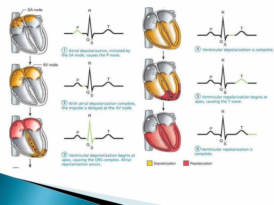
The general flow of electrical activity of the heart
It relates to the flow of depolarisation wave through the heart
This can change with position of the heart
Generally related to changes in electricalflow


Left ventricular hypertrophy
LBBB - bifasicular block
LBBB + 1st degree HB - trifasicular block
Mechanical shift of the heart
Normal variant

Right Ventricular Hypertrophy
RBBB
Dextrocardia
Mechanical shift
Normal variation
