editorial board editorial consultants · 2018-03-24 · (rimoin, 1997; covic 2004). the proportion...
TRANSCRIPT

JURNALUL PEDIATRULUI – Year IX, Vol. IX, Nr. 35-36, july-december 2006
1
Eugen Sorin BOIA
Liviu POP
Radu Emil IACOB
O Adam Marioara Boia
A Craciun M Gafencu
Daniela Iacob A Pirvan
Calin Popoiu Maria Puiu
I Velea
M Ardelean – Salzburg, Austria ES Boia – Timisoara, Romania
Maria Bortun – Timisoara, Romania V Botiu – Timisoara, Romania
S Garofallo – Milano, Italy DG Gotia – Iasi, Romania
C Ilie – Timisoara, Romania E Lazăr – Timisoara, Romania
J Mayr – Basel, Switzerland Eva Nemes – Craiova, Romania
L Pop – Timisoara, Romania I Popa – Timisoara, Romania
Maria Puiu – Timisoara, Romania GC Rogers – Greenville, USA J Schalamon – Graz, Austria
I Simedrea – Timisoara, Romania Rodica Stackievicz – Kfar Sava, Israel
H Stackievicz – Hadera, Israel C Tica – Constanta, Romania
JURNALUL PEDIATRULUI – Year IX, Vol. IX, Nr. 35-36, july-december 2006
EDITOR IN CHIEF
CO-EDITOR
SECRETARY
EDITORIAL BOARD
Timisoara, Romania Gospodarilor Street, nr. 42
Tel: +4-0256-439441 cod 300778
e-mail: [email protected]
ISSN p 1221-7212
ADDRESS
EDITORIAL CONSULTANTS
JURNALUL PEDIATRULUI – Year IX, Vol. IX, Nr. 35-36, july-december 2006
2
CONTENTS
I. GENETICS 1. THE OPTIMIZATION OF THE DIAGNOSIS AND MANAGEMENT OF THE PATIENTS AFFECTED BY MENTAL RETARDATION USING MLPA TEST IN THE EVALUATION PROTOCOL Maria Puiu, L Dehelean, Cristina Rusu, Cristina Skrypnyk, Ligia Barbarii, E Tomescu, Victoria Cret, Katalin Csep…….........................................................................................3
II. NEONATOLOGY 2. INTERCEREBRAL CYSTIC FORMATIONS Marioara Boia, Daniela Iacob, Aniko Manea, Mirabela Dima, Nicoleta Pavel..............................................................6
III. PEDIATRICS
3. CLINICAL, DEVELOPING AND THERAPEUTIC STAGES IN A KAWASAKI’S DISEASE CASE REPORT Rodica Urtila, Eulalia Boceanu, Dana Metea, Adrian Lacatusu.....................................................................................9 4. EVOLUTION OF A VARIANT DANDY-WALKER SYNDROME Adriana Moisa, Luminita Chiutu, Ileana Petrescu, Simona Cosoveanu.......................................................................15 5. ALLERGIC BRONCHOPULMONARY ASPERGILLOSIS (ABPA) CHARACTERISTIC OVERVIEW IN CYSTIC FIBROSIS
Laura Dracea.................................................................................................................................................................18 6. CYSTIC FIBROSIS ASSOCIATED WITH HEPATOBILIARY DISEASE Ioana Ciuca Popa, I Popa, L Pop, Zagorca Popa...........................................................................................................21 7. HISTOLOGICAL FEATURES OF DENDRITIC CELLS IN ALLERGIC ASTHMA - EXPERIMENTAL MODEL C Oancea, Liliana Vasile, V Ordodi, Janina Jiga, Carmen Bunu, V Tudorache, Georgeta Mihalas............................25 8. SEVERE JUNCTIONAL BRADYCARDIA BY DESTRUCTION OF THE SINUS NODE AT A PATENT WITH COMPLEX SURGICAL CORRECTION OF A CYANOGENIC CONGENITAL HEART DEFECT – THE FONTAN PROCEDURE A Lacatusu, Sonia Tanasescu, Erika Zborovszky.........................................................................................................29 9. CLINICAL STUDY IN PERIPHERAL ADENOPATHIES IN CHILDREN Ileana Puiu, D Bulucea, Polixenia Stancu, Veronica Nicolescu, M Dicu.....................................................................33
IV. PEDIATRIC SURGERY
10. SURGICAL TREATMENT OF GASTROSCHISIS USING SILIMED GASTROSCHISIS CONTAINER - CASE REPORT ES Boia, RE Iacob, O Adam, A Pavel, Maria Trailescu, Marioara Boia, Nicoleta Pavel............................................39 11. CONGENITAL LUNG MALFORMATIONS – A REVIEW VL David, A Radulescu, MC Popoiu............................................................................................................................46 12. INDUCED NECROTIZING ENTEROCOLITIS BY ISCHEMIA IN ANIMAL MODELS A Radulescu..................................................................................................................................................................53 13. DEPLETED URANIUM INDUCED FETAL MALFORMATIONS - LITERATURE REVIEW A Radulescu, ES Boia, RE Iacob, V David...................................................................................................................56
V. DENTISTRY 14. CARIES RISK FACTORS IN CHILDREN Roxana Oancea, Elisabeta Bratu, Daniela Jumnaca, Atena Galuscan, Ruxandra Sava-Rosianu, Angela Codruta Podariu.......................................................................................................61
MANUSCRIPT REQUIREMENTS............................................................................................................................66
JURNALUL PEDIATRULUI – Year IX, Vol. IX, Nr. 35-36, july-december 2006
3
THE OPTIMIZATION OF THE DIAGNOSIS AND MANAGEMENT OF THE PATIENTS AFFECTED BY
MENTAL RETARDATION USING MLPA TEST IN THE EVALUATION PROTOCOL
Maria Puiu1, L Dehelean2, Cristina Rusu3, Cristina Skrypnyk4, Ligia Barbarii5, E Tomescu6, Victoria Cret7, Katalin Csep8
1Medical genetics, University of Medicine and Pharmacy “V. Babes”, Timisoara 2Children Hospital “L. Turcanu”, Timisoara 3Medical genetics, University of Medicine and Pharmacy, Iasi 4Medical genetics, Faculty of Medicine and Pharmacy, Oradea 5Medical Institute, “M. Minovici”, Bucuresti 6Children Hospital, (IOMC), Bucuresti 7Pediatric Clinic I, Cluj-Napoca 8Faculty of Medicine and Pharmacy, Tg. Mures Abstract
Mental retardation (MR) is a relatively frequent disorder, with heterogeneous etiology and major social implications. Out of the genetic causes, an important part is represented by subtelomeric rearrangements (unidentified by classical analyses). MLPA (multiplex ligation-dependent probe amplification) is a new diagnostic method, cheap and very useful in identifying subtelomeric rearrangements. Key words: mental retardation, subtelomeric rearrangements, multiplex ligation-dependent probe amplification
Mental retardation (MR) is a very important public health problem, affecting 2-3 % of the population and causing major problems to affected individuals and their families and also to the society. Mental retardation’s etiology is various and includes genetic factors (chromosomal abnormalities, monogenic diseases – the most important being X-linked mental retardation and especially Fragile X Syndrome, multifactorial and mitochondrial disorders), environmental factors (infectious diseases, social agents) or simply unknown etiology (nonspecific MR) (Rimoin, 1997; Covic 2004). The proportion of different types of causes is different and in dependence with the degree of MR – so, for moderate and severe MR, genetic agents’ contribution is more important than for mild MR, where the environment is having a more important place (especially social agents). In both cases the contribution of unspecific MR is major (de Vries, 2001). Because of that, most of the present’s reserches in the field are focused on
the study of unspecific MR aiming to identify new genetic factors involved. Recently, it has been observed that subtelomeric rearrangements are having an important contribution (5%) in unspecific MR determinism (Flint, 1995).
Telomeric screening is interesting for 2 reasons : • Most of the translocations are involving telomeric regions, reason why an investigation of the chromosomal extremities will detect all the abnormalities, no matter the size; • Adjacent regions of the telomeres are enriched in gene number; rearrangements involving neighbouring DNA is more probably causing phenotypical consequences than other DNA regions (Knight, 2000). Until recently, screening for the telomeric rearrangements was not possible because of the complexity of the telomeric structure and because of the very expensive investigations. The researches were focused in 2 directions: clinical studies wanted to identify clinical signs that were associated to MR and could suggest the presence of a subtelomeric rearrangement; they also aimed to establish diagnostic scores in order to increase the efficiency of lab investigations (de Vries, 2001; Sandig, 2004); the laboratory work searched for new methods to show better the subtelomeric defect. Out of the clinical studies, we have to mention de Vries’ (2001) score projected to increase the efficiency of subtelomeric rearrangements’ identification (Table 1).
I. GENETICS

JURNALUL PEDIATRULUI – Year IX, Vol. IX, Nr. 35-36, july-december 2006
4
Table 1: Criteria for patients presenting with submicroscopic rearrangements (de Vries, 2000).
It is recommended that the pacient has a 3/> pts in order to identify an abnormality using MLPA method.
In the laboratory workfield, different techniques were suggested: classical or high resolution cytogenetic techniques (the study of prometaphase chromosomes), different fluorescent in situ hybridization methods – FISH (Knight, 1997; Knight, 2000) and more recently hybridization of the probe and multiplex amplification (MAPH) and the multiplex ligation-dependent probe amplification (MLPA) (Armour, 2002; Sellner, 2004). Standard cytogenetic techniques (a resolution of 400-500 bands) can detect only 5-10 Mb abnormalities, depending on the chromosomal region. High resolution techniques (850-1000 bands) last too long and they are useful only when we look for a specific abnormality in a specific region (it cannot be scanned the entire genome).
FISH techniques are very useful, but the test is very expensive. MAPH is being used for long genes, the method allowing you to study in the same time different parts of the gene (e.g.: the 23 exons of the BRCA1 gene). MLPA is a cheap method (6-10 Euros/test) and very useful for the detection of subtelomeric rearrangements, being considered as an election method in the field (Rooms, 2004). The technique has been recently introduced (2002) and many world specialists took over the technique to appreciate correctly the real frequency of the subtelomeric rearrangement (6,7% - Koolen, 2004), but also to start identifying the clinical picture of every rearrangement (Rossi, 2001).
The main characteristics of the MLPA technique (www.mlpa.com) are: • It allows the simultaneous testing of 40 different genomic DNA sequences by PCR; • It can identify sequences differing in a single base- pair; • The amount of DNA needed is very low (20 μg); • It needs a termocyclor and an electrophoretic system only; • The protocol is the same for different applications (detection of subtelomeric rearrangements; detection of aneuploidies - chromosomes 13, 18, 21, X and Y; detection of large deletions or duplications; detection of gene deletions
or duplications involved in cancer; detection of deletions / duplications of a single exon in specific genes – BRCA, NF; cuantification of the CpG islands methylation in the promoters of tumours suppression genes); • Because of the short sequence detection of the probe the method can be used also on the partially degraded DNA (e.g.: DNA extracted from fixed tissues or paraphin blocks); • The probe is amplified by PCR, not the DNA sample; • 2 probes are hybridized on the target sequences, then follows probe ligation and amplification of the target; • Amplification is achieved by multiplex-PCR – all the specific sequences are simultaneously amplified; • PCR protocol needs only one primer pair for the amplification of all fragments; • Product’s lenght is varying between 130 and 490 bp long, being analysed by electrophoresis.
Subtelomeric rearrangements represent a relatively new described category. The studies in this field are only a few and very simplistic because the methods used until now were very expensive. MLPA seems to be an ideal technique (for now) for the identification of subtelomeric rearrangements, being a cheap method (6-10 E/test) and fit for the identification of submicroscopic chromosomal abnormalities.
Because of the prohibitive price of the earlier methods, but also because of the recently (2002) introduced MLPA method, the number of researches including large patient groups is very limited. Only one study (Koolen, 2004) presented the MLPA results for 210 MR patients.
In the literature, after the identification of an abnormality in a MR child, the parents are tested; if they did not have the abnormality, the defect was considered to be a new mutation that produces the clinical picture of the child. If the abnormality was present also in the normal looking parent, the defect was considered as a polymorphism. If the parents were phenotypically abnormal, presenting the same subtelomeric rearrangement, the defect was considered as a familial one, but there were not many studies testing other members of the family that could be at risk.
Criteria Score Family history of MR • Compatible with monogenic inheritance • Uncompatible with monogenic inheritance (including discordant phenotypes)
1 2
Growth retardation with prenatal onset 2 Postnatal growth abnormalities (for each of them 1 point, with a maximum of 2 points) • Microcephaly • Short stature • Macrocephaly • Tall stature
1 1 1 1
2/> facial dysmorphies (especially hipertelorism, nasal or auricular malformations) 2 Extrafacial abnormalities (for each of them 1 point, with a maximum of 2 points); especially: • Hand malformations • Cardiac defects • Hipospadias +/- criptorchydism
1 1 1

JURNALUL PEDIATRULUI – Year IX, Vol. IX, Nr. 35-36, july-december 2006
5
There are no articles in the specialised literature to present genetic counselling offered to the family or if the prenatal diagnosis was achieved.
In the literature there are only 2 diagnostic scores (de Vries, 2001; Sandig, 2004), based on a relatively low number of cases.
Considering the small number of identified cases with subtelomeric rearrangements and that the modifications of each telomere is producing a different clinical picture, we
can appreciate the early stage in sketching the clinical aspects of MR determined by subtelomeric rearrangements, many clinicians from all over the world still working on this direction.
In Romania subtelomeric rearrangements as a cause of unspecific MR has not yet been studied by any method. The introduction of MLPA in this field would reduce the unidentified causes of mental retardation and increase the efficiency of specific MR diagnosis.
References: 1. Armour J.A.L., Barton D.E., Cockburn D.J., Taylor
G.R. – The Detection of Large Deletions or Duplications in Genomic DNA, Human Mutation 20: 325-337 (2002);
2. Covic Mircea, Stefanescu Dragos, Sandovici Ionel – Genetica Medicala, Retardul mental, p 457-472; Editura Polirom, 2004;
3. de Vries B.B.A., White S.M., Knight S.J.L., Regan R., Homfray T., Young I.D., Super M., McKeown C., Splitt M., Quarrell O.W.J., Trainer A.H., Niermeijer M.F., Malcom S., Flint J., Hurst J.A., Winter R.M. – Clinical studies on submicroscopic subtelomeric rearrangements: a checklist, J. Med. Genet 2001; 38: 145-150;
4. Flint J., Wilkie AOM, Buckle VJ, Winter RB, Holland AJ, McDermid HE. 1995. The detection of subtelomeric chromosomal rearrangenments in idiopathic mental retardation. Nat Genet 9: 132-139;
5. Knight J.L. Samantha, Lese M. Christa, Precht S. Kathrin, Kuc Julie, Ning Yi, Lucas Sarah, Regan Regina, Brenan Mary, Nicod Alison, Lawrie Martin N., Cardy L.N. Donald, Nguyen Huy, Hudson J. Thomas, Riethman C. Harold, Ledbetter H. David, Flint Jonathan – An Optimized Set of Human Telomere Clones for Studying Telomere Integrity and Architecture, Am. J. Hum. Genet. 67: 320-332, 2000;
6. Knight J.L. Samantha, Horsley W. Sharon, Regan Regina, Lawrie N. Martin, Maher E.J., Cardy L.N. Donald, Flint Jonathan, Kearney Lyndal – Development of Clinical Application of an Innovative Fluorescence in situ Hybridization Technique Which Detects Submicroscopic Rearrangements Involving Telomeres, Eur. J. Hum. Genet 1997; 5: 1-8;
7. Knight J.L. Samantha, Flint Jonathan – Perfect endings: a review of subtelomeric probes and their use in clinical diagnosis, J. Med. Genet 2000; 37: 401-409;
8. Koolen D.A., Nillesen W.M., Versteeg M.H.A., Merks G.F.M., Knoers N.V.A., Kets M., Vermeer S., van Ravenswaaij C.M.A., de Kovel C.G., Brunner H.G.,
Smeets D., de Vries B.B.A., Sistermans E.A. – Screening for subtelomeric rearrangements in 210 patients with unexplained mental retardation using multiplex ligation dependent probe amplification (MLPA), J. Med. Genet. 2004; 41: 892-899;
9. MLPA, MRC-Holland, Amsterdam (www.mlpa.com); 10. Rimoin L. David, Connor J. Michael, Pyeritz E. Reed –
Emery and Rimoin’s Principles and Practice of Medical Genetics, 3rd Edition, Churchill Livingstone 1997, Abnormal mental development p 725-736;
11. Rooms Liesbeth, Reyniers Edwin, van Luijk Rob, Scheers Stefaan, Wauters Jan, Ceulemans Berten, Van Den Ende Jenneke, Van Bever Yolande, Kooy R. Frank – Subtelomeric Deletions Detected in Patients With Idiopathic Mental Retardation Using Multiplex Ligation- Dependent Probe Amplification (MLPA), Human Mutation 23: 17-21 (2004);
12. Rossi Elena, Piccini Flavia, Zollino Marcella, Neri Giovanni, Caselli Desiree, Tenconi Romano, Castellan Claudio, Carrozzo Romeo, Danesino Cesare, Zuffardi Orsetta, Ragusa Angela, Castiglia Lucia, Galesi Ornella, Greco Donatella, Romano Corrado, Pierluigi Mauro, Perfumo Chiara, Di Rocco Maia, Faravelli Francesca, Bricarelli Franca Dagna, Bonaglia Maria Clara, Bedeschi Maria Francesca, Borgatti Renato – Cryptic telomeric rearrangements in subjects with mental retardation associated with dysmorphism and congenital malformations, J. Med. Genet 2001; 38: 417-420;
13. Sandig K.S. Walter, Hinkel G.K., Mitulla B., Ounap K., Sims G., Sitska M., Utermann B., Viertel P., Kalscgeuer V., Bartsch O. – Subtelomere FISH in 50 children with mental retardation and minor anomalies, identified by a checklist, detects 10 rearrangements including a de novo balanced translocation of chromosomes 17p13.3 and 20q13.33, Am J Med Genet A. 2004 Aug 1;128 (4): 364-73;
14. Sellner N. Loryn, Taylor R. Graham – MLPA and MAPH: New Techniques for Detection of Gene Deletions, Human Mutation 23: 413-419 (2004).
Correspondence to:
Puiu Maria, Martir O Munteanu Street, No. 9, Timisoara 300360, Romania Phone: +4-0256-226824, E-mail: [email protected]

JURNALUL PEDIATRULUI – Year IX, Vol. IX, Nr. 35-36, july-december 2006
6
INTERCEREBRAL CYSTIC FORMATIONS Marioara Boia1, Daniela Iacob1, Aniko Manea1, Mirabela Dima1, Nicoleta Pavel2 ¹University of Medicine and Pharmacy “Victor Babes” Timisoara 2Special Services for Children with Dizabilities Timisoara Abstract
Cerebral cystic formations occur more and more often in pediatric medical practice as a result of widespread using of modern imagistic techniques (cranial ultrasound, RMN, CT). Depending on the size, location, causes they can be asymptomatic or can be accompanied by a major clinical table.
In this abstract authors aim to overview the most frequent causes and also the correlation between specific ultrasound images and clinical signs intensity. Key words: cerebral cystic formations, causes, correlation between specific ultrasound images and clinical signs intensity. Discussions
Several cystic formations can be met at the level of cerebral tissue: congenital, inflammatory, neoplasic, traumatic or vascular. The most spread are the arachnoidian cysts which represents 1% from intracranial space replacing formations at child.
Regarding their etiology these can be congenital or achieved by accumulation of cerebrospinal fluid through adhesions, conglomeration of the arachnoid. The cyst is located between brain and dura mater and is lined by the arachnoid; contains cerebrospinal fluid and does not communicate with ventricles. Regarding the location, the most common are met at the level of sylvian, suprasselar fissure, plate quadrigeminal, cerebello-pontine and at the level of internal subtentorial cisterns. Rarely they are located in the interhemispheric fissure or at the level of cerebral convexity. From the echographical point of view they are visualized as formations filled with liquid (transsonics) with a well shaped contour which dislocates adyacent structures. (fig.1).
Intracranial congenital cysts can occur in several congenital malformations. Thus, corpus callosum agenesis and non lobar holoprosencephaly are two malformations often associated with medium line dorsal cysts. These can also be met in Dandy – Walker Complex, but in fact the cyst is a dilated IV ventricle. More often met clinical aspects and also other causes of the posterior cerebral fossa are illustrated in the Table Nr.1.
Another affection in which cystic formations can occur is Zellweger Syndrome (cerebral-hepatic-renal) and trysomy 13. In these two situations, cystic intercerebral
formations are followed by other placements, most frequently renal and hepatic.
Medium line cystic formations can also occur in
Arnold Chiari II malformation. Their origin is not exactly known but they appear, typically, near the quadrigeminal cisterns.
Pathological cystic formations must be differentiated from the normal cystic areas such as the great cistern, cavity of pellucid septum and cavum vergae. These structures are accompanied by normal dimension ventricles and have no mass effect.
Cystic lesions include areas of encefalomalacia and periventricular and subependymal cysts (fig.2). This type of lesion appears secondary to cerebral necrosis due to hemorrhage, infarct, infection and it is coming after the brain gets glial answer capacity.
Encephalomalacy can be unique – porencephaly or schizencephaly – or can be multifocal. The multifocal form appears when there is a diffused affection of brain. Echographical examination emphasizes a transonic formation, septate, non-homogeneous (Fig.3).
II. NEONATOLOGY
Fig. 1. The arachnoidian cysts.

JURNALUL PEDIATRULUI – Year IX, Vol. IX, Nr. 35-36, july-december 2006
7
Table Nr.1. Clinical aspects in the posterior cerebral fossa cystic.
AFFECTIONS CLINICAL ASPECTS Dandy-Walker Syndrome Hidrocephaly Familial vermian agenesis – S. Joubert Polypnea episodes, ptosis, abnormal eye
movements. Ventricle IV dislocation Widening of the IV ventricle followed by
disfunction of cerebral trunk or increasing intracranial pressure after intraventricular hemorrhage or meningitis
Widening of the magna cistern Asymptomatic or hypotonic, mioclonii, non progressive macrocephaly
Arachnoidian cyst Dilatation of the posterior fossa, hydrocephaly
In the diffuse forms there are visualized
supratentorial cavities, bilateral, glial septate. Usually they are placed in the cortex and peripheral white matter. Generally they are not placed in periventricular white matter, inferior temporal lobes and cerebellum.
Typical for these diffuse lesions is the ultrasound finding of these multiple cysts, bilateral, with several shapes and dimensions, which are not communicating with dilated ventricular system.
Conclusions
1. Cerebral cystic formations can occur secondary to malformations.
2. Also they can be acquired from infections, hemorrhages or cerebral ischemia.
3. The most frequent and severe cerebral malformations accompanied by cysts are holoprosencephaly and Dandy Walker malformation.
4. Multicystic encephalomalacia occurs in diffuse affections of cerebral parenchyma. Depending on dimensions and location can appear neurological sequels, tightly correlated with the dimensions of these cystic structures.
References 1. Abbitt PL, Hurst RW, Ferguson RDG, McIlhenny J,
Alford BA.The role of ultrasound în the management of Galen anevrisms in infang. Neuroradiologiy 1990, 32:86-89
2. Alford CA Jr, Cronic congenital and perinatal infections. in Avery GB editor, Neonatology,
Pathophysiology and Management of the newborn, Philadelphia, JB Lippincott, 1987
3. Babcock D.S., Cranial ultrasonography of infants, Baltimore, Wiliams and Wilkins, 1981
4. Babcock D.S., Han B.K., The accuracy of the high resolution real-time ultrasonography of the head infancy, Radiology, 1981, 139, 664-670
Fig. 2. Porencephaly. Fig. 3. Encephalomalacy.
JURNALUL PEDIATRULUI – Year IX, Vol. IX, Nr. 35-36, july-december 2006
8
5. Babcock D.S, Bove KE, Han BK. Intracranial hemorrhage in premature infants: sonographic-pathologic correlation. AJNR 1982; 3: 309-317
6. Boţiu Valentin, Boia M., Aportul ecografiei transfontanelare în diagnosticul şi monitorizarea evoluţiei nou-născutului cu encefalopatie hipoxico-ischiemică neonatală, Conferinţa Naţională de Pediatrie, Sibiu, 23-26.09.1998
7. Fitz CR, Holoprosencephaly and related entities, Neuroradiology, 1983, 25 225-238
8. Grand EG, Kerner M, Schellinger D et al, Evolution of porencephalic cysts from intraparenchymal hemorrhage in neonates sonographic evidence, AJR, 1982, 138, 467-470
9. Grand EG, Schellinger D, Richardson JD, Real time ultrasonography of the posterior fosa, J Ultrasound Med, 1983, 2, 73-87
10. Lucas G, Gruenewald SM, Lui K, Neonatal subependymal cysts detected by sonography; prevalence, sonographic findings and clinical significance, AJR, Clinical Perinatol, 1977, 4, 3-30
Correspondence to:
Marioara Boia, Gospodarilor Street, No. 42, Timisoara 300778, Romania E-mail: [email protected]
JURNALUL PEDIATRULUI – Year IX, Vol. IX, Nr. 35-36, july-december 2006
9
CLINICAL, DEVELOPING AND THERAPEUTIC STAGES IN A KAWASAKI’S DISEASE CASE REPORT
Rodica Urtila1, Eulalia Boceanu1, Dana Metea1, Adrian Lacatusu1 12nd Clinic of Pediatrics, University of Medicine and Pharmacy “V. Babes” Timisoara Abstract Kawasaki disease ( Kawasaki syndrom-KS ) is an acute self-limited vasculitis of childhood that is characterized by fever, bilateral nonexudative conjunctivitis, erythema of the lips and oral mucosa, changes in the extremities, rash and cervical lymphadenopathy(1,2). Coronary artery aneurysms or ectasia develop in 15% to 25% of untreated children and may lead to ischemic heart disease or sudden death.
We present a case of Kawasaki disease in a small child, using a modern algorithm of diagnosis. The patient received the classical therapy with Immunoglobulin and Aspirin. The repeated cardiac ultrasound made it possible to rule out the presence of the dreaded complication that is the coronary artery aneurysms disease. Key words: Kawasaki disease, coronary artery aneurysms, child Introduction Kawasaki disease was first described in Japan in 1967 by Tomisaku Kawasaki. The disease is now known to occur in both endemic and community – wide epidemic forms in the Americas, Europe and Asia in children of all races(6). In the United States, Kawasaki disease has surpassed acute rheumatic fever as the leading cause of acquired heart disease in children. Treatment of Kawasaki disease in the acute phase is directed at reducing inflammation in the coronary artery wall and preventing coronary thrombosis, whereas long – term therapy in individuals who develop coronary aneurysms is aimed at preventing myocardial ischemia or infarction(3,4,5). Recommendations of initial evaluation, treatment in the acute phase and long – term management of patients with Kawasaki disease are intended to assist physicians in understanding the range of acceptable approaches for caring for patients with Kawasaki disease. Ultimately, management decisions must be individualized to a patient’s specific circumstances(10,11,12). Case report
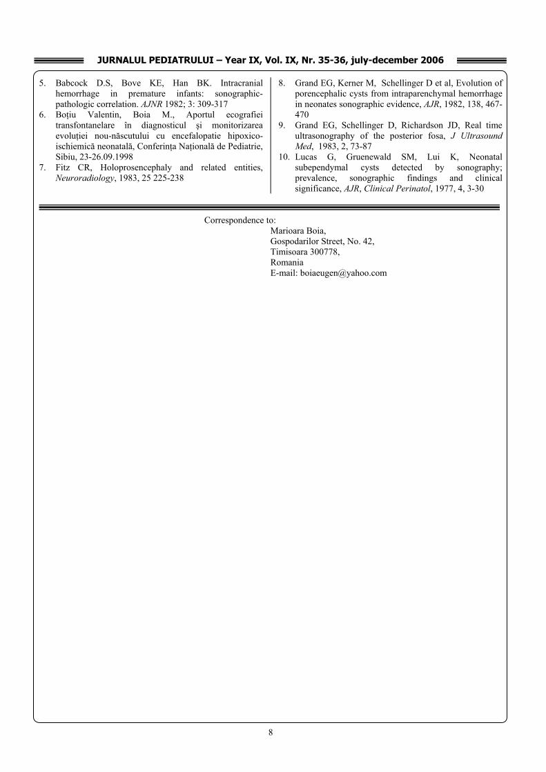
The authors present the patient A.R., 2 years old male, hospitalized in the Bega 2nd Clinic of Pediatrics for: drowsiness alternating with irritability, inappetence, fever, paleness, oral enanthema, fingers presenting lamellar desquamation, nonexudative conjunctival congestion
accompanied by pruritus, aching edema at superior and inferior limbs level.
History: the patient is the 2nd born child from normal pregnancy, normally delivered at full term, weigh at birth = 3500g, height at birth = 50cm, with no neonatal distress, breast fed from birth, current adequate vaccinations.
The patient came to our unit in its 8th day of illness, being hospitalized for erithemato - pultaceous tonsillitis accompanied by fever and for polymorphic erythema and previously treated with antibiotics (Ampicillin, Gentamicin, one day, and then Ceftriaxone 4 more days) and with HSH for 6 days. Under the treatment, the patient’s condition did not improve, febrile peeks 2-3 times a day (over 390C), skin erythema gradually diminishing.
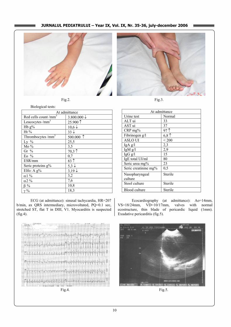
Physical examination: • Bad general state, drowsiness, inappetence • fever (39,50C) • intense paleness, lamellar desquamation of the fingers
in the superior limbs, red-carmine colored oral enanthema, strawberry, lucid appearance of the tongue, dry cracked lips (fig.1)
• bilateral nonexudative conjunctival congestion
accompanied by pruritus • aching edema at superior and inferior limbs level (fist
joint, tibio-tarsal joint – fig.2,3) • left subangulo-mandibular adenopathy, 2cm in
diameter, mobile, slightly sensible at palpation • rhythmical cardiac sounds, tachycardia, HR=200b/min
III. PEDIATRICS
Fig. 1.
JURNALUL PEDIATRULUI – Year IX, Vol. IX, Nr. 35-36, july-december 2006
10
Biological tests:
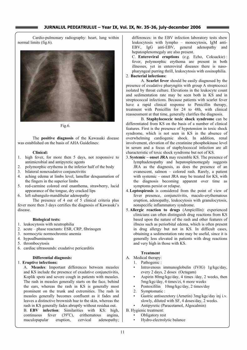
ECG (at admittance): sinusal tachycardia, HR=207
b/min, ax QRS intermediary, microvoltated, PQ=0.1 sec, stretched ST, flat T in DIII, V1. Myocarditis is suspected (fig.4).
Ecocardiography (at admittance): Ao=14mm, VS=18/24mm, VD=10/17mm, valves with normal ecostructure, thin blade of pericardic liquid (1mm). Exudative pericarditis (fig.5).
At admittance Urine test Normal ALT ui 33 AST ui 37 CRP mg% 97 ↑ Fibrinogen g/l 6,8 ↑ ASLO UI < 200 IgA g/l 2,3 IgM g/l 2,9 IgG g/l 15 IgE total UI/ml 80 Seric urea mg% 23 Seric creatinine mg% 0,5 Nasopharyngeal culture
Sterile
Stool culture Sterile
Blood culture Sterile
At admittance Red cells count /mm3 3.800.000 ↓ Leucocytes /mm3 25.900 ↑ Hb g% 10,6 ↓ Ht % 33 ↓ Thrombocytes /mm3 500.000 ↑ Ly % 25,5 Mo % 3,5 Gr % 70,3 ↑ Eo % 0,7 ESR/mm 83 ↑ Seric proteins g% 5,3 ↓ Elfo: A g% 3,19 ↓ α1 % 3,2 α2 % 7,6 β % 10,8 γ % 18,3
Fig.2. Fig.3.
Fig.4. Fig.5.
JURNALUL PEDIATRULUI – Year IX, Vol. IX, Nr. 35-36, july-december 2006
11
Cardio-pulmonary radiography: heart, lung within normal limits (fig.6).
The positive diagnosis of the Kawasaki disease
was established on the basis of AHA Guidelines: Clinical:
1. high fever, for more then 5 days, not responsive to antimicrobial and antipyretic agents
2. polymorphic erythema in the inferior half of the body 3. bilateral nonexudative conjunctivitis 4. aching edema at limbs level, lamellar desquamation of
the fingers in the superior limbs 5. red-carmine colored oral enanthema, strawberry, lucid
appearance of the tongue, dry cracked lips 6. left subangulo-mandibular adenopathy
The presence of 4 out of 5 clinical criteria plus fever more then 3 days certifies the diagnosis of Kawasaki’s disease.
Biological tests:
1. leukocytosis with neutrophilia 2. acute – phase reactants: ESR, CRP, fibrinogen 3. normocytic normochromic anemia 4. hypoalbuminemia 5. thrombocytosis 6. cardiac ultrasounds: exudative pericarditis
Differential diagnosis:
1. Eruptive infections: A. Measles: Important differences between measles and KS include the presence of exudative conjunctivitis, Koplik spots and severe cough in patients with measles. The rash in measles generally starts on the face, behind the ears, whereas the rash in KS is generally most prominent on the trunk and extremities. The rash in measles generally becomes confluent as it fades and leaves a distinctive brownish hue to the skin, whereas the rash in KS generally fades abruptly without residua out. B. EBV infection: Similarities with KS: high, continuous fever (39oC), erithematous angina, maculopapular eruption, cervical adenopathy;
differences: in the EBV infection laboratory tests show leukocytosis with lympho – monocytosis, IgM anti-EBV, IgG anti-EBV, general adenopathy and hepatosplenomegaly are also present. C. Enteroviral eruptions (e.g. Echo, Coksackie) : fever, polymorphic erythema are present in both illnesses, yet in enteroviral diseases there is naso-pharyngeal purring thrill, leukocytosis with eosinophilia.
2. Bacterial infections: A. Scarlet fever should be easily diagnosed by the
presence of exudative pharyngitis with group A streptococci isolated by throat culture. Elevations in the leukocyte count and sedimentation rate may be seen both in KS and in streptococcal infections. Because patients with scarlet fever have a rapid clinical response to Penicillin therapy, treatment with Penicillin for 24 to 48h, with clinical reassessment at that time, generally clarifies the diagnosis.
B. Staphylococcic toxic shock syndrome can be differentiated from KS on the basis of a number of clinical features. First is the presence of hypotension in toxic shock syndrome, which is not seen in KS in the absence of overwhelming cardiogenic shock. In addition, renal involvement, elevation of the creatinine phosphokinase level in serum and a focus of staphylococcal infection are all characteristic of toxic shock syndrome but not of KS. 3. Systemic – onset JRA may resemble KS. The presence of
lymphadenopathy and hepatosplenomegaly suggests JRA as the diagnosis, as does the presence of an evanescent, salmon – colored rash. Rarely, a patient with systemic – onset JRA may be treated for KS, with the diagnosis becoming apparent over time as symptoms persist or relapse.
4. Leptospirosis is considered from the point of view of fever presence, conjunctivitis, maculo-erythematous eruption, adenopathy, leukocytosis with granulocytosis, nonspecific inflammatory syndrome.
5. Allergic reaction to drugs (Ampicillin): experienced clinicians can often distinguish drug reactions from KS based upon the nature of the rash and other features of illness such as periorbital edema, which is often present in drug allergy but not in KS. In difficult cases, obtaining a sedimentation rate may be useful, since it is generally less elevated in patients with drug reactions and very high in those with KS.
Treatment
A. Medical therapy: 1. Pathogenic : • Intravenous immunoglobulin (IVIG) 1g/kgc/day,
every 2 days, 2 doses (Octagam) • Aspirin 80mg/kgc/day, 4 times /day, 2 weeks, then
5mg/kgc/day, 4 times/zi, 6 more weeks • Pentoxifilin 10mg/kgc/day, 2 times/day 2. Symptomatic : • Gastric antisecretory (Arnetin) 3mg/kgc/day inj i.v.
slowly, diluted with SF, 4 doses/day, 2 weeks. • Antipyretic (Paracetamol, Algocalmin)
B. Hygienic treatment: • Obligatory rest • Hydro-electrolytic balance
Fig.6.
JURNALUL PEDIATRULUI – Year IX, Vol. IX, Nr. 35-36, july-december 2006
12
Complications: 1. of the illness:
• Coronary artery aneurysms (5% in treated patients) • Thrombosis of the aneurismal coronary artery • Myocardium infarction • Cardiomegaly • Arrhythmia • Late coronary atherosclerosis • Sudden death (4%)
2. of the therapy: • Reye Syndrome (following Aspirin)
Monitoring showed a decrease of the inflammatory syndrome, with the thrombocytes value going back to normal, hemoleukogram and electrophoresis normalization, remission of pericarditis, lack of cardiac complications.
The tables below presents the most significant biological events during hospitalization:
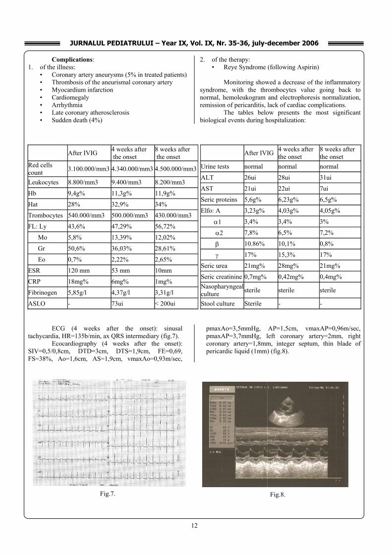
ECG (4 weeks after the onset): sinusal
tachycardia, HR=135b/min, ax QRS intermediary (fig.7). Ecocardiography (4 weeks after the onset):
SIV=0,5/0,8cm, DTD=3cm, DTS=1,9cm, FE=0,69, FS=38%, Ao=1,6cm, AS=1,9cm, vmaxAo=0,93m/sec,
pmaxAo=3,5mmHg, AP=1,5cm, vmaxAP=0,96m/sec, pmaxAP=3,7mmHg, left coronary artery=2mm, right coronary artery=1,8mm, integer septum, thin blade of pericardic liquid (1mm) (fig.8).
After IVIG 4 weeks after the onset
8 weeks after the onset
Red cells count 3.100.000/mm3 4.340.000/mm3 4.500.000/mm3
Leukocytes 8.800/mm3 9.400/mm3 8.200/mm3
Hb 9,4g% 11,3g% 11,9g%
Hat 28% 32,9% 34%
Trombocytes 540.000/mm3 500.000/mm3 430.000/mm3
FL: Ly 43,6% 47,29% 56,72%
Mo 5,8% 13,39% 12,02%
Gr 50,6% 36,03% 28,61%
Eo 0,7% 2,22% 2,65%
ESR 120 mm 53 mm 10mm
CRP 18mg% 6mg% 1mg%
Fibrinogen 5,85g/l 4,37g/l 3,31g/l
ASLO - 73ui < 200ui
After IVIG 4 weeks after the onset
8 weeks after the onset
Urine tests normal normal normal
ALT 26ui 28ui 31ui
AST 21ui 22ui 7ui
Seric proteins 5,6g% 6,23g% 6,5g%
Elfo: A 3,23g% 4,03g% 4,05g%
α1 3,4% 3,4% 3%
α2 7,8% 6,5% 7,2%
β 10.86% 10,1% 0,8%
γ 17% 15,3% 17%
Seric urea 21mg% 28mg% 21mg%
Seric creatinine 0,7mg% 0,42mg% 0,4mg% Nasopharyngeal culture sterile sterile sterile
Stool culture Sterile - -
Fig.7. Fig.8.
JURNALUL PEDIATRULUI – Year IX, Vol. IX, Nr. 35-36, july-december 2006
13
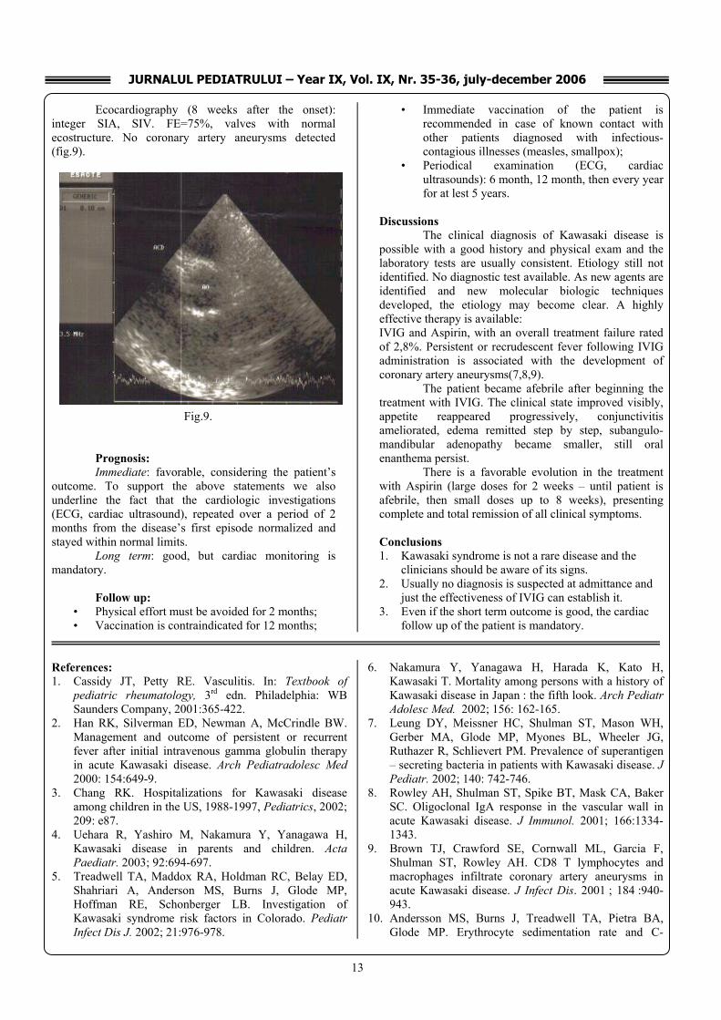
Ecocardiography (8 weeks after the onset): integer SIA, SIV. FE=75%, valves with normal ecostructure. No coronary artery aneurysms detected (fig.9).
Prognosis: Immediate: favorable, considering the patient’s
outcome. To support the above statements we also underline the fact that the cardiologic investigations (ECG, cardiac ultrasound), repeated over a period of 2 months from the disease’s first episode normalized and stayed within normal limits.
Long term: good, but cardiac monitoring is mandatory.
Follow up:
• Physical effort must be avoided for 2 months; • Vaccination is contraindicated for 12 months;
• Immediate vaccination of the patient is recommended in case of known contact with other patients diagnosed with infectious-contagious illnesses (measles, smallpox);
• Periodical examination (ECG, cardiac ultrasounds): 6 month, 12 month, then every year for at lest 5 years.
Discussions
The clinical diagnosis of Kawasaki disease is possible with a good history and physical exam and the laboratory tests are usually consistent. Etiology still not identified. No diagnostic test available. As new agents are identified and new molecular biologic techniques developed, the etiology may become clear. A highly effective therapy is available: IVIG and Aspirin, with an overall treatment failure rated of 2,8%. Persistent or recrudescent fever following IVIG administration is associated with the development of coronary artery aneurysms(7,8,9).
The patient became afebrile after beginning the treatment with IVIG. The clinical state improved visibly, appetite reappeared progressively, conjunctivitis ameliorated, edema remitted step by step, subangulo-mandibular adenopathy became smaller, still oral enanthema persist.
There is a favorable evolution in the treatment with Aspirin (large doses for 2 weeks – until patient is afebrile, then small doses up to 8 weeks), presenting complete and total remission of all clinical symptoms.
Conclusions 1. Kawasaki syndrome is not a rare disease and the
clinicians should be aware of its signs. 2. Usually no diagnosis is suspected at admittance and
just the effectiveness of IVIG can establish it. 3. Even if the short term outcome is good, the cardiac
follow up of the patient is mandatory. References: 1. Cassidy JT, Petty RE. Vasculitis. In: Textbook of
pediatric rheumatology, 3rd edn. Philadelphia: WB Saunders Company, 2001:365-422.
2. Han RK, Silverman ED, Newman A, McCrindle BW. Management and outcome of persistent or recurrent fever after initial intravenous gamma globulin therapy in acute Kawasaki disease. Arch Pediatradolesc Med 2000: 154:649-9.
3. Chang RK. Hospitalizations for Kawasaki disease among children in the US, 1988-1997, Pediatrics, 2002; 209: e87.
4. Uehara R, Yashiro M, Nakamura Y, Yanagawa H, Kawasaki disease in parents and children. Acta Paediatr. 2003; 92:694-697.
5. Treadwell TA, Maddox RA, Holdman RC, Belay ED, Shahriari A, Anderson MS, Burns J, Glode MP, Hoffman RE, Schonberger LB. Investigation of Kawasaki syndrome risk factors in Colorado. Pediatr Infect Dis J. 2002; 21:976-978.
6. Nakamura Y, Yanagawa H, Harada K, Kato H, Kawasaki T. Mortality among persons with a history of Kawasaki disease in Japan : the fifth look. Arch Pediatr Adolesc Med. 2002; 156: 162-165.
7. Leung DY, Meissner HC, Shulman ST, Mason WH, Gerber MA, Glode MP, Myones BL, Wheeler JG, Ruthazer R, Schlievert PM. Prevalence of superantigen – secreting bacteria in patients with Kawasaki disease. J Pediatr. 2002; 140: 742-746.
8. Rowley AH, Shulman ST, Spike BT, Mask CA, Baker SC. Oligoclonal IgA response in the vascular wall in acute Kawasaki disease. J Immunol. 2001; 166:1334-1343.
9. Brown TJ, Crawford SE, Cornwall ML, Garcia F, Shulman ST, Rowley AH. CD8 T lymphocytes and macrophages infiltrate coronary artery aneurysms in acute Kawasaki disease. J Infect Dis. 2001 ; 184 :940-943.
10. Andersson MS, Burns J, Treadwell TA, Pietra BA, Glode MP. Erythrocyte sedimentation rate and C-
Fig.9.
JURNALUL PEDIATRULUI – Year IX, Vol. IX, Nr. 35-36, july-december 2006
14
rective protein discrepancy and high prevalence of coronary artery abnormalities in Kawasaki disease. Pediatr Infect Dis J. 2001; 20:698-702.
11. Tseng CF, Fu YC, Fu LS, Betau H, Chi CS. Clinical spectrum of Kawasaki disease in infants. Zhonghua Yi Xue Za Zhi (Taipei).2001;64:168-173.
12. Kurotobi S, Nagai T, Kawakami N, Sano T. Coronary diameter in normal infants, children and patients with Kawasaki disease. Pediatr Int.2002;44:1-4.
Correspondence to: Rodica Urtila, 2nd Clinic of Pediatrics, University of Medicine and Pharmacy “V. Babes” Timisoara Str. Gh. Doja nr. 36, Timisoara E-mail: [email protected]
JURNALUL PEDIATRULUI – Year IX, Vol. IX, Nr. 35-36, july-december 2006
15
EVOLUTION OF A VARIANT DANDY-WALKER SYNDROME
Adriana Moisa1, Luminita Chiutu2, Ileana Petrescu3, Simona Cosoveanu3 1 2nd Pediatric Clinic, County Emergency Hospital Craiova; 2 County Emergency Hospital Craiova; UMF Craiova; 3 2nd Pediatric Clinic, County Emergency Hospital Craiova; UMF Craiova Abstract The paper presents the evolution of a twenty-one- month-old male child, who was diagnosed, since he was an infant (5 months), with an incomplete Dandy-Walker syndrome. Key words: incomplete Dandy-Walker syndrome, child, evolution. Introduction Classically, the incomplete Dandy-Walker syndrome is characterized by the vermis agenesis, the cyst dilatation of the fourth ventricle due to the rostral movement and the absence or atresia of the Magendie and Luschka foramens. Hydrocephaly is not usually present from the congenital point of view, but it develops in the first months of life. 90% of the patients who develop a hydrocephaly are registered before the age of 1 year. There are also incomplete variants, where the cerebellar hypogenesis is present without the dilatation of the forth ventricle and hydrocephaly. An ataxic syndrome occurs in less than 20% of the patients and it is usually late. The diagnosis of the Dandy-Walker malformation is confirmed by CT or skull RMN. Case presentation We present the case of the patient J.F.A., a male infant aged 1 year and 9 months, diagnosed ever since he was 5 months with variant Dandy-Walker syndrome, and who was admitted in 2nd Pediatric Clinic of the County Emergency Hospital, in February 2006 (O.F. 9442/2006). The occurrence of balance and clinical-biological assessment dysfunctions represented the reasons of the admission. The anamnesis revealed that he is the only child of a young couple, apparently healthy, with a higher education level. At the antenatal ultrasonography (third semester) he was diagnosed with megacysterna magna, cerebellar asymmetry. The child was born at 32 weeks (premature level I) with 2,400g in weight, Apgar =7/8, (perinatal ischemic hypoxia without requiring advanced resuscitation procedures at birth).
L.P. was artificially fed, diversified at 6 months, he took his vaccines according to the WHO scheme, and he followed the rachitism prophylaxis. Belated psychomotor development: he held his head at 8 months, he could sit at 11 months, he could walk by himself at 1 year and 7 months, he uttered his first word at 1 year. APP: bilateral inguinal-scrotal hernia (since he was 3 months old), variant Dandy-Walker syndrome starting with the age of five. The anamnesis also mentions the fact that at the age of five he was diagnosed with incomplete Dandy-Walker syndrome using a skull CT which indicates the cerebellar vermis and hemispheres hypoplasia, especially on the left side, replaced by an arachnoidian cyst that communicates with the fourth ventricle [fig.1]. A ventricular system with occipital remote horns because of an interpontin cyst [fig.2]. Cerebellar spaces that are slightly symmetrically enlarged [fig.3].
The transfontanel ultrasonography, which was performed at the age of 5 months, shows the presence of a callous body, cerebellar hemispheres and vermis hypoplasia [fig.4]. The clinical picture at admission showed: relatively good general state, 11Kg in weight, without fever, cardio-circulatorily, respiratorily and digestively stabilized, inguinal-scrotal hernia; he also presented a normally conformed skull, closed FA, normal active and passive movements, muscular discreet hypotony, unsupported walk with balance dysfunctions, watching things, and poor vocabulary. Biological assessments: HLG: Hb=12,20g%, Ht=37%, T=190000/mm3, L=8000/mm3, Ns=28%, E=4%, Ly=62%, M=6%. F.O. (AO): normally outlined papilla, blood vessels and retina with a normal aspect. Skull X-ray: no modifications of the bony structures at the neurocranium level, closed fontanels, visible sutures. A skull CT at the age of one year and nine months was refused by the parents. Neurological consult: discreet axial hypotony, psychomotor retard. The treatment involved a neuro-motor recovery within the specialized service of the hospital (medical exercises).

JURNALUL PEDIATRULUI – Year IX, Vol. IX, Nr. 35-36, july-december 2006
16
Discussions Classically, the Dandy-Walker malformation can be defined by: the cyst dilatation of the fourth ventricle, vermis agenesis, the absence of the foramens Magendie and Luschka. However, there are variants of the Dandy-Walker malformation, with moderate dilatation of the fourth ventricle, the permeability of the Magendie foramen, and the partial vermis agenesis. In the presented case, the antenatal ultrasonography (third semester) on the pregnant uterus showed megacysterna magna and cerebellar asymmetry, without performing amniocentesis or placental villus biopsy for the genetic study of this anomaly. After birth, in the first year of life, the child did not develop an important hydrocephaly with its consequences: macrocephaly (PC at 5 months = 43cm, at 1 year and 9 months, PC=48cm, FA closed at 1 year and 2 months) or intracranial hypertension (ICH).
However, the evolution of the child presented a psycho-motor retard (he held his head at 8 months, he could sit at 11 months, he could walk by himself at 1 year and 7 months, he uttered his first word at 1 year). Two months after he could walk by himself, there appeared some balance
dysfunctions and we took into consideration an ataxic syndrome, which can be noticed in less than 20% of the patient with Dandy-Walker syndrome The presented case responds to the conditions of a variant Dandy-Walker syndrome where the cranial-cerebral exam described: hypogenesis (partial agenesis) of the cerebellar vermis and of the cerebellar hemispheres, especially the left ones, which were replaced by the arachnoidian cyst that communicates with the fourth ventricle, a ventricular system with occipital remote horns because of an interpontin cyst, cerebellar spaces that are slightly symmetrically enlarged. Conclusions
1. The case was presented because it can be included in a variant Dandy-Walker syndrome, with low incidence of the affection in practice.
2. It shows the importance of the antenatal ultrasonography on the pregnant uterus as well as of the study of the chromosomal anomalies through amniocentesis or placental villus biopsy for the genetic advice (the transmission is autosomal recessive in the case of the Dandy-Walker syndrome).
Fig. 1 Fig. 2
Fig. 3 Fig. 4

JURNALUL PEDIATRULUI – Year IX, Vol. IX, Nr. 35-36, july-december 2006
17
3. The psychomotor retard in the case of a variant Dandy-Walker syndrome, even without developing hydrocephaly, remains important.
4. The possibility of developing an ataxic syndrome described in less than 20% of the patients within the Dandy-Walker syndrome.
Bibliography
1. Journal of Neuropathology and Experimental Neurology, vol. 13, pg. 14-39.
2. Current Pediatric, William Hay J., Myron J.Suvo, pg.769, ediţia XVII.
3. Compendiu de Pediatrie, Georgescu Adrian, Editura Bic All, 2001, pg. 667-673
4. Pediatrie partea a-II-a, Boli Neurologice şi neuromusculare, Geormăneanu, Walter Roşianu Editura Didactică şi Pedagogică, 1996, pg. 712-716
Correspondence to: Simona Cosoveanu 60, Maresal Antonescu Street Craiova Romania Telephone: +40251502210 e-mail:[email protected]

JURNALUL PEDIATRULUI – Year IX, Vol. IX, Nr. 35-36, july-december 2006
18
ALLERGIC BRONCHOPULMONARY ASPERGILLOSIS (ABPA) – CHARACTERISTIC OVERVIEW
IN CYSTIC FIBROSIS
Laura Dracea1
1Children’s Hospital of Brasov, Medicine Faculty - Transilvania University Brasov Summary:
Recognizing allergic bronchopulmonary
aspergillosis (ABPA) in the context of cystic fibrosis
(because of overlapping clinical, radiographic, microbiologic, and immunologic features), is often difficult. Advances in understanding of the pathogenesis of allergic aspergillosis, new possibilities in therapy have been done recently. Unlike in asthma, pulmonary infiltrates, bronchiectasis and obstructive lung disease are common manifestations of Cystic Fibrosis (CF) lung diseases with or without ABPA resulting from recurrent and chronic bacterial infection. Atopy as well as an onmset of a variety of immune responses to Aspergillus fumigatus antigens early in life in patients with Cf complicates the interpretation of various serological parameters for the diagnosis of ABPA. Early diagnosis and treatment aiming to suppress the inflammation is, however, important to prevent irreversible lung tissue damage.
There is a short overview of main characteristics of ABPA in patients with CF. Key words: allergic bronchopulmonary aspergillosis, cystic fibrosis
Aspergillus fumigatus, a widely distributed spore bearing fungus, causes multiple diseases in humans. The diseases produced by A. fumigatus include invasive pulmonary aspergillosis, aspergilloma and different forms of hypersensitivity diseases. Pneumonia due to Aspergillus and systemic aspergillosis occur primarily in patients who have immunosupresion or T cell or phagocytic impairment. Although no protective antibody response was detected in these patients (1), a CD4+ Th1 cytokine pattern was suggested to be important in rendering protection. Hypersensitivity lung disease includes allergic asthma, hypersensitivity pneumonitis, and ABPA; all result from the exposure to allergens of A fumigatus.
Pathogenic Aspergillus generally grows easily and relatively quickly on routine bacteriologic and mycological media in the clinical laboratory. Only pathogenic species are capable of growth at 35°C 37°C and A. fumigatus in
particular is capable of growth at ≥ 50°C. Pseudomonas aeruginosa may inhibit the growth of Aspergillus.
Aspergillus spores or inhalation trigger an IgE-mediated allergic inflammatory response in the bronchial airways, leading to bronchial obstruction and asthma. The immune response to Aspergillus antigens in patients with ABPA, as well as in allergic asthmatic patients and patients
with CF, is a Th2 CD4+ cell response A central question, then, is how ABPA differs from Aspergillus sensitivity in atopic asthma and CF. It is proposed that ABPA develops in genetically susceptible asthmatic patients and patients with
CF because of increased frequency and/or activity of A. fumigatus specific Th2 CD4+ cells.
The allergic inflammatory response in patients with ABPA appears to be quantitatively greater than that in Aspergillus-sensitive atopic asthma patients and patients with CF. In the proposed model of the immunopathogenesis of ABPA, as illustrated in A. fumigatus spores are inhaled
into the bronchial airway, where they are trapped by the luminal mucus, germinate, and form mycelia. A. fumigatus mycelia release allergens that are processed by antigen-presenting cells bearing HLA-DR2 or -DR5 and presented to T cells within the bronchoalveolar lymphoid tissue (BALT). The T cell response to Aspergillus allergens becomes skewed toward a Th2 CD4+ cell response, with synthesis and secretion of cytokines IL-4, IL-5, and IL-13.
One of the characteristic features in patients with ABPA is that A. fumigatus is found bound to the surface epithelium and is growing on and between the epithelial cells without being efficiently killed by mononuclear and eosinophilic infiltrates. It has also been shown that spores of A. fumigatus are attached to epithelial surfaces cultured in vitro (17). The physical presence of A. fumigatus on and between the epithelial cells is possibly of importance for the
modulation of the immunologic response toward a Th2-type
response (18). Over the past decades, virulence factors of A. fumigatus that interfere with or even block normal functions of the humoral and cellular defense of the airways have been detected (19).Virulence factors were discussed above. Some of these virulence factors are the proteolytic enzymes of A. fumigatus.
ABPA is found in highest incidence among atopic patients with CF. It has been hypothesized that in CF, the abnormal mucus promotes the trapping of Af spores within the bronchial airway, permitting and perhaps promoting growth of Af mycelia and probably, in genetically susceptible individuals, stimulate a Th2 cell response with subsequent ABPA. Proteases of Af may play a role in facilitation of antigen transport across the epithelial cell layer by damaging the epithelial integrity and be a direct interaction with epithelial cell surface receptors. These mechanisms would result in production of proinflammatory cytokines and corresponding inflammatory responses.

JURNALUL PEDIATRULUI – Year IX, Vol. IX, Nr. 35-36, july-december 2006
19
The classic case of ABPA fulfills the following criteria:
1. asthma 2. chest roentgenogtaphic infiltrates – current or in the
past – may be detectable on CT when roentgenographic is unremarkable
3. immediate cutaneous reactivity to Aspergillus species
4. elevated total serum IgE >417 IU/ml (>1000ng/ml) 5. serum precipitins antibodies to AF 6. central bronchiectasis on chest CT 7. peripheral blood eosinophylia 8. elevated serum IgE and/or IgG to Af
It has been suggested that the minimal essential criteria for diagnosis of ABPA include the following: (2)
1. asthma 2. immediate cutaneous reactivity to Af species 3. elevated total serum IgE concentration 4. elevated serum IgE to Af and IgG to Af 5. central bronchiectasis
Other diagnostic elements that may be supportive include a history of coughing up either mucus plugs or sputum flecked with brown, black or green elements, culture of sputum yielding Af or a sputum smear in which Af was identified microscopically, a sputum smear showing eosinophils, or a chest radiographic sign suggesting bronchial inflammation with or without plugs.
The diagnosis of ABPA in CF is often delayed because many of the diagnostic criteria overlap with common manifestations of CF.
The Epidemiologic Study of Cystic Fibrosis (ESCF) proposes two of the following 3 criteria:
1. immediate cutaneous reactivity to Af 2. precipitating antibodies to Af 3. total serum IgE>1000IU/ml
In addition at least 2 of the following are required: 1. bronchoconstriction 2. peripheral blood eosinophilia >1000 eosinophils 3. history of pulmonary infiltrates 4. elevated serum anti-A. fumigatus IgE or IgG 5. A. fumigatus in sputum found by culture or smear 6. Response to steroids
The literature reviews mention several predisposing factors for ABPA in CF:
- the increase in frequency and severity of bacterial lung infections that lead to an increased use of antibiotics that “may pave the way” for fungal infections - the use of inhaled tobramycin - HLA-DR molecules DR2, DR5 and possibly DR4 or DR7 that contribute to susceptibility; whereas HLA DQ2 contributes to resistance – their combination may determine the outcome of ABPA in CF - atopy with different patterns of allergic response to Af compared to other allergens (3,4) - association with delayed onset of P.aeruginosa
Factors that have been found to be associated with ABPA in CF are: males, adolescents, lower levels of lung function, presence of wheezing/asthma, positive cultures for P.aeruginosa, atopy, lower clinical and radiographic scores..
There was also a significant association found in patients colonized with Staph. aureus and with an increased decline in lung function (greater than expected/year).
Therapy for ABPA involves prophylaxis against and treatment of acute exacerbations as well as prevention of end-stage fibrotic disease. There are two aspects of treatment: first attenuation of the inflammation and immunological activity –for which corticosteroids are the mainstay of therapy (5,6); second – the attenuation of the antigen burden arising from fungal colonisation of the bronchial tree (5).
Therapy of ABPA in CF is problematic. This is because of several reasons: first – several of the diagnostic criteria of ABPA overlap with common manifestations of CF (therefor treatment must be rigurous); second – both cause many of the same clinical and physiological derangements; third- systemic corticosteroids (the cornerstone of treatment for ABPA) have toxicities that are concerning patients who are prone to develop diabetes, osteopenia, infections. In this regard there are no specific indications for how long may corticosteroids be given in case of high IgE values; it has been accepted that efficacy of treatment may be judged when levels decrease more than 50%; different schemes were proposed: starting dose of 1-2 mg Prednisolone/kg/day (maximum 40 mg) for up to 2 weeks, followed by 1 mg/kg for the same period, before changing to alternate day therapy. Weaning should be guided by the clinical response over the following weeks (it may take months for a favourable response).
Although corticosteroids are the mainstay of therapy in ABPA because they attenuate inflammatory and immunological activity, they have no effect on the antigen burden arising from the fungal colonization of the bronchial tree. Reducing the fungal burden in the respiratory tract might decrease antigenic stimmulation, reduce inflammatory response, ameliorate symptoms and possibly reduce the long term risk of disease progression.
Itrakonazole which has been used in doses of 200-400 mg/day for 1-2 weeks with tappering over several months (minimum of 3 months for decreasing the antigen load in the bronchial tree) has the disadvantage of limited oral bioavailability, and this particularly for the capsule form, requiring an acidic environment for dissolution which is inhibited by antiacid therapies (20). The liquid formulation is better absorbed but not available in this country. Vorikonazole is a recently introduced triazole antifungal with superior oral bioavailability which has been aproved for the treatment of invasive aspergillosis (7). It is however expensive, has a high potential for drug interactions and has been associated with a number of adverse effects. It appears to be generally well tolerated, but a transient disturbance in vision has been reported to occur in up to 30% of patients (8). Skin reactions (rash or photosensitivity) are the next common adverse effect (reported in up to 15% of patients), and elevations in hepatic enzymes have been reported in up to 10% of patients (8,9).
Although several small case series have suggested
that inhaled corticosteroids are useful in treating patients

JURNALUL PEDIATRULUI – Year IX, Vol. IX, Nr. 35-36, july-december 2006
20
with ABPA without CF (11), a double-blind, multicenter
study conducted in the United Kingdom in the 1970s of beclomethasone, at 400 µg/day without a spacer (a volume holding chamber used with steroid metered-dose inhalers), failed to demonstrate clinical benefit (12). Inhaled corticosteroids have been shown minimally to reduce
bronchial hyperresponsiveness in patients with ABPA without CF (13). There are minimal data to formulate conclusive treatment recommendations for ABPA in CF
Every case has to be interpretated taking into consideration the clinical data, laboratory data and intensity of allergic response to Af.
Some authors have suggested that serum IgE levels be followed regularly in patients with ABPA (10) because
IgE levels correlate with disease activity, and that
corticosteroid therapy should be instituted even for asymptomatic patients if the serum IgE level doubles from
the baseline value (10). The great majority of IgE is not directed against Aspergillus antigens but is nonspecific (14). Although the serum IgE level remains part of a constellation of clinical parameters used to decide when corticosteroids should be given, it cannot be used in isolation to make that decision (14, 15,16).
References: 1. Kurup NP, Gronnig G, Knutsen AP, Murale PS.
Citokines in allergic bronchopulmonary aspergillosis. Res Immunol 1998;149:466-77
2. Greenberger PA, Patterson R. diagnosis and management of allergic bronchopulmonary aspergillosis: Ann Allergy 1986;56:444-52
3. Valletta EA, Braggion C, Mastella G. Sensitization to Aspergillus and allergic bronchopulmonary aspergillosis in a cystic fibrosis population. Pediatr Asthma Allergy Immunol 1993; 7:43 9.
4. Skov M, Koch C, Reimert CM, Poulsen LK. Diagnosis of allergic bronchopulmonary aspergillosis in cystic fibrosis. Allergy 2000; 55:50 8
5. Vlahakis NE, Aksamit TR. Allergic bronchopulmonary aspergillosis: diagnosis and treatment. Mayo Clin Proc 2001; 76:930 8.
6. Judson MA, Stevens DA. Current pharmacotherapy of allergic bronchopulmonary aspergillosis. Expert Opin Pharmacother 2001; 2:1065 71.
7. Johnson LB, Kauffmann PA. Vorikonazole: a new triazole antifungal agent. Clin Infect Dis 2003:36:630-7
8. Herbrecht R, Denning DW, Patterson TF, Bennett JE, Greene RE, Oestmann JW, et al. Vorikonazole versus amphotericin B for primary therapy of invasive aspergillosis. N Engl J Med 2002;347:408-15
9. Walsh TJ, Lutsar I, Driscoll T, dupont D, Roden M, Ghahramani P, et al. Vorikonazole in the treatment of aspergillosis, scedosporiosis and other invasive fungal infections in children. Pediatr Infect Dis J 2002;21:240-8
10. Patterson R, Greenberger PA, Halwig JM, Liotta JL, Roberts M. Allergic bronchopulmonary aspergillosis: natural history and classification of early disease by serologic and roentgenographic studies. Arch Intern Med 1986; 146:916 8.
11. Imbeault B, Cormier Y. Usefulness of inhaled high-dose corticosteroids in allergic bronchopulmonary aspergillosis. Chest 1993; 103:1614 7
12. Report to the Research Committee of the British Thoracic Association. Inhaled beclomethasone dipropionate in allergic bronchopulmonary aspergillosis. Br J Dis Chest 1979; 73:349 56.
13. Van Haren EHJ, Lammers JWJ, Festen J, Heijerman HGM, Groot CAR, Van Herwaarden CLA. The effects of the inhaled corticosteroid budesonide on lung function and bronchial hyperresponsiveness in adult patients with cystic fibrosis. Respir Med 1995; 89:20914
14. Schuyler MR. Allergic bronchopulmonary aspergillosis. Clin Chest Med 1983; 4:15 22
15. Mroueh S, Spock A. Allergic bronchopulmonary aspergillosis in patients with cystic fibrosis. Chest 1994; 105:32 6.
16. Marchant JR, Warner JO, Bush A. Rise in total IgE as an indicator of allergic bronchopulmonary aspergillosis in cystic fibrosis. Thorax 1994; 49:1002 5
17. Paris S, Boisvieux-Ulrich E, Crestani B, et al. Internalization of Aspergillus fumigatus conidia by epithelial and endothelial cells. Infect Immun 1997; 65:1510 4.
18. Kurup VP, Seymour BW, Choi H, Coffman RL. Particulate Aspergillus fumigatus antigens elicit a TH2 response in BALB/c mice. J Allergy Clin Immunol 1994; 93:1013 20.
19. Tomee JF, Kauffman HF. Putative virulence factors of Aspergillus fumigatus. Clin Exp Allergy 2000; 30:47684
20. Stevens DA, Moss RB, Kurup VP, Kerutsen AP, Greenberger P, Judson MA, et al. Allergic bronchopulmonary aspergillosis in Cystic Fibrosis – state of the art: Cystic Fibrosis Foundation Consensus Conference. Clin Infect Dis 2003;37:S225-64
Correspondence to: Laura Dracea, Nicopole Street, No. 45, Brasov 500063, Romania Phone: +4-0268-415130.

JURNALUL PEDIATRULUI – Year IX, Vol. IX, Nr. 35-36, july-december 2006
21
CYSTIC FIBROSIS ASSOCIATED WITH HEPATOBILIARY DISEASE
Ioana Ciuca Popa1, I Popa1, L Pop1, Zagorca Popa2 1Pediatric IInd Department – University of Medicine and Pharmacy „Victor Babes” Timisoara 2National Centre of Cystic Fibrosis, Timisoara, Romania Abstract
Cystic fibrosis (CF) is the most frequent monogenic disease in population with Caucasian origin, potentially lethal, with marked clinical variability. With improved life expectancy of CF patients, it has become clear that cystic fibrosis associated hepatobiliary disease is a relatively frequent and serious complication which can affect quality of life and survival of affected patients. Data from literature mention the association of LD with, male gender, history of meconium ileus and severe genotype.
The paper aim is to review data concerning cystic fibrosis associated liver disease (CFLD) epidemiology, diagnosis and natural history. Key words: cystic fibrosis, liver disease, children. Background
In recent years, clinical attention for cystic fibrosis associated hepatobiliary disease (CFHD) has significantly increased from a small proportion (2-5%) of patients with end-stage multilobular biliary cirrhosis and portal hypertension, to the increasingly recognized asymptomatic patients with focal biliary cirrhosis. Cystic fibrosis associated hepatobiliary disease (CFHD) include entities like: liver disease, microgallbladder, cholelithiasis, neonatal cholestasis, common bile duct stenosis, the most important clinical expression is liver disease (LD).
Screening for liver disease (LD) has indicated that in the majority of affected patients, LD becomes clinically apparent by the end of the first decade of life, suggesting that this is a relatively early complication of CF. A slow progression is characteristic, hepatocellular failure is a delayed episode, whereas development of portal hypertension and related complications tends to occur earlier and more frequently.
Treatment with ursodeoxicholic acid (UDCA) is widely employed in these patients, even if its impact on the natural history of the disease remains to be defined. CFLD is the initial diagnostic finding in 1.5% of patients, suggesting that all patients with unexplained cirrhosis should have a sweat test as part of their diagnostic assessment. CFHD diagnosis
Detection of hepatobiliary disease, particularly LD, at an early stage, when therapeutic intervention is likely to be more effective, is a significant clinical problem.
Even though the pathologycal injuries are present since birth, the liver diseases become clinically evident in
unpredictable period of time. Early diagnosis of this complication allows much efficient therapeutical intervention. Expression of CFTR at the hepatobiliary level has been shown to occur exclusively at the apical membrane of epithelial cells lining intra and extra-hepatic bile ducts and gallbladder.
CF-associated liver disease is the first congenital liver disease in which the primary defect affects cholangiocytes rather than hepatocytes. Deficiency or dysfunction of CFTR at this level results in decreased bile fluidity and alkalinity and abnormalities in mucin secretion.
Bile duct obstruction and the development of mucus plugs, followed by cholangiocyte injury stimulate the development of focal biliary cirrhosis, the pathognomonic lesion of CF; extension of the initially focal fibrogenic process may lead to multilobular biliary cirrhosis (Fig.1).
LD is defined by the presence of at least 2 of the subsequent findings:
(1) abnormal values of liver tests (AST, ALT, gamma-GT).;
(2) hepatosplenomegaly detected on physical examination;
(3) ultrasound (US) changes consistent with LD.
Liver biochemistry tests (cholestasis and hepatocitolisis analysis) do not correlate with liver histology.
On liver biopsy even early changes are detectable, but is an invasive procedure, being rarely performed in children (Fig..1). The risk of error sampling is increase because of the focal distribution of hepatic lesions in CF.
Ultrasonography(+/- Doppler color) is an accurate method for LD assessment, detecting early structure changes like steatosis, but is also useful in the evolution assessment , detecting sign of cirrhosis, portal hypertension, ascitis. Abdominal US diagnose also gallbladder CF associated disease, like cholelithiasis, microgallbladder. Hepatobiliary scintigraphy (with iminodiacetic acid) (Fig.2) provides morphologic and functional information; can document a picture of biliary drainage impairment, the progression of liver disease and response to treatment. MR-cholangiography is a method more accurate for the diagnosis and assessment of hepatobiliary disease, but need sedation for children and is expensive.
Our aim study is to establishing the incidence of CFHD, evaluate risk factors for its development, and assessing the clinical course management of CFHD patients.

JURNALUL PEDIATRULUI – Year IX, Vol. IX, Nr. 35-36, july-december 2006
22
Epidemiology and risk factors Incidence of liver disease (LD) associated with
cystic fibrosis (CF) and its clinical description is still unsettled, being also difficult to determine the prevalence of the disease. Autopsy studies have shown this lesion to be present in over 70% of patients over the age of 20 years (Vouter & Shwachman, 1979). The development of multilobular biliary cirrhosis occurs in 2 - 5% of patients.
There is no current explanation why liver disease develops in some patients and not in others. Some studies have shown a four fold risk for the development of liver
disease in patients with a history of meconium ileus, male sex, and severe genotype.
We have assessed the prevalence and risk factors of this complication, and its impact on the clinical course of CF.
Material and methods
Lot study included 85 patients with typical CF: 56 children, younger than 10 years 29 patients, older than 10 years (Fig.3).
Patients characteristics: age ranging from 1 month
→ 18 years; median age at diagnosis was 10,5 years. CF studied lot included 56 female and 29 male. Patients were followed-up by: clinical examination biochemical markers, ultrasound examinations, hepatobiliary scintigraphy, liver biopsy and MRI cholangiography (in some cases).
Over a median follow-up period of 5 years, cumulative incidence of CFHD was 42,35% (36 patients); age at diagnosis of LD was 10.5 yrs, ranging from 1 mo. to 18 yr, with no incidence peak in any age group. Concerning the sex distribution, male predominated (66,6%). At present
regular physical examination, liver biochemistry and abdominal ultrasound are recommended.
CFHD was diagnosed in 36 patients,11,76% from all patients (10 children), were younger than 10 years, twenty-six (30,59%) aged over 10 yrs. Liver disease occurred in 63,88%(23 patients), microgallblader was found in 19,44% (7 patients) and cholelitiasis in 16,66% (6 patients).
Multilobular chirosis with severe liver disease was diagnosed in 6 patients (16,66%) and focal biliary cirrhosis in 13,88% (5 patients). Neonatal cholestasis occurred in 2 neonates (5,55%) (Fig. 4).
Fig.1. Fig.2.
Lot study=85 patients
57,65% CFHD42,35%
11,76%
30,59%
1100 children), age<10 yrs
2266 children), age>10 yrs
Fig.3

JURNALUL PEDIATRULUI – Year IX, Vol. IX, Nr. 35-36, july-december 2006
23
Multiple hepatobiliary conditions were associated in one patient, creating miscellaneous CFHD entities.
All patients were genetically tested for the most common 29 CF mutations.
Genotype structure of the 85 patients: 32 ΔF508 homozygous genotypes (5 with CFHD), 22 Δ F508/x (13
with CFHD), 5 non - Δ F508/x, 4 with CFHD, 26 unknown (x/x), 14 with CFHD (Fig. 5).
The frequency of Δ F508 allela was 50,58%.Among 32 ΔF508 homozygous genotypes, only 15,62% had a hepatobiliary expression. We could not establish a specific correlation between HD expressions and a certain mutation.
The heterogeneous phenotypes in CF patients
having the same genotype suggest that other environmental and/or genetic factors are implicated. The actual role of possible risk factors for development of LD remains controversial.
Recent observations suggest that clinical expression of LD in CF may be influenced by genetic modifiers; their identification is an important issue because it may allow recognition of patients at risk for the development of LD at the time of diagnosis of CF and early institution of prophylactic strategies. Several examples show that other nondisease causing genes can alter the course of monogenic disorders.
Natural history and CFHD management
In the majority of affected patients liver disease becomes clinically apparent by the end of the first decade of life; patients are initially asymptomatic. Rate of progression
may differ markedly: the majority of patients show a slow progression with a limited impact on the outcome of the disease, but in a few patients, often in the pediatric age, liver disease may represent the main clinical problem and its progression may be unusually rapid. When this disturbed liver architecture is apparent, major clinical problems occur, like portal hypertension with the development of splenomegaly and oesophageal varices; massive enlargement of the spleen causing abnormal pain, dyspnoea, signs of hypersplenism and upper gastrointestinal bleeding secondary to esophageal varices .
According to literature data concerning the therapy with ursodeoxicholic acid(UDCA), especially for patients with risk factors for the developing of liver disease, early treatment registered favorable effect in the majority of cases. Oral bile acid therapy, aimed at improving biliary secretion in terms of bile viscosity and bile acid composition, is currently the only available therapeutic approach for CFLD.
Hepatobiliary CF associated entities
Mulilobular cirrhosis16,66%
Liver disease, 63,88%
Neonatal cholestasis,5,5%
Focal cirrhosis 13,88%
Cholelit iasis 16,66%
Microgallbladder19,44%
Fig.4.
Genotype structure in CFHD patients
nonDF508/x, 11%
D508/x, 36,89%
DF508/non DF508, 11,11%
DF508/D508, 13,89%
x|x, 38,89%
Genotype structure in CFHD patients
Fig. 5.

JURNALUL PEDIATRULUI – Year IX, Vol. IX, Nr. 35-36, july-december 2006
24
The effect of UDCA consist in replacement/displacement of toxic endogenous bile acids, having a cytoprotective, antiapoptotic and immunomodulatory effect. UDCA has been employed in CFHD at the dose of 20/mg/kg/day, with beneficial effects on liver biochemistry, hepatic excretory function and biliary drainage, liver histology and nutritional status, but data on long-term efficacy on clinically relevant endpoints are still deficient. A European randomized, placebo controlled trial to evaluate the preventive efficacy of UDCA is presently underway.
Liver transplantation is increasingly performed in CF-patients with end-stage liver disease; indications often differ from other chronic liver diseases because liver function may be relatively preserved, portal hypertension and its complications (varices, hypersplenism, ascites) are the main clinical problems and there is a concomitant involvement of other organs, particularly the lungs. Liver transplantation should be offered to CF patients with progressive liver failure and/or with life threatening
sequelae of portal hypertension, who also have mild pulmonary involvement that is expected to support long-term survival.
Future therapies will likely be directed at gene transfer. A recent study showed delivery of adenoviral human CFTR gene to the biliary tree via ERCP with successful gene expression in bile duct cells. This would represent the ideal response to all liver problems in CF.
Conclusions
Liver disease is an early complication involving more than one fourth of CF patients. Active follow-up directed at its detection should be focused at the first decade of life. Better tools are needed for detection of liver disease at an early stage when therapeutic intervention is more likely to be effective, and for this purpose a test to detect cholangiocyte damage would be desirable. Until the most advanced stages are attained, presence of liver disease does not implicate a different clinical course of CF in terms of respiratory complications or nutritional problems.
References 1. Westaby D. :Liver and biliary disease in cystic fibrosis,
cap.in Cystic Fibrosis- M.E.Hodson, Red Chapmann ans Hall Medical, 1st ed., London, 1995, pg.281-293.
2. Colombo C.-“Gastrointestinal Disease”-Interactive Course on Cystic Fibrosis-ERS School Courses 2004-Viena
3. Popa I., Pop L., Popa Z. - „Fibroza chistica (Mucoviscidoza)”- Ed. Viata Medicala Romaneasca, 1998
4. I.M. Popa, I.Popa, L.Pop, Z.Popa, S.Turcu-Hepatobiliary disease in cystic fibrosis patients , European Journal of Cystic Fibrosis, Volume 5, Suplement 1, ISSN 1569, S 60/ P 258
5. Vouter GF, Shwachman H. Cystic fibrosis in adults: an autopsy study. Pathol Ann 1979; 14: 357-382
Correspondence to: Ioana Popa, Clinica II Pediatrie, Paltinis Nr. 1-3, Timisoara, E-mail: [email protected]
JURNALUL PEDIATRULUI – Year IX, Vol. IX, Nr. 35-36, july-december 2006
25
HISTOLOGICAL FEATURES OF DENDRITIC CELLS IN ALLERGIC ASTHMA - EXPERIMENTAL MODEL
C Oancea1, Liliana Vasile2, V Ordodi1, Janina Jiga1, Carmen Bunu1, V Tudorache3, Georgeta Mihalas1 1Department of Physiology and Immunology, 2Departament of Histology , 3Department of Pneumology, University of Medicine and Pharmacy “Victor Babes” Timisoara Abstract
It has been already shown that allergic reactions are driven by the continuous flow of antigen take-up and presentation processes, which are maintained mainly by the dendritic cells (DCs). The ability of allergens to cause allergic inflammation is conditioned by the presence of an immunological mean and of a micro-milieu that either favors Th2 responses, or hampers these reactions by inducing anti-inflammatory contra-regulatory activities of the immune system. The contact with allergens initiates a series of events that offers DCs the necessary “equipment” to migrate to regional lymph nodes and to activate allergen-specific Th2 cells (N. Novak). As immune system guardians, APC circulate from blood to peripheral tissue in order to capture self or non-self cells. After that, they migrate to the lymph drainage organs in order to transform naïve T cells into Th1 or Th2 effector cells. In the human immune system two functionally different subtypes of DCs were discovered: myeloid DCs that preferentially direct naïve T cells differentiation into Th1 cells, thus called DC1, and plasmacytoid DCs, which represent the type 2 DCs, namely plasmacytoid dendritic cells (pDC) and have a Th2 polarization profile. Key words: DCs, pulmonary, der p1, morfology Introduction Asthma is a very frequent disease that is present in all age groups, but mainly in children. It is estimated that up to 5% of United States population is affected. In Romania, bronchic asthma prevalence is of 7-8% of population, which accounts for over one million patients. Estimative, 1 out of 20 school-age children has bronchic asthma (diagnosed or not). Almost half of asthma cases occur before the age of 10 years and another third before the age of 40 years. In children, the male/female ratio is 2:l, but this ratio equalizes until the age of 30 years. Allergy is one of the factors that favor asthma installation. The tendency to be allergic is usually inherited. If one of the parents is allergic, the child presents 30% chances to develop an allergy; if both parents are allergic, the probability increases to 50-60%, and the child may have allergies that are not present in any of the parents. Although today we have excellent medicines and treatment schemes, the number of children that suffer from asthma at 7 or 10 years after the initial diagnosis varies between 26 and 78 %, with an average of 46%, while the
proportion of those that continue to present severe forms is of 6 up to 19%. The main feature of asthma diathesis is represented by the unspecific hyper-excitability of tracheobronchic tree. In both asthmatic and normal children, bronchic reactivity increases after viral infections of respiratory tract and after exposure to oxidant atmospheric pollutants. The viruses have a more important consequence, and after an apparently benign infection of upper respiratory airways, the reactivity may remain high for more weeks. The allergens may cause the increase of airways reactivity in minutes and can maintain it for several weeks. If antigen dose is sufficiently high, acute obstruction episodes may occur daily for a longer period of time, after a single exposure. Aims
Our main aim was to identify and mark the pulmonary DCs. The use of experimental animal models played and essential role in the understanding of mechanisms involved in the pathogenesis of bronchic asthma. Some animal species can be sensitized to various proteins and, during the time, they can develop atopic asthma that displays bronchic hyper-reactivity. Setting up of bronchic hyper-reactivity animal models was an important way to investigate the possible mechanisms involved in the pathogenesis of this phenomenon. Material and methods
The experiment was realized on two groups of eight Spraque-Dawley rats each, weighting between 250 – 300 grams. One of the groups was the control group. The sensitization consisted in two phases, after the following protocol (see figure 1). In the first phase, the rats were injected intra-peritoneal with der p1 solution (10.000AU/ml, 99%, ph=7.4) in aluminum hydroxide suspension (allergen extracts are usually applied as aqueous solutions or they are adsorbed to the adjuvants with deposit role, as aluminum hydroxide; while aqueous solutions present a high risk of anaphylactic reactions, the aluminum hydroxide is known as a potent agent that induces Th2 and stimulates the IgE synthesis) in the day 0 at the beginning of the experiment, followed by another intra-peritoneal injection in the day 12. The second phase consisted in the direct nebulization (with aerosols - Happyneb I) for an hour of 1 ml allergen in 10 ml of physiologic serum.

JURNALUL PEDIATRULUI – Year IX, Vol. IX, Nr. 35-36, july-december 2006
26
After the sensitization period, the animals were
sacrificed by decapitation, after intra-peritoneal administration of sodium pentobarbital, dose of 50 mg / body kg.
Trachea fragments sampled were washed of blood and maintained in a recipient with Krebs – Henseleit solution, at 40C, at most 15 minutes after harvesting. Macroscopic integer parts were chosen and spiral-type preparations were made, with a length of 15 mm and a width of 2-3 mm. The preparation was put in the 10 ml volume organ bath, containing Krebs – Henseleit solution, at 370C, continuously aerated with a mixture of O2 95% and CO2 5%. The Krebs – Henseleit solution used had the following composition: NaCl 118 mM, KCl 4.7 mM, CaCl2 2.5 mM, MgSO4 and KH2PO4 1.2 mM, NaHCO3 25 mM, glucose 5.55 mM. The solution pH was verified both in the beginning and the end of the experiment, as well as every 30 minutes during the experiment (pH = 7.4). Trachea spirals were pretensioned at 1.5 g and they were left to equilibrate 60 – 90 minutes, while the liquid in the bath was replaced every 15 minutes. Before the experiments were performed, the contractile response of trachea preparation was verified at 10-5 M acetylcholine. Preparation in which two similar contraction were not obtained were excluded from study. Between experiments, the preparation was washed with Krebs-Henseleit solution for three times at 1-2 minutes so that to regain the initial tonus.
Preparation was put in the organ bath with the inferior end fixed to a metallic ring by an inextensible silk lock. The superior end of preparation was connected through a similar lock to an isometric force transductor type FORT 10 (World Precision Instruments, WPI Inc.). The results were represented graphically by connecting the transductor to a unit of data acquisition in computerized system BIOPAC MP100, while the data processing and graphic representation were performed using „AQKNOWLEDGE” version 3,72 soft. The curves dose-effect for acetylcholine and methacoline at concentrations between 10-7 M and 10-4 M were determined. The preparation was incubated for 60 min with Der p1. After the incubation solution was removed, the preparation was repeatedly washed with Krebs – Henseleit solution and preparation reactivity to Mch was tested again.
After the animals were sacrificed, integer parts of trachea, lung and ganglions were harvested from both the sensitized lot and the control one. The fragments were
studied histologically, through simple (HE), trichromatic colorations or immuno-histochemical marking (S100). Results
Under physiological conditions, the tracheo-bronchic smooth muscles are in a state of permanent tonus, induced by the complex interaction of various regulatory factors. The main effector of bronchic hyper-reactivity is the tracheo-bronchic smooth musculature that presents an increased reactivity under conditions of disturbance of one or more musculature tonus regulatory factors.
An animal model adequate for this disease study has to ensure a high similarity with the disease pathology in humans, to allow the objective measurement of physiological parameters and to present sensitivity and reproducibility. Therefore, the ideal model of asthma should reproduce paroxysmal broncho-constriction, development of early and delayed response to allergen challenge, respiratory airways inflammation, including eosinophilia, bronchic obstruction variability, as well as pulmonary remodeling with the impairment of pulmonary function. Histological, der p1 sensitization induced the activation of lymphatic follicles from the lymph organs, as a sign of hyper-sensitivity reaction.
Histo-pathological changes consisted in inflammatory infiltrate rich in lymphocytes and rare eosinophils under basal membrane, glandular epithelium metaplasia and absence of significant changes of muscular layer. In the pulmonary tissue an aspect of alveolitis developed, with rich alveolar infiltrate. On the transversal section of trachea wall, in the unsensitised lot it can be seen a discrete edema in the submucosa (fig.2). In the sensitized lot it can be seen the immune marking of leucocytes, numerous in lamina propria, disposed subepithelial and perivascular (fig.3).
In the pulmonary areas in the sensitized lot we evidenced an abundant interstitial and perivascular lympho-plasmocitary infiltrate and immune marking with S100 anti-protein antibodies of some antigen-presenting cells, with stellar aspect in the periphery of lymphoid infiltrate (fig. 4). Also, in the bronchiolar wall of the lung allergenized with abundant lymphoid infiltrate, with nodular organization here and there, with numerous dendritic cells at the periphery of infiltrate, intra- and sub-epithelial in lamina propria of bronchic mucosa (fig. 5).
Fig.1 Sensitization protocol (der p1).

JURNALUL PEDIATRULUI – Year IX, Vol. IX, Nr. 35-36, july-december 2006
27
Conclusions Chronic inflammatory processes in the respiratory
tract of asthmatic patients turned the attention to the mechanisms involved in the induction and maintaining of allergic immune response in these tissues. Distinct DCs subpopulations were identified at all the levels of respiratory tree, including epithelium and airways submucosa, lung interstitium, parenchyma and tissues surrounding blood vessels in the pleura and alveolar surface. Unlike DCs in the skin that display a turnover rate of about three weeks in animal models, respiratory tree DCs turnover rate is more rapid, ranging between three and ten days.
As response to an allergen challenge, airways DCs are immediately recruited from blood myeloid by releasing chemotactic factors like MIP-3α and epithelial β-defensines or MDC, TARC, IL-8 and RANTES. This process involves an essential role of DCs in the allergen-induced immune
responses, as DCs are able to induce a pulmonary inflammatory reaction mediated through the activation of T cells and eosinophils that infiltrate the airways and are responsible for the increased production of Th2 IL-4 and IL-5 cytokines found in the bronchial lavage fluid. They are also responsible for an increased local production of IgE in mucosa by the plasmatic cells. During their migration, DCs undergo maturation and increase their stimulatory capacity towards T cells. Later, the antigens processed and presented to T cells activate naïve T cells into effector cells, which is a unique feature of DCs in the immune system. Polarized effector cells leave the lymph nodes and migrate to peripheral inflammatory tissue like airways, where they take part in the allergic inflammatory process. It can be hypothesized that allergen exposure normally induces the development of cells for T cell tolerance mediated by regulatory T cells. As an important
Fig.2 Transversal section trough trachea wall in unsensitized rats (trichromatic
coloration; objective x200).
Fig. 3 Trachea in allergized lot (anti LCA antibodies, chromogen DAB; objective x200).
Fig. 4 Lung section in allergized lot; (chromogen DAB, objective x400).
Fig. 5 Bronchiolar wall in allergized lung, (immune marking with S100anti-protein
antibodies, chromogen DAB; objective x200).

JURNALUL PEDIATRULUI – Year IX, Vol. IX, Nr. 35-36, july-december 2006
28
therapeutic action mean, increased expression of TARC in the airways epithelium of asthmatic patients can be down-regulated by the glycocorticoids treatment. This indicates that effective therapeutic strategies intervene in the initial stages of ongoing allergic cascade, like inflammatory DCs
recruitment to airways. Taken together, these discoveries show that respiratory tract DCs represent the engine of allergic-inflammatory immune responses acceleration, as well as of inhibition of these processes.
References 1. Banchereau, J., R. M. Steinman. 1998. Dendritic cells
and the control of immunity. Nature 392:245 2. Black J.L., Johnson P.R. Am J Resp Crit Car Med –
2000 3. Chapman M D. ´Environmental Allergen Monitoring
and Control´,Allergy, 1998, vol. 53: p48 to 53 4. Chung Y, Cho J, Chang YS, Cho SH, Kang CY.
Preventive and therapeutic effects of oral tolerance in a murine model of asthma. Immunobiology 2002; 206:408–23.
5. Duez, C., H. Akoum, P. Marquillies, J. Y. Cesbron, A. B. Tonnel, J. Pestel. 1998. Allergen-induced migration of human cells in allergic severe combined immunodeficiency mice. Scand. J. Immunol. 47:110
6. Haneda K, Sano K, Tamura G, Sato T, Habu S, Shirato K. TGFbeta induced by oral tolerance ameliorates experimental tracheal eosinophilia. J Immunol 1997; 159:4484–90.
7. Holt, P.G., P.A. Stumbles. 2000. Regulation of immunologic homeostasis in peripheral tissues by dendritic cells: the respiratory tract as a paradigm. J. Allergy Clin. Immunol. 105:421
8. Holt, P.G., S. Haining, D. J. Nelson, J. D. Sedgwick. 1994. Origin and steady-state turnover of class II MHC-bearing dendritic cells in the epithelium of the conducting airways. J. Immunol. 153:256
9. Hoyne GF, O’Hehir RE, Wraith DC, Thomas WR, Lamb JR. Inhibition of T cell and antibody responses to house dust mite allergen by inhalation of the dominant T cell epitope in naïve and sensitized mice. J Exp Med 1993; 178:1783–8.
10. Janssen EM, van Oosterhout AJM, van Rensen AJML, van Eden W, Nijkamp FP, Wauben MHM. Modulation of Th2 responses by peptide analogues in a murine model of allergic asthma: amelioration or deterioration of the disease process depends on the Th1 or Th2 skewing characteristics of the therapeutic peptide. J Immunol 2000; 164:580–8.
11. Lambrecht, B.N., R. A. Peleman, G. R. Bullock, R. A. Pauwels. 2000. Sensitization to inhaled antigen by
intratracheal instillation of dendritic cells. Clin. Exp. Allergy 30:214
12. Lambrecht BN. Dendritic cells and the regulation of the allergic immune response. Allergy 2005; 60:271–82.
13. McMillan SJ, Lloyd CM. Prolonged allergen challenge in mice leads to persistent airway remodelling. Clin Exp Allergy 2004; 34:497–507.
14. Nakao A, Kasai M, Kumano K, Nakajima H, Kurasawa K, Iwamoto I. High-dose oral tolerance prevents antigen-induced eosinophil recruitment into the mouse airways. Int Immunol 1998; 10:387–94.
15. Platts-Mills T A E, J A Woodfolk. ´Dust Mites and Asthma´. Allergy and Allergic Disease, 1997, Chapter 52, A. B. Kay Editor
16. Pynaert G, Rottiers P, Haegeman A et al. Antigen presentation by local macrophages promotes nonallergic airway responses in sensitized mice. Am J Respir Cell Mol Biol 2003; 29:634–41.
17. Robinson D.S, Am J Resp Cell Mol Biol – 1999 18. Takabayashi K, Libet L, Chisholm D, Zubeldia J,
Horner AA. Intranasal immunotherapy is more effective than intradermal immunotherapy for the induction of airway allergen tolerance in Th2-sensitized mice. J Immunol 2003; 170: 3898–905.
19. Temelkovski J, Hogan SP, Shepherd DP, Foster PS, Kumar RK. An improved murine model of asthma: selective airway inflammation, epithelial lesions and increased methacholine responsiveness following chronic exposure to aerosolised allergen. Thorax 1998; 53:849–56.
20. Tsitoura DC, Blumenthal RL, Berry G, DeKruyff RH, Umetsu DT. Mechanisms preventing allergen-induced airways hyperreactivity: role of tolerance and immune deviation. J Allergy Clin Immunol 2000; 106:239–146.
21. Tsitoura DC, Yeung VP, DeKruyff RH, Umetsu DT. Critical role of B cells in the development of T cell tolerance to aeroallergens. Int Immunol 2002; 14:659–6717. 22. Wiedermann U, Herz U, Vrtala S et al. Mucosal tolerance induction with hypoallergenic molecules in a murine model of allergic asthma. Int Arch Allergy Immunol 2001; 124:391–4.
Correspondence to: Assist. Prof. Cristian Oancea, MD Eftimie Murgu Squ. No.2, Timisoara, Romania Phone: +40722793572 E-mail: [email protected]

JURNALUL PEDIATRULUI – Year IX, Vol. IX, Nr. 35-36, july-december 2006
29
SEVERE JUNCTIONAL BRADYCARDIA BY DESTRUCTION OF THE SINUS NODE
AT A PATENT WITH COMPLEX SURGICAL CORRECTION OF A CYANOGENIC CONGENITAL
HEART DEFECT – THE FONTAN PROCEDURE A Lacatusu1, Sonia Tanasescu1, Erika Zborovszky1
1Pediatric Clinic II – The University of Medicine and Pharmacy, Timisoara Abstract
The Fontan procedure, in the case of the unique ventricle, can affect the specialized circulation system, starting with the sinus node. The incidence of the arrhythmias increases with the passing of time from the surgical event. The authors present the case of a 12-year-old patient, with severe junctional bradycardia by destruction of sinus node, following a the Fontan procedure performed for the surgical correction of a cyanogenic congenital heart defect. Key words: Fontan procedure, jonctional bradycardia, desctruction of the sinus node. Case presentation
We shall present the case of 12-year-old patient, B.R., with a known complex cyanogenic congenital heart disease. During his first 5 years of life, his evolution manifested the progressive intensification of the cyanosis, staturoponderal retard, and after this age were additionally present crises of hypoxia, of medium severity, with a mild initial evolution, followed by a progressive aggravation.
In the 6th year of life he is explored by the Institute of Cardiac Diseases: the physical objective exam shows generalized cyanosis, more obvious around the nose and in the nailbed, Hippocratic fingers, presternal extension, apexial shock in the 6th intercostal space, left, on the anterior axial line, intensive presternal murmur, rough intensive murmur with a maximum of intensity in the 2nd-3rd intercostal space, left parasternal.
Paraclinic investigation: Electrocardiography: Sinus rhythm, AV = 90 beats/ min, electric axis with left deviation, left ventricular
hypertrophy (fig. 1).
Fig: 1 EKG
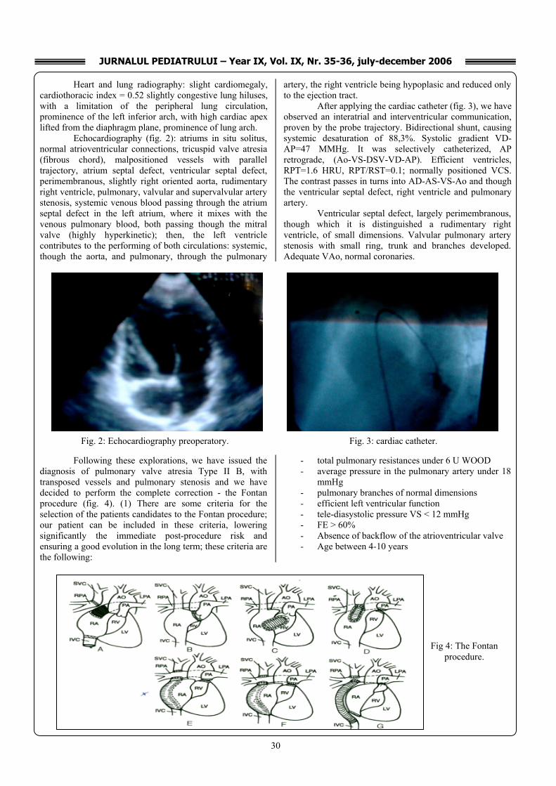
JURNALUL PEDIATRULUI – Year IX, Vol. IX, Nr. 35-36, july-december 2006
30
Heart and lung radiography: slight cardiomegaly, cardiothoracic index = 0.52 slightly congestive lung hiluses, with a limitation of the peripheral lung circulation, prominence of the left inferior arch, with high cardiac apex lifted from the diaphragm plane, prominence of lung arch.
Echocardiography (fig. 2): atriums in situ solitus, normal atrioventricular connections, tricuspid valve atresia (fibrous chord), malpositioned vessels with parallel trajectory, atrium septal defect, ventricular septal defect, perimembranous, slightly right oriented aorta, rudimentary right ventricle, pulmonary, valvular and supervalvular artery stenosis, systemic venous blood passing through the atrium septal defect in the left atrium, where it mixes with the venous pulmonary blood, both passing though the mitral valve (highly hyperkinetic); then, the left ventricle contributes to the performing of both circulations: systemic, though the aorta, and pulmonary, through the pulmonary
artery, the right ventricle being hypoplasic and reduced only to the ejection tract.
After applying the cardiac catheter (fig. 3), we have observed an interatrial and interventricular communication, proven by the probe trajectory. Bidirectional shunt, causing systemic desaturation of 88,3%. Systolic gradient VD-AP=47 MMHg. It was selectively catheterized, AP retrograde, (Ao-VS-DSV-VD-AP). Efficient ventricles, RPT=1.6 HRU, RPT/RST=0.1; normally positioned VCS. The contrast passes in turns into AD-AS-VS-Ao and though the ventricular septal defect, right ventricle and pulmonary artery.
Ventricular septal defect, largely perimembranous, though which it is distinguished a rudimentary right ventricle, of small dimensions. Valvular pulmonary artery stenosis with small ring, trunk and branches developed. Adequate VAo, normal coronaries.
Following these explorations, we have issued the
diagnosis of pulmonary valve atresia Type II B, with transposed vessels and pulmonary stenosis and we have decided to perform the complete correction - the Fontan procedure (fig. 4). (1) There are some criteria for the selection of the patients candidates to the Fontan procedure; our patient can be included in these criteria, lowering significantly the immediate post-procedure risk and ensuring a good evolution in the long term; these criteria are the following:
- total pulmonary resistances under 6 U WOOD - average pressure in the pulmonary artery under 18
mmHg - pulmonary branches of normal dimensions - efficient left ventricular function - tele-diasystolic pressure VS < 12 mmHg - FE > 60% - Absence of backflow of the atrioventricular valve - Age between 4-10 years
Fig. 3: cardiac catheter. Fig. 2: Echocardiography preoperatory.
Fig 4: The Fontan procedure.

JURNALUL PEDIATRULUI – Year IX, Vol. IX, Nr. 35-36, july-december 2006
31
Immediately after the procedure, the patient’s electrocardiography modified (fig. 5): the negative P wave,
preceding the QRS complex, FC=65-80 beats/ min, aspect of junctional rhythm with the efficient adaptation to effort.
In time, the patient gradually lost the cyanotic
effect, the perioperatory recovery was good; the patient was released from hospital 9 days after the procedure, having the surgical plaque already healed; as unique medication, it was recommended the anticoagulant treatment, under the supervision of INR. During the periodic medical examinations performed, a marked tendency to bradycardia was observed, with FC rest = 55-60 beats/min in the first 4 years, then FC rest tends to lower below 50 beats/min in the last 2 years, this being interpreted as a significant bradycardia and determining the exploration with a view to determine the suitability of permanent electro-stimulation. (2,3)
On the electrocardiography appears junctional rhythm, FC=43 beats/min, electrical axis QRS left deviated,
wave P negative in DII, DIII, AVF, wave QS in V1, wave T negative in V1, V2; rare supraventricular extrasystoles.
Holter-EKG: average FC = 40 beats/min, minimum FC = 26 beats/min, maximum FC = 86 beats/min, without TPSV or TV episodes, with 306 sinus pauses > 2.5 seconds, the longest pause being of 3.2 seconds, inferior atrial rhythm (wave P negative in DII, DIII, aVF), short periods of sinus rhythm (wave P positive in DII, DIII, aVF).
The effort test: TA start = 120/70mmHg, FC = 47 beats/min, inferior atrial rhythm (wave P negative in DII, DIII, AVF) at 100w, TA = 150/80 mmHg, FC = 125 beats/ min at 75w, FC = 90 beats/ min, aspect of sinus rhythm (wave P positive in DII, DIII, AVF – fig. 6) with the reappearance of the inferior atrial rhythm during rest, at the same time with the lowering of the cardiac frequency.
Fig. 5: EKG after the procedure.
Fig 6: Wave P positive in the effort test.

JURNALUL PEDIATRULUI – Year IX, Vol. IX, Nr. 35-36, july-december 2006
32
The patient can be included in the 2nd indication class of diffuse electro-stimulation, asymptomatic sinus bradycardia for a child with complex heart disease and heart frequency during rest under 40 beats/min or ventricular pauses > 3seconds. In this situation, were considered the type and way of electrostimulation. The only logical procedure in this case is the permanent electrostimulation. The ideal stimulation would have been an atrioventricular bicameral stimulation, as the patient had a good atrioventricular circulation; the preservation of the atrial pump would have been beneficial. Due to the complex morphological aspect resulted after the Fontan procedure, the accession of the endovascular probe in the right atrium is extremely difficult from the technical point of view and, for this reason, the patient was explored morphologically. The status after the Fontan procedure for tricuspid atresia, subaortal septal defect – 1.3 cm; right ventricle small, hypoplasic, without communication between the left and right atriums; without communication between right and left atriums, separated by a fibrous chord; Ao at valvular level = 2.12 cm, Ao ascending = 2.9 cm, DTDVS = 47 mm, PPVS = SIV = 1cm, FE = 64% (fig. 7).
The x-ray venography and the right cardiac catheterization were performed by injecting the contrast substance though the right femoral vein at the level of the right atrium; this showed the communication between the atrium and the inferior caval vein and with the right pulmonary artery though a duct; the injection of the contrast
substance through the brachial vein at the level of the superior caval vein shows its communication with the pulmonary artery, without communication between the right atrium and the superior caval vein.
This patient’s case is extremely interesting, both
morphologically and from the point of view of the arrhythmia evolution of post-surgical origin. The more complex the congenital heart defect is, the greater the possibility of a complex arrhythmia; this makes necessary the interdisciplinary collaboration.
References: 1. Klein H, Anricchio A, Reek S, et al. New Primary
prevention trials of sudden cardiac death in patients with left ventricular dysfunction: SCD-HEFT and MADIT – II. Am J Cardiol.1999; 83: 91D-97D.
2. Pescariu S, Dragulescu SI, Luca C. Ghid clinic de electrofiziologie cardiaca, Ed. Brumar, 2000.
3. Podrid PJ, KoweyPR. Cardiac Arrhytmia, Mechanisms, Diagnosis and Management, Williams & Wilkins, 1995.
Correspondence to: Adrian Lacatusu, Clinica II Pediatrie, Paltinis 1-3, Timisoara, Romania
Fig 7: ECO after the Fontan procedure.

JURNALUL PEDIATRULUI – Year IX, Vol. IX, Nr. 35-36, july-december 2006
33
CLINICAL STUDY IN PERIPHERAL ADENOPATHIES IN CHILDREN
Ileana Puiu1, D Bulucea2, Polixenia Stancu3, Veronica Nicolescu4, M Dicu5 1Head of Workings, Discipline Pediatrics, UMF Craiova 2University Professor, Discipline Pediatrics, UMF Craiova 3Reader, Discipline Pediatrics, UMF Craiova 4Intern, Pediatrics, County Clinical Emergency Hospital of Craiova 5Intern, Family Medicine, County Clinical Emergency Hospital of Craiova Abstract
Adenopathy, a common problem during childhood, may be the only clinical sign of disease, but it may as well represent one of the multiple typical signs of other illnesses. Sometimes, randomly finding an adenopathy might lead us to a severe disease. In the study we performed during the 11 years period, we noticed that infectious adenopathy is the most frequent since it was found in 694 cases (62,4%), followed by malignancy in 86 cases (7,7%) and collagen disease in 39 cases (3,5%). We also analized the onset and the course of the adenopathy, the frequency depending on the child′s age and the localization of the adenopathy. Key words: peripheral adenopathy, etiology, clinical features, child.
Introduction
In children, the lymphatic ganglions are normally touchable, save for when they are newly born children, when the ganglions are hard to notice. Following exposure to various antigen factors of the environment, a reaction of the lymphoid tissue emerges resulting in the gradual increase of the lymphatic ganglions. The fact that adenopathies appear more commonly in children may be explained by the special reaction way of the system which is immune to infections and by the persistence of the pathogenic agents in the lymphatic ganglions even after their disappearance from the initial centre.
It is often difficult to establish whether the adenopathy is a normal response to the inter-current infections that are frequent in a certain age category, or it is serious enough to suggest the existence of a more severe disease. So, the real incidence of adenopathy in children is hard to determine. This is why it was our aim to study the main diseases that develop with peripheral adenopathy in children and the differential diagnosis problems it raises. Taking into account the high number of diseases that can trigger adenomegalic syndrome in children, this diagnostic must be rapidly replaced by the diagnostic of the causal disease in order to allow for a specific treatment to be established.
Patients and Methods
This study was conducted on children with various diseases that presented significant peripheral adenopathy,
between 0 and 16 years of age, hospitalized in the Clinical Emergency Hospital and the Infectious Diseases Hospital of Craiova, for 11 years (01.01.1994 – 31.12.2004).
In all the cases, the common anamnesis, clinical and paraclinical exams were performed in order to establish the etiologic diagnosis. Generally, ganglions with the diameter of ≥ 1 cm were considered pathologic. However, in some cases, ganglions with the diameter of less than 1 cm were also considered pathologic in case they presented certain characteristics (hard texture, conglomerated, adherent, round-shaped, over-clavicle location).
The following parameters were examined in the studied group:
• the frequency of the main causes of peripheral adenopathy that required hospitalization;
• the development type of the adenopathy: acute or chronic;
• anamnestic peculiarities in connection with the age, gender;
• distribution of the cases depending on the extension of the adenopathy (localized or generalized).
Outcome and discussion
Our study was conducted only on children with significant peripheral adenopathy that required hospitalization for diagnosis and treatment.
The study on the span of 11 years revealed that the adenopathy of infectious etiology rank as first, with 694 cases (62.4%), followed by malignant diseases with 86 cases (7.7%) and auto-immune diseases with 39 cases (3.5%).
Among the serious diseases that manifest themselves by adenopathy, the most worrying both for the patients and the doctor is the possibility of a malignity. However, the presence of malignancy in patients with adenopathy is low.
Two studies conducted in the US by Allhiser (1981) and Williamson (1999) analyzing the risk of malignancy in patients with adenopathy, 3 children were found out of 238, and no case was found for the 80 children with adenopathy of undetermined adenopathy.
In the reference centers, the prevalence of malignancy in the biopsies performed on lymphatic ganglions was 40 – 60% (Lee, 1998). But these statements overestimate the probability of the existence of malignancy
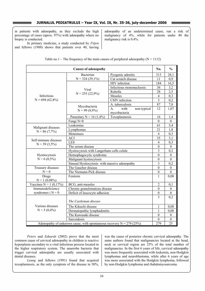
JURNALUL PEDIATRULUI – Year IX, Vol. IX, Nr. 35-36, july-december 2006
34
in patients with adenopathy, as they exclude the high percentage of cases (aprox. 97%) with adenopathy where no biopsy is conducted.
In primary medicine, a study conducted by Fitjen and fellows (1988) shows that patients over 40, having
adenopathy of an undetermined cause, run a risk of malignancy of 4%, while for patients under 40 the malignancy risk is 0.4%.
Table no.1 – The frequency of the main causes of peripheral adenopathy (N = 1112)
Causes of adenopathy No. %
Pyogenic adenitis 313 28,1 Bacterian N = 324 (29,1%) Cat scratch disease 11 0,9
HIV infection 184 16,5 Infectious mononucleosis 36 3,2 Rubella 28 2,5 Measles 4 0,3
Viral N = 255 (22,9%)
CMV infection 3 0,2 A. tuberculosis 87 7,8 Mycobacteria
N = 99 (8,9%) A. with non-typical mycobacteria
12 1,07
Parasitary N = 16 (1,4%) Toxoplasmosis 16 1,4
Infectious N = 694 (62,4%)
Fungi N=0 0 0 Leukemias 61 5,4 Lymphomas 21 1,8 Malignant diseases
N = 86 (7,7%) Metastases 4 0,3 ACJ 35 3,1 LES 4 0,3 Self-immune diseases
N = 39 (3,5%) The serum disease 0 0 Hystiocytosis with Langerhans cells celule 3 0,2 Hemophagocytic syndrome 0 0 Malignant hystiocytosis 0 0
Hystiocytosis N = 6 (0,5%)
Sinusal Hystiocytosis with massive adenopathy 3 0,2 The Gaucher disease 0 0 Treasury diseases
N = 0 The Niemann-Pick disease 0 0 Drugs
N = 1 (0.08%) Fenitoin 1 0,08
Vaccines N = 1 (0,17%) BCG, anti-measles 2 0,1 Chronic granulomatous disease 0 0 Immunodeficiency
syndromes i N = 0 Deficit of leucocyte adhesion 0 0
The Castleman disease 3 0,2
The Kikuchi disease 1 0,08 Dermatopathic lymphadenitis 1 0,08 The Kawasaki disease 0 0
Various diseases N = 5 (0,4%)
Sarcoidosis 0 0 Adenopathy of unknown cause, with spontaneous recovery N = 279 (25%) 279 25
Peters and Eduards (2002) prove that the most
common cause of cervical adenopathy in children is reactive hyperplasia secondary to a viral infectious process located in the higher respiratory system. The anaerobe bacteria that trigger cervical adenopathy are usually associated with dental diseases.
Leung and fellows (1991) found that acquired toxoplasmosis, as the only symptom of the disease in 50%,
was the cause of posterior chronic cervical adenopathy. The same authors found that malignancies located at the head, neck or cervical region are 25% of the total number of malignancies. In the first 6 years of life, cervical adenopathy was more frequently associated with leukemia, non-Hodgkin lymphomas and neuroblastoma, while after 6 years of age was more associated with the Hodgkin lymphoma, followed by non-Hodgkin lymphoma and rhabdomyosarcoma.
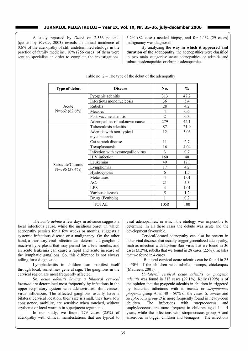
JURNALUL PEDIATRULUI – Year IX, Vol. IX, Nr. 35-36, july-december 2006
35
A study reported by Dutch on 2,556 patients (quoted by Ferrer, 2003) reveals an annual incidence of 0.6% of the adenopathy of still undetermined etiology in the practice of family medicine. 10% (256 cases) of them were sent to specialists in order to complete the investigations,
3.2% (82 cases) needed biopsy, and for 1.1% (29 cases) malignancy was diagnosed.
By analyzing the way in which it appeared and duration of the adenopathy, the adenopathies were classified in two main categories: acute adenopathies or adenitis and subacute adenopathies or chronic adenopathies.
Table no. 2 – The type of the debut of the adenopathy
Type of debut Disease No. %
Pyogenic adenitis 313 47,2 Infectious mononucleosis 36 5,4 Rubella 28 4,2 Measles 4 0,6 Post-vaccine adenitis 2 0,3
Acute N=662 (62,6%)
Adenopathies of unknown cause 279 42,1 Tuberculosis adenitis 87 21,9 Adenitis with non-typical mycobacteria
12 3,03
Cat scratch disease 11 2,7 Toxoplasmosis 16 4,04 Infection with cytomegallic virus 3 0,7 HIV infection 160 40 Leukemias 49 12,3 Lymphomas 17 4,2 Hystiocytosis 6 1,5 Metastases 4 1,01 ACJ 21 5,3 LES 4 1,01 Various diseases 5 1,2
Subacute/Chronic N=396 (37,4%)
Drugs (Fenitoin) 1 0,2 TOTAL 1058 100
The acute debute a few days in advance suggests a
local infectious cause, while the insidious onset, in which adenopathy persists for a few weeks or months, suggests a systemic infectious disease or a malignancy. On the other hand, a transitory viral infection can determine a ganglionic reactive hyperplasia that may persist for a few months, and an acute leukemia can cause a rapid and acute increase of the lymphatic ganglions. So, this difference is not always telling for a diagnostic.
Lymphadenitis in children can manifest itself through local, sometimes general sign. The ganglions in the cervical region are most frequently affected.
So, acute adenitis having a bilateral cervical location are determined most frequently by infections in the upper respiratory system with adenoviruses, rhinoviruses, virus influenzae. The affected ganglions usually have a bilateral cervical location, their size is small, they have low consistence, mobility, are sensitive when touched, without erythema or local warmth in superjacent teguments.
In our study, we found 279 cases (25%) of adenopathy with clinical manifestations that are typical to
viral adenopathies, in which the etiology was impossible to determine. In all these cases the debute was acute and the development favourable.
Cervical-located adenopathy can also be present in other viral diseases that usually trigger generalized adenopathy, such as infection with Epstein-Barr virus that we found in 36 cases (3.2%), rubella that we found in 28 cases (2.5%), measles that we found in 4 cases.
Bilateral cervical acute adenitis can be found in 25 – 50% of the children with rubella, mumps, chickenpox (Maureen, 2001).
Unilateral cervical acute adenitis or pyogenic adenitis was found in 313 cases (29.1%). Kelly (1998) is of the opinion that the pyogenic adenitis in children in triggered by bacterian infections with s. aureus or streptococus piogenes group A, in 40 – 80% of the cases. S. aureus and streptococus group B is more frequently found in newly-born children. The infections with streptococcus and staphylococcus are more frequent in children aged 1 – 4 years, while the infections with streptococcus group A and anaerobes in bigger children and teenagers. The infections

JURNALUL PEDIATRULUI – Year IX, Vol. IX, Nr. 35-36, july-december 2006
36
with anaerobe germs usually trigger unilateral adenitis with acute debut, in case of dental diseases.
The Kawasaki disease is another cause of unilateral bacterial acute adenitis that appears more often in suckling and the smaller children. It is an acute fever disease of unknown cause that can start by fever and acute cervical adenitis with unilateral location (50 – 70% of cases). This disease is rarely found in our area; no case was found during our study.
Subacute or chronic adenopathies have countless causes that can be grouped in infectious and non-infectious ones. The most common causes of infectious chronic adenopathy that we have noticed were the followings: HIV infection – 160 cases (16.5%); tuberculose adenitis – 87 cases (7.8%); toxoplasmosis – 16 cases; adenitis with non-typical mycobacteria – 12 cases; the cat scratch disease – 11 cases; cytomegalic virus infection – 3 cases. Other more seldom causes of infectious chronic adenitis are the followings: • fungi infections: aspergillosis, histoplasmosis, blastomycosis, paracoccidiomycosis; • bacteria infections: actinomycetes, anthrax, brucellosis, leptospirosis, syphilis, tularemia, inguinal granuloma. The non-infectious etiology of the chronic adenopathies consisted of:
• malignancies: leucemia (49 cases), lymphomas (17 cases), ganglionic metastases (4 cases); • collagen diseases (ACJ - 21 cases, LES - 4 cases) ; • hystiocytosis (6 cases); • various diseases (5 cases).
The subacute or chronic adenopathies are more often than not caused by the infection with mycobacteria, the cat scratch disease and toxoplasmosis. More rare causes include infection with Epstein-Barr virus and cytomegalic virus (Margileth, 1995, Malley, 2000).
The frequency of peripheral adenopathy depending on age and gender
Adenopathy is frequently noticed in children due to the immune system that reacts to the regular infections in this period. Adenopathy is rare in newly-born children who are hardly exposed to infectious agents, whereas other diseases that are associated with the adenopathy are seldom at this age.
Age is an important factor in assessing patients with adenopathy, as the frequency of the adenopathy increases with age and certain diseases appear more often in certain categories of age (Moore, 2001).
We noticed in the studied group that infectious adenopathies were more frequent in the small children, while the malignant ones and the collagen diseases were more frequent in bigger children.
Table no. 3 – The distribution of the main categories of adenopathy on etiology, age and gender.
Age groups Gender
0-1 years N=60
1-3 years N=173
3-6 years N=163
6-10 years N=159
10-16 years N=207
Types of adenopathy
Nr. % Nr. % Nr. % Nr. % Nr. %
M/F No. of cases
IInnffeeccttiioouuss
aaddeennooppaatthhiieess 56 93,3 156 90,1 144 88,3 137 86,1 166 80,1 484/275
Malignant diseases 4 6,6 15 8,6 14 8,0 15 9,4 18 8,6 38/28
Self-immune diseases 0 0 1 0,5 1 0,6 6 3,7 17 8,2 14/11
PPoosstt--vvaacccciinnee 0 0 1 0,5 0 0 1 0,6 0 0 2/0 Hystiocytosis 0 0 0 0 3 1,8 0 0 3 1,4 4/2 PPoosstt--ddrruugg
aaddeennooppaatthhiieess 0 0 0 0 1 0,6 0 0 0 0 1/0
Various diseases 0 0 0 0 0 0 0 0 5 2,4 4/1 In the 0 – 1 year of age group, infectious adenopathies
prevailed (93.3%), particularly the pyogenic adenitis (85%); the malignant ones were noticed in 6.6% cases of acute leukemia.
In the 1 – 3 years of age group, infectious adenopathies also prevail (90.1%), out of which 67% pyogenic adenitis, 8% tuberculosis adenitis and 6.9% HIV adenopathy. We hardly noticed cases of infections with non-typical
mycobacteria (2.3%) and infectious mononucleosis (1.1%). Malignant diseases were present in 8.6% cases.
In the 3 – 6 years of age group, there were 88.3% cases of infectious adenopathies, 8.1% of malignant adenopathies, and 1.8% of hystiocytosis.
In the 6 – 10 years of age group, there were 86.1% infectious adenopathies. We noticed an increase in the frequency of the cases with tuberculosis adenitis (13.8%).

JURNALUL PEDIATRULUI – Year IX, Vol. IX, Nr. 35-36, july-december 2006
37
Malignant diseases were present in 9.4% cases, while self-immune diseases in 3.7% cases.
In the 10 – 16 years of age group, there were 80.1% of infectious adenopathies. It was noticed an increase in the frequency of the cases of tuberculosis adenitis (16.4%), HIV infection (32.8%), infectious mononucleosis (10.6%), and toxoplasmosis (3.8%). There were 8.6% malignant cases, and 8.2% of self-immune diseases. In bigger children we also
noticed rare diseases of unknown etiology: the Castleman disease (3 cases), the Kukuchi disease (1 case).
The distribution of adenopathy cases in children,
based on its location After their extension, the adenopathies were classified
as: localized adenopathies and generalized adenopathies.
Table no. 4 – The connection between etiology and the location of adenopathy.
Localized Adenopathy
N = 474 (60,2%)
Cervical N=278 (58,6%)
Submento-nier; N=85
(17,9%)
Preauricullar;
N=34 (7,1%)
Supraclavicular; N=6
(1,2%)
Axillar N=41 (8,6%)
Inguinal N=30 (6,3%)
Generalized adenopathy
N = 313 (39,7%) Etiology of the adenopathy
No % No
% No % No
% No
% No
% No %
Infectious 262 94,2 85 100 34 100 2 33,3 37 90,2 30 100 226 72,2
Malignant diseases 12 4,3 - - - - 4 66,6 3 7,3 - - 51 16,2
Immunologic diseases - - - - - - - - - - - - 26 8,3
Post-drugs - - - - - - - - - - - - 1 0,3
Post-vaccine 1 0,3 - - - - - - 1 2,4 - - - -
Hystiocytosis - - - - - - - - - - - - 6 1,9
Various diseases 3 1,01 - - - - - - - - - 2 0,6
Localized or regional adenopathy was noticed in
60.2% cases, and generalized adenopathy was noticed in 39.8% cases.
In localized or regional adenopathies, each ganglionic group drains the lympha in a certain region of the body. So, the ganglions are grouped in the cervical, axillary, inguinal, mediastinal, and abdominal areas.
This study does not focus on deep mediastinal or abdominal adenopathies.
The anatomical location of the adenopathy guides us to locating the primary lesions that trigger the increase in the lymphatic ganglions.
Localized adenopathy was most of the time noticed in the cervical region (58.5%), followed by submandibulary and submentonier region (17.9%).
Cervical adenopathies were primarily benign (94.2%), caused by pyogenic adenitis (63.6%), tuberculosis adenitis (15.4%), infectious mononucleosis (10.7%).
Malignant etiology was present in 4.3% cases, being triggered by the Hodgkin disease, non-Hodgkin lymphoma and ganglionic metastases.
The adenopathy located exclusively submentoneric, pre-auricular or inguinal had benign, infectious etiology.
The super-clavicle adenopathy was rarely found (6 cases): 2 cases of tuberculosis adenitis, 3 cases with non-Hodgkin lymphomas and one case of ganglionic metastasis.
Axillary adenopathy had an infectious etiology in 37 cases (90.3%), malignant in 3 cases (7.3%), and postvaccinal in 1 case (2.4%). Benign generalized adenopathy (72.2%) was found in viral infections (HIV infection, infectious mononucleosis, rubella, smallpox, cytomegallic virus infection),

JURNALUL PEDIATRULUI – Year IX, Vol. IX, Nr. 35-36, july-december 2006
38
tuberculosis, toxoplasmosis and immunologic diseases. Herpetic virus, chickenpox-zoosterian virus and adenoviruses can also determine generalized adenopathy.
Generalized malignant adenopathy (16.2%) was found in acute leukemia and the Hodgkin disease.
Making the difference between localized and generalized adenopathy is important in setting the differential diagnosis and guides us in conducting the next investigations.
According to the studies performed by Allhiser (1981), ¾ of the studies cases presented localized adenopathy, with the rest of them having generalized adenopathy.
Herzog (1983) underlines that retro-auricular and occipital adenopathy are mostly present in sucklings and small children, while the cervical and submandibular ones are frequently present in bigger children. Cervical adenopathy, mostly noticed in children aged 2 – 12, is a response to various antigenic stimulations. Conclusions
1. Judging by the distribution of cases with peripheral adenopathy based on etiology, we noticed that infectious diseases rank first (62.4%), followed by malignant diseases (7.7%) and self-immune diseases (3.5%).
Reactive adenopathy with favourable development, with unknown etiology, was found in 25% cases.
2. The acute debute was noticed in 62.6% cases in the following diseases: pyogenic adenitis, infectious mononucleosis, rubella, smallpox, post-vaccine adenitis and acute adenopathies of unknown cause. The subacute or chronic debut was present in 37.4% cases in: tuberculosis adenitis with non-typical mycobateria, the cat scratch disease, toxoplasmosis, infection with cytomegallic virus, leukemias, lymphomas, hystiocytosis, metastases, immunologic diseases and various diseases.
3. Judging by the frequency of the peripheral adenopathies depending on age, we have noticed that infectious adenopathies were more frequent at the 0 – 1 years of age group (93.3%), malignant adenopathies at the 6 – 10 years of age group (9.4%), and adenopathy in self-immune diseases at the 10 – 16 years of age group (8.2%).
4. The distribution of the cases with peripheral adenopathy depending on location highlights the presence of localized adenopathy in 60.2% cases, and generalized adenopathy in 39.8% cases. As for the localized adenopathies, the most frequently found were peripheral adenopathies with a cervical location (58.6%), followed by submandibular location (17.9%).
References: 1. Allhiser JN, McKnight TA, Shank JC. Lymphadenopathy
in a family practice. J Fam Pract. 1981; 12: 27 - 32. 2. Ferrer R. Lymphadenopathy: differential diagnosis and
evaluation. Am Fam Physician 2003; 58: 1313 - 20. 3. Fitjen GH, Blijham GH. Unexplained lymphadenopathy
in family practice. An evaluation of the probability of malignant causes and the effectiveness of physicians workup. J Fam Pract 1988; 27: 373 - 6.
4. Herzog LW. Prevalence of lymphadenopathy of the head and neck in infants and children. Clinical Pediatrics 22 (7): 485 - 487.
5. Kelly CS, Kelly RE. jr. Lymphadenopathy in children; Pediatric Clinics of North America. 1998; 45 (4): 875 - 888.
6. Leung AK, Robson WL. Childhood cervical lymphadenopathy. Journal of Pediatrics Health Care 2004; 18 (1): 3 - 7.
7. Lee KC, Taim TA, et al. Contemporary management of cervical tuberculosis. Laryng 1998; 106: 60 -4.
8. Maureen MC. Cervical Lymphadenitis, Pediatr Rev 2001; 21 (12): 399 - 405.
9. Margileth AM. Sorting out the causes of lymphadenopathy. Contemporary Pediatrics 1995; 12: 23 - 40.
10. Moore SW. A clinical approach to Cervical adenopathy. The Medicine Journal. 2001; 43 (4).
11. Peters TR, Edwards KM. Cervical Lymphadenopathy and Adenitis. Pediatr Rev 2002; 21 (12): 399 - 405.
12. Williamson H. A jr. Lymphadenopathy in a family practice : a descriptive study of 249 casses. J. Farm. Pract; 1999; 20: 449 - 458.
Correspondence to:
Ileana Petrescu 60, Mareşal Antonescu Street Craiova Romania Telephone: +40251502210 E-mail:[email protected]
JURNALUL PEDIATRULUI – Year IX, Vol. IX, Nr. 35-36, july-december 2006
39
SURGICAL TREATMENT OF GASTROSCHISIS USING SILIMED GASTROSCHISIS CONTAINER - CASE REPORT
ES Boia¹, RE Iacob¹, O Adam², A Pavel2, Maria Trailescu², Marioara Boia1, Nicoleta Pavel3
¹University of Medicine and Pharmacy “Victor Babes” Timisoara ²Clinical Emergency Hospital for Children”Louis Turcanu” Timisoara - Pediatric Surgery and Orthopedics Department 3Special Services for Children with Dizabilities Timisoara Abstract.
Gastroschisis is a defect of the abdominal wall, on the right side of the umbilical cord, where eviscering intestinal loops.[2] The vertical opening is approximately 2 to 5 cm in size, with the umbilicus normally developed and properly positioned.[4,5] The small and large intestines are usually the only organs protruding outside the abdominal wall. The spleen and liver may also be involved, but with a much lower incidence.[6] Malformations of other major organ systems are infrequently associated with gastroschisis; however, if malformations occur, they are commonly related to infarction or atresia of the herniated bowel.[7]
Key words: gastroschisis, silimed gastroschisis container. Introduction
The etiology of gastroschisis is uncertain, but it has been reported to be of nongenetic origin.[8] Four hypotheses for the etiology of the defect have been proposed. The first is that gastroschisis may result from a vascular disruption of the right lateral fold allowing the abdominal contents to herniate outside the abdominal cavity.[2] The second is that the defect results from occlusion of the omphalomesenteric artery in utero. This occlusion may weaken the abdominal wall causing it to rupture.[4] The third hypothesis is that premature atrophy or abnormal persistence of the right umbilical vein leads to mesenchymal damage and failure of the epidermis to differentiate.[9] This damage or differentiation failure results in a defect of the abdominal wall. The fourth and last hypothesis is that a gastroschisis defect may be the end result of an intrauterine rupture of a small omphalocele with the absorption of the sac.[7]
The incidence of gastroschisis ranges from 1.4 to 2.5 per 10,000 live births and has no gender predilection.[7,9,10,12] Factors associated with an increased risk for gastroschisis include maternal age, parity, and maternal use of selected drugs. The incidence of gastroschisis is higher in young mothers and declines markedly with increasing maternal age.[1,2,13] Women less than 20 years of age are 11 times more likely to have an affected infant.[5] Low parity has also been shown to increase the risk for gastroschisis.[2] Drugs taken during the first trimester including nicotine, pseudo-
ephedrine alone or in combination with acetaminophen, phenylpropanolamine, cocaine, aspirin, and acetaminophen are associated with an increased incidence of gastroschisis.[5,15-17] With advancements in prenatal testing and ultrasonography, abdominal wall defects are commonly diagnosed in utero as early as 12 to 14 weeks gestation. The differential diagnosis of abdominal wall defects includes gastroschisis and omphalocele. Gastroschisis is a defect in the abdominal wall lateral to the umbilical cord, whereas omphalocele is a defect in which the intestines are enclosed within the umbilical cord. It may be difficult to distinguish between the two diagnoses if the protective sac of the omphalocele has been ruptured.[3,19] It is important to remember that gastroschisis defects do not involve the umbilical cord. It is also essential to distinguish between the two defects because there is a higher incidence of major congenital/chromosomal anomalies associated with omphalocele.[7] The incidence of chromosomal anomalies associated with gastroschisis is less than 5%.[4]
The infant with gastroschisis typically presents with a small, underdeveloped abdominal cavity caused by evisceration of the intestines. Although the distal portions of the colon, liver, and other solid organs have the potential to protrude through the abdominal wall defect, these organs usually remain in the abdominal cavity.[19] Malrotation occurs almost universally because of the protrusion of the intestines outside the abdominal wall.[20] Exposure to amniotic fluid can cause the uncovered bowel to become inflamed, thickened, and edematous. The affected bowel can also appear as a matted mass with no identifiable loops. A peel over the serosal surface of the bowel can occur as a result of amniotic fluid exposure. This, in conjunction with a chemical peritonitis, may impede reduction of the intestine into the abdominal cavity.[4,19,21]
There is a lower incidence of associated anomalies with gastroschisis compared with other abdominal wall defects. A 10-year review of infants with gastroschisis found a 30% incidence of associated anomalies with intestinal atresia and cryptorchidism or undescended testes being the most common.[22] Intestinal atresia was noted in 22% of affected infants, while cryptorchidism was noted in 55%.[22]
IV. PEDIATRIC SURGERY

JURNALUL PEDIATRULUI – Year IX, Vol. IX, Nr. 35-36, july-december 2006
40
In a second 10-year review of infants with gastroschisis, ileal atresia occurred in 5.4% of affected infants and cryptorchidism occurred in 24%.[14] Cryptorchidism in infants with gastroschisis has an estimated occurrence of 31%.[23] Cryptorchidism is considered a minor anomaly that usually requires conservative management.[23]
While the goal of delivery of the newborn with gastroschisis is to optimize their outcome by minimizing trauma to the exposed gastrointestinal contents, the best mode of delivery for these infants remains controversial. From a theoretical standpoint, one might assume delivery by cesarean section would be more advantageous than vaginal delivery for several reasons. The first reason is a cesarean delivery is thought to produce less compromise to the mesenteric circulation because there may be less compression and twisting of the bowel during uterine contractions and passage through the birth canal. Another reason is that the risk of infection to the exposed bowel is decreased by cesarean delivery with intact membranes. The last theoretical disadvantage to vaginal delivery is if a large defect is present with possible liver involvement, there may be an increased risk for avulsion injury.
While the rationale to promote cesarean delivery of the newborn with gastroschisis makes sense from a theoretical standpoint, none of these assumptions have been confirmed by clinical data.[24] No significant differences in outcomes between cesarean and vaginal delivery were noted in several studies of morbidity associated with gastroschisis and type of delivery.[22,25,26] The measures of morbidity in these studies included time to full oral feedings, duration of parenteral nutrition, age at discharge, incidence of complications, and number of hospital days.[22,25] Presurgical management. Stabilization and preoperative management of the newborn with gastroschisis must take into consideration many factors, including thermoregulation, fluid volume status, gastric distention and intestinal compromise, infection, respiratory status, and preparation for surgery. Stability of the aforementioned factors is necessary before the impending surgical repair to optimize the infant's outcome.
The infant must be monitored for signs of hypothermia, respiratory distress, and shock. A thorough physical examination should also be performed to determine the presence of other anomalies.[3,7]
Delivery room management of the infant with gastroschisis has included the use of saline-soaked gauze dressings to prevent damage to the exposed intestines.[29] The bowel bag is the most appropriate alternative.[28] Bowel bags provide a sterile environment for the exposed intestine and reduce the risk for contamination and tissue trauma. In addition, the bowel bag helps to prevent evaporative heat and fluid losses and enables pooling of fluid within the bag. This pooling of fluid can be measured to provide a more accurate assessment of fluid loss.[28]
Once initial stabilization in the delivery room is achieved, the newborn is admitted to the neonatal intensive care unit (NICU) for further evaluation and stabilization before surgical repair. Because the newborn with gastroschisis is at an increased risk for fluid loss because of the large surface area of exposed bowel, the newborn may
present with symptoms of shock.[30] Fluid resuscitation with isotonic solutions such as normal saline or Ringer's lactate is recommended for the newborn in shock.[31,32] Fluid resuscitation is usually continued until the infant's urine output normalizes and/or blood gases indicate normal acid-base balance.[31]
The infant must be continually assessed for signs of gastrointestinal compression before surgical repair. A naso/orogastric tube should be inserted and placed to intermittent suction to keep the bowel and stomach decompressed.[29,33]
Decompression is important because it helps to prevent partial or total obstruction of blood flow and oxygenation to the bowel. If decompression does not occur, there is an increased risk for bowel necrosis secondary to the constriction of the exteriorized intestine through the small visceral defect. Decompression will also reduce the infant's risk for emesis and thus aspiration.[33]
Bowel compromise can occur during positioning of the infant. Infants with gastroschisis should be positioned on their right side in a lateral decubitus position to enhance venous blood return from the gut.[29] The right lateral decubitus position also decreases the risk of decreased perfusion caused by compression or kinking of mesenteric vessels.[20]
Diagnostic testing and antibiotic prophylaxis are the last two areas of presurgical management. While the specific tests may vary from NICU to NICU, the most common presurgical studies ordered include a baseline chest x-ray, complete blood count (CBC) with differential and platelets, serum electrolytes, blood glucose level, total protein, and a blood type and cross match.[3] Broad-spectrum antibiotics such as ampicillin and gentamicin are started to decrease the risk of infection from bacterial contamination of the exposed bowel.[33,34]
Surgical management of the infant with gastroschisis remains controversial. While primary closure of the abdominal defect is the preferred surgical approach, each pediatric surgeon must subjectively assess the degree of abdominal wall tension anticipated before deciding the nature of the repair.[8,22] If primary closure cannot be obtained, the alternative management strategy is a staged silo repair.[31] In a small or medium size gastroschisis, one staged repair includes returning the bowel contents into the abdomen and closing the skin. If the gastroschisis is large, or there are other problems, it may need to be closed in a staged procedure over 7-10 days. A silastic sheet (silo) is placed around the exposed bowel. Every day, the silo is tightened to push more bowels into the baby's abdomen. The silo is then removed and the skin on the baby's abdomen is closed.
Because of the increased risk of sepsis and hypovolemic shock, primary closure is considered in all cases where reduction does not cause hemodynamic or respiratory compromise.[7,33] Airway and intra-abdominal pressures should be kept less than 25 and 20 mm Hg, respectively, to prevent adverse hemodynamic consequences to other organs and tissues.[7] Strategies to achieve primary repair include stretching of the abdominal wall, evacuating the contents of the stomach and small bowel, irrigating

JURNALUL PEDIATRULUI – Year IX, Vol. IX, Nr. 35-36, july-december 2006
41
meconium from the intestines, and enlarging the defect by leaving a fascial hernia.[20,35] If primary closure is attempted without sufficient space in the abdominal cavity, potential complications secondary to abdominal compartment syndrome may occur.[36,37] If the surgeon is unable to achieve primary closure or if a primary closure leads to hemodynamic and/or ventilatory compromise, an alternative method of closure must be used. Currently, most surgeons use a silastic silo for gradual reduction of herniated abdominal contents. Secondary closure occurs at a later time when the intestinal contents fit within the abdominal cavity.[7,20] Closure of the silo is usually performed in stages over 7 to 10 days, with reduction of the silo occurring one to two times daily.
A variety of methods including umbilical tape ties, sutures, clamps, or staples are used for silo reduction.[20,38] While slow reduction of the silo reduces the risk of abdominal compartment syndrome, the nurse must remember that the infant remains at risk for complications associated with abdominal compartment syndrome. The infant should be carefully assessed during and immediately after the reduction for complications. Initial postsurgical management of the infant with gastroschisis includes monitoring of vital signs, cardiovascular and respiratory status, fluid and electrolyte balance, and pain. After the repair, intra-abdominal pressure increases and can result in venous compression. Venous compression may compromise renal blood flow and the glomerular filtration rate, resulting in decreased urine output. A urinary catheter may be necessary to relieve bladder distention and to allow for a more accurate measurement of urine output.[4] Maintenance fluid requirements may need to be increased because of third spacing into the distended bowel and abdominal cavity.[29-31] Alterations in electrolyte balance may ensue from this shift of fluids. The postoperative infant may require anywhere from 120 to 170 mL/kg/d of a crystalloid solution that is adjusted to provide for adequate tissue perfusion and urine output.[20] A large-bore naso/orogastric tube placed to intermittent suction is needed to prevent gastrointestinal distention caused by hypoperistalsis. Hypoperistalsis or adynamic ileus is frequently seen in the postoperative period and may persist for several weeks.[29] The initial gastrointestinal drainage is characteristically green because of the back up of biliary and pancreatic secretions in the immediate postoperative period. As gut motility improves, the drainage becomes clear in appearance. Volume loss from the gastric tube must be monitored, because it is possible for the infant to lose up to 100 mL/kg/d.[3] Replacement of these losses is necessary to maintain homeostasis.
Because of the increased intra-abdominal pressure, close monitoring of respiratory status is essential for the first 48 to 72 hours postsurgery. Respiratory support, as indicated, is provided to optimize oxygenation and ventilation. The increase in abdominal pressure may interfere with optimal expansion of the diaphragm and venous return impeding both ventilation and oxygenation.[42] Some infants may benefit from mechanical ventilation or continuous positive airway pressure (CPAP) to maximize lung expansion, lung volume, and oxygenation.[33] Other
infants may not tolerate CPAP because of increased abdominal distention from the increased airflow to the gastrointestinal track. A properly functioning naso/orogastric tube will minimize this risk.
In the immediate postsurgical period, the infant with gastroschisis is usually returned to the NICU from the operating room intubated and on mechanical ventilation. While most infants can be extubated within 24 to 48 hours after surgery, infants who are small for gestational age, preterm, and/or have significantly increased intra-abdominal pressure may require a longer period of ventilator support.[31]
After the initial stabilization period, the main goal of management is to provide adequate nutrition and pain management. Initially, the infant will require parenteral nutrition. Gut motility is delayed because of the chemical peritonitis that occurred when the intestinal contents were exposed to amniotic fluid. Delayed gut motility may persist for weeks after surgical repair and is often influenced by the severity of the defect and other associated anomalies such as intestinal atresia.[29,31]
Because of the postoperative ileus, total parenteral nutrition (TPN) is needed in all infants with gastroschisis and is usually initiated within 24 to 48 hours after surgery.[29,33] Because these infants may require TPN for weeks after surgery,[31] a central line is recommended. The minimal daily requirements for postoperative TPN are 90 to 100 kcal/kg/d, 3 g/kg/d of protein, 3 to 4 g/kg/d of intravenous lipids, and dextrose to maintain euglycemia.[43] Because of protein losses from the surgical stress, wound healing, and/or third spacing, additional protein in the TPN may be necessary.[43] Once gut motility returns, it is important to be proactive with the initiation of enteral feedings. A retrospective study found the age of initial enteral feeding was positively correlated with the time of discharge.[14] These investigators also noted that for every additional day enteral feedings were delayed, hospital length of stay was increased by 1 day. Infants with gastroschisis have a tendency toward malabsorption of substrates and possible allergies secondary to gut inflammation. The use of elemental formulas, expressed human milk, or preterm formulas are indicated because they are more easily digested.[3,4,43] Typically, small volume feedings are initiated and advanced by 10 to 20 mL/kg/d as tolerated.[43] TPN is usually decreased as the feedings increase. Manipulation of bowel and the increase in intra-abdominal pressure postrepair may increase the need for analgesia in the first 48 to 72 hours after surgery.[30] Infants should be routinely assessed for pain using a validated pain assessment tool, and analgesia should be provided as needed according to established pain guidelines.[44] Pain may be controlled with analgesics such as morphine sulfate or fentanyl as a continuous intravenous drip or as a bolus at regularly scheduled intervals. The nurse should keep in mind that these medications may result in respiratory depression and slow gut motility.[3]
Outcomes for the infant with gastroschisis are usually affected by a number of complications, including
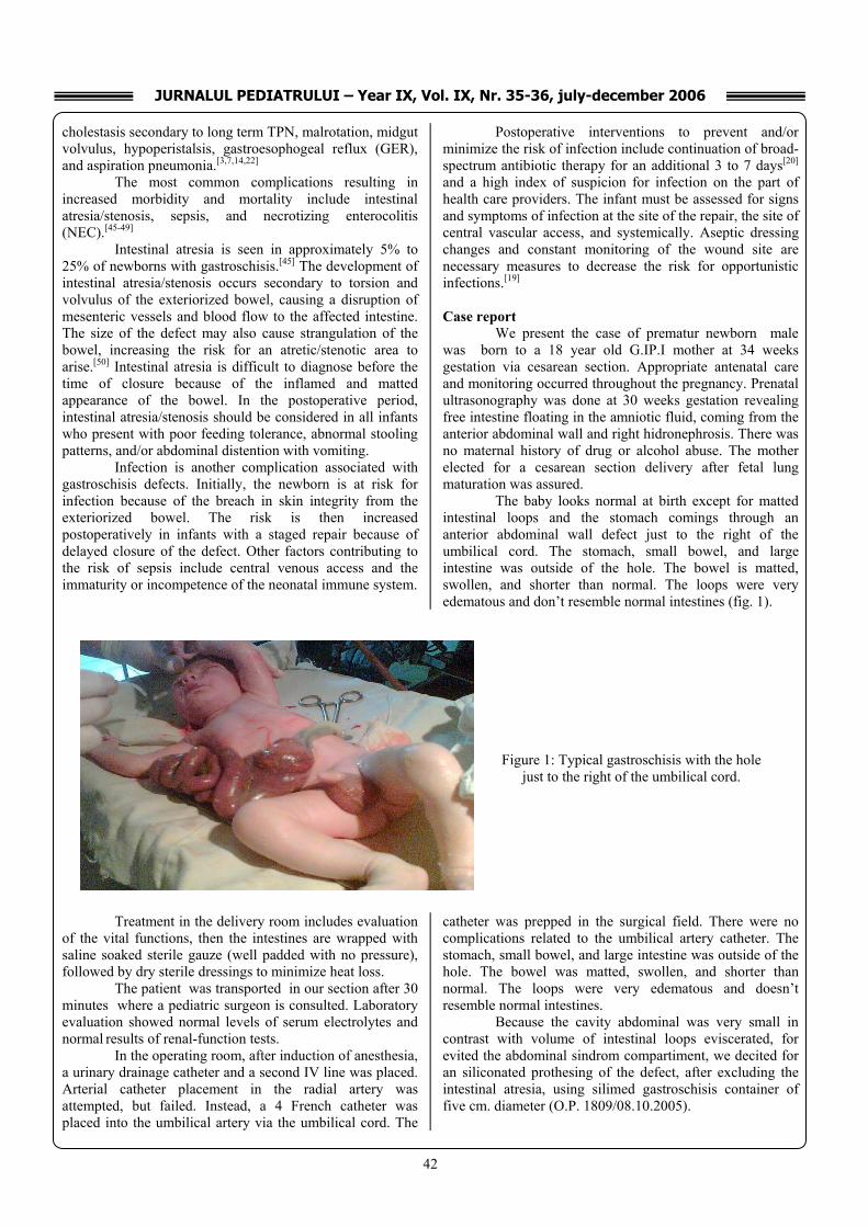
JURNALUL PEDIATRULUI – Year IX, Vol. IX, Nr. 35-36, july-december 2006
42
cholestasis secondary to long term TPN, malrotation, midgut volvulus, hypoperistalsis, gastroesophogeal reflux (GER), and aspiration pneumonia.[3,7,14,22]
The most common complications resulting in increased morbidity and mortality include intestinal atresia/stenosis, sepsis, and necrotizing enterocolitis (NEC).[45-49] Intestinal atresia is seen in approximately 5% to 25% of newborns with gastroschisis.[45] The development of intestinal atresia/stenosis occurs secondary to torsion and volvulus of the exteriorized bowel, causing a disruption of mesenteric vessels and blood flow to the affected intestine. The size of the defect may also cause strangulation of the bowel, increasing the risk for an atretic/stenotic area to arise.[50] Intestinal atresia is difficult to diagnose before the time of closure because of the inflamed and matted appearance of the bowel. In the postoperative period, intestinal atresia/stenosis should be considered in all infants who present with poor feeding tolerance, abnormal stooling patterns, and/or abdominal distention with vomiting. Infection is another complication associated with gastroschisis defects. Initially, the newborn is at risk for infection because of the breach in skin integrity from the exteriorized bowel. The risk is then increased postoperatively in infants with a staged repair because of delayed closure of the defect. Other factors contributing to the risk of sepsis include central venous access and the immaturity or incompetence of the neonatal immune system.
Postoperative interventions to prevent and/or minimize the risk of infection include continuation of broad-spectrum antibiotic therapy for an additional 3 to 7 days[20] and a high index of suspicion for infection on the part of health care providers. The infant must be assessed for signs and symptoms of infection at the site of the repair, the site of central vascular access, and systemically. Aseptic dressing changes and constant monitoring of the wound site are necessary measures to decrease the risk for opportunistic infections.[19]
Case report
We present the case of prematur newborn male was born to a 18 year old G.IP.I mother at 34 weeks gestation via cesarean section. Appropriate antenatal care and monitoring occurred throughout the pregnancy. Prenatal ultrasonography was done at 30 weeks gestation revealing free intestine floating in the amniotic fluid, coming from the anterior abdominal wall and right hidronephrosis. There was no maternal history of drug or alcohol abuse. The mother elected for a cesarean section delivery after fetal lung maturation was assured.
The baby looks normal at birth except for matted intestinal loops and the stomach comings through an anterior abdominal wall defect just to the right of the umbilical cord. The stomach, small bowel, and large intestine was outside of the hole. The bowel is matted, swollen, and shorter than normal. The loops were very edematous and don’t resemble normal intestines (fig. 1).
Treatment in the delivery room includes evaluation
of the vital functions, then the intestines are wrapped with saline soaked sterile gauze (well padded with no pressure), followed by dry sterile dressings to minimize heat loss.
The patient was transported in our section after 30 minutes where a pediatric surgeon is consulted. Laboratory
evaluation showed normal levels of serum electrolytes and normal results of renal-function tests.
In the operating room, after induction of anesthesia, a urinary drainage catheter and a second IV line was placed. Arterial catheter placement in the radial artery was attempted, but failed. Instead, a 4 French catheter was placed into the umbilical artery via the umbilical cord. The
catheter was prepped in the surgical field. There were no complications related to the umbilical artery catheter. The stomach, small bowel, and large intestine was outside of the hole. The bowel was matted, swollen, and shorter than normal. The loops were very edematous and doesn’t resemble normal intestines.
Because the cavity abdominal was very small in contrast with volume of intestinal loops eviscerated, for evited the abdominal sindrom compartiment, we decited for an siliconated prothesing of the defect, after excluding the intestinal atresia, using silimed gastroschisis container of five cm. diameter (O.P. 1809/08.10.2005).
Figure 1: Typical gastroschisis with the hole just to the right of the umbilical cord.

JURNALUL PEDIATRULUI – Year IX, Vol. IX, Nr. 35-36, july-december 2006
43
The silo has a flexible ring at the bottom which is inserted inside the abdomen while the intestines sit inside of the bag. The bag is made smaller little by little which causes the intestines to go back into the abdomen. The bag is sterile, impermeable to micro-organisms, transparent, flexible, resistant, internally smooth, does not adhere to the bowel loops, readily available, and inexpensive, properties
that make it an excellent alternative as a prosthesis for staged surgical treatment of congenital anomalies of the abdominal wall such as gastroschisis.
The intestines was returned to the abdomen gradually by gentle pressure and placing the string which ties off the top of the silo gradually lower on the silo at the bedside in the NICU (fig.2,3).
Once the intestines are almost all back inside (this
process was completed after 9 days in this case), the infant was returned to the operating room for closure of the gastroschisis (O.P. 1874/17.10.2005)
The silo was removed (fig. 4) and the hole in the abdomen closed, but because still was tension by the suture line we decided for lateroabdominal incisions (fig.5).
Antibiotics are discontinued shortly after the silo is removed.
The patient was supported with a ventilator for about 12 hours, then weaned to supplemental nasal oxygen at 1 week. The infant was initially maintained on peripheral total parenteral nutrition (TPN). Nasogastric suction was discontinued at 1 week and gesol solution begun, with the
use of a feeding tube. Two weeks before admission, fever, vomiting, shortness of breath, productive cough, and generalized weakness developed. The oxygen saturation was 88 percent while the patient was breathing ambient air.
Figure 2: A newborn with gastroschisis with a silimed gastroschisis container of five cm.
diameter- a 6-th day postsurgical.
Figure 3: A newborn with gastroschisis with a silimed gastroschisis container of five cm.
diameter- a 8-th day postsurgical.
Figure 4: A newborn with gastroschisis after container removed - a 9-th day postsurgical. Figure 5: Closure of the abdominal wall.

JURNALUL PEDIATRULUI – Year IX, Vol. IX, Nr. 35-36, july-december 2006
44
This was progressed to half-strength, then full-strength, breast milk over a 2-week period. He was gradually weaned from continuous feeds to bolus and regular breastfeeding. The patient was discharged at 25 days of life. Her weight was 2400 grams at time of discharge. At 6 month of age, he had experienced normal growth and development. Conclusions: 1. Gastroschisis is a rare, but complex, defect of the
abdominal wall. 2. Surgical treatment using silimed gastroschisis container
is an effective treatment in gastroschisis if the surgeon is unable to achieve primary closure leads to hemodinamic and/or ventilatory compromise.
3. While primary closure of the abdominal defect is the preferred surgical approach, each pediatric surgeon must subjectively assess the degree of abdominal wall tension anticipated before deciding the nature of the repair and the alternative management strategy is a staged silo repair.
4. There are numerous complications that may occur secondary to the evisceration of the intestines, requiring long-term follow-up.
5. With the advances seen in neonatal medicine, including surgical techniques, parenteral nutrition, respiratory support, and control of infection, these infants may go on to lead healthy and productive lives.
References 1. Molik K, Gingalewski CA, West KW, et al:
Gastroschisis: A plea for risk categorization. J Pediatr Surg 36:51-55, 2001.
2. Torfs C, Curry C, Roeper P: Gastroschisis. J Pediatr 116:1- 6, 1990.
3. Howell KK: Understanding gastroschisis: An abdominal wall defect. Neonatal Network 17:17-25, 1998 .
4. Glasser JG: Omphalocele and Gastroschisis. eMedicine 2001;2: 1-23. Accessed February 25, 2002.
5. Martin RW: Screening for fetal abdominal wall defects. Obstet Gynecol Clin North Am 25:517-526, 1998.
6. Luton D, DeLaguasie P, Guibourdenche J, et al: Effect of amnioinfusion on the outcome of prenatally diagnosed gastroschisis. Fetal Diagnos Ther 14:152-155, 1999.
7. Puri A, Bajpai M: Gastroschisis and omphalocele. Ind J Pediatr 66:773-789, 1999.
8. Komuro H, Imaizumi S, Hirata A, et al: Staged silo repair of gastroschisis with preservation of the umbilical cord. J Pediatr Surg 33:485- 488, 1998.
9. Khan AN, Thomas N: Gastroschisis. Accessed December 27, 2002. Di Tanna GL, Rosano A, Mastroiacovo R: Prevalence of gastroschisis at birth: Retrospective study. BMJ 325:389 -390, 2002.
10. Curry J, McKinney P, Thornton J, et al: The aetiology of gastroschisis. Br J Obstet Gynecol 107:1339 -1346, 2000
11. Boyd P, Bhattacharjee A, Gould S, et al: Outcome of prenatally diagnosed anterior abdominal wall defects. Arch Dis Childhood 78: F209-F213, 1998.
12. Stoll C, Alembik Y, Dott B, et al: Risk factors in congenital abdominal wall defects (omphalocele and gastroschisi): A study in a series of 265, 858 consecutive births. Ann Genet 44:201-208, 2001.
13. Sharp M, Bulsara M, Gollow I, et al: Gastroschisis: Early enteral feeds may improve outcome. J Paediatr Child Health 36:472- 476, 2000.
14. Kozer E, Nikfar S, Costei A, et al: Aspirin consumption during the first trimester of pregnancy and congenital anomalies: A meta-analysis. Am J Obstet Gynecol 187:1623-1630, 2002.
15. Werler MM, Sheehan JE, Mitchell AA: Maternal medication use and risks of gastroschisis and small intestinal atresia. Am J Epidemiol 155:26 -31, 2002.
16. Hume RF Jr, Martin LS, Bottoms SF, et al: Vascular disruption birth defects and history of prenatal cocaine exposure: a case control study. Fetal Diagn Ther 12:292-295, 1997.
17. Weaver LT: Anatomy and embryology, in Craven L (ed) Pediatric Gastrointestinal Disease: Pathophysiology, Diagnosis, & Management (ed 2). St Louis, MO, Mosby, 1996, p 9.
18. Brandt ML: Gastrointestinal surgical emergencies of the newborn, in Taeusch HW, Ballard RA (eds): Avery's Diseases of the Newborn (ed 7). Philadelphia, PA, WB Saunders, 1998, pp 979-994.
19. Kurkchubasche AG: The fetus with an abdominal wall defect. Med Health RI 84:159 -161, 2001
20. Ortiz VN, Villareal DH, Olmo J, et al: Gastroschisis: A ten-year review. Bolivian Assoc Med Periodical Rev 90:69 -73, 1998.
21. Lawson A, de la Hunt MN: Gastroschisis and undescended testis. J Pediatr Surg 36:366 -367, 2001
22. Blakelock RT, Harding JE, Kolbe A, et al: Gastroschisis: Can the morbidity be avoided? Pediatr Surg Intensivist 12:276 -282, 1997.
23. Anteby E, Yagel S: Route of delivery of fetuses with structural anomalies. Eur J Obstet Gynecol Reprod Biol 106:5-9, 2003.
24. Segel SY, Marder SJ, Parry S, et al: Fetal abdominal wall defects and mode of delivery: A systematic review. Obstet Gynecol 98(5 Pt 1):867- 873, 2001.
25. Strodtbeck F: Abdominal wall defects. Neonatal Network 17:51- 53, 1998
26. Rescorla FJ: Surgical emergencies in the newborn, in Polin RA, Yoder MC, Burg FD (eds): Workbook in Practical Neonatology (ed 3). Philadelphia, PA, WB Saunders, 2001, pp 423- 459
27. Hartman GE, Boyajian MJ, Choi SS, et al: General surgery, in Avery GB, Fletcher MA, MacDonald MG (eds): Neonatology: Pathophysiology and Management of the Newborn (ed 5). Philadelphia, PA, Lippincott, Williams & Wilkins, 1999, pp 1005-1044
28. Gaines BA, Holcomb GW, Neblett WW: Gastroschisis and omphalocele, in Ashcraft KW (ed): Pediatric

JURNALUL PEDIATRULUI – Year IX, Vol. IX, Nr. 35-36, july-december 2006
45
Surgery (ed 3). Philadelphia, PA, WB Saunders, 2000, pp 639- 653.
29. Langer JC: Gastroschisis and omphalocele. Semin Pediatr Surg 5:124 -128, 1996.
30. Diaz JH: Intraoperative management, in Goldsmith JP, Karotkin EH (eds): Assisted Ventilation of the Neonate (ed 3). Philadelphia, PA, WB Saunders, 1996, pp 409- 435
31. Berseth CL: Disorders of the umbilical cord, abdominal wall, urachus, and omphalomesenteric duct, in Taeusch HW, Ballard RA (eds): Avery's Diseases of the Newborn (ed 7). Philadelphia, PA, WB Saunders, 1998, pp 933-940.
32. Dolgin SE, Midulla P, Shlasko E: Unsatisfactory experience with 'minimal intervention management' for gastroschisis. J Pediatr Surg 35:1437-1439, 2000.
33. Watson RA, Howdieshell TR: Abdominal compartment syndrome. South Med J 91:326 -332, 1998.
34. Vanamo K: Silo reduction of giant omphalocele and gastroschisis utilizing continuous controlled pressure. Pediatr Surg Intensivist 16:536 -537, 2000.
35. Kimble RM, Singh SJ, Bourke C, et al: Gastroschisis reduction under analgesia in the neonatal unit. J Pediatr Surg 36:1672-1674, 2001.
36. Bianchi A, Dickson AP: Elective delayed reduction and no anesthesia: "minimal intervention management" for gastroschisis. J Pediatr Surg 33:1338 -1340, 1998
37. Davies MW, Kimble RM, Woodgate PG: Ward reduction without general anesthesia versus reduction and repair under general anesthesia for gastroschisis in newborn infants. Cochrane Database Syst Rev 3:CD003671, 2002
38. Dimitriou G, Greenought A, Griffin F, et al: Temporary impairment of lung function in infants with anterior abdominal wall defects who have undergone surgery. J Pediatr Surg 31:670 -672, 1996
39. Valentine CJ: Congenital anomalies of the alimentary tract, in Groh-Wargo S, Thompson M, Cox J (eds): Nutritional Care for High-Risk Neonates, Chicago, IL, Precept Press, 2000, pp 439- 455
40. Walden M: Pain Assessment and Management: Guideline for Practice. Glenview, IL, National Association of Neonatal Nurses, 2001
41. Driver CP, Bruce J, Bianchi A, et al: The contemporary outcome of gastroschisis. J Pediatr Surg 35:1719 -1723, 2000
42. Fleet MS, de la Hunt MN: Intestinal atresia with gastroschisis: A selective approach to management. J Pediatr Surg 35:1323-1325, 2000.
43. Sai Prasad TR, Bajpai M: Intestinal atresia. Ind J Pediatr 67:671- 678, 2000
44. Snyder CL: Outcome analysis for gastroschisis. J Pediatr Surg 34:1253-1256, 1999.
45. Ogunyemi D: Gastroschisis complicated by midgut atresia, absorption of bowel, and closure of the abdominal wall defect. Fetal Diagn Ther 16:227-230, 2001.
46. Jayanthi S, Seymour PL, Puntis JW, et al: Necrotizing enterocolitis after gastroschisis repair: A preventable complication? J Pediatr Surg 33:705-707, 1998.
47. Oldham KT, Coran AG, Drongowski RA, et al: The development of necrotizing enterocolitis following repair of gastroschisis: a surprisingly high incidence. J Pediatr Surg 23:945-949, 1988
48. Tunell WP, Puffinbarger NK, Tuggle DW, et al: Abdominal wall defects in infants. Survival and implications for adult life. Ann Surg 221:525-530, 1995
49. Koivusalo A, Lindahl H, Rintala RJ: Morbidity and quality of life in adult patients with a congenital abdominal wall defect: a questionnaire survey. J Pediatr Surg 37:1594 -1601, 2002
50. E.S.Boia, Marioara Boia: Urgenţe chirurgicale neonatale.Ed. Popa’s Art,1995.
Correspondence to:
Eugen Boia, Gospodarilor Street, No. 42, Timisoara 300778, Romania, E-mail: [email protected]
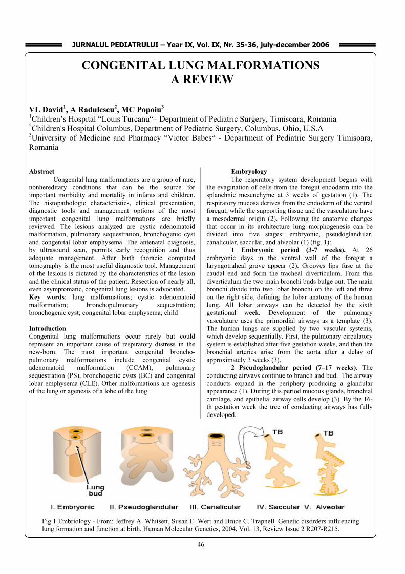
JURNALUL PEDIATRULUI – Year IX, Vol. IX, Nr. 35-36, july-december 2006
46
CONGENITAL LUNG MALFORMATIONS A REVIEW
VL David1, A Radulescu2, MC Popoiu3
1Children’s Hospital “Louis Turcanu“– Department of Pediatric Surgery, Timisoara, Romania 2Children's Hospital Columbus, Department of Pediatric Surgery, Columbus, Ohio, U.S.A 3University of Medicine and Pharmacy “Victor Babes“ - Department of Pediatric Surgery Timisoara, Romania Abstract
Congenital lung malformations are a group of rare, nonhereditary conditions that can be the source for important morbidity and mortality in infants and children. The histopathologic characteristics, clinical presentation, diagnostic tools and management options of the most important congenital lung malformations are briefly reviewed. The lesions analyzed are cystic adenomatoid malformation, pulmonary sequestration, bronchogenic cyst and congenital lobar emphysema. The antenatal diagnosis, by ultrasound scan, permits early recognition and thus adequate management. After birth thoracic computed tomography is the most useful diagnostic tool. Management of the lesions is dictated by the characteristics of the lesion and the clinical status of the patient. Resection of nearly all, even asymptomatic, congenital lung lesions is advocated. Key words: lung malformations; cystic adenomatoid malformation; bronchopulmonary sequestration; bronchogenic cyst; congenital lobar emphysema; child Introduction Congenital lung malformations occur rarely but could represent an important cause of respiratory distress in the new-born. The most important congenital broncho-pulmonary malformations include congenital cystic adenomatoid malformation (CCAM), pulmonary sequestration (PS), bronchogenic cysts (BC) and congenital lobar emphysema (CLE). Other malformations are agenesis of the lung or agenesis of a lobe of the lung.
Embryology The respiratory system development begins with
the evagination of cells from the foregut endoderm into the splanchnic mesenchyme at 3 weeks of gestation (1). The respiratory mucosa derives from the endoderm of the ventral foregut, while the supporting tissue and the vasculature have a mesodermal origin (2). Following the anatomic changes that occur in its architecture lung morphogenesis can be divided into five stages: embryonic, pseudoglandular, canalicular, saccular, and alveolar (1) (fig. 1):
1 Embryonic period (3-7 weeks). At 26 embryonic days in the ventral wall of the foregut a laryngotraheal grove appear (2). Grooves lips fuse at the caudal end and form the tracheal diverticulum. From this diverticulum the two main bronchi buds bulge out. The main bronchi divide into two lobar bronchi on the left and three on the right side, defining the lobar anatomy of the human lung. All lobar airways can be detected by the sixth gestational week. Development of the pulmonary vasculature uses the primordial airways as a template (3). The human lungs are supplied by two vascular systems, which develop sequentially. First, the pulmonary circulatory system is established after five gestation weeks, and then the bronchial arteries arise from the aorta after a delay of approximately 3 weeks (3).
2 Pseudoglandular period (7–17 weeks). The conducting airways continue to branch and bud. The airway conducts expand in the periphery producing a glandular appearance (1). During this period mucous glands, bronchial cartilage, and epithelial airway cells develop (3). By the 16-th gestation week the tree of conducting airways has fully developed.
Fig.1 Embriology - From: Jeffrey A. Whitsett, Susan E. Wert and Bruce C. Trapnell. Genetic disorders influencing
lung formation and function at birth. Human Molecular Genetics, 2004, Vol. 13, Review Issue 2 R207-R215.
JURNALUL PEDIATRULUI – Year IX, Vol. IX, Nr. 35-36, july-december 2006
47
3 Canalicular period (16–26 weeks). Tubules expand to form saccules widening the airway lumina. The mesenchyme thins and the airspaces come into increasingly close apposition to capillary network to form the gas exchange region (1). During this period the type I cells differentiate and surfactant components start to be produced by the cuboidal type II cells (1). After 23-24 gestation weeks despite the pulmonary immaturity preterm babies could survive with intensive care support (1-3).
4 Saccular period (22–36 weeks). During this stage the lung tissue maturate in preparation for birth. The
acinar tubules continue to proliferate and expand. The surface area of the gas exchange region increases and additional alveoli septae form. At 30 week of gestation surfactant is detectable in the amniotic fluid (3).
5 Alveolar period (36 weeks to maturity). The alveolar septation is completed in this period. Alveolar structures can be recognize histological after 32 weeks and are uniformly present at 36 gestation weeks (3). Most of the alveoli are formed after birth in the first two years of life (3).
Each of this stages associate specific disorders in correspondence with the developmental processes (Table1).
Embryonic Pseudoglandular Canalicular Saccular Alveolar Bronchogenic cysts
Pulmonary hypoplasia
Pulmonary hypoplasia
Pulmonary hyperplasia
Lobar emphysema
Pulmonary/lobar atresia
Cystic adenomatoid malformation
Respiratory distress syndrome
Pulmonary hypertension
Extralobar sequestration
Intralobar sequestration
Alveolar capillary dysplasia
Bronchopulmonary dysplasia
Pleural effusions
Tracheal/bronchial atresia/stenosis
Pulmonary lymphangiectasis
Alveolar simplification
Laryngeal/ esophageal atresia/ stenosis
Diaphragmatic hernia
Table1. Lung formation stages and specific lesions, Congenital cystic adenomatoid malformation (CCAM)
Is a rare pulmonary malformation with an estimated incidence between 1/25000 and 1/35000 (4). It is produced by the abnormal and extensive overgrowth of the bronchiolar structures. The cause for appearance is unknown but the error occurs sometimes between the fifth to the sixth week of gestation in the pseudoglandular stage of lung development (2). Stocker described in 1977 three distinct histological subtypes which later were expanded to five in
2002 (5). Each type was related with specific symptoms and radiographic findings (Table 2).
Type 1 is present in 50-70 % of cases and has the best prognosis (2). Type 2 has a poor prognosis and associates frequently other structural and chromosomal abnormalities (2). A more useful classification seems to be the one introduced by Adzick et al (4-5) using prenatal ultrasound examination. This classification simply differentiates antenatally lung lesions into macrocystic and microcystic.
0 Involvement of all lung lobes, incompatible with life 1 Single or multiple cysts, more 2 cm, lined by pseudostratified columnar epithelium 2 Single or multiple cysts (under 2 cm). Cuboidal or columnar epithelial lining 3 Predominantly solid lesions, with small (under 0.5 cm) cysts, lined by cuboidal epithelium 4 Large air-filled cysts, lined by flattened epithelial cells Table 2. Stocker’s classification (5).
Clinical presentation is variable. About 10%
become symptomatic during fetal life (4). The large mass inside the lung can restrict lung growth, can produce mediastinal shift, cardiovascular compromise and cava obstruction leading to non-immune hydrops fetalis (HF). 60% of patients become symptomatic within one month of life, another 10% between one and six months and 15 % by adolescence (6). The key symptom in neonatal period is the
respiratory distress. Outside of the neonatal period recurrent pulmonary infections in the affected lung with poor response to medical treatment is the commune presentation mode (6-7).
Postnatal the diagnostic is made on X-ray showing multiple air filled spaces (macrocystic) or a solid area (microcystic) (6). It is essential to take the X-ray with an in situ nasogastric tube in order to avoid mistaken with a

JURNALUL PEDIATRULUI – Year IX, Vol. IX, Nr. 35-36, july-december 2006
48
diaphragmatic hernia (2). Computed tomography (CT) is the most utile technique for diagnostic and pre-therapeutic evaluation (5-7). It show large air filled, fluid filled cysts or
containing air fluid levels (type 1); solid mass with multiple small cysts (type3) (7) (fig. 2).
Other diagnostic means include magnetic
resonance (MRI) and bronchoscopy. One of the most recent developments in the area is virtual bronchoscopy (VB). VB is a non-invasive three-dimensional (3D) technique that uses multidetector CT generated image. It can evaluate the airways down to the sixth- to seventh-order bronchial subdivisions (8).
Antenatal diagnostic it’s made by ultrasound scan (US) at 20-th gestation week (2-4-5-6). After the condition is detected the fetus is evaluated and monitored using ultrasound or MRI. MRI is more accurately providing besides structural, functional information also (3). The natural history of antenatal diagnosed CCAM is difficult to determine accurately. Although largely silent during fetal life and rare spontaneous regression of CCAM has been reported there are certain cases when fetal intervention is required: high CCAM volume ratio, presence of HF, mediastinal shift, cardiovascular compression, lung hypoplasia (4). The optimal type of fetal intervention is still a debate subject. Treatment options include: fetal surgical resection, cyst-amniotic shunting, thoracentesis, and steroid therapy (4). In some cases of CCAM associated whit HF antenatal steroid therapy was sufficient. Cyst-amniotic shunting may be the treatment of choice for macrocystic CCAM with HF while microcistic CCAM require surgical resection. Termination of pregnancy should be considered by the family when the fetus has insufficient pulmonary tissue to support life after birth (4).
Even if complications do not occur during intrauterine life there are still certain risks that require a delivery at a unit with appropriate neonatal and surgical expertise. After birth management is dictated by the clinical status of the new-born (5). If the early respiratory distress is present immediate management is required: appropriate management of the respiratory distress followed by surgical resection of the lesion (2).
Surgical resection of CCAM lesions is necessary even for asymptomatic patients in order to prevent infection and avoid malignant transformation of the lesion (5).
Excision of the CCAM is accomplished by lobectomy or segmentectomy and in certain cases even pneumonectomy (7). The usual intervention is lobectomy via thoracotomy or thoracoscopical approach. Lobectomy is preferred because of potential early air-leak after segmentectomy and long-term complications after pneumonectomy (9). Thoracoscopy provides several potential advantages over thoracotomy: lower pain, better postoperative pulmonary mechanics (10). Moreover up to 30% of neonates develop scoliosis after thoracotomy (10). Anesthetic management includes tracheal intubation and ventilation, central venous line, isoflurane/ oxygen analgesia, muscle relaxation. For a better surgical access, protection of the normal lung and reduced blood loss one-lung ventilation (OLV) should be used (17).
Opened ore thoracoscopical lobectomy follows the same principles. After the exposure of lung hilus the visceral pleura is opened carefully. The hilar vessels and lobar bronchus are exposed. The main segmental artery branches are identified, ligated with 3-0 silk and divided. The second stage consists in the identification, ligation and division of the segmental vein branches. The last to be approached is the lobar bronchus which is dived after a vascular clamp is placed proximal from the resection. The bronchus is sutured with continuous 3-0 silk and the closure is tested for air leak. After a lobectomy it is important to divide the inferior pulmonary ligament in order to enable the remaining lung to develop freely in the thoracic cavity. Postoperative a drain tube is leaved inside the pleural cavity for 48 h (2).
Early complications include sepsis, air leaks with pneumothorax, bronchopleural fistula, wound infections. Later complications and sequelae are incomplete excision, asthma, pneumonia (2-5).
Prognosis depends of several factors such as: cyst volume, histological type (type I has the best prognosis), the presence of complications during fetal life, the presence and severity of early respiratory distress (2-4-5). Neonates tolerate well lobectomy and the remaining lung usually evolves and expands into the remaining cavity replacing the lost pulmonary tissue (2).
CT: CCAM in the right lower lobe.

JURNALUL PEDIATRULUI – Year IX, Vol. IX, Nr. 35-36, july-december 2006
49
Pulmonary sequestration (PS) Pulmonary sequestration is a rare congenital lung
malformation characterized by a mass of nonfunctioning lung tissue with no connection with the normal tracheobronchial tree (2-11-12). The lesion has distinguished blood supply originating from the systemic artery system. The term “sequestration,” was first used in the medical literature by Pryce in 1946 (5-11-13). It represents 0.15- 6% of all pulmonary malformations (11). The origin of the lesion is uncertain but the most widely accepted hypothesis is that it results from an accessory lung bud developing inferior to the normal lung bud (11). There are 2 distinctive forms of pulmonary sequestration: intralobar and extralobar.
Intralobar sequestration is contained within the normal visceral pleura and it is usually located into the posterior basal segments of the lower left lobe (2-13). The arterial supply is from the descending thoracic aorta through inferior pulmonary ligament in 90% of cases (2-13). Other arterial sources could be the intercostal artery, subclavian
arteries, internal thoracic arteries and sometimes coronary arterial circulation which predispose to myocardial ischemia (13). Venous drainage is via the pulmonary veins in 98% and sometimes into the azygos or hemiazygos veins (2-13).
Extralobar sequestration is three times less frequent than the intralobar type (2-12) and usually is associated with other congenital anomalies (congenital diaphragmatic hernia, pectus excavatum, vertebral anomalies, and pericardial defects) (2). It is a mass of abnormal lung tissue invested in its own pleural covering. It is located usually in the left costophrenic groove nearby left suprarenal gland (2). About one-sixth are located below the diaphragm (13). The arterial supply is in most of the cases directly from the thoracic or abdominal aorta (2-13). Venous drainage is almost always in the azygos or hemiazygos veins (13). Rarely venous drainage is in the pulmonary veins or portal vein (2).
Clinical features of the 2 types of PS are different (Table 3).
Feature Intralobar sequestration Extralobar sequestration Age at diagnosis Child to adult Neonate Sex distribution Equal 80% male Location Posterior basal left segment Independent of lung Associated anomalies Uncommon common Venous drainage Pulmonary Systemic Bronchial communications Present none Table 3. Clinical features.
Intralobar sequestration is rarely symptomatic and
when present symptoms are nonspecific (chest pain, pleuritic pain, shortness of breath, and wheezing, recurrent infection) (2-11). The recurrent localization of the pathology in the lower lobe suggests the diagnostic.
Extralobar sequestration is confined to neonates because of the high frequency of concomitant congenital anomalies but usually it is asymptomatic and discovered incidentally on a routine X-ray (11). Extralobar sequestration may cause severe respiratory distress in new-born (2). Older infants and children may present with congestive heart failure, mitral regurgitation. In some particular uncommon cases the sequestration can produce even franc hemoptysis (13).
Diagnostic is made on plain X-ray: ill-defined consolidation inside the lung or a soft tissue mass with well- or ill-defined borders (11). A cystic area or air-fluid level can also be seen. There are certain difficulties in diagnosing from chest plain X-ray extralobar sequestration. The image could be obscured by cardiac or vertebral shadow. Angiography is a really useful mean for diagnostic and pre-therapeutic evaluation (2). A higher value has CT with contrast medium when the abnormal blood supply can be identified (11). CT can also give useful information about regional anatomy, the relations between the sequestration and the surrounding structures, about other concomitant
anomalies. Bronchoscopy and bronchography are not useful (2).
The key of pulmonary sequestration diagnostic is to identify the aberrant systemic blood supply.
Differential diagnostic refer to other congenital lung malformations (CCAM type 1 and 3, bronchogenic cyst, diaphragmatic hernia), lung infections (pneumonia, tuberculosis), malignant or nonmalignant mediastinal masses (13).
Antenatal diagnostic can be established by ultrasound scan (US) from the 20-th gestation week (12) and MRI seems to be the best evaluation and monitoring technique in the second and third trimester (3-14).
The management of a pulmonary sequestration diagnosed during fetal life involve in the first stage conservative treatment and ultrasound or MRI follow-up. Partial or complete regression during sequential scanning throughout pregnancy is possible (13). Fetal intervention and excision of the lesion during fetal life is required only if complications like surface exudation and a pleural effusion, signs of fetal distress, cardiac or cava compression appears (5-13).
After birth therapy will have to be individualized depending on symptoms, the nature of the sequestration, and the presence of any associated malformation. Small intralobar asymptomatic lesions benefit in many cases of conservative treatment with careful long-term CT scan

JURNALUL PEDIATRULUI – Year IX, Vol. IX, Nr. 35-36, july-december 2006
50
surveillance (13). Other experts advocate that even asymptomatic lesions should be surgical removed (11). Reasons for surgical excision of an asymptomatic lesion include the risk of recurrent infections, an increase in the arterio-venous shunt, pressure effect on adjacent normal lung, airway compression (11-12). When symptoms are present the consensus is that surgical treatment is required. Surgery usually involves lobectomy via thoracotomy or thoracoscopy (2-11). Special care must be taken when de vessels are handled because of their increased fragility. Care must be taken also during dissection not to produce injury to the phrenic nerve. If the risk of surgery is too high alternative treatment options like catheter-based embolization or ligation of the feeding artery are considered (2).
Extralobar sequestration is treated by sequestrectomy without sacrificing normal lung tissue (2).
In both cases intra- and extralobar sequestration the key for a successful surgical treatment is the correct and complete visualization of all supply vessels. For this reason a careful preoperative CT scan evaluation is imperative (11-13).
The most apprehensive intraoperative accident is to injure the arterial supply with massive bleeding. Other accidents and complications are the usual one for thoracic surgery.
Prognosis after surgical treatment is good. Exception is made by extralobar sequestration associated whit other congenital malformations. Bronchogenic cysts (BC)
It is a rare congenital lung malformation resulting from an abnormal growth of the aerial conducts. The lesion occurs sometime between the third and the sixth gestation week. It is usually unilocular, has no communication with the bronchial tree and filled with clear mucous secretion (2-15). It is almost always closely attached to major airways or the esophagus by dense fibrous tissue. Inside it is lined with ciliated columnar epithelium and the wall contains fibrous and elastic tissue, smooth muscle, and cartilage (2-15). In 65% it is located in the mediastinum (2). Intrapulmonary cysts are in most of the cases located in the lower lobes. Less frequent locations are pericardium, pleural cavity, cervical, paravertebral and occasional in extra-thoracic positions (2).
The clinical course of the cyst is strongly influenced by the presence or absence of communication with the parent bronchia. Non-communicating cysts cause symptoms by local compression but in most of the cases are asymptomatic (15). On the other hand a cyst-bronchial communication can cause two complications: tension cyst and infection. A tension cyst produces by rapid expansion acute cardio-respiratory embarrassment that necessitate urgent therapeutic intervention (2). Cyst infection is more frequent for the intrapulmonary cysts and have typical clinical presentation (fever, cough, sputum even hemoptysis) (2). Infections are recurrent and have poor response to treatment.
Diagnostic is made on plain chest X-ray or with more accuracy using thoracic computed tomography (15).
Beyond CT there are no diagnostic means that can bring additional significant information. Intrapulmonary bronchogenic cyst appears as a solitary, sharply defined round mass with water density. An air-fluid image indicates a communication between the cyst and the bronchial tree (14).
Treatment options include evacuations trough percutaneous or transbronchial needle aspirations and complete or partial resection by thoracoscopical or open surgery.
Needle aspiration is required in case of a tension cyst that expand rapid and can lead to death by acute cardio-respiratory embarrassment (14). Otherwise the cyst should be surgical removed. For intrapulmonary cysts the excision options are segmentectomy, lobectomy or simple cyst removal. Extrapulmonary cysts are removed with best results using thoracoscopic surgery (14). The precise vascular supply to these lesions is difficult to determine. Both extra- and intrapulmonary bronchogenic cysts are supplied by numerous small branches. Therefore special attention should be paid during the dissection of the cyst. Other peculiar situation is when cyst is adherent to a vital structure (trachea, main vessels, heart) (14). In this situation the resection is partial and the cyst epithelium must be destroyed using electrocautery to prevent recurrence or malignant degeneration (2-14).
Postoperative evolution is usually favorable with low mortality and morbidity.
Congenital lobar emphysema (CLE)
Congenital lobar emphysema is a rare condition characterized by the overinflation and distension of one or more pulmonary lobes (6). The basic defect is the inability of the affected lobe to deflate normally (2). The overexpanded portion compresses the rest of the lung affecting the normal respiration. By mediastinal shift can also compress the contralateral lung. The cause of the disease is represented by an intrinsic or extrinsic bronchial narrowing (16). Intrinsic narrowing can be produced by the weakness or absence of bronchial cartilage so that there is air entry but collapse of the narrow bronchial lumen during expiration (6). A large pulmonary artery, a prehilar bronchogenic cyst, an enlarged mediastinal node, an aneurismal ductus arteriosus could compress the bronchial tree and affect the cartilage rings. The affected cartilage rings become malformed, soft, and collapsible in response to the long-term in utero extrinsic effect. In about 50% of cases the etiology remains uncertain. The upper left lobe is the most frequent affected (42%) followed by right middle (35%) and right upper lobe (21%) (6). Multiple lobar involvements are possible but usually there is only one lobe affected. In about 10% of patients associated congenital anomalies are present, primarily congenital heart disease (16). Male to female ratio is about 3 to 1 (6).
Symptoms are the result of the compression of the normal lung tissue and vary according to the size of the affected lobe and the degrees of compression. Respiratory distress in varying severity is present at 50% of the cases at birth (2). The remaining 50% develop symptoms in the first 4 months (2). The typical postnatal presentation is that of a

JURNALUL PEDIATRULUI – Year IX, Vol. IX, Nr. 35-36, july-december 2006
51
new-born showing signs of progressive respiratory distress (progressive dyspneea, cyanosis and tachypnoea) (6). Physical examination shows asymmetry of chest, abdominal retractions on inspiration, hyper resonance and diminished air or absent breath sounds in the affected area (2-6-16).
The diagnostic is made on X-ray of the chest showing a hyperlucent overexpanded area with attenuated but defined vascularity and compression of the remaining lung on that side (2-16). It can show also widening of the ribs spaces, depression of the diaphragm, mediastinal shift and compression of the contralateral lung (16). A lateral X-ray will show anterior herniation of the expended lobe and the posterior displacement of the heart (2). Computed tomography scanning can provide more accurate information of the overdistended lobe and its vascularity, as well as information about the remaining lung (16). It shows a hyperlucent, overexpanded lobe with compression of the remaining lung and mediastinal shift. Bronchoscopy may be done in order to exclude a foreign body inside the bronchi but only in the nearby of a surgery facility because of the high risk of sudden respiratory deterioration (6). Other diagnostic tools are: pulmonary scintigraphy, MRI and ultrasound. Ultrasound is particular useful for antenatal diagnostic of the disease. It shows a large, fluid-filled lobe that compresses the rest of the lung (16). Antenatal diagnostic is not made as frequently as in other intrapleural fetal masses.
Treatment options are in concordance with the clinical presentation of the disease. If early severe and progressive respiratory distress is present prompt surgical intervention and resection of the affected lobe is required (2-6). The surgical procedure in most of the cases is lobectomy and sometimes segmentectomy by open or thoracoscopic approach (6). It is important that prior to pulmonary resection to perform a careful mediastinal exploration in order to exclude an eventually extrinsic cause of bronchial
compression (2). Patients who present milder symptoms could benefit from conservatively management (5).
Conclusions
Congenital lung malformations are a group of rare, nonhereditary conditions that can be the source for important morbidity and mortality in infants and children. They are an important source for early respiratory distress. Outside neonatal period they can produce recurrent and hardly curable pulmonary infections. Antenatal diagnostic is possible using ultrasound scan (US) from 20-th gestation week. After birth thoracic computed tomography (CT), with or without contrast, is the most useful diagnostic tool. It can provide high resolution images of the lesion and surrounding tissue and it is also an important pre-therapeutic and follow-up evaluation tool. New diagnostic techniques include fetal MRI, virtual bronchoscopy (VB). Ideal management include antenatal ultrasound diagnostic, follow-up during pregnancy using MRI, controlled delivery in a unit with appropriate neonatal and surgical expertise. If complications occur during fetal life prompt intervention is required and, in some cases, even pregnancy termination should be considered. After birth management is dictated by the type of the lesion and the clinical status of the patient. Except for CCAM (long-term high malignant potential), expectancy and careful CT follow-up is an option for small, peripheral asymptomatic lesions. Due to the inherent risk of infection or malignant transformation resection of nearly all, even asymptomatic, congenital lung lesions are advocated and lobectomy by thoracoscopic approach is the procedure of choice. Postoperative the remaining lung usually expands into the remaining cavity replacing the lost pulmonary tissue and yields excellent long-term results. With appropriate management, overall prognosis of congenital lung malformations is favorable with low long-term complications and sequelae ratio.
References 1. Jeffrey A. Whitsett, Susan E. Wert and Bruce C.
Trapnell. Genetic disorders influencing lung formation and function at birth. Human Molecular Genetics, 2004, Vol. 13, Review Issue 2 R207-R215
2. Prem Puri. Newborn Surgery. Ed. Butterworth-Heinemann 1996, 196-207
3. Gregor Kasprian, Csilla Balassy, Peter C. Brugger, Daniela Prayer. MRI of normal and pathological fetal lung development. European Journal of Radiology 57 (2006) 261–270
4. Koushi Asabe, Yoichiro Oka, Takayuki Shirakusa. Fetal case of congenital cystic adenomatoid malformation of the lung: Fetal therapy and a review of the published reports in Japan. Congenital Anomalies 2005; 45, 96–101
5. Michael Stanton, Mark Davenport. Management of congenital lung lesions. Early Human Development (2006) 82, 289—295
6. Kumar Arun, Bhatnagar V. Respiratory Distress in Neonates. Symposium Vol. 72, Issue 5 (2005) 425-428
7. Yolanda Herrero, et al. Cystic Adenomatoid Malformation of the Lung Presenting in Adulthood. Ann Thorac Surg 2005;79:326–9)
8. W. De Wever, J. Bogaert, J.A. Verschakelen. Virtual Bronchoscopy: Accuracy and Usefulness—An Overview. Semin Ultrasound CT MRI (2005)26:364-373
9. Shanmugam G, MacArthur K, Pollock JC. Congenital lung malformations – antenatal and postnatal evaluation and management. Eur J Cardiothorac Surg 2005; 27: 45–52
10. Keith E. Georgeson and Daniel J. Robertson. Minimally invasive surgery in the neonate: review of current evidence. Seminars in Perinatology, Vol 28, No 3 (June), 2004: pp 212-220
11. Colin R Cooke. Bronchopulmonary Sequestration. Respiratory care. June 2006 Vol. 51 no 6
12. Heather Zar et al. Congenital lung mass in an asymptomatic patient. South African Medical Journal Vol. 96(6) 2006: 512-513
JURNALUL PEDIATRULUI – Year IX, Vol. IX, Nr. 35-36, july-december 2006
52
13. Robert M. Freedom et al. The bronchopulmonary foregut malformation complex. Cardiol Young 2006; 16: 229–251
14. Ganesh Shanmugam. Adult congenital lung disease. European Journal of Cardio-thoracic Surgery 28 (2005) 483–489
15. Bradley M. Rogers, P. Kent Harman, Alan M. Johnson. Bronchopulmonary Foregut Malformations. Presented at the 97th Annual Meeting of the Southern Surgical
Association, Hot Springs, Virginia, December 1-4, 1985.
16. Beverly P. Wood. Congenital Lobar Emphysema. http://www.emedicine.com/Radio/topic188.htm
17. V. Guruswamy, S. Roberts, P. Arnold and F. Potter. Anaesthetic management of a neonate with congenital cyst adenoid malformation. British Journal of Anaesthesia 95 (2): 240–2 (2005)
Correspondence to: Vlad Laurentiu David Dr. Iosif Nemoianu Street, No. 2, Timisoara, Romania Phone: +40722 647 312 E-mail: [email protected]
JURNALUL PEDIATRULUI – Year IX, Vol. IX, Nr. 35-36, july-december 2006
53
INDUCED NECROTIZING ENTEROCOLITIS BY ISCHEMIA IN ANIMAL MODELS
A Radulescu1
1Center for Cell and Vascular Biology, Columbus Children’s Research Institute - Columbus, Ohio, USA Abstract Research into necrotizing enterocolitis has evolved recently from the gross and microscopical level to the biochemical and genetic level. For this new type of research there are many animal models used mostly in rats and mice that use ischemia, hypothermia and feeding protocols, all associated or used in individual matter, as an inducing factors for necrotizing enterocolitis. Animal models of necrotizing enterocolitis suggest both indirect and direct participation of ischemia in tissue damage, among many other factors. Key words: animal model, ischemia, necrotizing enterocolitis, colon Abbreviations NEC - necrotizing enterocolitis SMA – superior mesenteric artery PAF – platelet activating factor I/R – ischemia/reperfusion NEC is the most common disease of infancy, afflicting 5% of infants where birth-weight is less than 1500 grams. This disease is manifested by intestinal necrosis that often leads to the death of the patient. In the United States of America, there are more then 2,000 newborns diagnosed with NEC each year. The mortality of this disease is up to 20-50% resulting in more than 1,000 deaths each year.1 Risk factors may roughly be grouped into four main categories: prematurity, transient ischemia of the intestine, local/systemic inflammation predisposing the bowel to injury and therapeutic interventions.9 Several agents with the potential to alter intestinal hemodynamics have been considered as mechanisms responsible for NEC-related ischemia including platelet activating factor (PAF) inducible nitric oxide synthase, leukotrienes, prostaglandins and cytokines.9 The concept of ischemia as a preliminary cause of tissue damage in NEC generally assumes that this damage is mediated by a profound compromise of parenchymal O2 delivery; hence, blood flow and tissue O2 delivery fall below the “critical “ level necessary to maintain mitochondrial pO2, leading to cell demise.1 The most salient and distinguishing feature of the newborn intestinal circulation are its very low resting vascular resistance and hence high rate of blood flow when compared with older subjects. There are two major and important factors that contribute to the control of the vascular resistance within the
newborn intestine. The principal constrictor stimulus in the newborn intestine circulation is the peptide endothelin-1 or ET-1, as evidenced by the significant vasodilatation that occurs when ET-1 decreases.1 ET-1 is produced in endothelial cells in a constitutive fashion and exerts its constrictor effect by binding to ETa receptors on adjacent vascular smooth muscle, thus acting in a paracrine fashion.1 The constriction generated by ET-1 is sustained and profound and a significant increase in ET-1 production generates in the absence of counterbalancing vasodilator stimuli, leads to tissue hypoxia. Although constitutively produced, ET-1 production can be increased by a wide range of stimuli, including reduced blood flow rate, hypoxia and inflammatory cytokines. 1 The principal dilator stimulus in newborn intestine is nitric acid NO. The NO, relevant to vascular regulation is generated during the reduction of L-arginine to L-citrulline by the endothelial isoform of nitric oxide synthase (eNOS). eNOS is a constitutive enzyme but its activity can be increased by chemical agonists and mechanical stimuli, particularly flow rate.1 Perhaps of greatest importance to regulation of the newborn intestinal circulation is that developmental regulation of eNOS occurs during perinatal life. The eNOS expression within the mesenteric artery is low during fetal life, increases after birth in association with feeding, then decreases with subsequent maturation.1 The low vascular resistance characteristics of newborn intestine reflects substantial generation of eNOS-derived NO when compared with ET-1, a circumstance that mirrors the increased expression of eNOS within the newborn intestinal circulation. This balance favoring dilation facilitates an increased basal rate of blood flow and hence O2 delivery to the newborn intestine and is designed to meet the substantial oxidative demand of newborn intestine.1 Clearly, ischemia can cause intestinal damage: alternatively however, it is possible that intestinal damage, caused by factors other than ischemia, reduces the need for perfusion and reduces intestinal blood flow.1 Ischemia is certainly not the sole basis for NEC- related tissue damage, while it is very likely that ischemia occurs at some time before complete tissue destruction. Unfortunately, existing data from human and animal studies fail to provide significant insight into this issue. Touloukian et al.2, described 25 infants with NEC, 18 of which were <2500 grams who had evidence of significant birth asphyxia The authors suggest that asphyxia induces a
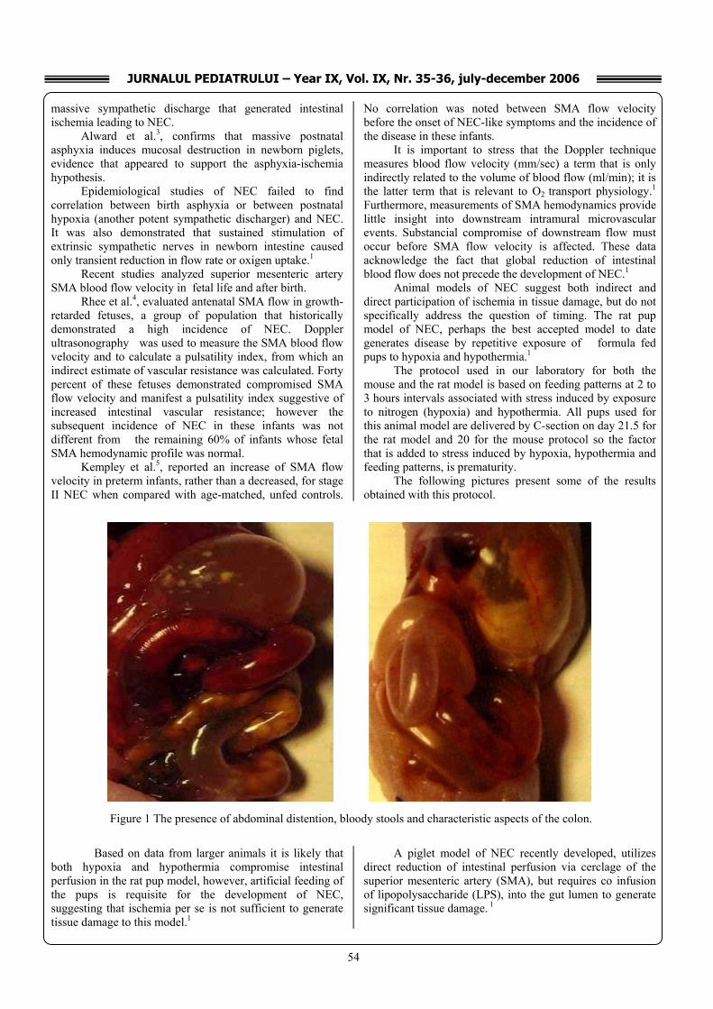
JURNALUL PEDIATRULUI – Year IX, Vol. IX, Nr. 35-36, july-december 2006
54
massive sympathetic discharge that generated intestinal ischemia leading to NEC. Alward et al.3, confirms that massive postnatal asphyxia induces mucosal destruction in newborn piglets, evidence that appeared to support the asphyxia-ischemia hypothesis. Epidemiological studies of NEC failed to find correlation between birth asphyxia or between postnatal hypoxia (another potent sympathetic discharger) and NEC. It was also demonstrated that sustained stimulation of extrinsic sympathetic nerves in newborn intestine caused only transient reduction in flow rate or oxigen uptake.1 Recent studies analyzed superior mesenteric artery SMA blood flow velocity in fetal life and after birth. Rhee et al.4, evaluated antenatal SMA flow in growth-retarded fetuses, a group of population that historically demonstrated a high incidence of NEC. Doppler ultrasonography was used to measure the SMA blood flow velocity and to calculate a pulsatility index, from which an indirect estimate of vascular resistance was calculated. Forty percent of these fetuses demonstrated compromised SMA flow velocity and manifest a pulsatility index suggestive of increased intestinal vascular resistance; however the subsequent incidence of NEC in these infants was not different from the remaining 60% of infants whose fetal SMA hemodynamic profile was normal. Kempley et al.5, reported an increase of SMA flow velocity in preterm infants, rather than a decreased, for stage II NEC when compared with age-matched, unfed controls.
No correlation was noted between SMA flow velocity before the onset of NEC-like symptoms and the incidence of the disease in these infants. It is important to stress that the Doppler technique measures blood flow velocity (mm/sec) a term that is only indirectly related to the volume of blood flow (ml/min); it is the latter term that is relevant to O2 transport physiology.1 Furthermore, measurements of SMA hemodynamics provide little insight into downstream intramural microvascular events. Substancial compromise of downstream flow must occur before SMA flow velocity is affected. These data acknowledge the fact that global reduction of intestinal blood flow does not precede the development of NEC.1 Animal models of NEC suggest both indirect and direct participation of ischemia in tissue damage, but do not specifically address the question of timing. The rat pup model of NEC, perhaps the best accepted model to date generates disease by repetitive exposure of formula fed pups to hypoxia and hypothermia.1 The protocol used in our laboratory for both the mouse and the rat model is based on feeding patterns at 2 to 3 hours intervals associated with stress induced by exposure to nitrogen (hypoxia) and hypothermia. All pups used for this animal model are delivered by C-section on day 21.5 for the rat model and 20 for the mouse protocol so the factor that is added to stress induced by hypoxia, hypothermia and feeding patterns, is prematurity. The following pictures present some of the results obtained with this protocol.
Based on data from larger animals it is likely that both hypoxia and hypothermia compromise intestinal perfusion in the rat pup model, however, artificial feeding of the pups is requisite for the development of NEC, suggesting that ischemia per se is not sufficient to generate tissue damage to this model.1
A piglet model of NEC recently developed, utilizes direct reduction of intestinal perfusion via cerclage of the superior mesenteric artery (SMA), but requires co infusion of lipopolysaccharide (LPS), into the gut lumen to generate significant tissue damage. 1
Figure 1 The presence of abdominal distention, bloody stools and characteristic aspects of the colon.
JURNALUL PEDIATRULUI – Year IX, Vol. IX, Nr. 35-36, july-december 2006
55
The sequence of the insult, LPS then ischemia or vice versa, does not affect the final degree of tissue damage; however, both are necessary. Therefore, while it is clear that reduction of flow through the mesenteric artery by 60% for 6 hours is required for tissue damage in this model, the relative role of this ischemia with respect to luminal LPS remains unclear.
Nonetheless, the etiology of NEC remains elusive and no specific treatment or prevention approaches have been successful yet. Great hope is nowadays given to the protective action of heparin binding epidermal growth factor (HB-EGF) on the bowel discovered and still under investigation by Besner10 in our laboratory.
References 1. Philip T. Nowicki, “Ischemia and necrotizing
enterocolitis: where, when and how “Seminars of Ped. Surg. (2005) 14, 152-158
2. Touloukian RJ, Berdon WE et al., “Surgical experience with necrotizing enterocolitis in the infant “J.Ped. Surg. 1967;2;289-401
3. Alward CT, Hook JB et al., “Effects of asphyxia on cardiac output and organ blood flow in the newborn piglet “ J. Ped. Resear. 1978;12;824-7
4. Rhee E., Detti L.,Mari G., “Superior mesenteric artery flow velocity wave forms in small gestational age fetuses“ J. Matern. Fetal Med. 1998;7;120-3
5. Kempley ST, Gamsu HR, “Superior mesenteric artery blood flow velocity in necrotizing enterocolitis“ Arch. Dis. Child. 1992;67;793-6
6. Granger HJ, Nyhof RA, “Dynamics of intestinal oxygenation: interaction between oxygen supply and uptake“ Am. J. Physiol. 1982;243:G91-6
7. Barlow B,Santulli TV, “ Importance of multiple episodes of hypoxia or cold stress on the development of enterocolitis in an animal model “ Surg. 1975; 77; 687-90
8. Caplan MS, Hedlung E., Adler L. et al., “Role of asphyxia and feeding in a neonatal rat model of necrotizing enterocolitis“ Pediatr. Pathol. 1994; 14; 1017-28
9. Treszl A, Tulassay T, Vasarhelyi B., “ Genetic basis for necrotizing enterocolitis--risk factors and their relations to genetic polymorphisms “Fron. Biosci. 2006 Jan 1; 11: 570-80.
10. N.Ann Kuhn, Veela B.Mehta, Sandra Glenn, M.P.Michalsky, G.Besner “ Heparin binding EGF-like growth factor (HB-EGF) decreases oxygen free radical production in vitro and in vivo “ – Antiox.&Red.Sign. 4:4:2002
Correspondence to: Andrei Radulescu, M.D. Ph.D. 644 Ann Street Columbus, OHAIO 43205, USA E-mail address: [email protected]

JURNALUL PEDIATRULUI – Year IX, Vol. IX, Nr. 35-36, july-december 2006
56
DEPLETED URANIUM INDUCED FETAL MALFORMATIONS - LITERATURE REVIEW
A Radulescu1, ES Boia2, RE Iacob2, V David3
1Center for Cell and Vascular Biology, Columbus Children’s Research Institute - Columbus, Ohio, USA 2University of Medicine and Pharmacy “Victor Babes“ - Timisoara 3Children’s Hospital “ Louis Turcanu “– Department of Pediatric Surgery, Timisoara, Romania Abstract
Depleted uranium is a byproduct of the enrichment process of uranium for its more radioactive isotopes to be used in nuclear energy .Because depleted uranium is pyrophoric and a dense metal with unique features when combined with alloys ,it is used by the military in armor and ammunition and frequently used in combat scenarios. This review reports on uranium uses and its published health effects, with major focus on its effects on reproduction and fetal development. Key words: uranium toxicity, fetal toxicity, congenital malformations
Depleted uranium (DU) is a man-made, radioactive, heavy metal derived from natural uranium (fig. 1). It reacts with most non-metallic elements; it has pyrophoric properties and may spontaneously ignite at room temperature in air, oxygen and water. These unique properties make it appealing for use in many civilian and military applications.
DU is used as X-ray radiation shielding in hospitals, as counter weights for rudders and flaps in
commercial aircrafts, in keels of sailing yachts and as ballast in both military and non-military airplanes.1
It is used by the military for the production of distinctly powerful projectiles (e.g., bullets/ penetrators, missile nose cones) and also as a protective armor for tanks. As a projectile, a DU penetrator ignites on impact under high temperature; it has a low melting point.
Large quantities of DU and/or radioactive decay products and other radioactive impurities can lead to substantial external exposure. A Geiger counter measurement by a correspondent in the recent Iraq war show that radiation emitting from a DU bullet fragment registered nearly 1000 – 1900 times the normal background radiation level.
A three-foot long DU fragment from a 12 mm tank shell registered radiation 1300 times the background level. A DU tank found by the U.S Army radiological team emitted 260 – 270 millirads of radiation per hour compared to the safety limit of 100 millirads per year. A pile of jet-black dust registered a count of 9839 emissions in one minute, a level more than 300 times the average background level.2
The U.S. military deployed DU munitions for the
first time during the 1991 Gulf War. DU weapons were also used in the 1994– 1995 war in Bosnia, the 2002 U.S. invasion of Afghanistan and in Iraq in 2003.1
However, until recent years little attention was paid to the potential toxic effects of uranium on reproduction and development. Moreover, most experimental studies on uranium-induced developmental toxicity have been performed in a sole species of mammals, mice.4
Fig. 1 Uranium forms and isotopes.6
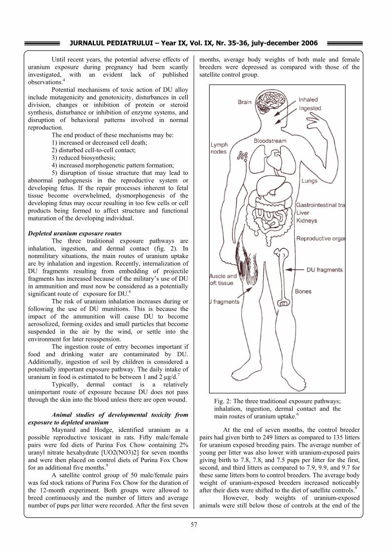
JURNALUL PEDIATRULUI – Year IX, Vol. IX, Nr. 35-36, july-december 2006
57
Until recent years, the potential adverse effects of uranium exposure during pregnancy had been scantly investigated, with an evident lack of published observations.4
Potential mechanisms of toxic action of DU alloy include mutagenicity and genotoxicity, disturbances in cell division, changes or inhibition of protein or steroid synthesis, disturbance or inhibition of enzyme systems, and disruption of behavioral patterns involved in normal reproduction.
The end product of these mechanisms may be: 1) increased or decreased cell death; 2) disturbed cell-to-cell contact; 3) reduced biosynthesis; 4) increased morphogenetic pattern formation; 5) disruption of tissue structure that may lead to
abnormal pathogenesis in the reproductive system or developing fetus. If the repair processes inherent to fetal tissue become overwhelmed, dysmorphogenesis of the developing fetus may occur resulting in too few cells or cell products being formed to affect structure and functional maturation of the developing individual.
Depleted uranium exposure routes
The three traditional exposure pathways are inhalation, ingestion, and dermal contact (fig. 2). In nonmilitary situations, the main routes of uranium uptake are by inhalation and ingestion. Recently, internalization of DU fragments resulting from embedding of projectile fragments has increased because of the military’s use of DU in ammunition and must now be considered as a potentially significant route of exposure for DU.6
The risk of uranium inhalation increases during or following the use of DU munitions. This is because the impact of the ammunition will cause DU to become aerosolized, forming oxides and small particles that become suspended in the air by the wind, or settle into the environment for later resuspension.
The ingestion route of entry becomes important if food and drinking water are contaminated by DU. Additionally, ingestion of soil by children is considered a potentially important exposure pathway. The daily intake of uranium in food is estimated to be between 1 and 2 μg/d.7
Typically, dermal contact is a relatively unimportant route of exposure because DU does not pass through the skin into the blood unless there are open wound.
Animal studies of developmental toxicity from
exposure to depleted uranium Maynard and Hodge, identified uranium as a
possible reproductive toxicant in rats. Fifty male/female pairs were fed diets of Purina Fox Chow containing 2% uranyl nitrate hexahydrate [UO2(NO3)2] for seven months and were then placed on control diets of Purina Fox Chow for an additional five months.9
A satellite control group of 50 male/female pairs was fed stock rations of Purina Fox Chow for the duration of the 12-month experiment. Both groups were allowed to breed continuously and the number of litters and average number of pups per litter were recorded. After the first seven
months, average body weights of both male and female breeders were depressed as compared with those of the satellite control group.
At the end of seven months, the control breeder
pairs had given birth to 249 litters as compared to 135 litters for uranium exposed breeding pairs. The average number of young per litter was also lower with uranium-exposed pairs giving birth to 7.8, 7.8, and 7.5 pups per litter for the first, second, and third litters as compared to 7.9, 9.9, and 9.7 for these same litters born to control breeders. The average body weight of uranium-exposed breeders increased noticeably after their diets were shifted to the diet of satellite controls.9
However, body weights of uranium-exposed animals were still below those of controls at the end of the
Fig. 2: The three traditional exposure pathways; inhalation, ingestion, dermal contact and the main routes of uranium uptake.6
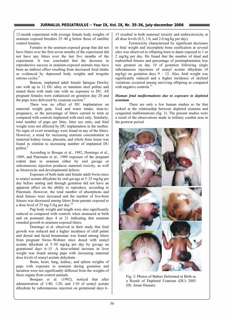
JURNALUL PEDIATRULUI – Year IX, Vol. IX, Nr. 35-36, july-december 2006
58
12-month experiment with average female body weights of uranium exposed breeders 25–40 g below those of satellite control females.
Females in the uranium-exposed group that did not have litters over the first seven months of the experiment did not have any litters over the last five months of the experiment. It was concluded that the decrease in reproductive success in uranium-exposed animals may have been an indirect effect resulting from decreased food intake as evidenced by depressed body weights and irregular estrous cycles.9
Benson, implanted adult female Sprague–Dawley rats with up to 12 DU alloy or tantalum steel pellets and mated them with male rats with no exposure to DU. All pregnant females were euthanized on gestation day 20 and the pups were delivered by cesarean section.8
There was no effect of DU implantation on maternal weight gain, food and water intake, time-to-pregnancy, or the percentage of litters carried to term as compared with controls implanted with steel only. Similarly, total number of pups per litter, litter sex ratio, and fetal weight were not affected by DU implantation in the mother. No signs of overt teratology were found in any of the litters. However, a trend for increasing uranium concentration in maternal kidney tissue, placenta, and whole fetus tissue was found in relation to increasing number of implanted DU pellets.8
According to Bosque et al., 1992, Domingo et al., 1989; and Paternain et al., 1989 exposure of the pregnant rodent dam to uranium either by oral gavage or subcutaneous injection produces maternal toxicity, as well as fetotoxicity and developmental defects.
Exposure of both male and female adult Swiss mice to uranyl acetate dihydrate by oral gavage at 5–25 mg/kg per day before mating and through gestation did not have an apparent affect on the ability to reproduce, according to Paternain. However, the total number of absorptions and dead fetuses were increased and the number of live-born fetuses was decreased among litters from parents exposed to a dose level of 25 mg U/kg per day.10
Pup body weight and length were also significantly reduced as compared with controls when measured at birth and on postnatal days 4 or 21 indicating that uranium retarded growth in uranium-exposed litters.
Domingo et al. observed in their study that fetal growth was reduced and a higher incidence of cleft palate and dorsal and facial hematomas was found among litters from pregnant Swiss–Webster mice dosed with uranyl acetate dihydrate at 5–50 mg/kg per day by gavage on gestational days 6–15 .A dose-related increase in liver weight was found among pups with increasing maternal dose levels of uranyl acetate dehydrate.
Brain, heart, lung, kidney, and spleen weights of pups with exposure to uranium during gestation and lactation were not significantly different from the weights of these organs from control animals.
Bosques et al. (1992), noticed that after administration of 1/40, 1/20, and 1/10 of uranyl acetate dihydrate by subcutaneous injection on gestational days 6–
15 resulted in both maternal toxicity and embryotoxicity at all dose levels (0.5, 1.0, and 2.0 mg/kg per day).
Fetotoxicity characterized by significant decreases in fetal weight and incomplete bone ossification at several sites was observed in offspring born to dams exposed to 1 or 2 mg/kg per day. He found that the number of dead and reabsorbed fetuses and percentage of postimplantation loss was greatest on day 10 of gestation following single subcutaneous injections of uranyl acetate dihydrate (4 mg/kg) on gestation days 9 – 12. Also, fetal weight was significantly reduced and a higher incidence of skeletal variations occurred among surviving offspring as compared with negative controls.11
Human fetal malformations due to exposure to depleted uranium
There are only a few human studies so far that looked at the relationship between depleted uranium and congenital malformations (fig. 3). The present studies were a result of the observations made in military combat area in the postwar period.
Fig. 3: Photos of Babies Deformed at Birth as a Result of Depleted Uranium (DU) 2003 (Dr. Jenan Hassan).
JURNALUL PEDIATRULUI – Year IX, Vol. IX, Nr. 35-36, july-december 2006
59
The Nuclear Policy Research Institute, USA reports that as early as 1995-96, Iraqi doctors suspected a rise in leukemia and birth defects among children born or treated at the Women and Children’s Hospital in central Basra, Iraq’s second largest city.
The hospital diagnoses all children less than 15 years of age in the whole government of Basra with a malignancy or suspected malignancy.
The Iraqi studies, the only population-based studies available, have their limitations including a lack of independent measures of exposure such as tissue and urine samples, no control city for comparison, mobile population so that some exposed individuals moved from the area while unexposed people moved into the area and, as a retrospective study, a question of assessment bias.
The findings can be summarized as following: • 1990 - 2001 rate of malignancies per 100,000
children < 5 years of age has tripled. • 1993-2000 rate of malignant diseases in children
compared to 1990 has quadrupled. • Children under 5 with leukemia: o 2 cases reported in 1990 o 41 cases reported in 2000 • Congenital Malformations: Incidence per 1000
births o 3.04 cases reported in 1990 o 17.6 cases reported in 2000 Additional information comes from, Imad Al-
Sadoon et al., who performed an analysis of registered congenital malformation among births in Basrah, Iraq for the period from 1990 to 2000.12
In general there was an apparent increase in the incidence rate from 1995 upwards. In 2000 such incidence was almost six folds higher than in 1991. To improve statistical efficiency of the data collected and overcome small numbers of cases recorded, the pattern and incidence of congenital malformations are grouped into three periods, 1991 to 1994, 1995 to 1998 and 1999 to 2000.12
The incidence rate for the first period was 2.5 congenital malformations per 1000 births while the respective figure for the second period is 4.57 and for the third period were 13.49.
Congenital heart diseases and chromosomal aberrations were reported at a higher frequency during the
latter years. Such unusual malformations as phocomelia and icthyosis, which were not reported in 1990 have been recorded later though in small numbers. The frequency of cleft lip and palate followed a similar trend. No apparent trends were observed in the remaining malformations.12
Sumanovic-Glamuzina et al., investigated the prevalence of major congenital malformations in West Herzegovina, a part of Bosnia and Herzegovina, immediately and five years after 1991-1995 military activities, which allegedly included the use of weapons with depleted uranium.13
The study included all live-born and stillborn neonates and excluded all aborted fetuses in two one-year cohorts (1995 and 2000) of neonates in the Maternity Ward of the Mostar University Hospital.
Major malformations were found in 40 (2.16%) out of 1,853 neonates in 1995 (95% confidence interval [CI], 1.49-2.82%) and in 33 (2.26%) out of 1,463 neonates five years later (95% CI, 1.50-3.01%), ie, at comparable prevalence.13
Anomalies of the musculoskeletal system were the most common, followed by anomalies of the digestive system (in 1995) and the cardiovascular system (in 2000).
Authors of this study concluded that against all the current studies that show the teratological influence of depleted uranium, the alleged environmental pollution in some regions of the former Yugoslavia, which was attributed to military activities and the presence of depleted uranium (the "Balkan syndrome"), there was no significant postwar increase in the prevalence of congenital malformations.13
In a review on the chemical toxicity of uranium, Stopps and Todd mentioned that during World War II two studies were carried out, one of which featured exposure to high levels of the metal and another involved only a brief 24-hr exposure. Although it was reported that in both studies significant effects on reproduction were observed, the results were not repeated or extended by other investigators.4
Acknowledgements – the future investigation of the effects of depleted uranium on fetal malformations is supported by NATO, grant PDD (CP)-(CBP.EAP.RIG 982293) to Andrei Radulescu.
References 1. Rita Hindin, Doug Brugge, Bindu Panikkar,
“Teratogenicity of depleted uranium aerosols: A review from an epidemiological perspective“ Environmental Health: A Global Access Science Source 2005, 4:17
2. Peterson S: Remains of toxic bullets litter Iraq. Christian Science Monitor [http://www.csmonitor.com /2003/0515/p01s02-woiq.html].2003, May 15
3. Stopps GJ, Todd M. “The Chemical Toxicity of Uranium with Special Reference to Effects on the Kidney and the Use for Biological Monitoring” Research Report, Atomic Energy Control Board. Ottawa,Canada; 1982.
4. M. Luisa Albina, Montserrat Belles et al. “Influence of Maternal Stress on Uranium-Induced Developmental Toxicity in Rats”, Experimental Biology and Medicine 06/2003
5. George C.Jiang, M.Aschner, “ Neurotoxicity of Depleted Uranium Reasons for Increased Concern “,Biological Trace Element Research vol.110,2006
6. N. H. Harley, E. C. Foulkes, L. H. Hilborne, A. Hudson, and C. R. Anthony, Depleted Uranium, RAND, Santa Monica, CA (1999).
7. Benson, K.A. and McBride, S.A. 1997: Uranium levels in the fetus and placenta of female rats implanted with depleted uranium pellets prior to breeding. The Toxicologist 36, 258.(1997)
JURNALUL PEDIATRULUI – Year IX, Vol. IX, Nr. 35-36, july-december 2006
60
8. Maynard, E. and Hodge, H. 1949: Studies of the toxicity of various uranium compounds when fed to experimental animals. In Voeglen, C., editor, Pharmacology and toxicology of uranium compounds, Volume 1. New York: McGraw-Hill, 309;76.
9. Paternain, J.L., Domingo, J.L., Ortega, A. and Llobe, J.M. 1989: The effects of uranium on reproduction, gestation and postnatal survival in mice. Ecotoxicol Environ Saf 17, 291; 96.
10. Bosque, M.A., Domingo, J.L. and Corbella, J. 1992: Embryotoxicity of uranium in mice. Variability with the day of exposure. Rev Toxicol 9, 107; 10.
11. Imad Al-Sadoon, Genan G. Hassan, Alim A-H. Yacoub, Depleted uranium and health of people in Basrah: Incidence and pattern of congenital anomalies among births in Basrah during the period of 1990- 2000.
12. Sumanovic-Glamuzina D, Saraga-Karacic V, Roncevic Z, Milanov A, Bozic T, Boranic M. , Incidence of major congenital malformations in a region of Bosnia and Herzegovina allegedly polluted with depleted uranium , Croat Med J. 2003 Oct;44(5):579-84
Correspondence to: Andrei Radulescu, M.D. Ph.D. 644 Ann Street Columbus, OHAIO 43205, USA E-mail address: [email protected]
JURNALUL PEDIATRULUI – Year IX, Vol. IX, Nr. 35-36, july-december 2006
61
CARIES RISK FACTORS IN CHILDREN
Roxana Oancea1, Elisabeta Bratu2, Daniela Jumnaca1, Atena Galuscan1, Ruxandra Sava-Rosianu1, Angela Codruta Podariu1 1Department of Preventive, Community Dentistry and Oral Health
2Department of Pedodontics and Orthodontics
Faculty of Dentistry Victor Babes University of Medicine and Pharmacy, Timisoara Abstract
The preoccupation for maintaining dental health, resistant to the action of microorganisms starts from the 5-6 week of intrauterine life, when the embryogenesis of the child's teeth begins. Related to that, each stage has to be well differentiated regarding dental care measures and an evaluation of the oral heath is necessary because of several facts: it is the period of temporary and mixed dentition when the frequency of the dental lesions is very high with negative impact on the development of the cranio-facial system but also with an impact on the development of the hole body. The understanding of the complex nature of caries depends on the multiple risk factors that have to be taken in consideration and the prophylactic and therapeutic measures are based on the accurency of the evaluation of all this factors. Key words: caries risk, children, fluoride, diet, oral health. The preoccupation for maintaining dental health, resistant to the action of microorganisms starts from the 5-6 week of intrauterine life, when begins the embryogenesis of the child's teeth. Related to that, each stage has to be well differentiated regarding dental care measures and an evaluation of the oral heath is necessary because of several facts: it is the period of temporary and mixed dentition when the frequency of the dental lesions is very high with negative impact on the development of the cranio-facial system but also with an impact on the development of the hole body. Caries can be viewed as an infectious disease and mutans streptococci are considered to be important bacteria for its development (Emilson & Krasse 1985, Loesche 1986), although no single type of micro-organism has been identified as the primary cause of either enamel, root or crown caries (Nyvad & Kilian 1987). The bacteria attach to the first primary teeth to erupt, especially to the fissures of the molars in 2–3-year-old children (Alaluusua & Renkonen 1983). However more recent studies indicate that the infection may occur at a younger age and those mutans streptococci can colonize the oral cavity of predentate children as young as 6 months of age. The time of contamination is of a certain importance, as the later a child is infected, the less caries lesions develop in early childhood and later on (Alaluusua &
Renkonen 1983, Köhler et al. 1988). Mutans streptococci are transmittable from the primary dentition to the permanent dentition (Gibbons 1984, Alaluusua et al. 1987), and also between individuals (Köhler & Bratthall 1978, Rogers 1981). The transmission occurs through contamination of the saliva (Rogers 1981) so that mothers are considered to transfer the infection to their child (Köhler & Bratthall 1978, Aaltonen et al. 1990). High levels of mutans streptoccoci in the mother’s mouth contribute to maternal transfer as does maternal dietary habits and poor oral hygiene. It has been found that 20–50% of mothers in the Scandinavian countries have high counts of salivary mutans streptococci (Berkowitz & Jones 1985, Paunio et al. 1988). Habitual xylitol consumption by mothers has been shown to lead to a significant reduction in mother-child mutans bacteria transmission when assessed in two-year-old children (Söderling et al. 2000). Carious lesions increase the counts of mutans streptococci, while reductions can be achieved by restriction of sucrose-sweetened products (Rugg-Gunn & Edgar 1984, Birkhed et al. 1990) and the use of xylitol (Isokangas et al. 1989, Söderling et al. 2000), together with anti-microbial preventive procedures (Loesche et al. 1989, Tenovuo 1992). Dietary habits and dental caries have shown to be of importance for caries development (Kleemola-Kujala & Räsänen 1979, Birkhed 1990), but the frequency of brushing the teeth was more related to caries than were dietary factors in some studies (Schröder & Granath 1983, Stecksén-Blicks 1985a, Stecksén- Blicks & Holm 1995). The frequency of consumption of sugar-containing products relates to caries, and the ingestion of fermentable carbohydrates is associated with its prevalence (Rugg-Gunn & Edgar 1984, Holbrook et al. 1995, Gibson & Williams 1999). Significant correlations between sugar consumption and caries increment have also been observed by Rugg-Gunn & Edgar (1984), while a clear correlation was observed between the occurrence of "rampant" caries in young children and the use of sweetened dummies and prolonged use of “dinky feeders” containing sugar (Walker 1987). Social and demographic factors such as race, knowledge, schooling and financial status have all been linked with the occurrence of caries (Hunt 1990, Powell 1998, Gibson & Williams 1999). In addition, the time needed for the development of caries is also an important
V. DENTISTRY

JURNALUL PEDIATRULUI – Year IX, Vol. IX, Nr. 35-36, july-december 2006
62
consideration (Alaluusua & Renkonen 1983, Köhler et al. 1988). Today the biological mechanisms of dental caries are well established. It is a disease with a number of important etiological factors – each of which must be simultaneously present to initiate and progress the disease. The factors are: fermentable carbohydrates (substrate); cariogenic microorganisms; susceptible tooth surface/host and the fourth factor time. Bacteria (mutans streptococci) in dental plaque metabolize sugars and produce acids, which lowers the pH in the mouth and promotes loss of minerals from the tooth surface. Minerals in the oral cavity including fluoride are redeposited on the tooth surface once the neutral pH is restored (normally after approximately 20min). This process is dynamic and as long as minerals are replaced the tooth surface remains sound and intact. However, a prolonged pH drop and frequent net loss of minerals lead to a weakening and eventual break down (cavity) in the tooth surface. Early childhood caries (ECC) appears to be a particularly virulent form of dental caries causing extensive destruction of the deciduous teeth, often very rapidly. This may be due to extremes in one or more of the three factors above. Much research into the etiology and prevention of ECC has focused on the dietary substrate component leading to the terms ‘baby bottle tooth decay’ and ‘nursing caries’. More recent research confirming the relative role of the microbial(plaque) and tooth resistance factors has fostered a better appreciation of the biological risk and protective factors in ECC. There is overwhelming evidence that sugars (such as sucrose, fructose and glucose) and other fermentable carbohydrates (such as highly refined flour) play a role in the initiation and development of dental caries. Sucrose is the most common sugar and is the only one that, when metabolized, produces dextrans which promote superior bacterial adhesion to teeth. Because of this it is considered the most important substrate in the establishment of cariogenic bacteria. It now appears that the frequency of intake of sucrose is more important than the total amount consumed. A review of the role of substrate in ECC by Reisine and Douglass found that the total weight of sugar in children’s diet was not predictive of dental caries; however, the frequency of sugar intake was. Frequent consumption of sugar favors the establishment of cariogenic bacteria and provides continuous substrate that influences the initiation and progression of the caries. Controversy exists as to whether infant formulas or bovine milk in bottles and breast milk given frequently to infants contribute to the development of ECC. The evidence of a relationship between bottle use and caries risk is weak and it is likely that the risk of caries may be sensitive to the interaction of multiple factors including other (non-bottle) dietary practices. Prolonged or on demand breast-feeding has been implicated in the development of ECC. The American Academy of Pediatric Dentistry’s policy (AAPD) on breast-feeding states that: “Although breast-feeding is essential in
providing the best possible nutrition to infants, the AAPD cautions that frequent breast-feeding at night and on demand after eruption of teeth may be implicated in contributing to the development of early childhood caries (ECC)”. There are controversies about this issue. The international dietary guidelines recommend exclusive breastfeeding until 6 months and then continuing breast-feeding with complementary foods until 2 years or more. More recent and methodologically stronger studies have suggested that breast-feeding per se is not significantly associated with ECC. Laboratory studies have noted that human breast milk does not appear to cause the drop in plaque pH required for the initiation and progress of dental decay and may in fact promote the deposition of calcium and phosphate ions on the tooth surface.
Several factors can predispose an individual or indeed a particular tooth to dental caries. These may include immunological factors, reduced saliva flow, immature enamel and defects of the tooth tissues.
Because enamel is immunologicaly inactive, the main immune defence against mutans streptoccoci is provided largely by Immunoglobulin A (IgA) or serum and gingival crevicular fluid. As children become infected with oral microorganisms, they develop salivary IgA antibodies. In addition to providing specific immunological factors, the saliva acts as an important protective factor. Saliva buffers plaque acids, aids in oral clearance and acts as a reservoir for minerals to assist in the re-mineralization of enamel.
Teeth erupt into the mouth with immature enamel. The process of enamel maturation continues following tooth eruption, so that teeth become less susceptible to decay over time. The enamel matures incorporating orally available ions including fluoride.
The ingestion of the fluoride, irrespective of the available form, acts preeruptive, during the mineralization period of the teeth, which is developed in 2 phases: -the mineralization of dental hard tissues - the preeruptive maturation of the enamel.
The period of time for this 2 phases, for both temporary and permanent dentition is 13 years and a half (without the third molar).
The mineralization of the temporary incisors starts at 3-4 months intrauterine life and is finished 4-5 months after birth. The mineralization of the permanent incisors starts at 3-4 months after birth, excepting the lateral incisor which begin the mineralization at 10-12 months and it ends at 4-5 years.
The mineralization of the temporary canine starts at 5 months of intrauterine life and it ends at 9 months, while the mineralization of the permanent canine starts at 4-5 months and it ends at 6-7 years.
The mineralization of the premolars starts at 1 ½ -2
½ and it ends at 5-7 years. The temporary molars begin their mineralization at
5 months of intrauterine life and it ends at 6 months for the first molar, while the second molar stars at 6-7 months and ends at 10-12 months.
The permanent first molar starts the mineralization at birth and ends at 2 ½ -3 years and the second molar achieve mineralization between 2 ½ -3 and 7-8 years.

JURNALUL PEDIATRULUI – Year IX, Vol. IX, Nr. 35-36, july-december 2006
63
The intervals presented are medium values for a certain population. There are variation conditioned by climacteric condition, race, region.
From this data it has to be revealed the fact that the preeruptive mineralization of the first molar is the shortest when it is compared with the other teeth. This explains the great susceptibility of this tooth to dental caries.
On the other hand, the time of mineralization of the temporary teeth is 6 time shorter comparative to the permanent teeth, fact that can explain the susceptibility to caries.
Regarding the utilization of prenatal fluoride the American Dental Association asked during a debate the following questions: - Is the passing of fluoride through the placenta safe? - Does the child benefit from the fluoride? - Is it a proven fact that fluoride in intrauterine is
involved in the maturation of enamel? The answer to the first question is that fluoride passes easily through the placenta from the 5th – 6th month of pregnancy.
Regarding the second question, the answer is given indirectly through the epidemiologically studies that revealed a decrease of carious lesions in temporary teeth at children who had benefit from an optimum fluoride income during intrauterine life.
At last, the answer to the third question is that fluoride is incorporated during the maturation of the enamel of temporary teeth, but at a lower concentration than in adult teeth because the mineralization stages are shorter. It looks like the incorporation of fluoride is not just the result of absorption but also of the concentration of preexistent fluoride in the bony tissue.
Ingestion of fluoride during the second half of the pregnancy, increasing the concentration of fluoride in the skeletal tissue of the child, is susceptible of influencing the ulterior incorporation of fluoride in the teeth. We can practically say that the ingestion of 1 mg F/day at young mothers is recommended from the fourth month of pregnancy.
Therefore, a tooth is most susceptible to caries immediately after eruption until final maturation.
Many studies have found a significant relationship between developmental defects of the tooth surface and dental caries. Developmental disturbances to the tooth germ during embryological development can result in loss of integrity of the surface enamel which in turn allows additional plaque accumulation on what would otherwise be a smooth surface. Such developmental disturbances may include premature birth or low-birth weight, pre- and postnatal infection/illness, nutritional deficiency and a variety of environmental pollutants including maternal smoking.
Much of the literature uses terms (including nursing caries and baby bottle tooth decay) or case definitions that imply that the inappropriate use of the baby bottle plays a central role in the development of dental decay in infants and young children, however, supporting epidemiological data is difficult to find. For this reason the term early childhood caries (ECC) is the term now used to
collectively refer to dental decay in infants and preschool children. In reality most babies are fed with a nursing bottle for at least some of the time and yet as Horowitz points out most of them do not develop ECC. Two bottle-related behaviours have attracted most interest in ECC research – the use of bottles at night/nap time and the use of the bottle beyond 12 months. Reisine and Douglass found little strong evidence to support either of these ideas and suggest that this paucity of evidence may be due to the use of retrospective parental self-reports. The alternate explanation they offer is that the critical period may be soon after the eruption of teeth into the mouth and that early use of the bottle containing sweet fluids supports the early establishment and dominance of cariogenic microflora. This may be more important than bottle use after 12 months. Litt et al. found that the use of the bottle at night-time was associated with sugar intake. The mothers who reported night-time bottle use were also more likely to have children with a higher sugar intake. Because of this aspect is very important that the treatment of the pregnant mother should include education regarding preventive oral care of the infant and toddler.
Too often a child’s first dental examination occurs after the deciduous dentition or even much of the permanent dentition has erupted. By this time, much opportunity for the prevention of dental pathology has passed. The expectant mother and family should be instructed in the importance of early dental examinations for the child. A child should receive the first dental examination between 6 to 12 months of age. Early examination enables the oral health professional to identify detrimental feeding habits, educate the parent about oral hygiene procedures, determine fluoride intake status, introduce the child to dentistry in a non threatening manner, and prepare the parents for the child’s future dental needs (Goepferd & Garcia-Godoy, 1999). Expectant mothers and their families should also be instructed in the care of the infant’s mouth beginning at birth. The infant’s mouth should be gently cleansed daily with a damp washcloth. As soon as teeth have erupted, they should be brushed daily with a soft toothbrush. The deciduous dentition is critical to proper phonetic development, space maintenance for permanent teeth, and the child’s self-image. The number of teeth and surfaces at risk varies with age (Hausen et al. 1983, Nordblad & Larmas 1985a, Vehkalahti et al. 1991, Virtanen 1997), as also does the maturation age of tooth (Nordblad & Larmas 1985b). Nowadays the eruption of teeth occurs at an earlier chronological age than earlier (Helm 1969, Virtanen 1994, Eskeli et al. 1999). Most caries attacks on fissures occur during the first three years after eruption, and the survival of the first and second permanent molars immediately after eruption and filling increments in the upper incisors are good indicators of dental health. A distinction should be made between chronological and dental age, however. The post-eruptive filling placement curves for individual teeth,
JURNALUL PEDIATRULUI – Year IX, Vol. IX, Nr. 35-36, july-december 2006
64
obtained using the survival analysis method, evidently follow the pattern of caries attack. (Virtanen 1997) The occlusal surfaces of permanent teeth are those most frequently attacked by caries (Nordblad & Larmas 1985b, Vehkalahti et al. 1990, Li et al. 1993, Virtanen & Larmas 1995), and more caries lesions have been demonstrated in pits and fissures of posterior teeth than on other surfaces (Dummer et al. 1990, Kingman 1993). The highest caries experience of all has been found in permanent molars (Nordblad & Larmas 1985b, Greenwell et al. 1990, Vehkalahti et al. 1990), whereas caries is seldom seen in teeth, such as canines, lower incisors and premolars (Nordblad 1986, Greenwell et al. 1990, Vehkalahti et al 1990, Virtanen & Larmas 1995). The risk of occlusal caries is highest during and after tooth eruption (Härkänen et al. 2002), between 6 and 9 years of age for the first permanent molars, and after the age of 13 for the second permanent molars (Nordblad 1986, Ripa et al. 1988, Vehkalahti et al. 1991, Larmas et al. 1995). Approximal surfaces of permanent molars have been found to become carious after 12 years of age (Nordblad 1986, Ripa et al. 1988, Virtanen & Larmas 1995), and a correlation has been shown between past caries on approximal surfaces and the developing of new approximal caries lesions (Mejàre et al. 2001). Fifteen is an important age because of newly erupted second molars and the increasing role of approximal decay in the dentition (Vehkalahti et al. 1990). The occurrence of caries has been declining in communities with and without organized preventive programs or fluoridation (Hargreaves 1987, Seppä et al. 2000). It is assumed that the reasons are related to the use of fluorides, to improvements in oral hygiene, or to a change in microbial, host and salivary factors, or to dietary changes (Marthaler 1984). It is suggested, however, that the most probable reason is related to the increased use of fluorides (Marthaler 1984, Bratthall et al. 1996), while according to Renson et al. (1985), the organized availability of dental resources and oral health education programs may be one explanation. The decline in caries may also have been due in part to new diagnostic and treatment criteria (Nadanowsky & Sheiham 1995). More recently, it has been reported that the decline in caries seems not to be associated with professional preventive measures performed in dental clinics (Seppä et al. 1998), but there is nevertheless a good deal of agreement on the preventive effect of fluoride toothpastes in this respect (Bratthall et al. 1996). Probably the most effective caries prevention treatment available today is fluoridation of municipal water supplies and the use of
fluoride toothpastes (Winston & Bhaskar 1998). According to Mandel (1996), the protective properties of fluoride dominate and host resistance wins out as an explanation for the decline in caries.
The use of the fluoride compounds on a large scale modified the evolution and progression of the cariuos lesions. For example, the occlusal caries become cavitary lesions much later and as a consequence the lesions that were cavitary sometimes ago, today is present only a slight modification in the enamel colour. This phenomen of hidden caries was described in the early 80 and it was demonstrated in various clinical trials.
In the case of the caries localised on aproximal surfaces there are modification in the relation between the depth of the lesion and the presence or absence of the cavity. If the lesion was considered cavitary at the moment that the radiotransparency reached the enamel-dentin jonction, today much of the lesions with this depth are not cavitated lesions. This sugests that the disponibility of the fluorid modifie the radiografic image of the aproximal lessions (Pitts, 1992).
When preventive procedure is planned it must be taken in consideration a few risk indicators: age risk, the period of risk, the teeth and surfaces with risk, the medical and social risk. (Bader et al. 1986, Nordblad & Larmas 1986, Virtanen 1997, Vehkalahti et al. 1997, Meurman 1997, Powell 1998).
Because of that, the colaboration with the pediatrician, wich has to oversee the geneal heath condition, is very important, any change in the general eqiuilibrum could affect the oral health status.
The use of fluoridation, the sealing of pits and fissures together with oral hygiene implamantation and healty dietary habits are the 4 methods indicated by WHO(World Health Organiyation) for prevention of dental caries. The dentist has to aplly the prophylactic measures, has to implement an adequate toothbrush teqniques contributing to the consolidation of oral health.
A healthy diet which must contain all the necessary nutrient elements is important from the pediatrician point of view for an adequate groth and development and also for the dentist in order to prevent the risk factor represented by the existence of a cariogenic diet –carbohidrate substrate.
The understanding of the complex nature of caries depends on the multiple risk factors that have to be taken in consideration and the prophylactic and therapeutic measures are based on the accurency of the evaluation of all this factors.
Bibliography 1. Alanen P, Isokangas P & Gutmann K (2000) Xylitol
candies in caries prevention: results of a field study in Estonian children. Community Oral Epidemiol 28: 218–224.
2. Batchelor P (2002) The limitation of a high-risk approach for the prevention of dental caries. Community Dent Oral Epidemiol 30:302-12
3. Belfrán-Aquilar ED, Goldstein JW & Lockwood SA (2000) Fluoride varnishes: A review of their clinical use, cariostatic mechanism, efficacy and safety. J Am Dent Assoc 131: 589–596.
4. Bratu Elisabeta (2005) Practica pedodontică .Ediţia aIIIa Editura Orizonturi Universitare
5. Gussy M. (2006) Early childhood caries: current evidence for etiology and prevention. Journal of Paediatrics and Child Health 42 :37-43

JURNALUL PEDIATRULUI – Year IX, Vol. IX, Nr. 35-36, july-december 2006
65
6. Hanganu C,Dănilă I, (2002) Stomatologie Comunitară, Editura Tehnică, Chişinău
7. Hausen H, Kärkkäinen S & Seppä L (2000) Application of the high- risk strategy to control dental caries. Community Oral Epidemiol 28: 26–34.
8. Hausen H, Kärkkäinen S & Seppä L (2001) Caries data collected from public health records compared with data based on examinations by trained examiners. Caries Res 35: 360–365.
9. McGlone P, Watt R & Sheiham A (2001) Evidence-based dentistry: an overview of the challenges in changing professional practice. Br Dent J 190: 636–639.
10. Mandel ID (1996) Caries prevention: current strategies, new directions. J Am Dent Assoc 127: 1477–1488.
11. Podariu A si colab. (2003) Tratat de preventie oro-dentară, ed. Waldpress, Timisoara, 178-181
12. Psoter W.(2003) Classification of dental caries patterns in the primary dentition-a multidimensional scaling analasys. Community Oral Epidemiol 31:231-8
13. Reich E, Lussi A & Newbrun E (1999) Caries risk assessment. Int Dent J 49: 15–26.
14. Seppä L, Kärkkäinen S & Hausen H (2000) Caries trends 1992-1998 in two low-fluoride Finnish towns formerly with and without fluoridation. Caries Res 34: 462–468.
15. Splieth C, Steffen H, Rosin M & Welk A (2000) Caries prevention with chlorhexidine-thymol varnish in high risk children. Community Dent Oral Epidemiol 28: 419–423.
16. Varsio S (1999) Caries-preventive treatment approaches for child and youth at two extremes of dental health. University of Helsinki. ISBN 952-91-1150-9.
Correspondence to: Roxana Oancea, Eftimie Murgu Squ. No.2, Timisoara, Romania Phone: +40721335788 E-mail: [email protected]

JURNALUL PEDIATRULUI – Year IX, Vol. IX, Nr. 35-36, july-december 2006
66
MANUSCRIPT REQUIREMENTS
The manuscript must be in
English, typed single space, one column on A4 paper, with margins: top – 3 cm, bottom – 2,26 cm, left – 1,5 cm, right – 1,7cm. A 10-point font Times New Roman is required.
The article should be organized in the following format: Title, Names of all authors (first name initial, surname), Names of institutions in which work was done (use the Arabic numerals, superscript), Abstract, Keywords, Text (Introduction, Purpose, Materials and Methods, Results, Discussions and/or Conclusions), References, and first author’s correspondence address.