education exhibit ileal pouch–anal anasto- mosis...
TRANSCRIPT
Note: This copy is for your personal non-commercial use only. To order presentation-ready copies for distribution to your colleagues or clients, contact us at www.rsna.org/rsnarights.
221EDUCATION EXHIBIT
Ileal pouch–anal anastomosis (IPAA) surgery preserves fecal continence for improved quality of life in patients who require proctocolectomy for treatment of severe bowel diseases such as inflammatory disease and familial adenomatous polyposis. In IPAA surgery, an ileal reservoir, or pouch, is created and anastomosed to the anal canal. Awareness of the surgical technique and the postoperative anatomy of the IPAA is im-portant to identify complications at computed tomography (CT), mag-netic resonance (MR) imaging, and fluoroscopy. Complications include anastomotic leak, abscess, pouchitis, venous thrombus, pouch fistula, and stricture. Leaks from the blind end of the pouch and the pouch-anal anastomosis often result in pelvic abscesses, which may require ultrasonography- or CT-guided drainage; judicious catheter manage-ment can help improve clinical outcomes and avoid excessive imaging. Pouchitis may be identified by the presence of a thickened enhancing pouch wall and associated inflammatory changes and lymphadenopathy. The venous system must be scrutinized for thrombi secondary to surgi-cal manipulation and sepsis. Fistulas are likely because of the presence of chronic inflammation or infection and may be seen at MR imaging, CT, or fluoroscopy. Strictures appear as areas of focal luminal narrow-ing with proximal dilatation, which can lead to obstruction. To avoid repeated exposure to radiation, MR imaging may be performed in pa-tients who must undergo frequent imaging.©RSNA, 2010
Ileal Pouch–Anal Anasto- mosis Surgery: Imaging and Intervention for Post- operative Complications1
LEARNING OBJECTIVES FOR TEST 4
Discuss the post-
operative anatomy of the IPAA as seen at CT, MR imaging, and fluoroscopy.
Describe the im-
aging features of complications of IPAA surgery seen at CT, MR imaging, and fluoroscopy.
Discuss the role of
image-guided inter-vention in treatment of complications of IPAA surgery.
CME FEATURESee accompanying
test at http://www.rsna.org
/education/rg_cme.html
Abbreviation: IPAA = ileal pouch–anal anastomosis
RadioGraphics 2010; Published online Content Code: 1From the Department of Radiology, Boston University Medical Center and Boston University, 820 Harrison Ave, FGH Bldg, 3rd Floor, Boston, MA 02118. Recipient of a Certificate of Merit award for an education exhibit at the 2008 RSNA Annual Meeting. Received April 6, 2009; revision requested June 11 and received July 21; accepted July 24. All authors have no financial relationships to disclose. Address correspondence to J.C.B. (e-mail: ).
The Editor has no relevant financial relationships to disclose.
©RSNA, 2010

222 January-February 2010 radiographics.rsna.org
Figure 1. Illustration shows the surgical technique for creation of an IPAA with a J configuration. The distal end of the ileum is closed with staples (black arrow), the ileum is folded, and a stapling device is inserted through the apical enterotomy (white ar-row). (Reprinted, with permission, from reference 1.)
IntroductionIleal pouch–anal anastomosis (IPAA) creation, sometimes referred to as ileoanal pull-through, is a surgical technique that was developed to im-prove the quality of life of patients who undergo proctocolectomy by allowing transanal defecation and fecal continence. The surgery involves colec-tomy with mucosal proctectomy and creation of an ileal reservoir or pouch, which is anastomosed to the anal canal. IPAA creation is indicated for patients with ulcerative colitis when their disease has progressed despite maximal therapy, when dysplasia or malignancy has been detected at endoscopy, or when growth retardation has been identified (in children). It also is indicated for patients with adenomatous polyposis, because the risk for colon cancer approaches 100% by the time they reach 45 years of age. Contraindications to IPAA creation are determined by the risk for pouch failure and include Crohn disease (because of the risk for inflammation in the bowel segment used to create the pouch), poor sphincter control, anorectal inflammation, advanced-stage anorectal carcinoma, and perianal disease. Complications of IPAA creation may lead to poor pouch function and incontinence, the need for surgical revision, and pouch failure requiring excision (1,2).
In this article, the surgical technique and post-operative anatomy of the IPAA are described, and the appearances of complications such as anasto-motic leak, abscess, pouchitis, venous thrombus, pouch fistula, and stricture at computed tomog-raphy (CT), magnetic resonance (MR) imaging, and fluoroscopy are discussed. The role of image-guided intervention for abscess drainage and proper catheter management also are discussed.
Surgical TechniqueAwareness of the surgical technique and postop-erative anatomy of the IPAA facilitates radiologic identification of complications. There are sev-eral techniques for forming the reservoir pouch, including S, W, and J configurations; however, the J configuration is used most often because it is associated with improved clinical outcomes and a more facile surgical technique (1).
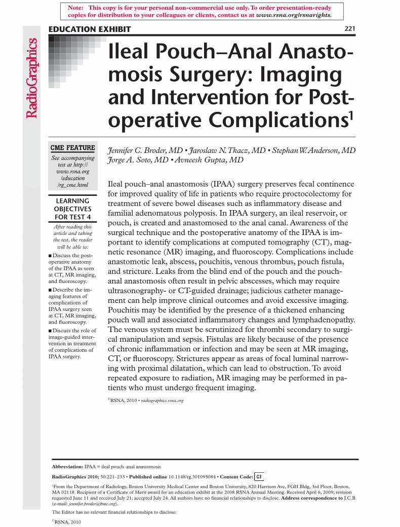
Several steps are involved in the formation of a J pouch. First, colectomy and proctectomy are performed. The distal end of the ileum is stapled closed to form a blind end (also referred to as the “stump,” “appendage,” or “efferent limb”). The distal ileum is then folded back on itself, and an apical enterotomy is created. A linear stapling de-vice is passed through the enterotomy defect (Fig 1). With the stapling device, a side-to-side anasto-mosis is made by cutting the ileal walls centrally. The two loops are then connected with two paral-lel staple lines to form a large pouch. At times, the over-sewn stump may not be fully attached to the pouch and may dangle off to the side. A circular stapling device is then passed through the anus to create an anastomosis between the anus and the enterotomy at the distal end of the pouch. Alter-
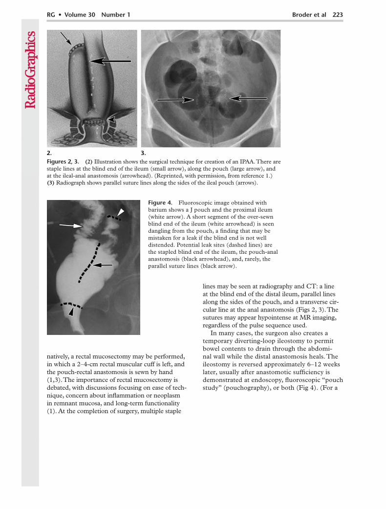
(2) Illustration shows the surgical technique for creation of an IPAA. There are staple lines at the blind end of the ileum (small arrow), along the pouch (large arrow), and at the ileal-anal anastomosis (arrowhead). (Reprinted, with permission, from reference 1.) (3) Radiograph shows parallel suture lines along the sides of the ileal pouch (arrows).
natively, a rectal mucosectomy may be performed, in which a 2–4-cm rectal muscular cuff is left, and the pouch-rectal anastomosis is sewn by hand (1,3). The importance of rectal mucosectomy is debated, with discussions focusing on ease of tech-nique, concern about inflammation or neoplasm in remnant mucosa, and long-term functionality (1). At the completion of surgery, multiple staple
lines may be seen at radiography and CT: a line at the blind end of the distal ileum, parallel lines along the sides of the pouch, and a transverse cir-cular line at the anal anastomosis (Figs 2, 3). The sutures may appear hypointense at MR imaging, regardless of the pulse sequence used.
In many cases, the surgeon also creates a temporary diverting-loop ileostomy to permit bowel contents to drain through the abdomi-nal wall while the distal anastomosis heals. The ileostomy is reversed approximately 6–12 weeks later, usually after anastomotic sufficiency is demonstrated at endoscopy, fluoroscopic “pouch study” (pouchography), or both (Fig 4). (For a
Figure 4. Fluoroscopic image obtained with barium shows a J pouch and the proximal ileum (white arrow). A short segment of the over-sewn blind end of the ileum (white arrowhead) is seen dangling from the pouch, a finding that may be mistaken for a leak if the blind end is not well distended. Potential leak sites (dashed lines) are the stapled blind end of the ileum, the pouch-anal anastomosis (black arrowhead), and, rarely, the parallel suture lines (black arrow).

224 January-February 2010 radiographics.rsna.org
Figure 5. Abscess after IPAA creation in a 24-year-old man with ulcerative colitis. (a) Axial CT im-age obtained with oral and intravenous contrast material shows parallel suture lines (arrows) demarcat-ing the pouch, which is displaced by a round fluid collection with thick enhancing walls (arrowhead). Contrast material had not reached the pouch at the time of imaging. (b) Coronal CT image shows that the staple line of the ileal stump (black arrowhead) is immediately adjacent to the abscess (white arrow-head), a finding suggestive of leakage from the suture line. The displaced pouch (arrows) is seen to the right of the abscess. Despite drainage, a persistent leak caused fluid to reaccumulate in the abscess mul-tiple times; surgical revision of the blind end of the ileum likely will be necessary in this patient.
with IPAA developed pelvic sepsis, which may result from anastomotic leakage or dehiscence, pelvic abscess, pelvic or perineal wound infection, or a combination thereof. Patients with ulcer-ative colitis have been shown to be at greater risk for pelvic sepsis within the first 4 months after surgery than those with familial adenomatous polyposis, a finding likely due to immunosup-pression and delayed healing because of preoper-ative treatment with systemic corticosteroids (8). There is a high association of pelvic sepsis with poor functional outcome and pouch failure (2).
When patients with an IPAA present with symptoms suggestive of a leak or abscess, several imaging modalities may be employed, depend-ing on the clinical manifestations. As mentioned earlier, fluoroscopic pouchography with water-sol-uble rectal contrast material may be performed to identify the presence and origin of a leak; however, CT is more sensitive than fluoroscopy in depicting abscesses, and it may depict other possible causes of the patient’s symptoms as well (9). An initial fluoroscopic evaluation may make subsequent performance of CT problematic if marked enteric contrast enhancement persists. At our institution, when patients present emergently with symptoms suggestive of pelvic sepsis, CT often is performed initially. CT protocols should include the use of oral and intravenous contrast material and review of multiplanar reformatted images. If possible, the amount of oral contrast material administered should be adequate to ensure opacification of the
more detailed discussion of the use of barium in fluoroscopic reservoir examinations, see Alfisher et al [4] and Crema et al [5].) The question of whether to create a temporary ileostomy is controversial. In the surgical literature, discus-sions center on the risk for complications with possible compromise of long-term function (6). Ileal diversion may not be necessary in elec-tive surgeries in which a technically competent pouch construction has been achieved in non–steroid-dependent patients with good nutritional status and a normal hematocrit level (1).
Leaks and AbscessesAnastomotic leaks tend to originate from poten-tially vulnerable areas within the pouch, espe-cially suture lines, which are prone to dehiscence. Suture lines must be carefully scrutinized at imaging of patients who are thought to have pouch complications such as leaks and abscesses. Vulnerable areas include the over-sewn blind end of the ileum (the ileal stump), the pouch-anal anastomosis, and, rarely, the parallel suture lines along the reservoir sides (Fig 4). Small leaks may not cause substantial inflammation or sepsis. Patients with a leak that has developed into an abscess present with focal or diffuse abdominal pain, fever, an elevated white blood cell count, or a combination of these symptoms. In a meta-analysis by Hueting et al (7), 9.5% of patients
TeachingPoint
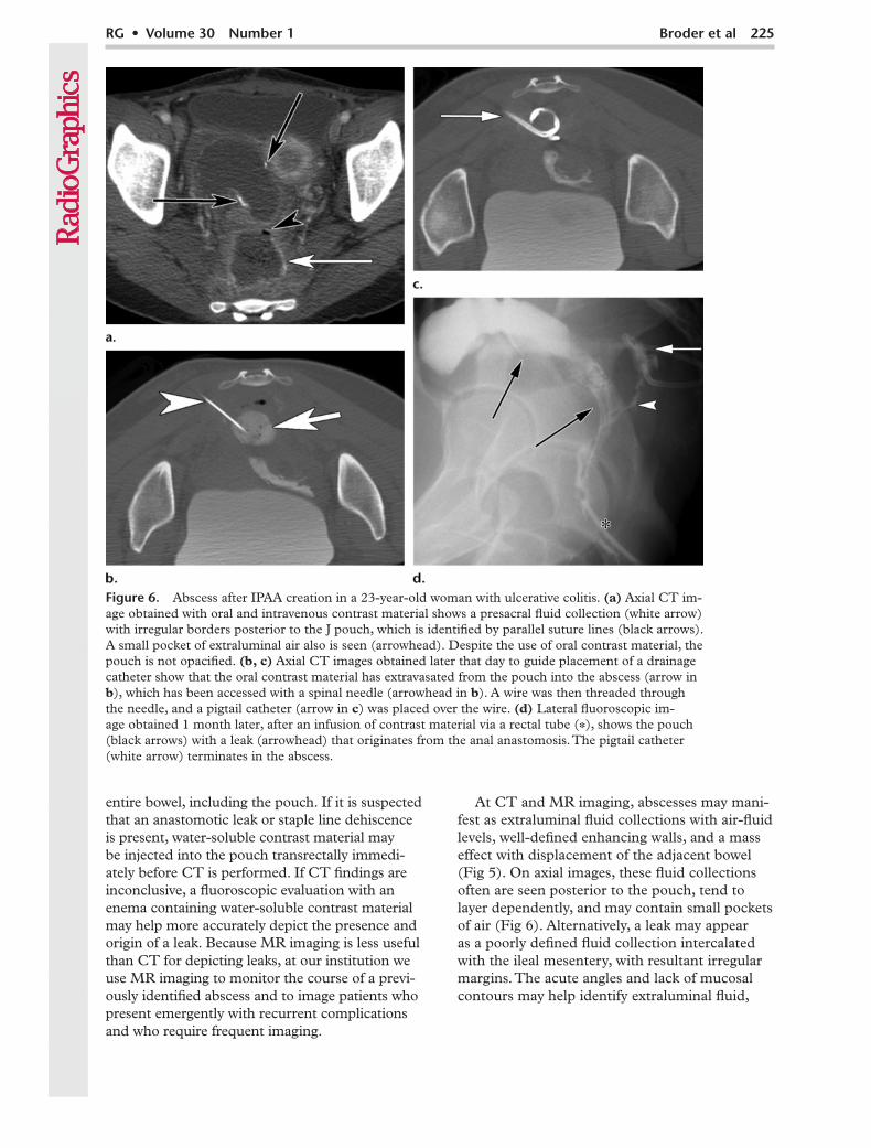
Figure 6. Abscess after IPAA creation in a 23-year-old woman with ulcerative colitis. (a) Axial CT im-age obtained with oral and intravenous contrast material shows a presacral fluid collection (white arrow) with irregular borders posterior to the J pouch, which is identified by parallel suture lines (black arrows). A small pocket of extraluminal air also is seen (arrowhead). Despite the use of oral contrast material, the pouch is not opacified. (b, c) Axial CT images obtained later that day to guide placement of a drainage catheter show that the oral contrast material has extravasated from the pouch into the abscess (arrow in b), which has been accessed with a spinal needle (arrowhead in b). A wire was then threaded through the needle, and a pigtail catheter (arrow in c) was placed over the wire. (d) Lateral fluoroscopic im-age obtained 1 month later, after an infusion of contrast material via a rectal tube (*), shows the pouch (black arrows) with a leak (arrowhead) that originates from the anal anastomosis. The pigtail catheter (white arrow) terminates in the abscess.
At CT and MR imaging, abscesses may mani-fest as extraluminal fluid collections with air-fluid levels, well-defined enhancing walls, and a mass effect with displacement of the adjacent bowel (Fig 5). On axial images, these fluid collections often are seen posterior to the pouch, tend to layer dependently, and may contain small pockets of air (Fig 6). Alternatively, a leak may appear as a poorly defined fluid collection intercalated with the ileal mesentery, with resultant irregular margins. The acute angles and lack of mucosal contours may help identify extraluminal fluid,
entire bowel, including the pouch. If it is suspected that an anastomotic leak or staple line dehiscence is present, water-soluble contrast material may be injected into the pouch transrectally immedi-ately before CT is performed. If CT findings are inconclusive, a fluoroscopic evaluation with an enema containing water-soluble contrast material may help more accurately depict the presence and origin of a leak. Because MR imaging is less useful than CT for depicting leaks, at our institution we use MR imaging to monitor the course of a previ-ously identified abscess and to image patients who present emergently with recurrent complications and who require frequent imaging.
226 January-February 2010 radiographics.rsna.org
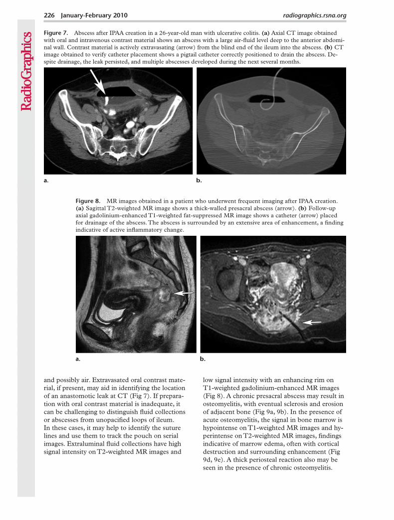
Figure 8. MR images obtained in a patient who underwent frequent imaging after IPAA creation. (a) Sagittal T2-weighted MR image shows a thick-walled presacral abscess (arrow). (b) Follow-up axial gadolinium-enhanced T1-weighted fat-suppressed MR image shows a catheter (arrow) placed for drainage of the abscess. The abscess is surrounded by an extensive area of enhancement, a finding indicative of active inflammatory change.
Figure 7. Abscess after IPAA creation in a 26-year-old man with ulcerative colitis. (a) Axial CT image obtained with oral and intravenous contrast material shows an abscess with a large air-fluid level deep to the anterior abdomi-nal wall. Contrast material is actively extravasating (arrow) from the blind end of the ileum into the abscess. (b) CT image obtained to verify catheter placement shows a pigtail catheter correctly positioned to drain the abscess. De-spite drainage, the leak persisted, and multiple abscesses developed during the next several months.
low signal intensity with an enhancing rim on T1-weighted gadolinium-enhanced MR images (Fig 8). A chronic presacral abscess may result in osteomyelitis, with eventual sclerosis and erosion of adjacent bone (Fig 9a, 9b). In the presence of acute osteomyelitis, the signal in bone marrow is hypointense on T1-weighted MR images and hy-perintense on T2-weighted MR images, findings indicative of marrow edema, often with cortical destruction and surrounding enhancement (Fig 9d, 9e). A thick periosteal reaction also may be seen in the presence of chronic osteomyelitis.
and possibly air. Extravasated oral contrast mate-rial, if present, may aid in identifying the location of an anastomotic leak at CT (Fig 7). If prepara-tion with oral contrast material is inadequate, it can be challenging to distinguish fluid collections or abscesses from unopacified loops of ileum. In these cases, it may help to identify the suture lines and use them to track the pouch on serial images. Extraluminal fluid collections have high signal intensity on T2-weighted MR images and

Figure 9. Abscess with osteomyelitis after IPAA creation for colon adenocarcinoma in a 66-year-old woman with ulcerative colitis. (a) Sagittal unenhanced CT image shows extraluminal air pockets (arrowhead) and a presacral abscess (white arrow) that contains hyperattenuating oral contrast material from an earlier imaging study. Sclerosis (black arrow) of the adjacent sacrum is seen, a finding suggestive of chronic osteomyelitis. (b, c) Lateral (b) and anteroposterior (c) fluoroscopic images obtained after the administration of an enema containing water-soluble contrast material show extravasation (arrowhead) into the presacral fluid collection (white arrows) from the blind end of the ileum (* in c). Sacral osteomyelitis (black arrow in b) also is seen. (d, e) Sagittal T2-weighted (d) and gadolinium-enhanced T1-weighted (e) fat-suppressed MR images show sacral bone marrow edema (arrow in d) and marked presacral soft-tissue enhancement (* in e). (f, g) Unenhanced axial CT images show transgluteal placement of a catheter for drainage of the abscess. A spinal needle (arrowhead in f) was used to access the collection, a wire (arrowhead in g) was threaded through the needle, and a pigtail catheter (arrow in g) was inserted over the wire.

228 January-February 2010 radiographics.rsna.org
solution to maintain patency. Premature removal of the catheter should be avoided because it may lead to reaccumulation of fluid in the abscess as a result of incomplete healing of the leakage site. Even after drainage has ceased or imaging has demonstrated resolution of the fluid collec-tion, complete healing of the source of the leak, commonly a dehiscent suture line, may not have occurred, and fluid may reaccumulate in the ab-scess soon after the catheter is removed. To check for a persistent leak before removing the catheter, we prefer to obtain an abscessogram or fistulo-gram after gently infusing water-soluble contrast material directly into the catheter. By filling the residual cavity with contrast material, we can detect any persistent communication between the fluid collection and bowel. In comparison, orally or transrectally administered contrast material of-ten follows the path of least resistance within the bowel lumen without extravasating through the leak and into the fluid collection, creating a false impression that the defect has completely healed.
Figure 10. Pouchitis in a 20-year-old woman who underwent IPAA creation 5 years earlier for intractable indeterminate colitis. (a, b) Ax-ial (a) and sagittal (b) T2-weighted MR im-ages show pouch wall thickening (arrow) and adenopathy (arrowhead in b). (c) Axial gado-linium-enhanced T1-weighted fat-suppressed MR image shows mucosal enhancement (black arrow), wall thickening, peripouch adenopa-thy (arrowhead), and inflammatory change (white arrows).
Management of an abscess often includes ultrasonography (US)- or CT-guided drainage and treatment with antibiotics. Deep abscesses are more accessible by way of a transgluteal ap-proach, which requires that the patient be in the prone position (10). At our institution, we often use the Seldinger technique to reduce the risks for hemorrhage and injury to adjacent structures. An 18-gauge needle is used to access the fluid collection, and a 0.035-inch J-tip guidewire is threaded through the needle. A 12-F pigtail cath-eter is then advanced over the wire and into the fluid collection after serial tract dilations (Figs 6b, 6c, 9f, 9g). Larger fluid collections may be drained safely by using a single-stick trocar tech-nique. The use of CT fluoroscopy helps increase the accuracy of catheter placement.
Judicious management of drainage catheters may improve clinical outcomes and minimize the need for further imaging. At our institution, we routinely suture all catheters to the skin to avoid their accidental removal, because replacing a catheter after partial drainage may be difficult or impossible. The catheter should be flushed several times a day with small amounts of saline

The infusion of contrast material via the catheter may be performed during fluoroscopy or imme-diately before CT. An abscess that persists despite repeated treatments with catheter drainage and antibiotics likely is the result of a continued leak and may require surgical intervention with pouch revision or excision (11).
PouchitisThe most common complication of IPAA is inflammation of the ileal pouch reservoir, com-monly referred to as pouchitis. Although the etiology of pouchitis is not fully understood, it is hypothesized to result from disequilibrium of intestinal flora. Approximately 20% of patients with an IPAA experience an episode of pouchitis within 1 year after surgery, and approximately 50% experience an episode of pouchitis within 10 years after surgery (12). These patients present with frequent watery or bloody stools, pain, fever, or a new onset of rectal bleeding. In many cases,
Figure 11. Severe pouchitis, 1 year later, in the same patient as in Figure 10, who is now thought to have Crohn disease. (a) Axial CT image obtained with intravenous contrast material shows dilatation of the J pouch (arrows). (b, c) Axial contrast-enhanced CT image (b) and axial gadolinium-enhanced T1-weighted fat-suppressed MR image (c) show wall thickening (black arrow), mucosal hyperenhancement (black arrowhead), and peripouch inflammatory changes (white arrows). Adjacent pelvic lymphade-nopathy (white arrowhead) also is seen.
the clinical history is suggestive of pouchitis and the diagnosis is achieved with direct endoscopic visualization. However, because the symptoms of pouchitis at presentation often raise concern about a possible leak or abscess, CT or MR imaging often is performed. At our institution, if a leak or abscess is suspected, we perform CT emergently, and we use MR imaging to follow the course of previously diagnosed pouchitis and for emergent evaluation of patients with recur- rent complications who require frequent imaging. Pouchitis is treated primarily with antibiotics; however, other drugs, such as steroids and inflix-imab, also are used to treat refractory cases (13). Patients with complicated, intractable pouchitis or resultant fistula or abscess formation ultimately may be found to have Crohn disease (9,14). CT and MR imaging features of pouchitis include pouch wall thickening, peripouch adenopathy and inflammatory changes (stranding and fluid), mucosal hyperenhancement, and pouch dilatation (Figs 10, 11). The diagnosis may be verified with endoscopy if clinically indicated (Fig 12).
TeachingPoint

radiographics.rsna.org
Venous thrombi after IPAA creation. (13) Axial CT im-age obtained with intravenous contrast material shows filling defects (ar-rows) in the right and left portal veins. (14) Coronal CT image obtained with intravenous contrast material in a different patient shows a segmen-tal thrombus (white arrow) with a resultant hepatic perfusion abnormal-ity (black arrows) secondary to preferential arterial perfusion.
Figure 12. Pouchitis after IPAA creation. Endoscopic image shows extensive mucosal erythema, ulcer-ation, and inflammatory changes. (Courtesy of Francis Farraye, MD, Boston University Medical Center, Boston, Mass.)
Venous ThrombusAt CT and MR imaging, the venous system must be scrutinized for thrombi, a complica-tion reported in as many as 45% of patients who undergo CT within 10 weeks after IPAA sur-gery (15). Venous thrombi likely are secondary to surgical manipulation of the mesentery and the heightened inflammatory status of patients with inflammatory bowel disease, which may be exacerbated in those with an abscess or pouchi-tis. Patients usually are asymptomatic, but they may present with abdominal pain or tenderness, fever, nausea, vomiting, ileus, and leukocytosis (15). Most thrombi are incidentally found at CT performed to determine the cause of these symptoms. Thrombi are treated with anticoagula-tion therapy, which usually results in complete recovery with no significant damage to long-term pouch function (16). Subtle, asymptomatic thrombi may not require treatment.
Thrombi often originate in the mesenteric veins and propagate downstream into the portal vein. At CT and MR imaging, venous thrombi most often are seen as multiple, peripheral, oc-clusive, tubular filling defects in the segmental hepatic portal veins (Fig 13). Central thrombi in the superior mesenteric, inferior mesenteric, main portal, and right and left portal veins tend to be nonocclusive, although they may occasion-ally be completely occlusive. Thrombi in the por-tal venous system may cause peripheral wedge-shaped hepatic perfusion abnormalities because of compensatory arterial hyperperfusion of the involved liver segments (Fig 14) (17).
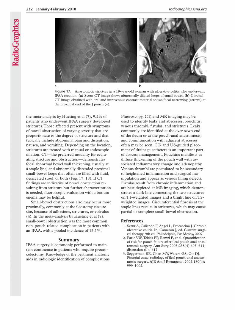
FistulasPatients who undergo IPAA surgery may de-velop fistulas as a result of chronic infection or bowel wall inflammation, likely from a persistent anastomotic leak or Crohn disease. An epithelial-ized tract forms between adjacent bowel walls or between the bowel and the abdominal wall, anal sphincter, or genitourinary tract. In the meta-analysis by Hueting et al (7), it was reported that an anal or vaginal fistula developed in 5.5% of patients who underwent IPAA surgery. Clini-cal manifestations and imaging features vary, depending on the location of the fistula. Fistulas usually are managed surgically but also may be treated medically with infliximab. Fistulas may be seen at fluoroscopy after an infusion of water-soluble contrast material into the pouch (Fig 15). However, pouchography has low sensitivity for the detection of subtle fistulas, perhaps because transrectal contrast material often follows the path of least resistance within the bowel lumen rather than opacifying the fistula (9). Similarly, CT may not always depict fistulas; sensitivity is reported to be as low as 33% (9). Fistulas may be easily identified and characterized at MR imaging; thus, it is the modality of choice at our institution. Fistulas appear as a hypointense line between the involved structures on T1-weighted images, a hyperintense line on T2-weighted im-ages, and usually have an enhancing rim on gado-linium-enhanced T1-weighted images (Fig 16).
Anastomotic Strictures and Small-Bowel Obstruction
Anastomotic strictures that are secondary to fibrosis occur at the site of the ileal-anal anasto-mosis or at the proximal end of the J pouch. In
TeachingPoint
TeachingPoint
TeachingPoint
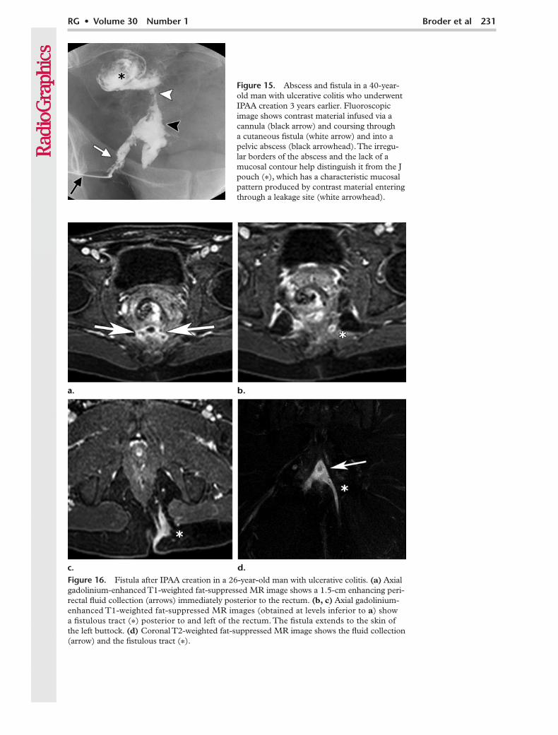
Figure 16. Fistula after IPAA creation in a 26-year-old man with ulcerative colitis. (a) Axial gadolinium-enhanced T1-weighted fat-suppressed MR image shows a 1.5-cm enhancing peri-rectal fluid collection (arrows) immediately posterior to the rectum. (b, c) Axial gadolinium-enhanced T1-weighted fat-suppressed MR images (obtained at levels inferior to a) show a fistulous tract (*) posterior to and left of the rectum. The fistula extends to the skin of the left buttock. (d) Coronal T2-weighted fat-suppressed MR image shows the fluid collection (arrow) and the fistulous tract (*).
Figure 15. Abscess and fistula in a 40-year-old man with ulcerative colitis who underwent IPAA creation 3 years earlier. Fluoroscopic image shows contrast material infused via a cannula (black arrow) and coursing through a cutaneous fistula (white arrow) and into a pelvic abscess (black arrowhead). The irregu-lar borders of the abscess and the lack of a mucosal contour help distinguish it from the J pouch (*), which has a characteristic mucosal pattern produced by contrast material entering through a leakage site (white arrowhead).
radiographics.rsna.org
Figure 17. Anastomotic stricture in a 19-year-old woman with ulcerative colitis who underwent IPAA creation. (a) Scout CT image shows abnormally dilated loops of small bowel. (b) Coronal CT image obtained with oral and intravenous contrast material shows focal narrowing (arrows) at the proximal end of the J pouch (*).
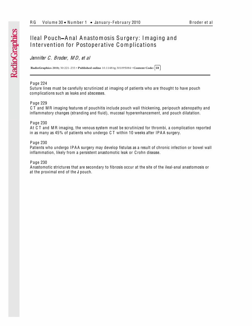
the meta-analysis by Hueting et al (7), 9.2% of patients who underwent IPAA surgery developed strictures. Those affected present with symptoms of bowel obstruction of varying severity that are proportionate to the degree of stricture and that typically include abdominal pain and distention, nausea, and vomiting. Depending on the location, strictures are treated with manual or endoscopic dilation. CT—the preferred modality for evalu-ating stricture and obstruction—demonstrates focal abnormal bowel wall thickening, usually at a staple line, and abnormally distended proximal small-bowel loops that often are filled with fluid, desiccated stool, or both (Figs 17, 18). If CT findings are indicative of bowel obstruction re-sulting from stricture but further characterization is needed, fluoroscopic evaluation with a barium enema may be helpful.
Small-bowel obstructions also may occur more proximally, commonly at the ileostomy closure site, because of adhesions, strictures, or volvulus (4). In the meta-analysis by Hueting et al (7), small-bowel obstruction was the most common non–pouch-related complication in patients with an IPAA, with a pooled incidence of 13.1%.
SummaryIPAA surgery is commonly performed to main-tain continence in patients who require procto-colectomy. Knowledge of the pertinent anatomy aids in radiologic identification of complications.
Fluoroscopy, CT, and MR imaging may be used to identify leaks and abscesses, pouchitis, venous thrombi, fistulas, and strictures. Leaks commonly are identified at the over-sewn end of the ileum or at the pouch-anal anastomosis, and communication with adjacent abscesses often may be seen. CT- and US-guided place-ment of drainage catheters is an important part of abscess management. Pouchitis manifests as diffuse thickening of the pouch wall with as-sociated inflammatory change and adenopathy. Venous thrombi are postulated to be secondary to heightened inflammation and surgical ma-nipulation and appear as venous filling defects. Fistulas result from chronic inflammation and are best depicted at MR imaging, which demon-strates a dark line connecting the two structures on T1-weighted images and a bright line on T2-weighted images. Circumferential fibrosis at the staple lines results in strictures, which may cause partial or complete small-bowel obstruction.
References 1. Serur A, Caliendo F, Angel L, Procaccino J. Chronic
ulcerative colitis. In: Cameron J, ed. Current surgi-cal therapy. 9th ed. Philadelphia, Pa: Mosby, 2007.
2. Fazio VW, Tekkis PP, Remzi F, et al. Quantification of risk for pouch failure after ileal pouch anal anas-tomosis surgery. Ann Surg 2003;238(4):605–614; discussion 614–617.
3. Seggerman RE, Chen MY, Waters GS, Ott DJ. Pictorial essay: radiology of ileal pouch-anal anasto-mosis surgery. AJR Am J Roentgenol 2003;180(4): 999–1002.
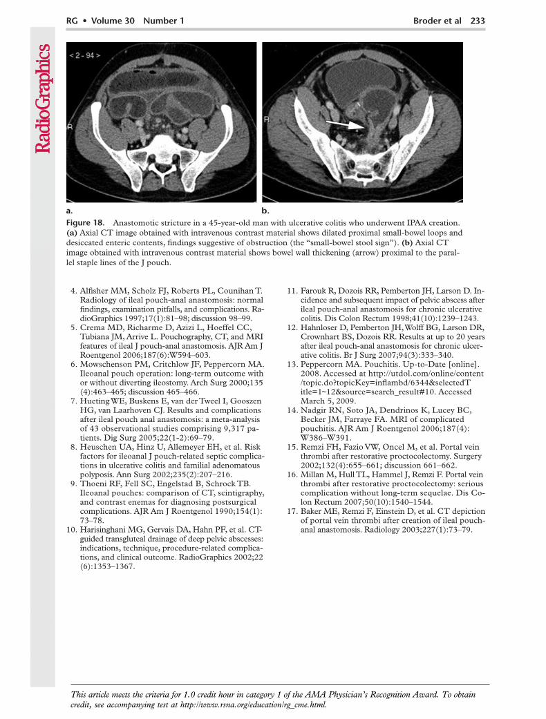
Figure 18. Anastomotic stricture in a 45-year-old man with ulcerative colitis who underwent IPAA creation. (a) Axial CT image obtained with intravenous contrast material shows dilated proximal small-bowel loops and desiccated enteric contents, findings suggestive of obstruction (the “small-bowel stool sign”). (b) Axial CT image obtained with intravenous contrast material shows bowel wall thickening (arrow) proximal to the paral-lel staple lines of the J pouch.
4. Alfisher MM, Scholz FJ, Roberts PL, Counihan T. Radiology of ileal pouch-anal anastomosis: normal findings, examination pitfalls, and complications. Ra-dioGraphics 1997;17(1):81–98; discussion 98–99.
5. Crema MD, Richarme D, Azizi L, Hoeffel CC, Tubiana JM, Arrive L. Pouchography, CT, and MRI features of ileal J pouch-anal anastomosis. AJR Am J Roentgenol 2006;187(6):W594–603.
6. Mowschenson PM, Critchlow JF, Peppercorn MA. Ileoanal pouch operation: long-term outcome with or without diverting ileostomy. Arch Surg 2000;135 (4):463–465; discussion 465–466.
7. Hueting WE, Buskens E, van der Tweel I, Gooszen HG, van Laarhoven CJ. Results and complications after ileal pouch anal anastomosis: a meta-analysis of 43 observational studies comprising 9,317 pa-tients. Dig Surg 2005;22(1-2):69–79.
8. Heuschen UA, Hinz U, Allemeyer EH, et al. Risk factors for ileoanal J pouch-related septic complica-tions in ulcerative colitis and familial adenomatous polyposis. Ann Surg 2002;235(2):207–216.
9. Thoeni RF, Fell SC, Engelstad B, Schrock TB. Ileoanal pouches: comparison of CT, scintigraphy, and contrast enemas for diagnosing postsurgical complications. AJR Am J Roentgenol 1990;154(1): 73–78.
10. Harisinghani MG, Gervais DA, Hahn PF, et al. CT- guided transgluteal drainage of deep pelvic abscesses: indications, technique, procedure-related complica-tions, and clinical outcome. RadioGraphics 2002;22 (6):1353–1367.
11. Farouk R, Dozois RR, Pemberton JH, Larson D. In-cidence and subsequent impact of pelvic abscess after ileal pouch-anal anastomosis for chronic ulcerative colitis. Dis Colon Rectum 1998;41(10):1239–1243.
12. Hahnloser D, Pemberton JH, Wolff BG, Larson DR, Crownhart BS, Dozois RR. Results at up to 20 years after ileal pouch-anal anastomosis for chronic ulcer-ative colitis. Br J Surg 2007;94(3):333–340.
13. Peppercorn MA. Pouchitis. Up-to-Date [online]. 2008. Accessed at http://utdol.com/online/content /topic.do?topicKey=inflambd/6344&selectedTitle=1~12&source=search_result#10. Accessed March 5, 2009.
14. Nadgir RN, Soto JA, Dendrinos K, Lucey BC, Becker JM, Farraye FA. MRI of complicated pouchitis. AJR Am J Roentgenol 2006;187(4): W386–W391.
15. Remzi FH, Fazio VW, Oncel M, et al. Portal vein thrombi after restorative proctocolectomy. Surgery 2002;132(4):655–661; discussion 661–662.
16. Millan M, Hull TL, Hammel J, Remzi F. Portal vein thrombi after restorative proctocolectomy: serious complication without long-term sequelae. Dis Co-lon Rectum 2007;50(10):1540–1544.
17. Baker ME, Remzi F, Einstein D, et al. CT depiction of portal vein thrombi after creation of ileal pouch-anal anastomosis. Radiology 2003;227(1):73–79.
This article meets the criteria for 1.0 credit hour in category 1 of the AMA Physician’s Recognition Award. To obtaincredit, see accompanying test at http://www.rsna.org/education/rg_cme.html.
R G Volu m e 30 N u m ber 1 Janua ry - F eb r ua ry 2010 B roder et a l
I lea l Pouch A nal A nasto m osis Su rgery: I m aging and I n ter ven t ion for Postoper a t ive C o m pl ica t ions Jennifer C . Broder, M D , et al
P age 224 Suture lines must be carefully scrutinized at imaging of patients who are thought to have pouch complications such as leaks and abscesses. P age 229 C T and M R imaging features of pouchitis include pouch wall thickening, peripouch adenopathy and inflammatory changes (strand ing and fluid), mucosal hyperenhancement, and pouch dilatation. P age 230 At C T and M R imaging, the venous system must be scrutinized for thrombi, a complication reported in as many as 45% of patients who undergo C T within 10 weeks after IPA A surgery. P age 230 Patients who undergo IPA A surgery may develop fistulas as a result of chronic infection or bowel wall inflammation, likely from a persistent anastomotic leak or C rohn disease. P age 230 Anastomotic strictures that are secondary to fibrosis occur at the site of the ileal-anal anastomosis or at the proximal end of the J pouch.
RadioGraphics 2010; Published online Content Code: