effect of ischemia on localization of heat shock protein 25 in kidney
TRANSCRIPT

Effect of ischemia on localization of heat shock protein 25 inkidney
ANDREAS SCHOBER, ANKE BURGER-KENTISCHER, EVA MULLER, and FRANZ-X. BECK
Department of Physiology, University of Munich, Munich, Germany
Effect of ischemia on localization of heat shock protein 25 inkidney. The effects of renal ischemia on the intracellular distri-bution of the low-molecular weight heat shock protein (HSP)25were examined using immunofluorescence microscopy. In allkidney zones, ischemia decreased HSP25 in the supernatant of thetissue homogenates and increased it in the pellet fraction (con-taining mainly nuclei and cytoskeletal components). This wasassociated with disappearance of HSP25 staining from the brushborder of proximal convoluted tubule (PCT) cells. Because nonuclear staining of cortical tubule cells was apparent either incontrol or ischemic kidneys, ischemia seems to cause a closerassociation of HSP25 with cytoskeletal components. HSP25 prob-ably participates in the postischemic restructuring of the cytoskel-eton of PCT cells.
Interruption of renal blood flow leads to intracellularredistribution of the low-molecular weight heat shockprotein 25 (HSP25) in the cells of the injured kidney [1]. Itis unknown whether this intracellular redistribution iscaused by HSP25 moving from the cytosol into the nucleusor/and by a closer association of this HSP with cytoskeletalstructures such as actin. We thus characterized in greaterdetail the ischemia-induced intracellular redistribution ofHSP25.
METHODS
Male Wistar rats (Charles River, Sulzfeld, Germany)kept on a standard diet (Alma, Kempten, Germany) wereanesthetized by i.p. injection of thiobutabarbitone sodium(Inactin, 100 to 120 mg/kg body weight; Byk-Gulden,Konstanz, Germany). The surgical protocol and prepara-tion of the left kidney have been described elsewhere [1].Ischemia of the left kidney (60 minutes) was achieved byoccluding the left renal artery by a weak clip. Both kidneyswere harvested immediately after the 60 minutes of isch-emia and were frozen in propane/isopentane (3:1, 2196°C).The contralateral nonischemic kidney served as the control.
For indirect immunofluorescence microscopy, 4-mm sec-tions were cut at 220°C (CM3050 cryotome; Leica, Nuss-loch, Germany) and were fixed immediately in 4% parafor-maldehyde. The sections were heated in a citric acid buffer(10 mM, pH 6) in a microwave oven for 10 minutes.Endogenous avidin binding was blocked with an avidin-biotin blocking kit (Zymed, San Francisco, CA, USA) andnonspecific antibody binding sites with 20% normal goatserum (Life Technologies, Eggenstein, Germany) and0.01% Tween-20. For immunodetection, sections wereincubated with a HSP25-specific antiserum [SPA-801;Stress Gen, Victoria, Canada; 1:500 dilution in phosphate-buffered saline (PBS)/goat serum/Tween-20; 1 hour],washed in PBS, and incubated with a biotin-SP-conjugatedgoat antirabbit IgG (Dianova, Hamburg, Germany; 1:500dilution in PBS/goat serum/Tween-20). The sections wererewashed and exposed to rhodamine (TRITC)-conjugatedstreptavidin (Dianova; 1:400 dilution in PBS/goat serum/Tween-20; 30 minutes) in a light-proof container. Negativecontrols for each antibody were processed in parallel by
Key words: proximal convoluted tubule, van Willebrand’s factor, cytoskel-eton, actin, microfilaments in endothelial cells, cytoskeletal restructuring.
© 1998 by the International Society of Nephrology
Fig. 1. Extractable (supernatant fraction, left) and nonextractable (pel-let fraction, right) heat shock protein 25 (HSP25) in cortex (CX), outermedulla (OM), and inner medulla (IM) in ischemic left kidneys (s) andin nonischemic right kidneys (controls, e; N 5 4). Data is taken from [1];means 6 SEM. *P , 0.05 versus corresponding control.
Kidney International, Vol. 54, Suppl. 67 (1998), pp. S-174–S-176
S-174
omitting the primary antibody. Finally, the sections weremounted under cover slips with FluorSave TN Reagent(Calbioch, Bad Soden, Germany) and were stored at 4°C inthe dark until visualization in an inverted microscope(IM35; Zeiss, Oberkochen, Germany). A separate series ofmethanol-fixed (220°C, 5 minutes) cryosections was usedfor double labeling with a monoclonal mouse antibody(Boehringer, Mannheim, Germany) against von Wille-brand’s factor (vWF) and the above antiserum againstHSP25. For vWF staining, a FITC-conjugated goat anti-mouse antibody (DAKO, Hamburg, Germany) was used assecondary antibody (primary and secondary antibody dilu-tion 1:10). After vWF staining, the sections were stained forHSP25 as mentioned earlier here. Stains were visualizedwith filter combinations appropriate for monitoring fluo-rescence of FITC or rhodamine.
RESULTS AND DISCUSSION
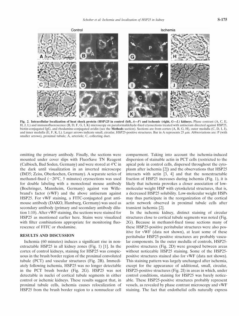
Ischemia (60 minutes) induces a significant rise in non-extractable HSP25 in all kidney zones (Fig. 1) [1]. In thecortex of control kidneys, staining for HSP25 was conspic-uous in the brush border region of the proximal convolutedtubule (PCT) and vascular structures (Fig. 2B). Immedi-ately following ischemia, HSP25 was no longer detectablein the PCT brush border (Fig. 2G). HSP25 was notdetectable in nuclei of cortical tubule segments in eithercontrol or ischemic kidneys. These results suggest that, inproximal tubule cells, ischemia causes relocalization ofHSP25 from the brush border region to a nonnuclear cell
comparment. Taking into account the ischemia-induceddispersion of stainable actin in PCT cells (restricted to theapical pole in control cells, dispersed throughout the cyto-plasm after ischemia [2]) and the observations that HSP25interacts with actin [3, 4] and that the nonextractablefraction of HSP25 increases during ischemia (Fig. 1), it islikely that ischemia provokes a closer association of low-molecular weight HSP with cytoskeletal structures, that is,a decreased HSP25 solubility. Low-molecular weight HSPsmay thus participate in the reorganization of the corticalactin network observed in proximal tubule cells aftertransient ischemia [2].
In the ischemic kidney, distinct staining of circularstructures close to cortical tubule segments was noted (Fig.2G). Because in methanol-fixed tissue sections many ofthese HSP25-positive peritubular structures were also pos-itive for vWF (data not shown), at least some of theseperitubular HSP25-positive structures are probably vascu-lar components. In the outer medulla of controls, HSP25-positive structures (Fig. 2D) were grouped between areaswithout noticeable HSP25 staining. Some of the HSP25-positive structures stained also for vWF (data not shown).This staining pattern was largely unchanged after ischemia,except for the appearance of additional, small, circular,HSP25-positive structures (Fig. 2I) in areas in which, undercontrol conditions, staining for HSP25 was barely notice-able. These HSP25-positive structures probably representvessels, as revealed by phase contrast microscopy and vWFstaining. The fact that endothelial cells naturally express
Fig. 2. Intracellular localization of heat shock protein (HSP)25 in control (left, A—F) and ischemic (right, G—L) kidneys. Phase contrast (A, C, E,H, J, L) and immunofluorescence (B, D, F, G, I, K) microscopy on paraformaldehyde-fixed cryosections treated with antiserum directed against HSP25,biotin-conjugated IgG, and rhodamine-conjugated avidin (see the Methods section). Sections are from cortex (A, B, G, H), outer medulla (C, D, I, J),and inner medulla (E, F, K, L). Larger arrows indicate small, circular, HSP25-positive structures. Bar in A represents 25 mm. Abbreviations are: P (withsmaller arrows), proximal tubule; A, arteriole; C, collecting duct.
Schober et al: Ischemia and localization of HSP25 in kidney S-175

high levels of the small HSP and that this HSP is involvedin the oxidative stress-induced actin reorganization in thesecells [5] supports the view that HSP25 may participate inthe microfilament response to ischemic stress also in endo-thelial cells of the kidney. As also shown in Figure 2,ischemia did not noticeably affect the intense HSP25staining of collecting duct cells in the inner medulla.Whether the ischemia-induced increase in nonextractableHSP25 in this kidney zone (Fig. 1) is due to closerassociation of HSP25 with cytoskeletal or other proteins orto multioligomerization of this HSP remains to be eluci-dated.
ACKNOWLEDGMENTS
This study was supported by the Deutsche Forschungsgemeinschaft (Be963/4-4). The presented material is part of A. Schober’s M.D. thesis.
Reprint requests to Professor F-X. Beck, Department of Physiology,University of Munich, Pettenkoferstr. 12, D-80336 Munich, Germany.
REFERENCES
1. SCHOBER A, MULLER E, THURAU K, BECK FX: The response of heatshock proteins 25 and 72 to ischaemia in different kidney zones.Pflugers Arch 434:292–299, 1997
2. MOLITORIS BA: Ischemia-induced loss of epithelial polarity: Potentialrole of the actin cytoskeleton. Am J Physiol 260:F769–F778, 1991
3. LAVOIE JN, HICKEY E, WEBER LA, LANDRY J: Modulation of actinmicrofilament dynamics and fluid phase pinocytosis by phosphorylationof heat shock proteins. J Biol Chem 268:24210–24214, 1993
4. BENNDORF R, HAYESS K, RYAZANTSEV S, WIESKE M, BEHLKE J,LUTSCH G: Phosphorylation and supramolecular organization of mu-rine small heat shock protein HSP25 abolish its actin polymerization-inhibiting activity. J Biol Chem 269:20780–20784, 1994
5. HUOT J, HOULE F, MARCEAU F, LANDRY J: Oxidative stress-inducedactin reorganization mediated by the p38 mitogen-activated proteinkinase/heat shock protein 27 pathway in vascular endothelial cells. CircRes 80:383–392, 1997
Schober et al: Ischemia and localization of HSP25 in kidneyS-176