effect of tri-n-butyltin on intracellular ca2+ concentration of rat cerebellar neurons
TRANSCRIPT

European Journal of Pharmacology - Environmental Toxicology and Pharmacology Section, 248 (1993) 89-93 89 © 1993 Elsevier Science Publishers B.V. All rights reserved 0926-6917/93/$06.00
EJPTOX 40051
Short communication
Effect of tri-n-butyltin on intracellular C a 2+ concentration of rat cerebellar neurons
Yasuo Oyama, Tosh iko U e h a and A k em i Hayash i
Di~,ision of Pharmacological Science, Department of Health Sciences, Faculty of Integrated Arts and Sciences, The University of Tokushima, Tokushima 770, Japan
Received 28 December 1992, revised MS received 17 February 1993, accepted 16 March 1993
The effect of tri-n-butyltin on the intracellular Ca 2+ concentration ([Ca2+] i) of cerebellar neurons dissociated from rats was examined using fluo-3 and a flow cytometer. Tri-n-butyltin at 100 nM or more (up to 1 txM) increased the [Ca2+]i in a dose-dependent manner. The effect of tri-n-butyltin on the [Ca2+]i was greatly reduced under external CaZ+-free ([CaZ+]o-free) conditions, suggesting its dependence on the presence of [Ca 2+ ]o. Lower trialkyltins, such as triethyltin and trimethyltin at 1 txM, exerted little or no action on the [Ca2+]i . Therefore, the cytotoxic action of tri-n-butyltin may be different from those of lower trialkyltins.
Tributyltin; Triethyltin; Trimethyltin; Intracellular Ca2+; Brain neurons; Cytotoxicity
1. Introduction
The possible environmental hazard of triorganotins has become a subject of concern because of the rapid expansion in their technical applications (Van der Kerk, 1978; Wilkinson, 1984). Of the triorganotins, tri- alkyltins have been described as exerting a variety of toxic actions on mammals, including neurotoxicity, hepatoxicity, and immunotoxicity (Snoeij et al., 1987). The dominant profile of the toxicity of lower tri- alkyltins, such as trimethyltin and triethyltin, was con- sidered to be neurotoxicity, while the toxicity of tri- alkyltins with intermediate chain length, such as tri-n- butyltin and tri-n-propyltin, was characterized by im- munotoxic action. However, recent studies have also revealed the toxic actions of tri-n-butyltin and bis(tri- n-butyltin) oxide on the mammalian central nervous system (O'Callaghan and Miller, 1986; Miyake et al., 1990).
We have reported that tri-n-butyltin in the hundred nanomolar range initiates an increase in intracellular Ca 2+ ([Ca2+]i) of mouse thymocytes, suggesting a type of cytotoxic action (Chikahisa and Oyama, 1992). It is,
Correspondence to: Y. Oyama, Division of Pharmacological Science, Department of Health Sciences, Faculty of Integrated Arts and Sciences, The University of Tokushima, Minami-Josanjima 1-1, Tokushima 770, Japan. Tel. (81X886)23-2311, ext. 2411; Fax (81)(886)55-2108.
therefore, essential to find out whether tri-n-butyltin also increases the [Ca2+]i of brain neurons because such an investigation may provide some insight into the cellular basis of neurotoxic action of tri-n-butyltin. In the present study, the effect of tri-n-butyltin on the [Ca2+]i of brain neurons dissociated from the rat has been examined in comparison with the effects of lower trialkyltins.
2. Materials and methods
2.1. Chemicals
Pentaacetoxymethyl ester of fluo-3, a fluorescent dye for [Ca2+]i, dissolved in dimethylsulfoxide was purchased from Dojindo Laboratory, Kumamoto, Japan. Dispase, a proteolytic enzyme for dissociation of neurons, was from Godo Shusei, Tokyo, Japan. Trialkyltins used in this study were tri-n-butyltin chlo- ride (Kishida Chemical, Osaka, Japan), triethyltin chlo- ride (Merck, Schuchardt, Germany) and trimethyltin chloride (Tokyo Kasei, Tokyo, Japan). Other chemical reagents were purchased from Katayama Chemical In- dustries, Osaka, Japan.
2.2. Dissociation o f brain neurons
The cerebellum, dissected from 12-14-day-old Wis- tar Kyoto strain rats (from Nissin, Tokushima, Japan),

90
was sliced at a thickness of 500 g m (Narishige, Tokyo, Japan). Brain slices were treated with dispase (1000 P U / m l ) at 35-36°C for 50-60 min (Oyama et al., 1992a,b), after which they were pipetted in Tyrode's solution to disperse the neurons. Tyrode's solution, containing dissociated neurons and residua, was passed through a filter (mesh diameter of 53 /xm) to remove residua and larger neurons because of application of dissociated neurons to a flow cytometer, described previously (Oyama et al., 1992a,b).
2.3. Fluorescence measurement
The neurons were incubated in Tyrode's solution with the pentaacetoxymethyl ester of fluo-3 at 300 nM for 60 min to allow fluo-3 to load into the neurons as previously described in rat brain neurons (Oyama et al., 1992a). Fluorescence measurement of live cell pop- ulation in the cell suspension (Oyama et al., 1992b) was performed on a flow cytometer (Cyto-ACE 150, Japan Spectroscopic Co., Tokyo, Japan). The excitation wave- length was 488 nm and the emission was detected at a wavelength of 530 +_ 20 nm. Fluorescence obtained from dissociated cerebellar neurons was analyzed on a personal computer (PC-8100XR, Nippon Electronics Co., Tokyo, Japan) with software (JASCO System pro- gram Ver 3.XX) developed by Japan Spectroscopic Co., Tokyo, Japan. Data acquisition from 4000-5000 neurons required about 20 s. Therefore, the acquisition started 10 s before the time described in the text and figures. Changes in the fluo-3 fluorescence to higher and lower intensities correspond to the increase and decrease in the [Ca2+]i, respectively. Fluorescence in- tensity was resolved into 255 channels (arbitrary num- ber) as shown in the abscissa of the histogram (see figs. 1 and 2). Tentative calibration of fluo-3 fluorescence for the [Ca2+]i was made by a method using ionomycin and MnC12 (Kao et al., 1989).
A. B. C. TBT 30nM 100 nM 300 nM
128r- [ ~ ~ ,
64
o L .J I . I .~ ~ . . ~ z I , I I ~ I L , I
75 255 75 255 75 255
D. F TBT lpM IONO I#M
128[- /1
64
z 0 L _ S < k I ~ I I , I
75 255 75 255 Fluorescence Intensity (Channel Number)
F. 220
E 200 ,/ z !I ~_ -4:3 - - - 0
m / 0 Ionomycin 1 #M / 0 /El [ ] Tri-n-butyttin I~M
1,10
5 10 rain Time After Drug Application
Fig. 1. Effect of tri-n-butyltin and ionomycin on the fluo-3 fluores- cence histogram in dissociated cerebellar neurons (4000 neurons). Histograms represent the fluo-3 fluorescence distribution in the neurons. Abscissa of (A-E) indicates the fluorescence intensity. Fluorescence intensity was resolved into 255 channels (arbitrary number). Ordinate shows the number of cells at respective channels. Histograms without and with the arrow were obtained in the control and 10 rnin after the following applications: (A) 30 nM tri-n-butyltin (TBT), (B) 100 nM, (C) 300 nM, (D) 1 p~M and (E) 1 ~M ionomycin (IONO), respectively. According to the calibration method described by Kao et al. (1989), the channel numbers 125, 165 (mid point of abscissa), 185 and 200 correspond to 10 nM, 100 nM, 300 nM and 1 ~xM [Ca 2+ ]2, respectively. (F) time courses of tri-n-butyltin- and ionomycin-induced changes in the mean channel number of fluores-
cence histogram indicated by circles and squares, respectively.
3. Results
3.1. Effect of tri-n-butyltin on the fluo-3 fluorescence
Tri-n-butyltin at concentrations of 30 nM or less did not appear to exert any action on fluo-3 fluorescence profile of dissociated cerebellar neurons (fig. 1A), sug- gesting no change in the [Ca2+] i. However, tri-n-butyl- tin at 100 nM slightly increased the intensity of fluores- cence, indicating a slight increase in the [Ca2+] i of dissociated neurons (fig. 1B). The steady-state of tri-n- butyltin action on the fluorescence profile was ob- tained 10-20 min after application. The mean channel number of the fluorescence histogram in the presence of 100 nM tri-n-butyltin increased from the control value of 143.2 + 0.3 to 149.6 _+ 0.3 (mean channel num-
ber _+ S.E. in 4000 neurons). These mean channel num- bers were calibrated to 27 nM and 41 nM of the [Ca2+]i, respectively. Increasing the tri-n-butyltin con- centration (up to 1 /xM) further augmented the fluo-3 fluorescence in a dose-dependent manner. As shown in figs. 1C and 1D, the fluorescence histogram in dissoci- ated neurons clearly moved in the direction of higher. The mean channel number of the fluorescence his- togram (also obtained from 4000 neurons) was in- creased from 141.1 + 0.3 to 165.6_+ 0.3 by 300 nM tri-n-butyltin, and from 144.2 _+ 0.3 to 180.4 _+ 0.3 by 1 /xM tri-n-butyltin, respectively. In the cases of 300 nM and 1 /xM tri-n-butyltin, the calibrated [Ca2+] i at re- spective mean channel number reached 102 nM and 306 nM (figs. 1C and 1D). Ionomycin, a calcium ionophore, at a concentration of 1 # M greatly in-
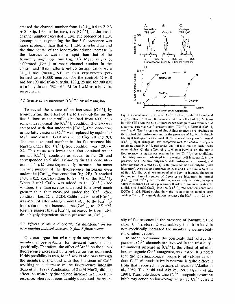
creased the channel number from 142.4 _+ 0.4 to 212.3 _+ 0.4 (fig. 1E). In this case, the [Ca2+]i at the mean channel number exceeded 1 I*M. The potency of 1 # M ionomycin in augmenting the fluo-3 fluorescence was more profound than that of 1 /.~M tri-n-butyltin and the time course of the ionomycin-induced increase in the fluorescence was more rapid than that of the tri-n-butyltin-induced one (fig. 1F). Mean values of calibrated [Ca2+]~ at mean channel number in the control and 10 min after tri-n-butyltin application were 31 + 3 nM (mean + S.E. in four experiments per- formed with 16,000 neurons) for the control, 67 + 18 nM for 100 nM tri-n-butyltin, 122 + 28 nM for 300 nM tri-n-butyltin and 562 + 61 nM for 1 /xM tri-n-butyltin, respectively.
3.2. Source of an increased [Ca 2 +]i by tri-n-butyltin
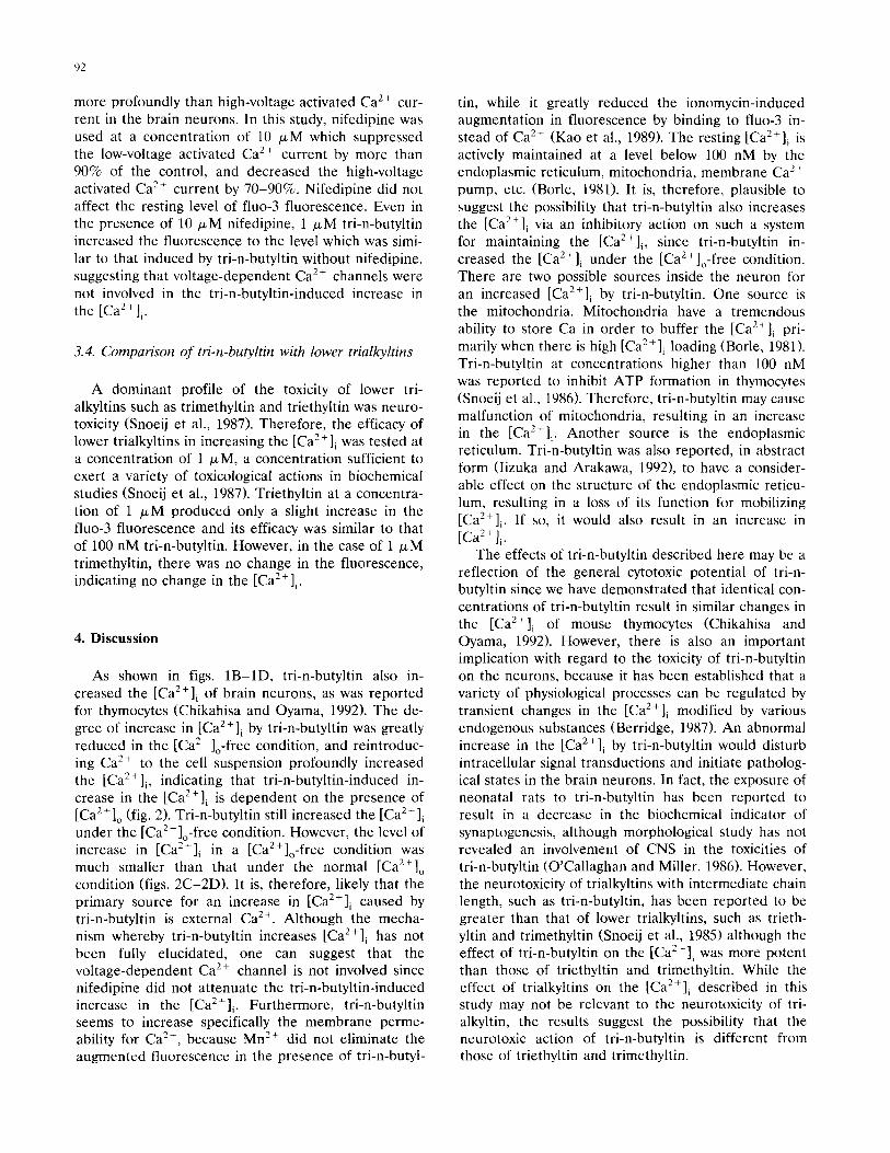
To reveal the source of an increased [ C a 2 + ] i by tri-n-butyltin, the effect of 1 /xM tri-n-butyltin on the fluo-3 fluorescence profile, obtained from 4000 neu- rons, under normal the [Ca 2÷]o condition (fig. 2A) was compared with that under the [Ca2+]o-free condition; in the latter, external Ca z+ was replaced by equimolar Mg 2÷ and 2 mM E G T A was added (figs. 2B and 2C). The mean channel number in the fluorescence his- togram under the [Ca2+]o-free condition was 120.5 + 0.2. This value was lower than that obtained under normal [Ca2+] o condition as shown in fig. 2B and corresponded to 9 nM. Tri-n-butyltin at a concentra- tion of 1 IxM time-dependently increased the mean channel number of the fluorescence histogram even under the [Cae+]o-free condition (fig. 2B). It reached 140.0 + 0.2, corresponding to 27 nM of the [Ca2+] i. When 2 mM CaCI 2 was added to the [CaZ+]o-free solution, the fluorescence increased to a level much greater than that measured under the [Ca2+]o-free condition (figs. 2C and 2D). Calibrated value of [Ca2+]i was 455 nM after adding 2 mM CaC12 to the [Ca2+] o- free solution that increased the [Ca2+]o to 12.5 txM. Results suggest that a [Ca2+]~ increased by tri-n-butyl- tin is highly dependent on the presence of [Ca2+]o.
3.3. Effects of Mn and organic Ca antagonist on the tri-n-butyltin-induced increase in fluo-3 fluorescence
One can argue that tri-n-butyltin may increase the membrane permeability for divalent cations non- specifically. Therefore, the effect of Mn 2+ on the fluo-3 fluorescence increased by tri-n-butyltin was examined. If this possibility is true, Mn 2+ would also pass through the membrane and bind with fluo-3 instead of Ca 2+ resulting in a decrease in the fluorescence intensity (Kao et al., 1989). Application of 2 mM MnC1 a did not affect the tri-n-butyltin-induced increase in fluo-3 fluo- rescence, whereas it considerably decreased the inten-
91
A. B. C. Normal Ca Ca-Free TBT I~M Controls TBT 1/aM
3 "6
z 0 I , I I L I I ~ I
75 255 75 255 75 255 Fluorescence Intensity (Channel Number)
D. 2OO
~ 150 ~ Ga-Fr~ j
0
120
5 10 1~3 min Time After Drug Application
Fig. 2. Contr ibution of external Ca 2+ to the tri-n-buty]tin-induced augmentation in fluo-3 fluorescence. A: the effect of 1 I*M tri-n- butyltin (TBT) on the fluo-3 fluorescence histogram was examined at a normal external Ca z+ concentration ([Ca 2÷]o). Normal [Ca2+]o was 2 mM. The histograms of fluo-3 fluorescence were obtained in the control (left histogram) and in the presence of 1 /xM tri-n-butyl- tin (right histogram with arrow). B: the control histogram in normal [ Ca2+ ]o (right histogram) was compared with the control histogram obtained under [Ca 2÷ ]o-free condition (left histogram indicated with open circle). C: the effect of 1 /xM tri-n-butyltin on the fluo-3 fluorescence histogram was examined under [Ca 2+ ]o-free conditions. The histograms were obtained in the control (left histogram), in the presence of 1 p,M tri-n-butyltin (middle histogram with arrow), and after addition of 2 mM CaCI 2 in the presence of tri-n-butyltin (right histogram). Abscissa and ordinate of A, B and C are similar to those of figs. 1A-IE. D: time courses of tri-n-butyltin-induced change in the mean channel number of fluorescence histogram in normal [ Ca2÷ ]o and [Ca 2÷ ]o-free conditions, respectively, indicated by open squares (Normal Ca) and open circles (Ca-Free). Arrow indicates the addition of 2 mM CaCI 2 into the [Ca2+]o-free solution containing EGTA 2 mM. Filled circles show the mean channel number after adding CaCI 2. This manipulation increased the [Ca 2+ ]o to 12.5/xM.
sity of fuorescence in the presence of ionomycin (not shown). Therefore, it was unlikely that tri-n-butyltin non-specifically increased the membrane permeability for divalent cations.
In order to examine the possibility that voltage-de- pendent Ca 2+ channels are involved in the tri-n-butyl- tin-induced increase in [Ca2+]i, the effect of nifedip- ine, an organic Ca 2+ antagonist, was tested. It is noted that the pharmacological property of voltage-depen- dent Ca 2÷ channels in brain neurons is quite different from that reported in peripheral neurons (Akaike et al., 1989; Takahashi and Akaike, 1991; Oyama et al., 1991). Thus, dihydropyridine Ca 2÷ antagonists exert an inhibitory action on low-voltage activated Ca 2÷ current
92
more profoundly than high-voltage activated Ca 2+ cur- rent in the brain neurons. In this study, nifedipine was used at a concentration of 10 /xM which suppressed the low-voltage activated Ca 2+ current by more than 90% of the control, and decreased the high-voltage activated Ca 2+ current by 70-90%. Nifedipine did not affect the resting level of fluo-3 fluorescence. Even in the presence of 10/xM nifedipine, 1 # M tri-n-butyltin increased the fluorescence to the level which was simi- lar to that induced by tri-n-butyltin without nifedipine, suggesting that voltage-dependent Ca 2 + channels were not involved in the tri-n-butyltin-induced increase in the [Ca2+] i.
3.4. Comparison of tri-n-butyltin with lower trialkyltins
A dominant profile of the toxicity of lower tri- alkyltins such as trimethyltin and triethyltin was neuro- toxicity (Snoeij et al., 1987). Therefore, the efficacy of lower trialkyltins in increasing the [Ca2+]i was tested at a concentration of 1 /xM, a concentration sufficient to exert a variety of toxicological actions in biochemical studies (Snoeij et al., 1987). Triethyltin at a concentra- tion of 1 txM produced only a slight increase in the fluo-3 fluorescence and its efficacy was similar to that of 100 nM tri-n-butyltin. However, in the case of 1 /xM trimethyltin, there was no change in the fluorescence, indicating no change in the [Ca2+]~.
4. Discussion
As shown in figs. 1B-1D, tri-n-butyltin also in- creased the [Ca2+] i of brain neurons, as was reported for thymocytes (Chikahisa and Oyama, 1992). The de- gree of increase in [Ca2+]i by tri-n-butyltin was greatly reduced in the [CaZ+]o-free condition, and reintroduc- ing Ca 2+ to the cell suspension profoundly increased the [Ca2+]i, indicating that tri-n-butyltin-induced in- crease in the [Ca2+]i is dependent on the presence of [Ca2+]o (fig. 2). Tri-n-butyltin still increased the [Ca2+] i under the [Ca2+]o-free condition. However, the level of increase in [Ca2+]i in a [CaZ+]o-free condition was much smaller than that under the normal [Ca2+] o condition (figs. 2C-2D). It is, therefore, likely that the primary source for an increase in [Ca2+]i caused by tri-n-butyltin is external Ca 2+. Although the mecha- nism whereby tri-n-butyltin increases [Ca2+] i has not been fully elucidated, one can suggest that the voltage-dependent Ca 2+ channel is not involved since nifedipine did not attenuate the tri-n-butyltin-induced increase in the [Ca2+]i . Furthermore, tri-n-butyltin seems to increase specifically the membrane perme- ability for Ca 2+, because Mn 2+ did not eliminate the augmented fluorescence in the presence of tri-n-butyl-
tin, while it greatly reduced the ionomycin-induced augmentation in fluorescence by binding to fluo-3 in- stead of Ca 2÷ (Kao et al., 1989). The resting [Ca2+] i is actively maintained at a level below 100 nM by the endoplasmic reticulum, mitochondria, membrane Ca :÷ pump, etc. (Borle, 1981). It is, therefore, plausible to suggest the possibility that tri-n-butyltin also increases the [Ca2+]i via an inhibitory action on such a system for maintaining the [Ca2+]i , since tri-n-butyltin in- creased the [Ca2+]i under the [Ca2+]o-free condition. There are two possible sources inside the neuron for an increased [Ca2+] i by tri-n-butyltin. One source is the mitochondria. Mitochondria have a tremendous ability to store Ca in order to buffer the [Ca?+] i pri- marily when there is high [Ca2+]i loading (Borle, 1981). Tri-n-butyltin at concentrations higher than 100 nM was reported to inhibit ATP formation in thymocytes (Snoeij et al., 1986). Therefore, tri-n-butyltin may cause malfunction of mitochondria, resulting in an increase in the [Ca2+]i . Another source is the endoplasmic reticulum. Tri-n-butyltin was also reported, in abstract form (Iizuka and Arakawa, 1992), to have a consider- able effect on the structure of the endoplasmic reticu- lum, resulting in a loss of its function for mobilizing [Ca2+] i. If so, it would also result in an increase in [ C a 2 + ] i .
The effects of tri-n-butyltin described here may be a reflection of the general cytotoxic potential of tri-n- butyltin since we have demonstrated that identical con- centrations of tri-n-butyltin result in similar changes in the [Ca2+]i of mouse thymocytes (Chikahisa and Oyama, 1992). However, there is also an important implication with regard to the toxicity of tri-n-butyltin on the neurons, because it has been established that a variety of physiological processes can be regulated by transient changes in the [Ca2+] i modified by various endogenous substances (Berridge, 1987). An abnormal increase in the [Ca2+]i by tri-n-butyltin would disturb intracellular signal transductions and initiate patholog- ical states in the brain neurons. In fact, the exposure of neonatal rats to tri-n-butyltin has been reported to result in a decrease in the biochemical indicator of synaptogenesis, although morphological study has not revealed an involvement of CNS in the toxicities of tri-n-butyltin (O'Callaghan and Miller, 1986). However, the neurotoxicity of trialkyltins with intermediate chain length, such as tri-n-butyltin, has been reported to be greater than that of lower trialkyltins, such as trieth- yltin and trimethyltin (Snoeij et al., 1985) although the effect of tri-n-butyltin on the [Ca2+]~ was more potent than those of triethyltin and trimethyltin. While the effect of trialkyltins on the [Ca2+]~ described in this study may not be relevant to the neurotoxicity of tri- alkyltin, the results suggest the possibility that the neurotoxic action of tri-n-butyltin is different from those of triethyltin and trimethyltin.

Acknowledgements
This study was supported by the research grant from the Nippon Life Insurance Foundation. The initial stage of the experiment was carried out by the Grant-in-Aid for Scientific Research (no. 03807009) from the Minister of Education, Science and Culture, Japan.
References
Akaike, N., P.G. Kostyuk and Y.V. Ospichuk, 1989, Dihydro-pyri- dine-sensitive low-threshold calcium channels in isolated rat hy- pothalamic neurones, J. Physiol. 412, 181.
Berridge, M., 1987, Inositol triphosphate and diacylglycerol: two interacting second messengers, Ann. Rev. Biochem. 56, 159.
Borle, A.B., 1981, Control, modulation and regulation of cell cal- cium, Rev. Physiol. Biochem. 90, 13.
Chikahisa, L. and Y. Oyama, 1992, Tri-n-butyltin increases intracel- lular Ca 2+ in mouse thymocytes, Pharmacol. Toxicol. 71, 190.
Iizuka, T. and Y. Arakawa, 1992, Effect of organotin compounds on the structure and function of endoplasmic reticulum, Jpn. J. Hyg. 47, 249.
Kao, J.P.Y., A.T. Harootunian, A.T. and R.Y. Tsien, 1989, Photo- chemically generated cytosolic calcium pulses and their detection by fluo-3, J. Biol. Chem. 264, 8179.
Miyake, K., T. Misawa and S. Shigeta, 1990, Toxicity of bis(tri-n- butyltin)oxide on learning and development in the rat, Jpn. J. Hyg. 45, 926.
O'Callaghan, J.P. and D.B, Miller, 1986, Acute exposure of the neonatal rat of tributyltin results in decreases in biochemical
93
indicators of synaptogenesis and myelinogenesis, J. Pharm. Exp. Ther. 246, 394.
Oyama, Y., T. Nakaye, N. Akaike and K. Tsuchida, 1991, Effect of CD-349, a dihydropyridine derivative Ca-antagonist, on the volt- age-dependent Ca 2+ currents in isolated mammalian CNS neu- rons, Arch. Int. Pharmacodyn. 313, 47.
Oyama, Y., L. Chikahisa, A. Hayashi, T. Ueha, T, Sato and H. Matoba, 1992a, Triphenyltin-induced increase in the intracellular Ca 2+ of dissociated mammalian CNS neuron: Its independence from voltage-dependent Ca :+ channels, Jpn. J. Pharmacol. 58, 467.
Oyama, Y., A. Hayashi, T. Ueha and K. Noda, 1992b, Flow cytomet- ric estimation of the Ginkgo biloba extract on the content of hydrogen peroxide in dissociated mammalian brain neurons, Jpn. J. Pharmacol. 60, 385.
Snoeij, N.J., A.A.J. Van Iersel, A.H. Pennink and W. Seinen, 1985, Toxicity of triorganotin compounds: Comparative in vivo studies with a series of trialkyltin compounds and triphenyltin chloride in male rats, Toxicol. Appl. Pharmacol. 81,274.
Snoeij, N.J., P.M. Punt, A,H. Penninks, and W. Seinen, 1986, Effects of tri-n-butyltin chloride on energy metabolism, macromolecular synthesis, precursor uptake and cyclic AMP production in iso- lated rat thymocytes, Biochim. Biophys. Acta. 852, 234.
Snoeij, N.J., A.H. Penninks and W. Seinen, 1987, Biological activity of organotin compounds - an overview, Environ. Res. 44, 335,
Takahashi, K. and N. Akaike, 1991, Calcium antagonist effects on low-threshold (T-type) calcium current in rat isolated hippocam- pal CAI pyramidal neurons, J. Pharm. Exp. Ther. 265, 169.
Van der Kerk, G.J.M., 1978, The organic chemistry of tin, Chem. Tech. 8, 356.
Wilkinson, R.R., 1984, Technoeconomic and enviromental assess- ment of industrial organotin compounds, Neurotoxicology 5, 141.