p/q-type ca2 channel 1a regulates synaptic … ca2 channel 1a regulates synaptic competition on...
TRANSCRIPT
Development/Plasticity/Repair
P/Q-Type Ca2� Channel �1A Regulates SynapticCompetition on Developing Cerebellar Purkinje Cells
Taisuke Miyazaki,1* Kouichi Hashimoto,2* Hee-Sup Shin,3 Masanobu Kano,2 and Masahiko Watanabe1
1Department of Anatomy, Hokkaido University School of Medicine, Sapporo 060-8638, Japan, 2Department of Cellular Neurophysiology, Graduate Schoolof Medical Science, Kanazawa University, Takara-machi, Kanazawa 920-8640, Japan, and 3National Creative Research Initiative Center for Calcium andLearning, Korea Institute of Science and Technology, Seongbuk-ku, Seoul 136-791, Korea
Synapse formation depends critically on the competition among inputs of multiple sources to individual neurons. Cerebellar Purkinjecells have highly organized synaptic wiring from two distinct sources of excitatory afferents. Single climbing fibers innervate proximaldendrites of Purkinje cells, whereas numerous parallel fibers converge on their distal dendrites. Here, we demonstrate that the P/Q-typeCa 2� channel �1A, a major Ca 2� channel subtype in Purkinje cells, is crucial for this organized synapse formation. In the �1A knock-outmouse, many ectopic spines were protruded from proximal dendrites and somata of Purkinje cells. Innervation territory of parallel fiberswas expanded proximally to innervate the ectopic spines, whereas that of climbing fibers was regressed to the basal portion of proximaldendrites and somata. Furthermore, multiple climbing fibers consisting of a strong climbing fiber and one or a few weaker climbingfibers, persisted in the majority of Purkinje cells and were cowired to the same somata, proximal dendrites, or both. Therefore, the lack of�1A results in the persistence of parallel fibers and surplus climbing fibers, which should normally be expelled from the compartmentinnervated by the main climbing fiber. These results suggest that a P/Q-type Ca 2� channel �1A fuels heterosynaptic competition betweenclimbing fibers and parallel fibers and also fuels homosynaptic competition among multiple climbing fibers. This molecular functionfacilitates the distal extension of climbing fiber innervation along the dendritic tree of the Purkinje cell and also establishes climbing fibermonoinnervation of individual Purkinje cells.
Key words: cerebellum; Purkinje cell; climbing fiber; parallel fiber; P/Q-type calcium channel; �1A subunit; development; synapseformation
IntroductionThe cerebellar cortex receives two excitatory afferents, climbingfibers (CFs) and mossy fibers (Palay and Chan-Palay, 1974). Anenormous amount of information is transferred at the mossyfiber– granule cell synapses and then to Purkinje cells (PCs) viaparallel fibers (PFs), the granule cell axons. Each PC forms10 5�10 6 PF synapses on its distal dendrites called spinybranchlets, accounting for �95% of the total PC synapses (So-telo, 1978). In contrast, each PC is innervated by a single CF,which forms hundreds of synapses onto proximal shaft dendrites.Hence, CF activity can cause strong depolarization of the inner-vating PCs and trigger Ca 2� entry into them through voltage-dependent Ca 2� channels (VDCCs) (Kano et al., 1992; Konnerthet al., 1992; Regehr and Mintz, 1994). When coactivated with
CFs, PF synapses undergo long-term depression, a form of syn-aptic plasticity thought to underlie motor learning in the cerebel-lum (Ito, 2001).
The monoinnervation by CFs is preceded by the stage of mul-tiple innervation. During the first week of the rodent’s life, mul-tiple CFs innervate the soma of each PC (Crepel et al., 1981;Mariani and Changeux, 1981a,b) and form the “pericellularnests” (Altman, 1972). During the second week, supernumeraryCFs decrease until a one-to-one relationship is achieved. Simul-taneously, the pericellular nests are displaced progressively to-ward “peridendritic” innervation (Chedotal and Sotelo, 1992).Analyses of classical agranular animal models elucidate thatPF–PC synaptogenesis is a prerequisite for developmental elimi-nation of surplus CFs (Woodward et al., 1974; Crepel et al., 1980;Mariani, 1982; Bravin et al., 1995; Sugihara et al., 2000). Studiesusing gene knock-out mice have extended our knowledge thatglutamate receptors play important roles in the excitatory synap-tic wiring to PCs (Kano et al., 1995, 1997, 1998; Kashiwabuchi etal., 1995; Kurihara et al., 1997; Offermanns et al., 1997; Ichise etal., 2000; Hashimoto et al., 2001; Ichikawa et al., 2002). In partic-ular, the glutamate receptor GluR�2, although its native ligandsand channel properties remain elusive, is crucial for PF–PC syn-aptogenesis (Guastavino et al., 1990; Kashiwabuchi et al., 1995;Kurihara et al., 1997; Lalouette et al., 2001) and for restricting CFinnervation to proximal dendrites (Hashimoto et al., 2001;
Received Sept. 13, 2003; revised Dec. 9, 2003; accepted Dec. 10, 2003.This work was supported by Grants for Scientific Research on Priority Areas (to M.W., K.H., and M.K.), Special
Coordination Funds for Promoting Science and Technology (M.K., K.H.), and a Grant-in-Aid for Scientific Research(M.W., K.H., M.K.) provided by the Ministry of Education, Culture, Sports, Science, and Technology, and the JapaneseGovernment. This work was also supported in part by Takeda Science Foundation (M.W.) and Ichiro KaneharaFoundation (M.W.). We thank Dr. Ryoichi Ichikawa (Sapporo Medical University) for his technical support to antero-grade neuronal tracing.
*T.M. and K.H. contributed equally to this work.Correspondence should be addressed to Masahiko Watanabe, Department of Anatomy, Hokkaido University
School of Medicine, Sapporo 060-8638, Japan. E-mail: [email protected]:10.1523/JNEUROSCI.4208-03.2004
Copyright © 2004 Society for Neuroscience 0270-6474/04/241734-10$15.00/0
1734 • The Journal of Neuroscience, February 18, 2004 • 24(7):1734 –1743
Ichikawa et al., 2002). Thus, the developmental wiring of PFs andthat of CFs are competitive with each other, and GluR�2 supports PFsynaptogenesis. Intriguingly, reciprocal changes (i.e., expanded PFinnervation and regressed CF innervation) are induced in the adultcerebellum when afferent activities are silenced by tetrodotoxin(Bravin et al., 1999). These results suggest that counter mechanismsthat support CFs over PFs in an acitivity-dependent manner shouldexist to structure properly organized innervation.
To test this hypothesis, we examined the cerebellum lackingthe �1A subunit of P/Q-type Ca 2� channels. We found that inthe �1A knock-out mouse, CF innervation was regressed proxi-mally, whereas PF innervation was expanded reciprocally. Fur-thermore, multiple CFs persisted in the majority of mutant PCsby innervating the same somatodendritic compartments. There-fore, VDCC �1A subunit consolidates innervation territory by asingle main CF and expels surplus CFs and PFs from its territory.
Materials and MethodsAnimals and sections. The null knock-out mouse defective in VDCC �1Asubunit was produced by homologous recombination as described pre-viously (Jun et al., 1999). Heterozygous pairs were mated to obtain thehomozygous mutant (�1A �/�) and control (�1A �/�) offspring. Thegenotype was determined by PCR using a mixture of primers CCa1AF1(5�-ataataagtcacctctcgttctaaag-3�), CCa1AR1 (5�-ccagcttgagtggccgcagca-cacg-3�), and PGK-neo (5�-ctgactaggggaggagtagaag-3�), which yieldscDNA fragments of 280 and 360 base pairs for the wild-type or knock-outallele, respectively.
In each morphological analysis, we used three mutant and three con-trol mice at postnatal day (P) 21 unless otherwise noted. Under deeppentobarbital anesthesia, mice for light microscopic analyses (i.e., histol-ogy, immunohistochemistry, and anterograde tracer labeling) were per-fused transcardially with 4% paraformaldehyde in 0.1 M sodium phosphatebuffer, pH 7.2. After excision from the skull, brains were further immersedovernight in the same fixative and processed for preparation of parasagittalmicroslicer sections (50 �m in thickness; VT1000S; Leica, Wien, Austria) orfor parasagittal paraffin sections (4 �m; SM2000R, Leica). For conventionalelectron microscopy, mice were perfused with 2% paraformaldehyde–2%glutaraldehyde in 0.1 M sodium cacodylate buffer, pH 7.2. Microslicer sec-tions (400 �m) were postfixed with 1% osmium tetroxide in 0.1 M cacodylatebuffer for 1 hr, dehydrated in graded alcohols, and embedded in Epon 812for the preparation of ultrathin sections (70 nm in thickness) using an Ul-tracut ultramicrotome (Leica).
Antibody. We used rabbit calbindin antiserum (1:10,000), guinea pigvesicular glutamate transporter 1 (VGluT1) antibody (1 �g/ml), andguinea pig VGluT2 antibody (1 �g/ml), the specificities of which havebeen reported previously (Nakagawa et al., 1998; Miyazaki et al., 2003).For immunofluorescence, FITC-, indocarbocyanine-, and aminometh-ylcoumarin acetate-labeled species-specific secondary antibodies wereused at a dilution of 1:200 (Jackson Immunoresearch, West Grove, PA).For immunoperoxidase, immunoreaction was visualized with 3,3�-diaminobenzidine (DAB) using a Histofine SAB-PO(R) kit (Nichirei,Tokyo, Japan).
Histology. A midsagittal microslicer section was selected from eachmouse such that the section had the smallest medullar zone at the base ofthe cerebellar lobules. After staining with hematoxylin, images of thecerebellar histology were taken with a light microscope (AX-70; OlympusOptical, Tokyo, Japan) equipped with a digital camera DP11 (OlympusOptical).
Immunofluorescence. Microslicer sections were immunoreacted over-night with calbindin antiserum, followed by incubation with fluorescentsecondary antibody for 2 hr. Images were taken with a confocal laser-scanning microscope (Fluoview, Olympus Optical). To quantitativelyevaluate somatic association or dendritic translocation of CFs, doubleimmunofluorescence for calbindin and VGluT2 was applied to paraffinsections at P10, P15, P18, and P21, and images were taken with a fluores-cence microscope (AX-70, Olympus Optical) equipped with a CCD cam-era (Sensys; Nippon Roper, Chiba, Japan) and analyzed with Metamorph
software (Nippon Roper). Somatic association by CFs was judged by thepresence of at least three VGluT2-positive puncta on the somatic surfaceof PCs. Dendritic translocation of CFs was evaluated by measuring thedistance from the base of the molecular layer to the tips of VGluT2-positive puncta relative to the total vertical length of the molecular layer.
Electron microscopy. For qualitative and quantitative analyses by con-ventional electron microscopy, ultrathin sections cut in the parasagittalor transverse plane were prepared from the straight portion of the lobules4/5 and stained with 2% uranyl acetate for 5 min and mixed lead solutionfor 2 min. In each mouse, 10 electron micrographs were taken randomlyfrom the neuropil of the molecular layer. In addition, 10 electron micro-graphs containing thick PC dendrites (�2 �m in caliber) were takenfrom each mouse to measure the spine formation at proximal shaft den-drites. All electron micrographs were taken at an original magnificationof 4000� and printed at the final magnification of 16,000�. For immu-noperoxidase electron microscopy, microslicer sections that had beenimmunoreacted with VGluT1 or VGluT2 antibody and visualized withDAB were postfixed with 1% osmium tetroxide in 0.1 M cacodylate bufferfor 15 min, dehydrated in graded alcohols, and embedded in Epon 812.
Anterograde labeling. Under anesthesia with chloral hydrate (350mg/kg of body weight, i.p.), a glass pipette (G-1.2; Narishige, Tokyo,Japan) filled with 2–3 �l of 10% solution of biotinylated dextran amine[BDA; 10,000 molecular weight (MW); Molecular Probes, Eugene, OR]or dextran Texas red (DTR; 3000 MW; Molecular Probes) in PBS, pH 7.4,was inserted stereotaxically to the inferior olive by the dorsal approach.The tracer was injected by air pressure at 20 psi with 5 sec intervals for 1min (Pneumatic Picopump; World Precision Instruments, Tokyo, Ja-pan). After 2 d of survival, mice were anesthetized and fixed by transcar-dial perfusion.
For bright-field light microscopy, BDA-labeled CFs were visualized byovernight incubation with avidin-biotin-peroxidase complex (Elite ABCkit; Vector Laboratories, Burlingame, CA) and colored in black usingDAB and cobalt. Some sections were further processed for immunoper-oxidase for calbindin to label PCs with brown DAB products. For fluo-rescence microscopy by combined anterograde and immunofluores-cence labelings, DTR-labeled microsclicer sections were incubated withcalbindin antiserum or with a mixture of calbindin antiserum andVGluT2 antibody followed by incubation with fluorescent secondaryantibodies for 2 hr. Images of double labeling were taken with a confocallaser-scanning microscope, and those of triple labeling were taken with afluorescence microscope and deconvoluted using MetaMorph (NipponRoper) and AutoDeblur software (AutoQuant, Watervliet, NY).
For electron microscopy by combined anterograde and immunoelec-tron labelings, BDA-labeled microslicer sections were incubated over-night with a mixture of VGluT2 antibody and avidin-biotin-peroxidasecomplex, which were diluted with Tris-buffered saline containing 1%BSA and 0.004% saponin. Sections were then incubated with 1.4 nm goldparticle-conjugated anti-guinea pig antibody (1:200; Nanogold; Nano-probes, Stony Brook, NY) for 3 hr. Immunogold for VGluT2 was firstsilver-enhanced using an HQ-silver enhance kit (Nanoprobes), and thenBDA was visualized with DAB. Sections were postfixed with 1% osmiumtetroxide for 15 min, dehydrated in graded alcohols, and embedded inEpon 812. Serial ultrathin sections were prepared in the transverse plane(i.e., parallel to the pial surface to reconstruct innervation patterns fromthe base of Purkinje cell dendrites upwards).
Electrophysiology. Parasagittal cerebellar slices (250 �m thickness)were prepared from wild-type (�1A �/�), heterozygous (�1A �/�), andhomozygous mutant (�1A �/�) mice P18 –P29 as described previously(Kano et al., 1995, 1997). Because we have not found any electrophysio-logical difference so far between �1A �/� and �1A �/� mice, we usedboth genotypes as controls in electrophysiological analyses. Whole-cellrecordings were made from visually identified PCs using an upright mi-croscope (BX51WI; Olympus Optical) at 31°C. Resistances of patch pi-pettes were 3– 6 M� when filled with an intracellular solution composedof (in mM): 60 CsCl, 10 Cs D-gluconate, 20 TEA-Cl, 20 BAPTA, 4 MgCl2,4 ATP, 0.4 GTP, and 30 HEPES, pH 7.3, adjusted with CsOH. The pipetteaccess resistance was compensated by 70 – 80%. The composition of thestandard bathing solution was (in mM): 125 NaCl, 2.5 KCl, 2 CaCl2, 1MgSO4, 1.25 NaH2PO4, 26 NaHCO3, and 20 glucose, bubbled with 95%
Miyazaki et al. • Ca2� Channel �1A and Synapse Formation J. Neurosci., February 18, 2004 • 24(7):1734 –1743 • 1735
O2 and 5% CO2. Bicuculline (10 �M) was al-ways added to block inhibitory synaptic trans-mission. Ionic currents were recorded with anAxopatch 1D (Axon Instruments, Foster City,CA) patch-clamp amplifier. The signals werefiltered at 2 kHz and digitized at 20 kHz. On-line data acquisition and off-line data analysiswere performed using PULSE software (HekaElektronik, Lambrecht/Pfalz, Germany). Stim-ulation pipettes (5–10 �m tip diameter) werefilled with standard saline and used to applysquare pulses for focal stimulation (duration,0.1 msec; amplitude, 0 –90 V). CFs were stimu-lated in the granule cell layer 50 –100 �m awayfrom the Purkinje cell soma.
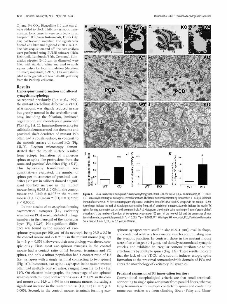
ResultsHyperspiny transformation and alteredsynaptic morphologyAs reported previously (Jun et al., 1999),the mutant cerebellum defective in VDCC�1A subunit was slightly reduced in sizebut grossly normal in the cerebellar anat-omy, including the foliation, laminatedorganization, and monolayer alignment ofPCs (Fig. 1A,C). Immunofluorescence forcalbindin demonstrated that the soma andproximal shaft dendrites of mutant PCsoften had a rough surface, in contrast tothe smooth surface of control PCs (Fig.1B,D). Electron microscopy demon-strated that the rough surface resultedfrom ectopic formation of numerousspines or spine-like protrusions from thesoma and proximal dendrites (Fig. 1E,F).This hyperspiny transformation wasquantitatively evaluated; the number ofspines per micrometer of proximal den-drites (�2 �m in caliber) showed a signif-icant fourfold increase in the mutantmouse, being 0.065 � 0.084 in the controlmouse and 0.240 � 0.107 in the mutantmouse (Fig. 1 I) (mean � SD; n 3; t test;p 0.0001).
In both strains of mice, spines formingasymmetrical synapses (i.e., excitatorysynapses on PCs) were distributed in largenumbers in the neuropil of the molecularlayer (Fig. 1G,H). No significant differ-ence was found in the number of axo-spinous synapses per 100 �m 2 of the neuropil, being 26.5 � 3.7 inthe control mouse and 27.0 � 3.7 in the mutant mouse (Fig. 1 J)(n 3; p 0.856). However, their morphology was altered con-spicuously. First, most axo-spinous synapses in the controlmouse had a contact ratio of 1:1 between terminals and PCspines, and only a minor population had a contact ratio of 1:2(i.e., synapses with a single terminal connecting to two spines)(Fig. 1G). In contrast, axo-spinous synapses in the mutant mouseoften had multiple contact ratios, ranging from 1:2 to 1:6 (Fig.1H). On electron micrographs, the percentage of axo-spinoussynapses with multiple contact ratios was 2.4 � 1.0% in the con-trol mouse and 14.9 � 4.9% in the mutant mouse, indicating asignificant increase in the mutant mouse (Fig. 1K) (n 3; p 0.005). Second, in the control mouse, terminals forming axo-
spinous synapses were small in size (0.5–1 �m), oval in shape,and contained relatively few synaptic vesicles accumulating nearthe synaptic junction. In contrast, those in the mutant mousewere often enlarged (�1 �m), had densely accumulated synapticvesicles, and exhibited an irregular contour attributable to theattachments by multiple spines (Fig. 1H). These results indicatethat the lack of the VDCC �1A subunit induces ectopic spineformation at the proximal somatodendritic domain of PCs andalters the morphology of excitatory PC synapses.
Proximal expansion of PF innervation territoryConventional morphological criteria are that small terminalsconnecting to single spines originate from parallel fibers, whereaslarge terminals with multiple contacts to spines and containingnumerous vesicles are from climbing fibers (Palay and Chan-
Figure 1. A–H, Cerebellar histology and Purkinje cell cytology in the VDCC �1A control (A, B, E, G) and mutant (C, D, F, H ) mice.A, C, Hematoxylin staining for midsagittal cerebellar sections. The lobule number is indicated by the numbers 1–10. B, D, Calbindinimmunofluorescece. E–H, Electron micrographs of proximal shaft dendrites of PCs (E, F ) and PC synapses in the neuropil (G, H ).Arrowheads indicate the neck of ectopic spines protruding from a shaft dendrite of a mutant. Asterisks indicate the head of PCspines forming asymmetric contact with axon terminals. I--K, Histograms showing the spine number per 1 �m of proximal shaftdendrites ( I ), the number of junctions at axo-spinous synapses per 100 �m 2 of the neuropil ( J), and the percentage of axonterminals contacting multiple spines ( K). *p 0.005; **p 0.0001. WT, Wild-type; KO, knock-out; PCD, Purkinje cell dendrite.Scale bars: A, 1 mm; B, 20 �m; E, 1 �m; G, 500 mm.
1736 • J. Neurosci., February 18, 2004 • 24(7):1734 –1743 Miyazaki et al. • Ca2� Channel �1A and Synapse Formation
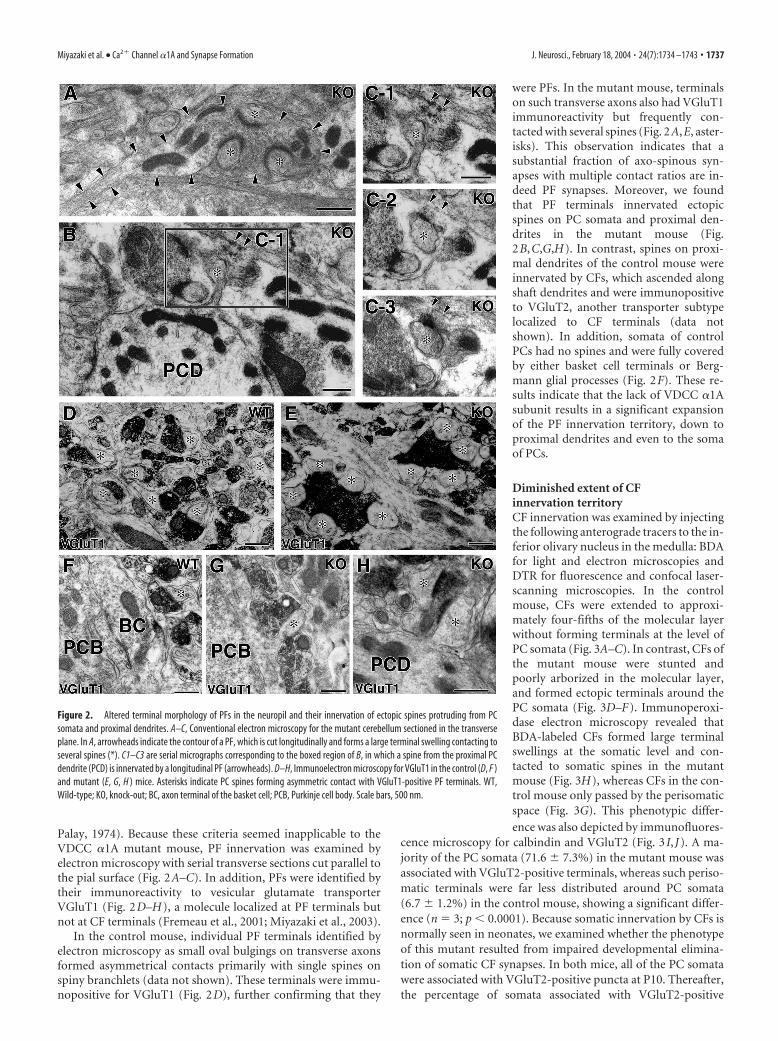
Palay, 1974). Because these criteria seemed inapplicable to theVDCC �1A mutant mouse, PF innervation was examined byelectron microscopy with serial transverse sections cut parallel tothe pial surface (Fig. 2A–C). In addition, PFs were identified bytheir immunoreactivity to vesicular glutamate transporterVGluT1 (Fig. 2D–H), a molecule localized at PF terminals butnot at CF terminals (Fremeau et al., 2001; Miyazaki et al., 2003).
In the control mouse, individual PF terminals identified byelectron microscopy as small oval bulgings on transverse axonsformed asymmetrical contacts primarily with single spines onspiny branchlets (data not shown). These terminals were immu-nopositive for VGluT1 (Fig. 2D), further confirming that they
were PFs. In the mutant mouse, terminalson such transverse axons also had VGluT1immunoreactivity but frequently con-tacted with several spines (Fig. 2A,E, aster-isks). This observation indicates that asubstantial fraction of axo-spinous syn-apses with multiple contact ratios are in-deed PF synapses. Moreover, we foundthat PF terminals innervated ectopicspines on PC somata and proximal den-drites in the mutant mouse (Fig.2B,C,G,H). In contrast, spines on proxi-mal dendrites of the control mouse wereinnervated by CFs, which ascended alongshaft dendrites and were immunopositiveto VGluT2, another transporter subtypelocalized to CF terminals (data notshown). In addition, somata of controlPCs had no spines and were fully coveredby either basket cell terminals or Berg-mann glial processes (Fig. 2F). These re-sults indicate that the lack of VDCC �1Asubunit results in a significant expansionof the PF innervation territory, down toproximal dendrites and even to the somaof PCs.
Diminished extent of CFinnervation territoryCF innervation was examined by injectingthe following anterograde tracers to the in-ferior olivary nucleus in the medulla: BDAfor light and electron microscopies andDTR for fluorescence and confocal laser-scanning microscopies. In the controlmouse, CFs were extended to approxi-mately four-fifths of the molecular layerwithout forming terminals at the level ofPC somata (Fig. 3A–C). In contrast, CFs ofthe mutant mouse were stunted andpoorly arborized in the molecular layer,and formed ectopic terminals around thePC somata (Fig. 3D–F). Immunoperoxi-dase electron microscopy revealed thatBDA-labeled CFs formed large terminalswellings at the somatic level and con-tacted to somatic spines in the mutantmouse (Fig. 3H), whereas CFs in the con-trol mouse only passed by the perisomaticspace (Fig. 3G). This phenotypic differ-ence was also depicted by immunofluores-
cence microscopy for calbindin and VGluT2 (Fig. 3 I, J). A ma-jority of the PC somata (71.6 � 7.3%) in the mutant mouse wasassociated with VGluT2-positive terminals, whereas such periso-matic terminals were far less distributed around PC somata(6.7 � 1.2%) in the control mouse, showing a significant differ-ence (n 3; p 0.0001). Because somatic innervation by CFs isnormally seen in neonates, we examined whether the phenotypeof this mutant resulted from impaired developmental elimina-tion of somatic CF synapses. In both mice, all of the PC somatawere associated with VGluT2-positive puncta at P10. Thereafter,the percentage of somata associated with VGluT2-positive
Figure 2. Altered terminal morphology of PFs in the neuropil and their innervation of ectopic spines protruding from PCsomata and proximal dendrites. A–C, Conventional electron microscopy for the mutant cerebellum sectioned in the transverseplane. In A, arrowheads indicate the contour of a PF, which is cut longitudinally and forms a large terminal swelling contacting toseveral spines (*). C1–C3 are serial micrographs corresponding to the boxed region of B, in which a spine from the proximal PCdendrite (PCD) is innervated by a longitudinal PF (arrowheads). D–H, Immunoelectron microscopy for VGluT1 in the control (D, F )and mutant (E, G, H ) mice. Asterisks indicate PC spines forming asymmetric contact with VGluT1-positive PF terminals. WT,Wild-type; KO, knock-out; BC, axon terminal of the basket cell; PCB, Purkinje cell body. Scale bars, 500 nm.
Miyazaki et al. • Ca2� Channel �1A and Synapse Formation J. Neurosci., February 18, 2004 • 24(7):1734 –1743 • 1737
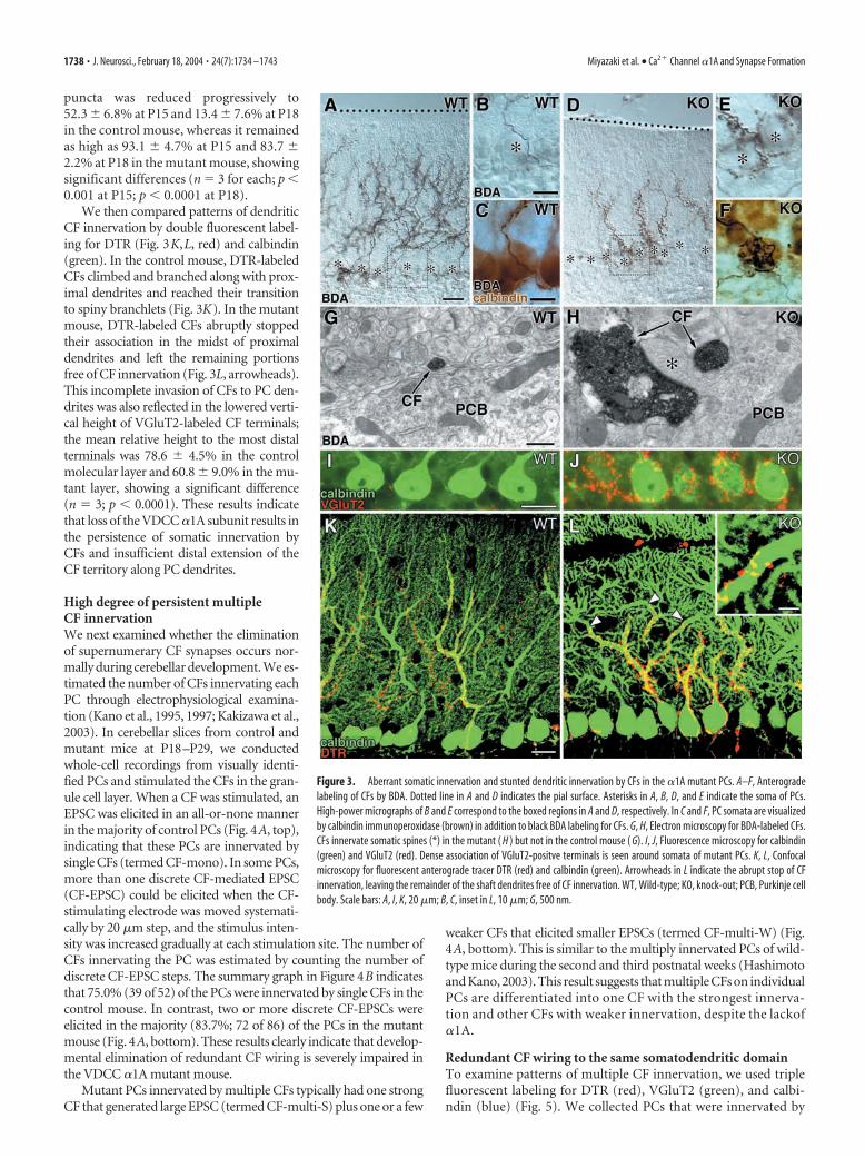
puncta was reduced progressively to52.3 � 6.8% at P15 and 13.4 � 7.6% at P18in the control mouse, whereas it remainedas high as 93.1 � 4.7% at P15 and 83.7 �2.2% at P18 in the mutant mouse, showingsignificant differences (n 3 for each; p 0.001 at P15; p 0.0001 at P18).
We then compared patterns of dendriticCF innervation by double fluorescent label-ing for DTR (Fig. 3K,L, red) and calbindin(green). In the control mouse, DTR-labeledCFs climbed and branched along with prox-imal dendrites and reached their transitionto spiny branchlets (Fig. 3K). In the mutantmouse, DTR-labeled CFs abruptly stoppedtheir association in the midst of proximaldendrites and left the remaining portionsfree of CF innervation (Fig. 3L, arrowheads).This incomplete invasion of CFs to PC den-drites was also reflected in the lowered verti-cal height of VGluT2-labeled CF terminals;the mean relative height to the most distalterminals was 78.6 � 4.5% in the controlmolecular layer and 60.8 � 9.0% in the mu-tant layer, showing a significant difference(n 3; p 0.0001). These results indicatethat loss of the VDCC �1A subunit results inthe persistence of somatic innervation byCFs and insufficient distal extension of theCF territory along PC dendrites.
High degree of persistent multipleCF innervationWe next examined whether the eliminationof supernumerary CF synapses occurs nor-mally during cerebellar development. We es-timated the number of CFs innervating eachPC through electrophysiological examina-tion (Kano et al., 1995, 1997; Kakizawa et al.,2003). In cerebellar slices from control andmutant mice at P18–P29, we conductedwhole-cell recordings from visually identi-fied PCs and stimulated the CFs in the gran-ule cell layer. When a CF was stimulated, anEPSC was elicited in an all-or-none mannerin the majority of control PCs (Fig. 4A, top),indicating that these PCs are innervated bysingle CFs (termed CF-mono). In some PCs,more than one discrete CF-mediated EPSC(CF-EPSC) could be elicited when the CF-stimulating electrode was moved systemati-cally by 20 �m step, and the stimulus inten-sity was increased gradually at each stimulation site. The number ofCFs innervating the PC was estimated by counting the number ofdiscrete CF-EPSC steps. The summary graph in Figure 4B indicatesthat 75.0% (39 of 52) of the PCs were innervated by single CFs in thecontrol mouse. In contrast, two or more discrete CF-EPSCs wereelicited in the majority (83.7%; 72 of 86) of the PCs in the mutantmouse (Fig. 4A, bottom). These results clearly indicate that develop-mental elimination of redundant CF wiring is severely impaired inthe VDCC �1A mutant mouse.
Mutant PCs innervated by multiple CFs typically had one strongCF that generated large EPSC (termed CF-multi-S) plus one or a few
weaker CFs that elicited smaller EPSCs (termed CF-multi-W) (Fig.4A, bottom). This is similar to the multiply innervated PCs of wild-type mice during the second and third postnatal weeks (Hashimotoand Kano, 2003). This result suggests that multiple CFs on individualPCs are differentiated into one CF with the strongest innerva-tion and other CFs with weaker innervation, despite the lackof�1A.
Redundant CF wiring to the same somatodendritic domainTo examine patterns of multiple CF innervation, we used triplefluorescent labeling for DTR (red), VGluT2 (green), and calbi-ndin (blue) (Fig. 5). We collected PCs that were innervated by
Figure 3. Aberrant somatic innervation and stunted dendritic innervation by CFs in the �1A mutant PCs. A–F, Anterogradelabeling of CFs by BDA. Dotted line in A and D indicates the pial surface. Asterisks in A, B, D, and E indicate the soma of PCs.High-power micrographs of B and E correspond to the boxed regions in A and D, respectively. In C and F, PC somata are visualizedby calbindin immunoperoxidase (brown) in addition to black BDA labeling for CFs. G, H, Electron microscopy for BDA-labeled CFs.CFs innervate somatic spines (*) in the mutant ( H ) but not in the control mouse ( G). I, J, Fluorescence microscopy for calbindin(green) and VGluT2 (red). Dense association of VGluT2-positve terminals is seen around somata of mutant PCs. K, L, Confocalmicroscopy for fluorescent anterograde tracer DTR (red) and calbindin (green). Arrowheads in L indicate the abrupt stop of CFinnervation, leaving the remainder of the shaft dendrites free of CF innervation. WT, Wild-type; KO, knock-out; PCB, Purkinje cellbody. Scale bars: A, I, K, 20 �m; B, C, inset in L, 10 �m; G, 500 nm.
1738 • J. Neurosci., February 18, 2004 • 24(7):1734 –1743 Miyazaki et al. • Ca2� Channel �1A and Synapse Formation
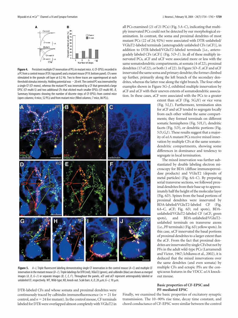
DTR-labeled CFs and whose somata and proximal dendrites werecontinuously traced by calbindin immunofluorescence (n 21 forcontrol, and n 24 for mutant). In the control mouse, CF terminalslabeled for DTR were overlapped almost completely with VGluT2 in
all PCs examined (21 of 21 PCs) (Fig. 5A–C), indicating that multi-ply innervated PCs could not be detected by our morphological ex-amination. In contrast, the soma and proximal dendrites of mostmutant PCs (22 of 24; 92%) were associated with DTR-unlabeled/VGluT2-labeled terminals [anterogradely unlabeled CFs (uCF)], inaddition to DTR-labeled/VGluT2-labeled terminals [i.e., antero-gradely labeled CFs (aCF)] (Fig. 5D–J). In all of these multiply in-nervated PCs, aCF and uCF were associated more or less with thesame somatodendritic compartments, at somata (4 of 22), proximaldendrites (17 of 22), or both (1 of 22). In Figure 5D–F, aCF and uCFinnervated the same soma and primary dendrite; the former climbedup further, primarily along the left branch of the secondary den-drites, whereas the latter rose along the right branch. The four otherexamples shown in Figure 5G–J, exhibited multiple innervation byaCF and uCF with their uneven extents of somatodendritic associa-tion. In these cases, aCF were associated with the PCs to a greater
extent than uCF (Fig. 5G,H) or vice versa(Fig. 5I,J). Furthermore, termination sitesfor aCF and uCF tended to segregate locallyfrom each other within the same compart-ments; they formed terminals on differentsomatic hemispheres (Fig. 5H,I), dendriticfacets (Fig. 5D), or dendritic portions (Fig.5D,G,J). These results suggest that a major-ity of �1A mutant PCs receive mixed inner-vation by multiple CFs at the same somato-dendritic compartments, showing somedifferences in dominance and tendency tosegregate in local termination.
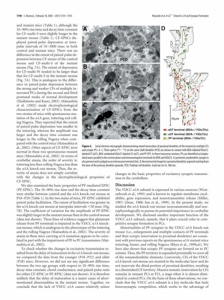
The mixed innervation was further sub-stantiated by double labeling electron mi-croscopy for BDA (diffuse immunoperoxi-dase products) and VGluT2 (deposits ofmetal particles) (Fig. 6A–C). By preparingserial transverse sections, we followed prox-imal dendrites from their base up to approx-imately half the height of the molecular layer(Fig. 6D). Spines from the basal portions ofproximal dendrites were innervated byBDA-labeled/VGluT2-labeled CF (Fig.6A–C, aCF; Fig. 6D, red spots), BDA-unlabeled/VGluT2-labeled CF (uCF, greenspots), and BDA-unlabeled/VGluT2-unlabeled terminals on transverse axons(i.e., PF terminals) (Fig. 6D, yellow spots). Inthis case, uCF innervated the basal portionsof proximal dendrites to a larger extent thanthe aCF. From the fact that proximal den-drites are innervated by single CFs but not byPFs in the adult wild-type PCs (Larramendiand Victor, 1967; Ichikawa et al., 2002), it isdeduced that the mixed innervations overthe same dendrites (and even somata) bymultiple CFs and ectopic PFs are the con-spicuous features in the VDCC �1A knock-out mouse.
Basic properties of CF-EPSC andPF-mediated EPSC
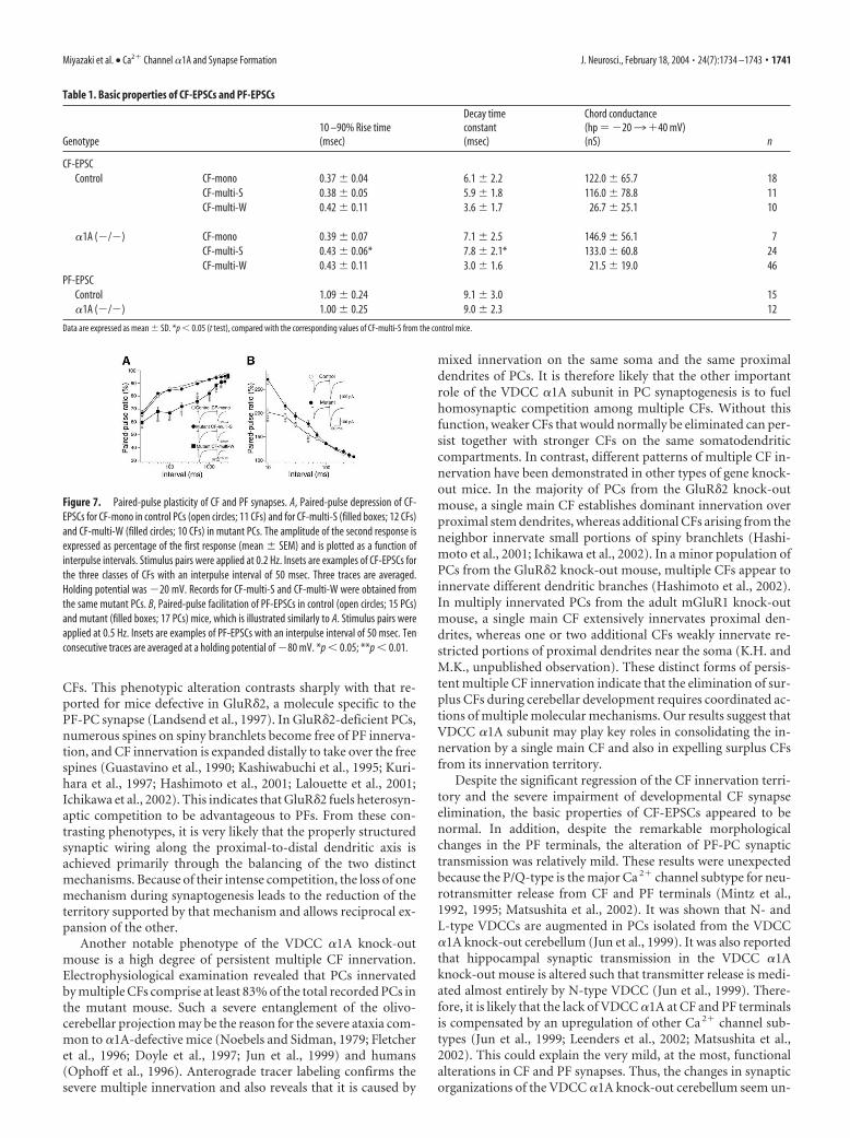
Finally, we examined the basic properties of excitatory synaptictransmission. The 10 –90% rise time, decay time constant, andchord conductance of CF-EPSC were similar between the control
Figure 4. Persistent multiple CF innervation of PCs in mutant mice. A, CF-EPSCs recorded ina PC from a control mouse (P29; top panel) and a mutant mouse (P19; bottom panel). CFs werestimulated in the granule cell layer at 0.2 Hz. Two to three traces are superimposed at eachthreshold stimulus intensity. Holding potential was �20 mV. The control PC was innervated bya single CF (CF-mono), whereas the mutant PC was innervated by a CF that generated a largeEPSC (CF-multi-S) and two additional CFs that elicited much smaller EPSCs (CF-multi-W). B,Summary histograms showing the number of discrete steps of CF-EPSCs from control mice(open columns; 4 mice, 52 PCs) and from mutant mice (filled columns; 7 mice, 86 PCs).
Figure 5. A–J, Triple fluorescent labeling demonstrating single CF innervation in the control mouse (A–C) and multiple CFinnervation in the mutant mouse (D–J ). Triple labelings for DTR (red), VGluT2 (green), and calbindin (blue) are shown as mergedimages (A, D, G–J ) or separate images (B, C, E, F ). Throughout the panels, aCF and uCF represent anterogradely labeled orunlabeled CF, respectively. WT, Wild-type; KO, knock-out. Scale bars: A, D, 20 �m; G--J, 10 �m.
Miyazaki et al. • Ca2� Channel �1A and Synapse Formation J. Neurosci., February 18, 2004 • 24(7):1734 –1743 • 1739
and mutant mice (Table 1), although the10 –90% rise time and decay time constantfor CF-multi-S were slightly longer in themutant mouse (Table 1). CF-EPSCs dis-played paired-pulse depression at inter-pulse intervals of 10 –3000 msec in bothcontrol and mutant mice. There was nodifference in the extent of paired-pulse de-pression between CF-mono of the controlmouse and CF-multi-S of the mutantmouse (Fig. 7A). The extent of depressionfor CF-multi-W tended to be larger thanthat for CF-multi-S in the mutant mouse(Fig. 7A). This is analogous to the differ-ence in paired-pulse depression betweenthe strong and weaker CFs of multiply in-nervated PCs during the second and thirdpostnatal weeks of normal development(Hashimoto and Kano, 2003). Matsushitaet al. (2002) made electrophysiologicalcharacterization of CF-EPSCs from thetwo strains of mice with spontaneous mu-tation of the �1A gene, tottering and roll-ing Nagoya. They reported that the extentof paired-pulse depression was smaller inthe tottering, whereas the amplitude waslarger and the decay time constant waslonger in the rolling Nagoya when com-pared with the control mice (Matsushita etal., 2002). Other aspects of CF-EPSC werenormal in these two spontaneous mutantmice (Matsushita et al., 2002). In terms ofcerebellar ataxia, the order of severity is:tottering less than rolling Nagoya less thanthe �1A knock-out mouse. Thus, the se-verity of ataxia does not simply correlatewith the changes in the electrophysiological properties ofCF-EPSC.
We also examined the basic properties of PF-mediated EPSC(PF-EPSC). The 10 –90% rise time and the decay time constantwere similar between control and the �1A knock-out mouse atP18 –P29 (Table 1). In the two stains of mice, PF-EPSC exhibitedpaired-pulse facilitation. The extent of facilitation was greater inthe �1A knock-out mouse at interpulse intervals 50 msec (Fig.7B). The coefficient of variation for the amplitude of PF-EPSCwas slightly larger in the mutant mouse than in the control mouse(data not shown). These lines of evidence suggest that glutamaterelease from PF terminals is impaired in the VDCC �1A knock-out mouse, which is analogous to the phenotypes of the totteringand the rolling Nagoya (Matsushita et al., 2002). The severity ofataxia in these mice carrying �1A mutation appears to be corre-lated in part with the impairment of PF to PC transmission (Mat-sushita et al., 2002).
To check whether the changes in excitatory transmission re-sulted from the delay of maturation in the �1A knock-out mouse,we compared the data from the younger (P18 –P22) and older(P28) mice. However, we did not see any significant differencebetween the two age groups with respect to 10 –90% rise time,decay time constant, chord conductance, and paired-pulse ratiofor either CF-EPSC or PF-EPSC (data not shown). It is thereforeunlikely that the delay of maturation is a major cause of afore-mentioned abnormalities in the mutant mouse. Together, weconclude that the lack of VDCC �1A causes relatively minor
changes in the basic properties of excitatory synaptic transmis-sion in the cerebellum.
DiscussionThe VDCC �1A subunit is expressed in various neurons (West-enbroek et al., 1995) and is known to regulate membrane excit-ability, gene expression, and neurotransmitter release (Miller,1987; Llinas, 1988; Jun et al., 1999). In the present study, westudied the �1A knock-out mouse neuroanatomically and neu-rophysiologically to pursue its potential importance in cerebellardevelopment. We disclosed another important function of theVDCC �1A subunit, namely, that it plays crucial roles in com-petitive synapse formation in PCs.
Abnormalities of PF synapses in the VDCC �1A knock-outmouse (i.e., enlargement and multiple contacts of PF terminalsand their ectopic innervation to proximal dendrites) are consis-tent with previous reports on the spontaneous �1A mutant micetottering, leaner, and rolling Nagoya (Rhyu et al., 1999a,b). Wehave also shown that ectopic PF synapses are formed on to PCsomata. Thus, the PF territory is expanded proximally to cover allof the somatodendritic elements. Conversely, CFs of the VDCC�1A knock-out mouse are stunted in the molecular layer and donot innervate the distal portion of proximal dendrites, resultingin a diminished CF territory. Massive somatic innervation by CFsremains in mutant PCs at P21, a stage when it is almost elimi-nated in control PCs. On the basis of these observations, we con-clude that the VDCC �1A subunit is a key molecule that fuelsheterosynaptic competition, which works to the advantage of
Figure 6. Serial electron micrographs demonstrating mixed innervation of proximal dendrites of the mutant by multiple CFsand ectopic PFs. A–C, Three spines (*1�*3) on the same shaft dendrite (PCD) are shown to contact with BDA-labeled/VGluT2-labeled CF (aCF), BDA-unlabeled/VGluT2-labeled CF (uCF), and PF (PF). In these transverse sections, PFs are identified as longitu-dinal axons parallel to the section plane and immunonegative terminals for BDA and VGluT2. A symmetric axodendritic synapse isalso present and is judged as an interneuron terminal (Int). D, Reconstructed image for a proximal dendritic segment starting fromthe base of the primary dendrite upwards. PCD, Purkinje cell dendrite. Scale bar (in A), 500 nm.
1740 • J. Neurosci., February 18, 2004 • 24(7):1734 –1743 Miyazaki et al. • Ca2� Channel �1A and Synapse Formation
CFs. This phenotypic alteration contrasts sharply with that re-ported for mice defective in GluR�2, a molecule specific to thePF-PC synapse (Landsend et al., 1997). In GluR�2-deficient PCs,numerous spines on spiny branchlets become free of PF innerva-tion, and CF innervation is expanded distally to take over the freespines (Guastavino et al., 1990; Kashiwabuchi et al., 1995; Kuri-hara et al., 1997; Hashimoto et al., 2001; Lalouette et al., 2001;Ichikawa et al., 2002). This indicates that GluR�2 fuels heterosyn-aptic competition to be advantageous to PFs. From these con-trasting phenotypes, it is very likely that the properly structuredsynaptic wiring along the proximal-to-distal dendritic axis isachieved primarily through the balancing of the two distinctmechanisms. Because of their intense competition, the loss of onemechanism during synaptogenesis leads to the reduction of theterritory supported by that mechanism and allows reciprocal ex-pansion of the other.
Another notable phenotype of the VDCC �1A knock-outmouse is a high degree of persistent multiple CF innervation.Electrophysiological examination revealed that PCs innervatedby multiple CFs comprise at least 83% of the total recorded PCs inthe mutant mouse. Such a severe entanglement of the olivo-cerebellar projection may be the reason for the severe ataxia com-mon to �1A-defective mice (Noebels and Sidman, 1979; Fletcheret al., 1996; Doyle et al., 1997; Jun et al., 1999) and humans(Ophoff et al., 1996). Anterograde tracer labeling confirms thesevere multiple innervation and also reveals that it is caused by
mixed innervation on the same soma and the same proximaldendrites of PCs. It is therefore likely that the other importantrole of the VDCC �1A subunit in PC synaptogenesis is to fuelhomosynaptic competition among multiple CFs. Without thisfunction, weaker CFs that would normally be eliminated can per-sist together with stronger CFs on the same somatodendriticcompartments. In contrast, different patterns of multiple CF in-nervation have been demonstrated in other types of gene knock-out mice. In the majority of PCs from the GluR�2 knock-outmouse, a single main CF establishes dominant innervation overproximal stem dendrites, whereas additional CFs arising from theneighbor innervate small portions of spiny branchlets (Hashi-moto et al., 2001; Ichikawa et al., 2002). In a minor population ofPCs from the GluR�2 knock-out mouse, multiple CFs appear toinnervate different dendritic branches (Hashimoto et al., 2002).In multiply innervated PCs from the adult mGluR1 knock-outmouse, a single main CF extensively innervates proximal den-drites, whereas one or two additional CFs weakly innervate re-stricted portions of proximal dendrites near the soma (K.H. andM.K., unpublished observation). These distinct forms of persis-tent multiple CF innervation indicate that the elimination of sur-plus CFs during cerebellar development requires coordinated ac-tions of multiple molecular mechanisms. Our results suggest thatVDCC �1A subunit may play key roles in consolidating the in-nervation by a single main CF and also in expelling surplus CFsfrom its innervation territory.
Despite the significant regression of the CF innervation terri-tory and the severe impairment of developmental CF synapseelimination, the basic properties of CF-EPSCs appeared to benormal. In addition, despite the remarkable morphologicalchanges in the PF terminals, the alteration of PF-PC synaptictransmission was relatively mild. These results were unexpectedbecause the P/Q-type is the major Ca 2� channel subtype for neu-rotransmitter release from CF and PF terminals (Mintz et al.,1992, 1995; Matsushita et al., 2002). It was shown that N- andL-type VDCCs are augmented in PCs isolated from the VDCC�1A knock-out cerebellum (Jun et al., 1999). It was also reportedthat hippocampal synaptic transmission in the VDCC �1Aknock-out mouse is altered such that transmitter release is medi-ated almost entirely by N-type VDCC (Jun et al., 1999). There-fore, it is likely that the lack of VDCC �1A at CF and PF terminalsis compensated by an upregulation of other Ca 2� channel sub-types (Jun et al., 1999; Leenders et al., 2002; Matsushita et al.,2002). This could explain the very mild, at the most, functionalalterations in CF and PF synapses. Thus, the changes in synapticorganizations of the VDCC �1A knock-out cerebellum seem un-
Table 1. Basic properties of CF-EPSCs and PF-EPSCs
Genotype10 –90% Rise time(msec)
Decay timeconstant(msec)
Chord conductance(hp �203�40 mV)(nS) n
CF-EPSCControl CF-mono 0.37 � 0.04 6.1 � 2.2 122.0 � 65.7 18
CF-multi-S 0.38 � 0.05 5.9 � 1.8 116.0 � 78.8 11CF-multi-W 0.42 � 0.11 3.6 � 1.7 26.7 � 25.1 10
�1A (�/�) CF-mono 0.39 � 0.07 7.1 � 2.5 146.9 � 56.1 7CF-multi-S 0.43 � 0.06* 7.8 � 2.1* 133.0 � 60.8 24CF-multi-W 0.43 � 0.11 3.0 � 1.6 21.5 � 19.0 46
PF-EPSCControl 1.09 � 0.24 9.1 � 3.0 15�1A (�/�) 1.00 � 0.25 9.0 � 2.3 12
Data are expressed as mean � SD. *p 0.05 (t test), compared with the corresponding values of CF-multi-S from the control mice.
Figure 7. Paired-pulse plasticity of CF and PF synapses. A, Paired-pulse depression of CF-EPSCs for CF-mono in control PCs (open circles; 11 CFs) and for CF-multi-S (filled boxes; 12 CFs)and CF-multi-W (filled circles; 10 CFs) in mutant PCs. The amplitude of the second response isexpressed as percentage of the first response (mean � SEM) and is plotted as a function ofinterpulse intervals. Stimulus pairs were applied at 0.2 Hz. Insets are examples of CF-EPSCs forthe three classes of CFs with an interpulse interval of 50 msec. Three traces are averaged.Holding potential was �20 mV. Records for CF-multi-S and CF-multi-W were obtained fromthe same mutant PCs. B, Paired-pulse facilitation of PF-EPSCs in control (open circles; 15 PCs)and mutant (filled boxes; 17 PCs) mice, which is illustrated similarly to A. Stimulus pairs wereapplied at 0.5 Hz. Insets are examples of PF-EPSCs with an interpulse interval of 50 msec. Tenconsecutive traces are averaged at a holding potential of �80 mV. *p 0.05; **p 0.01.
Miyazaki et al. • Ca2� Channel �1A and Synapse Formation J. Neurosci., February 18, 2004 • 24(7):1734 –1743 • 1741
likely to result primarily from dysfunctional transmitter releasefrom PFs or CFs.
We instead propose that the drastic alteration in the synapticorganization of the PCs is caused by impaired postsynaptic Ca 2�
channel functions, although information is still lacking as towhether and how the loss of postsynaptic P/Q-type VDCC iscompensated. The �1A subunit constitutes the high voltage-activated type of Ca 2� channels and is particularly abundant inPC somata and dendrites, constituting �90% of the total Ca 2�
current density (Mintz et al., 1992, 1995; Stea et al., 1994). CFactivity causes strong depolarization of PCs in an all-or-nonemanner and invariably elicits a characteristic complex spike(Eccles et al., 1966). Therefore, the Ca 2� influx into PCs duringCF activity is primarily attained through VDCC formed by the�1A subunit. We assumed that CF-evoked Ca 2� influx to a PCthrough �1A channels may consolidate coactivated CF synapses,whereas it may punish other excitatory synapses unrelated to theCa 2� influx. This proposed mechanism is reminiscent of theNMDA receptor-dependent synapse refinement in the visual andsomatosensory systems, in which NMDA receptors are thoughtto function as a coincidence detector that introduces Ca 2� influxin an activity-dependent manner. It is thought that the Ca 2�
influx through NMDA receptors strengthens synapses with cor-related activities, whereas it weakens those with uncorrelated ac-tivities, through which immature redundant connections are re-fined into functionally mature ones (Lisman, 1989; Li et al., 1994;Feldman et al., 1998). Because cerebellar PCs lack NMDA recep-tors (Yamada et al., 2001), the VDCC �1A subunit may functionas a substitute for the roles played by the NMDA receptors andmay function as a coincidence detector. Without such Ca 2�
channel function, activity-dependent shaping of functional syn-aptic circuitry is impaired; in the cerebellum, multiple CFs ofdifferent origins and PFs can be wired onto the same somatoden-dritic compartment. Therefore, we hypothesize that postsynapticP/Q-type Ca 2� channels in PCs fuel heterosynaptic competitionfor extending the CF innervation territory along the PC dendritictree and also fuel homosynaptic competition for establishing themonoinnervation by a main single CF of each PC. This should betested in future studies using the PC-specific knock out of the�1A subunit gene.
ReferencesAltman J (1972) Postnatal development of the cerebellar cortex in the rat. II.
Phases in the maturation of Purkinje cells and of the molecular layer.J Comp Neurol 145:399 – 463.
Bravin M, Rossi F, Strata P (1995) Different climbing fibres innervate sepa-rate dendritic regions of the same Purkinje cell in hypogranular cerebel-lum. J Comp Neurol 357:395– 407.
Bravin M, Morando L, Vercelli A, Rossi F, Strata P (1999) Control of spineformation by electrical activity in the adult rat cerebellum. Proc Natl AcadSci USA 96:1704 –1709.
Chedotal A, Sotelo C (1992) Early development of the olivocerebellar pro-jections in the fetal rat using CGRP-immunocytochemistry. Eur J Neuro-sci 4:1159 –1179.
Crepel F, Delhaye-Bouchaud N, Guastavino JM, Sampaio I (1980) Multipleinnervation of cerebellar Purkinje cells by climbing fibers in staggerermutant mouse. Nature 283:483– 484.
Crepel F, Delhaye-Bouchaud N, Dupont JL (1981) Fate of the multiple in-nervation of cerebellar Purkinje cells by climbing fibers in immature con-trol, X-irradiated and hypothyroid rats. Dev Brain Res 1:59 –71.
Doyle J, Ren X, Lennon G, Stubbs L (1997) Mutations in the Cacnl1a4 cal-cium channel gene are associated with seizures, cerebellar degeneration,and ataxia in tottering and leaner mutant mice. Mamm Genome8:113–120.
Eccles JC, Llinas R, Sasaki K (1966) The excitatory synaptic action of climb-
ing fibres on the Purkinje cells of the cerebellum. J Physiol (Lond)182:268 –296.
Feldman DE, Nicoll RA, Malenka RC, Isaac JTR (1998) Long-term depres-sion at thalamocortical synapses in developing rat somatosensory cortex.Neuron 21:347–357.
Fletcher CF, Lutz CM, O’Sullivan TN, Shaughnessy Jr JD, Hawkes R, FrankelWN, Copeland NG, Jenkins NA (1996) Absence epilepsy in totteringmutant mice is associated with calcium channel defects. Cell 87:607– 617.
Fremeau Jr RT, Troyer MD, Pahner I, Nygaad GO, Tran CH, Reimer RJ,Belloccio EE, Fortin D, Storm-Mathisen J, Edwards RH (2001) The ex-pression of vesicular glutamate transporters defines two classes of excita-tory synapse. Neuron 31:247–260.
Guastavino JM, Sotelo C, Damez-Kinselle I (1990) Hot-foot murine muta-tion: behavioral effects and neuroanatomical alterations. Brain Res523:199 –210.
Hashimoto K, Kano M (2003) Functional differentiation of multiple climb-ing fiber inputs during synapse elimination in the developing cerebellum.Neuron 38:785–796.
Hashimoto K, Ichikawa R, Takechi H, Inoue Y, Aiba A, Sakimura K, MishinaM, Hashikawa T, Konnerth A, Watanabe M, Kano M (2001) Roles ofglutamate receptor �2 subunit (GluR�2) and metabotropic glutamatereceptor subtype1 (mGluR1) in climbing fiber synapse elimination dur-ing postnatal cerebellar development. J Neurosci 21:9701–9712.
Ichikawa R, Miyazaki T, Kano M, Hashikawa T, Tatsumi H, Sakimura K,Mishina M, Inoue Y, Watanabe M (2002) Distal extension of climbingfiber territory and multiple innervation caused by abberant wiring toadjacent spiny branchlets in cerebellar Purkinje cells lacking glutamatereceptor �2. J Neurosci 22:8487– 8503.
Ichise T, Kano M, Hashimoto K, Yanagihara D, Nakao K, Shigemoto R,Katsuki M, Aiba A (2000) mGluR1 in cerebellar Purkinje cells essentialfor long-term depression synapse elimination and motor coordination.Science 288:1832–1835.
Ito M (2001) Cerebellar long-term depression: characterization, signaltransduction, and functional roles. Physiol Rev 81:1143–1195.
Jun K, Piedras-Renteria ES, Smith SM, Wheeler DB, Lee SB, Lee TG, Chin H,Adams ME, Scheller RH, Tsien RW, Shin HS (1999) Ablation of P/Q-type Ca 2� channel currents, altered synaptic transmission, and progres-sive ataxia in mice lacking the �1A-subunit. Proc Natl Acad Sci USA96:15245–15250.
Kakizawa S, Yamada K, Iino M, Watanabe M, Kano M (2003) Effects ofinsulin-like growth factor I on climbing fibre synapse elimination duringcerebellar development. Eur J Neurosci 17:545–554.
Kano M, Rexhausen U, Dreessen J, Konnerth A (1992) Synaptic excitationproduces a long-lasting rebound potentiation of inhibitory synaptic sig-nals in cerebellar Purkinje cells. Nature 356:601– 604.
Kano M, Hashimoto K, Chen C, Abeliovich A, Aiba A, Kurihara H, WatanabeM, Inoue Y, Tonegawa S (1995) Impaired synapse elimination duringcerebellar development in PKC� mutant mice. Cell 83:1223–1231.
Kano M, Hashimoto K, Kurihara H, Watanabe M, Inoue Y, Aiba A, TonegawaS (1997) Persistent multiple climbing fiber innervation of cerebellarPurkinje cells in mice lacking mGluR1. Neuron 18:71–79.
Kano M, Hashimoto K, Watanabe M, Kurihara H, Offermanns S, Jiang H, WuY, Jun K, Shin HS, Inoue Y, Simon MI, Wu D (1998) Phospholipase C�4is specifically involved in climbing fiber synapse elimination in the devel-oping cerebellum. Proc Natl Acad Sci USA 95:15724 –15729.
Kashiwabuchi N, Ikeda K, Araki K, Hirano T, Shibuki K, Takayama C, InoueY, Kutsuwada T, Yagi T, Kang Y, Aizawa S, Mishina M (1995) Impair-ment of motor coordination Purkinje cell synapse formation and cerebel-lar long-term depression in GluR�2 mutant mice. Cell 81:245–252.
Konnerth A, Dreessen J, Augustine GJ (1992) Brief dendritic calcium signalsinitiate long-lasting synaptic depression in cerebellar Purkinje cells. ProcNatl Acad Sci USA 89:7051–7055.
Kurihara H, Hashimoto K, Kano M, Takayama C, Sakimura K, Mishina M,Inoue Y, Watanabe M (1997) Impaired parallel fiber-Purkinje cell syn-apse stabilization during cerebellar development of mutant mice lackingthe glutamate receptor �2 subunit. J Neurosci 17:9613–9623.
Laloutette A, Lohof A, Sotelo C, Guenet J, Mariani J (2001) Neurobiologicaleffects of a mull mutation depend on genetic context: comparison be-tween two hotfoot alleles of the delta-2 ionotropic glutamate receptor.Neuroscience 105:443– 455.
Landsend AS, Amiry-Moghaddam M, Matsubara A, Bergersen L, Usami S,Wenthold RJ, Ottersen OP (1997) Differential localization of � gluta-
1742 • J. Neurosci., February 18, 2004 • 24(7):1734 –1743 Miyazaki et al. • Ca2� Channel �1A and Synapse Formation
mate receptors in the rat cerebellum: coexpression with AMPA receptorsin parallel fiber-spine synapses and absence from climbing fiber-spinesynapses. J Neurosci 17:834 – 842.
Larramendi LMH, Victor T (1967) Synapses on the Purkinje cell spines inthe mouse an electronmicroscopic study. Brain Res 5:15–30.
Leenders AGM, van den Maagdenberg AMJM, Lopes da Silva FH, Sheng ZH,Molenaar PC, Ghijsen WEJM (2002) Neurotransmitter release fromtottering mice nerve terminals with reduced expression of mutated P- andQ-type Ca 2�-channels. Eur J Neurosci 15:13–18.
Li Y, Erzurumlu RS, Chen C, Jhaveri S, Tonegawa S (1994) Whisker-relatedneuronal patterns fail to develop in the trigeminal brainstem nuclei ofNMDAR1 knock-out mice. Cell 76:427– 437.
Lisman J (1989) A mechanism for the Hebb and the anti-Hebb processesunderlying learning and memory. Proc Natl Acad Sci USA 86:9574 –9578.
Llinas RR (1988) The intrinsic electrophysiological properties of mamma-lian neurons: insights into central nervous system function. Science242:1654 –1664.
Mariani J (1982) Extent of multiple innervation of Purkinje cells by climb-ing fibers in the olivocerebellar system of weaver, reeler, and staggerermutant mice. J Neurobiol 13:119 –126.
Mariani J, Changeux JP (1981a) Ontogenesis of olivocerebellar relation-ships. I. Studies by intracellular recordings of the multiple innervation ofPurkinje cells by climbing fibers in the developing rat cerebellum. J Neu-rosci 1:696 –702.
Mariani J, Changeux JP (1981b) Ontogenesis of olivocerebellar relation-ships. II. Spontaneous activity of inferior olivary neurons and climbingfiber mediated activity of cerebellar Purkinje cells in developing rats.J Neurosci 1:703–709.
Matsushita K, Wakamori M, Rhyu IJ, Arii T, Oda S, Mori Y, Imoto K (2002)Bidirectional alterations in cerebellar synaptic transmission of totteringand rolling Ca 2� channel mutant mice. J Neurosci 22:4388 – 4398.
Miller RJ (1987) Multiple calcium channels and neuronal function. Science235:46 –52.
Mintz IM, Venema VJ, Swiderek KM, Lee TD, Bean BP, Adams ME (1992)P-type calcium channels blocked by the spider toxin �-Aga-IVA. Nature355:827– 829.
Mintz IM, Sabatini BL, Regehr WG (1995) Calcium control of transmitterrelease at a cerebellar synapse. Neuron 15:675– 688.
Miyazaki T, Fukaya M, Shimizu H, Watanabe M (2003) Subtype switchingof vesicular glutamate transporters at parallel fibre-Purkinje cell synapsesin developing mouse cerebellum. Eur J Neurosci 17:2563–2572.
Nakagawa S, Watanabe M, Isobe T, Kondo H, Inoue Y (1998) Cytologicalcompartmentalization in the staggerer cerebellum, as revealed by calbi-ndin immunohistochemistry for Purkinje cells. J Comp Neurol395:112–120.
Noebels JL, Sidman RL (1979) Inherited epilepsy: spike-wave and focal mo-tor seizures in the mutant mouse tottering. Science 204:1334 –1336.
Offermanns S, Hashimoto K, Watanabe M, Sun W, Kurihara H, ThompsonRF, Inoue Y, Kano M, Simon MI (1997) Impaired motor coordinationand persistent multiple climbing fiber innervation of cerebellar Purkinjecells in mice lacking G�q. Proc Natl Acad Sci USA 94:14089 –14094.
Ophoff RA, Terwindt GM, Vergouwe MN, Van Eijk R, Oefner PJ, HoffmanSMG, Lamerdin JE, Mohrenweiser HW, Bulman DE, Ferrari M, Haan J,Lindhout D, van Ommen GJB, Hofker MH, Ferrari MD, Frants RR(1996) Familial hemiplegic migraine and episodic ataxia type-2 arecaused by mutations in the Ca 2� channel gene CACNL1A4. Cell87:543–552.
Palay S, Chan-Palay V (1974) Cerebellar cortex. Cytology and organization,pp 63– 69, 242–287. New York: Springer.
Regehr WG, Mintz IM (1994) Participation of multiple calcium channeltypes in transmission at single climbing fiber to Purkinje cell synapses.Neuron 12:605– 613.
Rhyu IJ, Abbott LC, Walker DB, Sotelo C (1999a) An ultrastructual study ofgranule cell/Purkinje cell synapses in tottering (tg/tg), leaner (tgla/tgla),and compound heterozygous tottering/leaner (tg/tgla) mice. Neuro-science 90:717–728.
Rhyu IJ, Oda S, Uhm CS, Kim H, Suh YS, Abbott LC (1999b) Morphologicinvestigation of rolling mouse Nagoya (tgrol/tgrol) cerebellar Purkinje cells:anataxic mutant, revised. Neurosci Lett 266:49 –52.
Sotelo C (1978) Purkinje cell ontogeny: formation and maintenance ofspines. Prog Brain Res 48:149 –170.
Stea A, Tomlinson WJ, Soong TW, Bourinet E, Dubel SJ, Vincent SR, SnutchTP (1994) Localization and functional properties of a rat brain �1A cal-cium channel reflect similarities to neuronal Q- and P-type channels. ProcNatl Acad Sci USA 91:10576 –10580.
Sugihara I, Bailly Y, Mariani J (2000) Olivocerebellar climbing fibers in thegranuloprival cerebellum: morphological study of individual axonal pro-jections in the X-irradiated rat. J Neurosci 20:3745–3760.
Westenbroek RE, Sakurai T, Elliott EM, Hell JW, Starr TVB, Snutch TP,Catterall WA (1995) Immunochemical identification and subcellulardistribution of the �1A subunits of brain calcium channels. J Neurosci15:6403– 6418.
Woodward DJ, Hoffer BJ, Altman J (1974) Physiological and pharmacolog-ical properties of Purkinje cells in rat cerebellum degranulated by postna-tal x-irradiation. J Neurobiol 5:283–304.
Yamada K, Fukaya M, Shimizu H, Sakimura K, Watanabe M (2001) NMDAreceptor GluR�1, GluR�3 and GluR�1 are enriched at the mossy fibre-granule cell synapses in the adult mouse cerebellum. Eur J Neurosci 13:2025–2036.
Miyazaki et al. • Ca2� Channel �1A and Synapse Formation J. Neurosci., February 18, 2004 • 24(7):1734 –1743 • 1743