lrrk2 kinase activity regulates synaptic vesicle ... · original research article published: 27 may...
TRANSCRIPT

ORIGINAL RESEARCH ARTICLEpublished: 27 May 2014
doi: 10.3389/fnmol.2014.00049
LRRK2 kinase activity regulates synaptic vesicle traffickingand neurotransmitter release through modulation of LRRK2macro-molecular complexMaria D. Cirnaru1,2, Antonella Marte3, Elisa Belluzzi4, Isabella Russo4, Martina Gabrielli 2,5,
Francesco Longo6, Ludovico Arcuri6, Luca Murru2, Luigi Bubacco4, Michela Matteoli5,7,
Ernesto Fedele8, Carlo Sala2,5, Maria Passafaro2, Michele Morari6, Elisa Greggio4, Franco Onofri3 and
Giovanni Piccoli1,2*
1 Division of Neuroscience, San Raffaele Scientific Institute and Vita-Salute University, Milan, Italy2 Department of Molecular and Cellular Pharmacology, National Research Council, Neuroscience Institute, Milan, Italy3 Department of Experimental Medicine, University of Genova, Genova, Italy4 Department of Biology, University of Padova, Padova, Italy5 Department of Medical Biotechnology and Translational Medicine, University of Milan, Milan, Italy6 Department of Medical Science and National Institute of Neuroscience, University of Ferrara, Ferrara, Italy7 Humanitas Clinical and Research Center, Pharmacology and Brain Pathology, Rozzano, Italy8 Department of Pharmacy, University of Genoa, Genoa, Italy
Edited by:
Kirsten Harvey, University CollegeLondon, UK
Reviewed by:
Lu-Yang Wang, University ofToronto, CanadaNicola B. Mercuri, University ofRome, ItalyR. Jeremy Nichols, The Parkinson’sInstitute, USA
*Correspondence:
Giovanni Piccoli, San RaffaeleScientific Institute and Vita-SaluteUniversity, via Olgettina 58, 20132Milan, Italye-mail: [email protected]
Mutations in Leucine-rich repeat kinase 2 gene (LRRK2) are associated with familial andsporadic Parkinson’s disease (PD). LRRK2 is a complex protein that consists of multipledomains executing several functions, including GTP hydrolysis, kinase activity, and proteinbinding. Robust evidence suggests that LRRK2 acts at the synaptic site as a molecularhub connecting synaptic vesicles to cytoskeletal elements via a complex panel ofprotein-protein interactions. Here we investigated the impact of pharmacological inhibitionof LRRK2 kinase activity on synaptic function. Acute treatment with LRRK2 inhibitorsreduced the frequency of spontaneous currents, the rate of synaptic vesicle traffickingand the release of neurotransmitter from isolated synaptosomes. The investigationof complementary models lacking LRRK2 expression allowed us to exclude potentialoff-side effects of kinase inhibitors on synaptic functions. Next we studied whetherkinase inhibition affects LRRK2 heterologous interactions. We found that the bindingamong LRRK2, presynaptic proteins and synaptic vesicles is affected by kinase inhibition.Our results suggest that LRRK2 kinase activity influences synaptic vesicle release viamodulation of LRRK2 macro-molecular complex.
Keywords: LRRK2, kinase, presynaptic vesicle, synaptic activity, protein interaction
INTRODUCTIONParkinson’s disease (PD) is an age-related neurodegenerative dis-ease affecting 2% of the population above 65-years and is clini-cally characterized by bradykinesia, rigidity, and resting tremor.The neuropathological hallmark of the disease is the progressiveloss of dopaminergic neurons in the substantia nigra (Moore et al.,2005; Hardy et al., 2006). Although the majority of cases are idio-pathic, mutations in the Leucine-rich repeat kinase 2 (LRRK2)gene (PARK8; OMIM 609007) cause late-onset PD. LRRK2 muta-tions account for up to 13% of familial PD cases compatiblewith dominant inheritance (Paisan-Ruiz et al., 2004; Zimprichet al., 2004) and have been identified in 1–2% of sporadic PDpatients (Aasly et al., 2005; Berg et al., 2005). LRRK2 is a largeprotein encompassing several functional domains including akinase domain with feature similar to mitogen activated proteinkinase kinase kinases (MAPKKK) and receptor-interacting pro-tein kinases (RIPK) (Bosgraaf and Van Haastert, 2003; Guo et al.,2006). Several single nucleotide variants have been identified inLRRK2 (Brice, 2005). While only the common G2019S mutation,located in the kinase domain, has been consistently associated
with increased kinase activity in vitro (West et al., 2005; Gloeckneret al., 2006; Greggio et al., 2006), a recent study monitoringLRRK2 autophosphorylation at Ser 1292 suggested that otherpathogenic mutants possess augmented activity in the cellularcontext (Sheng et al., 2012). Up to now few LRRK2 substrates havebeen identified in in vitro studies, but none has been convinc-ingly proved in vivo, leaving the pathophysiological relevance ofthe kinase activity unclear. Instead, several lines of evidence sug-gest that kinase activity is linked to LRRK2 dimerization (Greggioet al., 2008; Sen et al., 2009; Civiero et al., 2012) as well as sub-cellular distribution (Berger et al., 2010) and regulates binding to14-3-3 proteins (Nichols et al., 2010). Accumulating data corre-late LRRK2 to synaptic functions. Several studies suggested thatLRRK2 is part of a protein complex that influences the traffickingof synaptic vesicles belonging to the recycling pool (Shin et al.,2008; Piccoli et al., 2011; Matta et al., 2012). The descriptionof synaptic phenotype in LRRK2 mutant models (Tong et al.,2009; Migheli et al., 2013; Yun et al., 2013) further underlines thetight link among LRRK2, synaptic vesicle trafficking and neuro-transmitter release. In this study we investigated the functional
Frontiers in Molecular Neuroscience www.frontiersin.org May 2014 | Volume 7 | Article 49 | 1
MOLECULAR NEUROSCIENCE

Cirnaru et al. LRRK2 at the presynaptic site
impact of LRRK2 kinase activity on presynaptic function and wedetermined functional properties of neurons upon LRRK2 phar-macological inhibition. A combination of electrophysiological,biochemical and imaging analyses suggested that LRRK2 inhibi-tion impacts synaptic transmission acting on the organization ofLRRK2 macro-molecular complex at the presynaptic site.
MATERIALS AND METHODSANIMALS, NEURON CULTURES, AND DRUGSNon-transgenic wild-type (WT) and LRRK2 knock-out (KO)mice, back-crossed on a C57BL/6J strain, were obtained fromMayo Clinic (Jacksonville, FL, USA) through a collaborationwith Prof. Matthew Farrer and Dr. Heather Melrose (Hinkleet al., 2012). Animals were kept following guidelines of Ministryof Education, Universities and Research (MIUR). Neuron cul-tures were prepared from either mouse cortexes or hippocampiobtained from embryonic day 15.5–16.5 mice (C57BL/6J).High-density (750–1000 cells/mm2) and medium-density (150–200 cells/mm2) neuron cultures were plated and grown asdescribed on 12-well plastic tissue culture plates (Iwaki; BibbySterilin Staffordshire, UK) or on 12 mm diameter coverslips putinto 24-well plastic tissue culture plates (Iwaki) (Piccoli et al.,2007). IN-1 and GSK-2578215A compounds (Tocris Bioscience,Bristol, UK) or DMSO were added to culture media at theconcentrations indicated through the text.
PLASMIDS AND PROTEIN PURIFICATIONN-terminal 3xFLAG and myc hLRRK2 full length (hereinafterFLAG-LRRK2 and myc-LRRK2), N-terminal FLAG hLRRK2A2106T (a kind gift of Prof. Dario Alessi, MRC, University ofDundee), LRRK2 silencing and control viral constructs vectorshave been already described (Bauer et al., 2009; Nichols et al.,2009; Civiero et al., 2012). FLAG-LRRK2 was purified via affinitychromatography using FLAG-M2 agarose beads (Sigma Aldrich)as previously described (Civiero et al., 2012) from HEK293Tcells transfected by lipofection using Lipofectamine 2000 (LifeTechnologies Carlsbad, CA, USA) according to manufacturer’sinstructions. Viral particles were produced as in (Bauer et al.,2009). Neurons were infected at DIV4 and processed whenindicated.
IMMUNO-PRECIPITATION AND ANTIBODIESImmunoprecipitation was performed as described previously(Onofri et al., 2007) using 25 µl of settled prewashed pro-tein G-Sepharose beads (GE-Healthcare, Freiburg, Germany) toprecipitate the immunocomplexes. NaCl 150 mM, Tris 50 mM(pH 7.4), NP-40 (1% v/v), SDS (0.1% v/v) and protease andphosphatase inhibitors extracts of Percoll-purified synaptosomesobtained from rat cerebral cortex were incubated for 2 h at RT inabsence or in presence of IN- 1 (1 µM) with anti-LRRK2 anti-bodies (10 µg/sample; MJFF C41-2 Abcam, Cambridge UK) ora control rabbit IGg (10 µg/sample; Sigma-Aldrich, St. Louis,MO, USA). The eluted proteins were separated by SDS-PAGE,transferred onto nitrocellulose membrane (GE-Healthcare) andanalyzed by western-blotting. Antibodies list includes rabbit antiLRRK2 1:500 MJFF C41-2, rabbit anti LRRK2 P-Ser 935 UDD210(12) (Abcam), rabbit anti synapsin I 1:500 (Synaptic System,
Goettingen, Germany), mouse anti actin 1:1000, mouse antiFLAG 1:1000, mouse anti myc 1:1000, mouse anti synaptophysin1:1000 (Sigma-Aldrich St. Louis, MO, USA). The secondaryantibodies (HRP-conjugated anti-mouse, anti-rabbit) (BIORAD,Hercules, CA, USA) were used in a ratio of 1:5000 coupledwith the ECL chemiluminescence detection system. Immunoblotswere quantified by densitometric analysis of the fluorograms(Quantity One software, Bio-Rad) obtained in the linear rangeof the emulsion response.
IN VITRO KINASE ASSAYGST-LRRK2970−2527 (Life technologies) at the concentration of30 nM were incubated with 500 µM LRRKtide, 100 µM 33P-ATP(0.5 µCi) in kinase reaction buffer consisting of 25 mM Tris-HCl (pH7.5), 5 mM beta-glycerophosphate, 2 mM dithiothreitol(DTT), 0.1 mM Na3VO4, 10 mM MgCl2 and increasing concen-trations of inhibitors at 30◦C for 1 h. Reactions were carried outin triplicate and spotted onto P81 phosphocellulose. Followingdifferent washing of phosphocellulose membranes with 75 mMphosphoric acid, 33P incorporation into LRRKtide was quantifiedwith Cyclone (Perkin Elmer, Alameda, CA, USA).
SIZE EXCLUSION CHROMATOGRAPHYCells transiently transfected with FLAG-LRRK2 wild-type weresolubilized in lysis buffer containing 20 mM Tris-HCl pH 7.5,150 mM NaCl, 1 mM EDTA, 1% Triton X-100, 2.5 mM sodiumpyrophosphate, 1 mM beta-glycerophosphate, 1 mM NaVO4,protease inhibitor cocktail (Sigma-Aldrich) and lysates werecleared for 30 min at 14,000 xg. When appropriate, proteins werefurther purified via FLAG immunoprecipitation as describedabove. Cleared lysates (0.5 ml; 5 mg total proteins) or purifiedproteins (0.5 ml; 1.3 µg of purified protein) were injected andseparated on a Superose 6 10/300 column (GE Healthcare). Thecolumn was preequilibrated with buffer (20 mM Tris-HCl pH7.5, 150 mM NaCl and 0.07% Triton X-100) and used at a flowrate of 0.5 ml/min. Elution volumes of standards were 7.5 mlfor Blue Dextran (V0), 11.5 ml for hemocyanin from Carcinusaestuarii (900 kDa), 12 ml for thyreoglobin (669 kDa), 14 ml forferritin (440 kDa). When appropriate, inhibitors (1 µM IN-1 and1 µM GSK-2578215A) were applied for 90 min before lysis andkept throughout the following purification steps, including equi-libration of chromatographic mobile phase. Chromatographicfractions were analyzed by dot blot. One microliter of each frac-tion from SEC was applied onto a nitrocellulose membrane.The membrane was blocked with 10% (w/v) milk in TBS plus0.1% Triton (TBS-T) for 1 h and subsequently incubated withmouse monoclonal anti-Flag M2-peroxidase (Sigma-Aldrich).Immunoreactive proteins were visualized using enhanced chemi-luminescence plus (ECL plus, GE Healthcare).
SYNAPTIC VESICLE PURIFICATION AND LRRK2 BINDING ASSAYSSynaptic vesicles (SV) were obtained from rats by homogeniza-tion of the isolated forebrains and finally purified through thestep of controlled-pore glass (CPG) chromatography (Huttneret al., 1983). After elution, purified SV were centrifuged for 2 hat 175,000 × g and resuspended at a protein concentration of 1–2 mg/ml in 0.3 M glycine, 5 mM HEPES, 0.02% sodium azide, pH
Frontiers in Molecular Neuroscience www.frontiersin.org May 2014 | Volume 7 | Article 49 | 2

Cirnaru et al. LRRK2 at the presynaptic site
7.4 (glycine buffer). Protein concentrations were determined bythe Bradford or BCA assays. SDS-PAGE was performed accordingto Laemmli (1970). For the dissociation of endogenously boundLRRK2 purified SV (40 µg/sample) were incubated for 1 h at30◦C with or without IN-1 (1 µM) in glycine buffer plus 30 mMNaCl, 25 mM Tris/HCl, 2 mM DTT, 10 mM MgCl2 protease andphosphatase inhibitors. After the incubation, LRRK2 bound toSV were separated by soluble LRRK2 by high-speed centrifuga-tion (400,000 × g for 45 min) (Messa et al., 2010). Aliquots ofthe resuspended pellets were subjected to SDS–PAGE and subse-quent Western blotting with anti LRRK2 MJFF C41-2 (Abcam)antibody. The recovery of SV, used to correct the amounts ofLRRK2 bound to SV, was determined by Western blotting withanti-synaptophysin antibody (kind gift of Prof. Paul GreengardThe Rockefeller University New York USA). The binding ofpurified FLAG-LRRK2 to native SV was performed like below.SV (10 µg/sample) were incubated for 1 h at 0◦C with FLAG-LRRK2 (50 nM) in glycine buffer plus 30 mM NaCl, 25 mMTris/HCl, 2 mM DTT, 10 mM MgCl2 protease and phosphataseinhibitors and 1.0 µg/ml bovine serum albumin in absence orin presence of IN-1 (1 µM). After incubation, SV-bound FLAG-LRRK2 was separated by high-speed centrifugation (400,000 gfor 45 min). Aliquots of the resuspended pellets were subjectedto immunoblotting with anti-FLAG (Sigma-Aldrich) antibodies.The recovery of SV was determinated like above.
EXO-ENDOCYTOTIC ASSAYThe endocytosis assay to monitor SV recycling was performedusing rabbit polyclonal antibodies directed against the intrav-esicular domain of synaptotagmin1 (Synaptic System), appliedfor 5 min if not indicated otherwise at RT on the cultures, asdescribed previously (Matteoli et al., 1992). Incubations withthe antibody (1:400) were performed in Tyrode solution con-taining 124 mM NaCl, 5 mM KCl, 2 mM MgCl2, 30 mM glucose,25 mM HEPES, pH 7.4 and 2 mM CaCl2. After fixation and per-meabilization, a synaptophysin counter staining with mouse antisynaptophysin, 1:400 (Sigma-Aldrich) visualized the totality ofSV. Acquired images were processed and quantitatively analyzedwith ImageJ software as previously described (Verderio et al.,1999). Briefly, GFP positive processes were manually tracked andthe number of synaptotagmin and synaptophysin positive clus-ters and synaptophysin positive clusters present in the region ofinterest were automatically counted.
NEUROTRANSMITTER RELEASESynaptosome were isolated from cerebral cortex (fronto-temporalareas) as described previously (Marti et al., 2003; Mela et al.,2004). The synaptosomal pellet was resuspended in oxygenated(95% O2, 5% CO2) Krebs solution (mM: NaCl 118.5, KCl 4.7,CaCl2 1.2, MgSO4 1.2, KH2PO4 1.2, NaHCO3 25, glucose 10).One millilitre aliquot of the suspension (∼0.35 mg protein) wasslowly injected into nylon syringe filters (outer diameter 13 mm,0.45 µM pore size, internal volume of about 100 µl; Teknokroma,Barcelona, Spain) connected to a peristaltic pump. Filters weremaintained at 36.5◦C in a thermostatic bath and superfusedat a flow rate of 0.4 ml/min with a preoxygenated Krebs solu-tion. Under the superfusion conditions adopted in the present
study, the fast and continuous removal of endogenous substancesreleased by nerve terminals rules out that endogenous glutamateis uptaken by glutamate transporters, or even activates autore-ceptors. Sample collection (every 3 min) was initiated after a20 min period of filter washout. The effect of IN-1 was evaluatedon both spontaneous efflux and K+-stimulated neurotransmitteroutflow. IN-1 (3 µM) was added to the perfusion medium 9 minbefore a 90 s pulse of 15 mM KCl, and maintained until the endof the experiment. In other experiments purified synaptosomeswere prepared on Percoll gradients (Sigma-Aldrich) and incu-bated at 37◦C for 15 min in presence of 0.03 µM [3H]D-aspartate(Marte et al., 2010). A 90 s period of depolarization was appliedat t = 39 min of superfusion with 15 mM KCl, substituting for anequimolar concentration of NaCl. IN-1 1 µM was added 9 minbefore depolarization. Fractions collected and superfused synap-tosomes were counted for radioactivity by liquid scintillationcounting. The efflux of radioactivity in each fraction has beenexpressed as a percentage of the total radioactivity present insynaptosomes at the onset of the fraction collected (fractionalrate). Depolarization-evoked neurotransmitter overflow was cal-culated by subtracting the transmitter content of the two 3-minfractions, representing the basal release, from that in the two3-min fractions collected during and after the depolarizationpulse.
SLICE ELECTROPHYSIOLOGYC57Bl/6J mice were anesthetized in a chamber saturated withchloroform and then decapitated. The brain was rapidly removedand placed in an ice-cold solution containing 220 mM sucrose,2 mM KCl, 1.3 mM NaH2PO4, 12 mM MgSO4, 0.2 mM CaCl2,10 mM glucose, 2.6 mM NaHCO3 (pH 7.3, equilibrated with95% O2 and 5% CO2). Coronal hippocampal slices (thickness,250–300 µm) were prepared with a vibratome VT1000 S (Leica,Wetzlar Germany) and then incubated first for 40 min at 36◦Cand then for 30 min at room temperature in artificial CSF (aCSF),consisting of (in mM) 125 NaCl, 2.5 KCl, 1.25 NaH2PO4, 1 mMMgCl2, 2 mM CaCl2, 25 mM glucose, and 26 mM NaHCO3 (pH7.3, equilibrated with 95% O2 and 5% CO2). Slices were thendivided into 2 experimental groups: the first one was the controlgroup and the second one was the group of slices incubated withthe inhibitor 1 at concentration of 2 µM for at least 2 h. Slices weretransferred to a recording chamber perfused with aCSF, wherethe concentration of CaCl2 was increased to 4 mM and MgCl2decreased to 0.5 mM, due to the low frequency of miniature exci-tatory post-synaptic currents (mEPSCs) in CA1 hippocampus,at a rate of ∼2 ml/min and at 38◦C. Whole-cell patch-clampelectrophysiological recordings were performed with an AxonMulticlamp 700 B amplifier (Molecular devices, Sunnyvale, CAUSA) and using an infrared-differential interference contrastmicroscope. Patch microelectrodes (borosilicate capillaries witha filament and an outer diameter of 1.5 µm; Sutter Instruments,Novato, CA USA) were prepared with a four-step horizontalpuller (Sutter Instruments) and had a resistance of 3–5 M�.
mEPSCs were recorded at a holding potential of −65 mVwith an internal solution containing: 126 mM K-gluconate, 4 mMNaCl, 1 mM EGTA, 1 mM MgSO4, 0.5 mM CaCl2, 3 mM ATP(magnesium salt), 0.1 mM GTP mM (sodium salt), 10 mM
Frontiers in Molecular Neuroscience www.frontiersin.org May 2014 | Volume 7 | Article 49 | 3
Cirnaru et al. LRRK2 at the presynaptic site
glucose, 10 mM HEPES (pH adjusted to 7.3 with KOH). Accessresistance was between 10 and 20 M�; if it changed by >20% dur-ing the recording, the recording was discarded. All glutamatergiccurrents were recorded in the presence of bicuculline (20 µM)in the external solution, to block the GABAergic transmission,and lidocaine (500 µM), to block the action potentials onset.Currents through the patch-clamp amplifier were filtered at 2 kHzand digitized at 20 kHz using Clampex 10.1 Software (MolecularDevices). Analysis was performed offline with Clampfit 10.1software (Molecular Devices).
ELECTROPHYSIOLOGICAL RECORDINGS OF CULTURED NEURONSWhole-cell voltage clamp recordings were performed using aMultiClamp 700 A amplifier (Molecular devices) coupled toa pCLAMP 10 Software (Molecular Devices), and using aninverted Axiovert 200 microscope (Zeiss, Oberkochen Germany).Patch electrodes, fabricated from thick borosilicate glasses (SutterInstruments) were pulled and fire-polished to a final resistance of3–5 M� using a two-stage puller (Narishige, Japan). Experimentswere performed at room temperature (20–25◦C) in the exter-nal control solution KRH (125 mM NaCl, 5 mM KCl, 1.2 mMMgSO4, 1.2 mM KH2PO, 2 mM CaCl2, 6 mM D-glucose, and25 mM HEPES/NaOH, pH 7.4). The age of the patched neuronsranged between 13 and 16 DIV. Recordings were performed keep-ing neurons at holding potential of -70 mV, in the presence of1 µM TTX and using the following internal solution (PotassiumGluconate—KGluc): 130 mM KGluc, 10 mM KCl, 1 mM EGTA,10 mM HEPES, 2 mM MgCl2, 4 mM MgATP, 0.3 mM Tris-GTP(pH 7.4, adjusted with KOH). Traces were acquired at 10 kHzand lowpass filtered at 4 kHz. Recordings with either leak currents>300 pA or series resistance >20 M� were discarded. Series resis-tance was monitored during experiments and recordings withchanges over 20% control during experiments were also dis-carded. mEPSC traces were analyzed using MiniAnalysis Program(Synaptosoft Decatur, GA USA) with a threshold of 10 pA. Onlyevents exceeding the baseline noise by >2 SDs were considered.The mean mEPSC frequency for CTRL neurons was 1.23646 ±0.13746 Hz (mean ± SE).
STATISTICAL ANALYSISAll data are expressed as mean ± standard error of the mean(SE). Data were analyzed with an unpaired Student’s t-test (twogroups) or ANOVA followed by Dunn’s post-hoc test (more thantwo groups). The indication of number of experiment (n) andlevel of significance (p) are indicated throughout the text.
RESULTSLRRK2 KINASE INHIBITION IMPAIRS SYNAPTIC TRANSMISSIONWe previously demonstrated that LRRK2 controls synaptic trans-mission acting as a presynaptic scaffold (Piccoli et al., 2011).Given that LRRK2 possesses an active kinase domain, we inves-tigated the impact of LRRK2 kinase inhibition on synaptic activ-ity. We modulated LRRK2 kinase activity taking advantage oftwo potent LRRK2 inhibitors, IN-1 (Deng et al., 2011) andGSK-2578215A (Reith et al., 2012) (hereinafter GSK). Thesetwo molecules proved to inhibit LRRK2 kinase activity whentested by in vitro assays where GST-LRRKtide was offered to
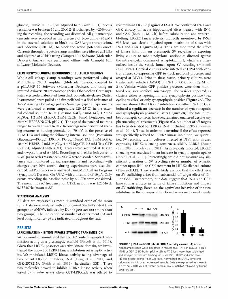
recombinant LRRK2 (Figures A1A–C). We confirmed IN-1 andGSK efficacy on acute hippocampal slices treated with IN-1and GSK (both 1 µM, 2 h) before solubilization and western-blotting. LRRK2 kinase activity, indirectly monitored by P-Ser935 level, was clearly impaired upon incubation of slices withIN-1 and GSK (Figures 1A,B). Thus, we monitored the effectof kinase inhibition on presynaptic SV recycling by exposingliving culture to rabbit polyclonal antibodies directed againstthe intravesicular domain of synaptotagmin1, which are inter-nalized inside the vesicle lumen upon SV recycling (Matteoliet al., 1992). Cortical cultures were infected at DIV4 with con-trol viruses co-expressing GFP to track neuronal processes andassayed at DIV14. Prior to these assays, primary cultures weretreated with vehicle (DMSO) or IN-1 or GSK (both 2 µM for2 h). Vesicles within GFP positive processes were then moni-tored via laser confocal microscopy. The vesicles appeared asclusters either synaptotagmin and synaptophysin positive (i.e.,cycling vesicles) or only synaptophysin positive (Figure 2A). Theanalysis showed that LRRK2 inhibition via either IN-1 or GSKinduced a significant decrease in the number of synaptotagminand synaptophysin positive clusters (Figure 2B). The total num-ber of synaptic contacts, however, remained unaltered despite anypharmacological treatments (Figure 2C). A number of off-targetshas been described for LRRK2 IN-1, including ERK5 (Luermanet al., 2014). Thus, in order to determine if the effect reportedwas specifically related to LRRK2 kinase inhibition, we quanti-fied SV recycling rate in cultures infected at DIV4 with virusesexpressing LRRK2 silencing constructs, siRNA LRRK2 (Baueret al., 2009; Piccoli et al., 2011). As previously reported, LRRK2silencing was associated to an increase in synaptotagmin uptake(Piccoli et al., 2011). Interestingly, we did not measure any sig-nificant alteration of SV recycling rate or number of synapticcontact upon IN-1 or GSK treatment in LRRK2 silenced culture(Figures 2D,E). These results likely exclude that the effect seenon SV trafficking arises from substantial off target effect of IN-1 or GSK. Furthermore, our data suggest that IN-1 and GSKhave similar efficacy in terms of kinase inhibition and impacton SV trafficking. Based on the equivalent behavior of the twoinhibitors, in the subsequent functional assays we focused mainly
FIGURE 1 | IN-1 and GSK inhibit LRRK2 activity ex-vivo. (A) Acutehippocampal slices were incubated in regular aCSF (NT) or in aCSF + IN-1(IN-1) or GSK (GSK) both 1 µM for 2 h at RT. Slices were then solubilizedand assayed by western blotting for P-Ser 935, LRRK2 and actin level.(B) The graph reports P-Ser 935 level, normalized on LRRK2 level andcalculated as fold over not treated sample. Data are expressed as mean ±s.e.m; ∗p < 0.01 vs. not treated sample, n = 4, ANOVA followed by Dunn’spost-hoc test.
Frontiers in Molecular Neuroscience www.frontiersin.org May 2014 | Volume 7 | Article 49 | 4

Cirnaru et al. LRRK2 at the presynaptic site
FIGURE 2 | LRRK2 kinase inhibition impairs SV trafficking. (A) Theexo-endocytotic assay was performed on cortical neurons infected at DIV4with virus expressing control siRNA and GFP and left untreated (NT) orincubated with IN-1 or GKS compound (both 2 µM for 2 h) before beingtested at DIV14. Cycling SV appear as synaptotagmin (s-tagmin) positiveclusters along neuron processes. Total SV pool was revealed by stainingwith anti-synaptophysin antibodies upon fixation and permeabilization.Images show signals acquired for synaptotagmin, synaptophysin and theirsuperimposition plus GFP (merge). (B) The percentage of s-tagmin ands-physin positive clusters within the totality of s-physin positive clustersreflects the pool of cycling vesicles. (C) Total number of SV pools was notaltered by treatment with IN-1 and GSK compound. The graph reportsnumber of synaptophysin-positive clusters per 10 µm of GFP-positiveprocess. (D) Similar experiments were performed on cortical neuronsinfected on DIV4 with viruses expressing LRRK2 siRNA and GFP. In LRRK2down-regulated culture SV cycling is not affected upon treatment with IN-1and GSK compound (both 2 µM for 2 h). (E) Total number of SV pools wasnot altered by treatment with IN-1 and GSK compound. The graph reportsnumber of synaptophysin-positive clusters per 10 µm of GFP-positiveprocess. Data are expressed as mean ± s.e.m.; ∗p < 0.05 vs. not treated,n = 20, ANOVA followed by Dunn’s post-hoc test. Panel size is 35 × 5 µm.
on IN-1. Given the impact of LRRK2 inhibition on SV traffick-ing, we next investigated the effect on IN-1 on neurotransmitterrelease. To this aim we measured glutamate release from isolatedsynaptosomes upon IN-1 treatment in either basal or stimu-lated condition (Figure 3A). A pulse of 15 mM K+ caused anapproximate three-fold, transient elevation of glutamate levels.IN-1 (3 µM) did not affect spontaneous glutamate efflux, butinhibited the K+-evoked glutamate overflow by about 60%. In
FIGURE 3 | IN-1 impairs neurotransmitter release from isolated
synaptosome. (A) Synaptosomes obtained from the cerebral(fronto-temporal) cortex of LRRK2 WT mice were perfused with Krebssolution, and stimulated with a 90 s pulse of 15 mM KCl. IN-1 (3 µM) wasperfused 9 min before KCl and maintained until the end of experiment. IN-1reduced K+-evoked glutamate overflow. Data are means ± s.e.m. of 5–6determinations per group, and are expressed as absolute glutamateconcentrations in the superfusate (in nM) or K+-evoked glutamate overflow(in pmol/mg protein/min; insets). (B) Similar experiments were executed oncortical synaptosome obtained from LRRK2 KO mice. IN-1 (3 µM) failed toimpair K+-evoked glutamate overflow. Statistical analysis was performed onoverflow values by the Student t-test for unpaired data. ∗∗p < 0.01 differentfrom KCl alone.
a complementary approach, we measured the basal and evoked(15 mM K+) release of [3H]D-aspartate in presence or not ofIN-1 (1 µM). Also in this model, IN-1 impaired the K+ evokedrelease by about 35% (calculated as fraction of overflow andexpressed as mean ± s.e.m.: K+ alone = 1.4 ± 0.01 K+ + IN-1 = 0.9 ± 0.01, p < 0.01, n = 7, Student’s t-test). To excludepotential off target effect of IN-1, we studied glutamate releasein synaptosomes obtained from LRRK2 KO mice (Figure 3B).Spontaneous and K+-evoked glutamate efflux was not differentbetween the two genotypes. However, IN-1 (3 µM) did not signif-icantly influence the K+-evoked glutamate release in LRRK2 KOmice. Robust evidence correlates synapsin I to the mobilizationof SV and release of neurotransmitter (Orenbuch et al., 2012).
Frontiers in Molecular Neuroscience www.frontiersin.org May 2014 | Volume 7 | Article 49 | 5
Cirnaru et al. LRRK2 at the presynaptic site
Thus, we verified whether the lack of effect of IN-1 on glutamaterelease we reported in LRRK2 KO mice could arise from disturbedsynapsin I level. Western blotting analysis of synaptosome fromwild-type and LRRK2 KO mice did not shown any significantdifference (Figure A1D). This evidence indicates that the impair-ment in neurotransmitter release arises from a specific effect ofIN-1 on LRRK2. Given the impact of LRRK2 kinase inhibition onpresynaptic functions, we next evaluated the functional outcomeof LRRK2 inhibition in terms of neuronal activity. To this aim, westudied the electrophysiological properties in two different neu-ronal models, namely acute hippocampal slices and hippocampalcultures. First we exposed acute hippocampal slices obtained
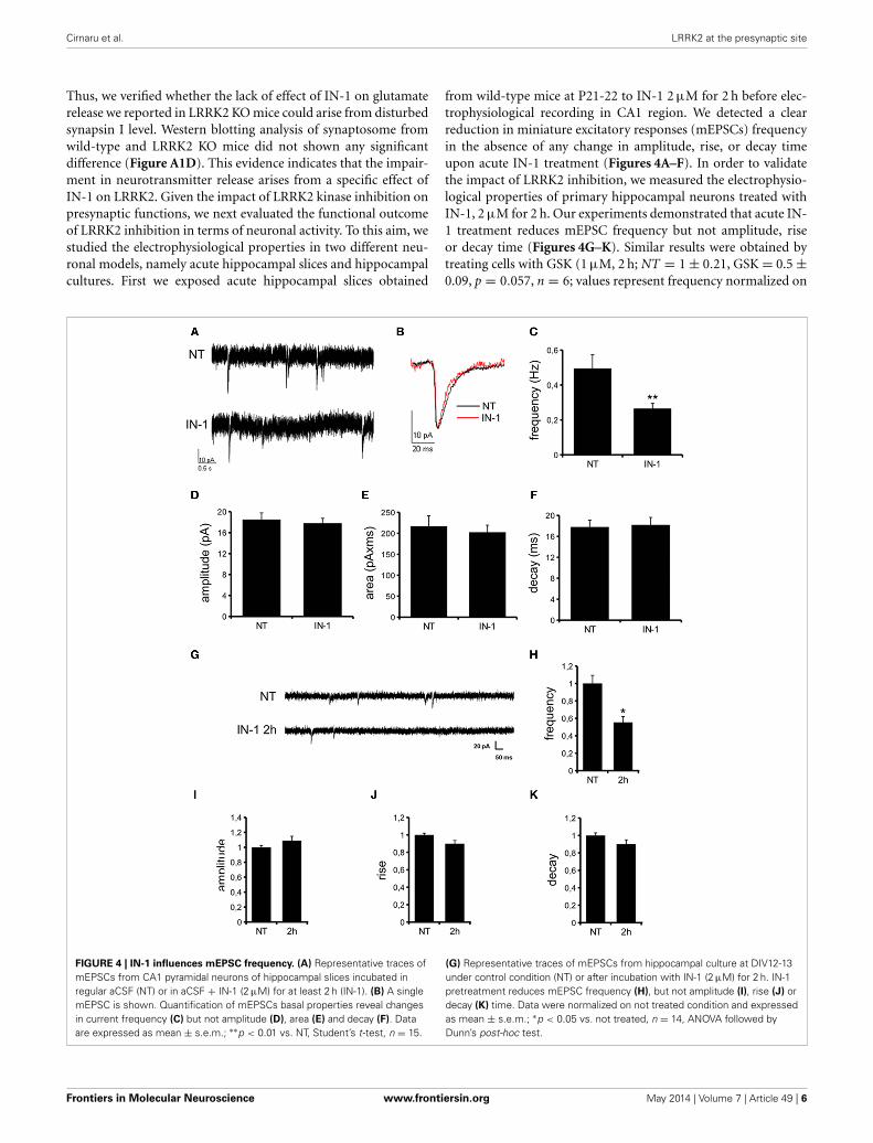
from wild-type mice at P21-22 to IN-1 2 µM for 2 h before elec-trophysiological recording in CA1 region. We detected a clearreduction in miniature excitatory responses (mEPSCs) frequencyin the absence of any change in amplitude, rise, or decay timeupon acute IN-1 treatment (Figures 4A–F). In order to validatethe impact of LRRK2 inhibition, we measured the electrophysio-logical properties of primary hippocampal neurons treated withIN-1, 2 µM for 2 h. Our experiments demonstrated that acute IN-1 treatment reduces mEPSC frequency but not amplitude, riseor decay time (Figures 4G–K). Similar results were obtained bytreating cells with GSK (1 µM, 2 h; NT = 1 ± 0.21, GSK = 0.5 ±0.09, p = 0.057, n = 6; values represent frequency normalized on
FIGURE 4 | IN-1 influences mEPSC frequency. (A) Representative traces ofmEPSCs from CA1 pyramidal neurons of hippocampal slices incubated inregular aCSF (NT) or in aCSF + IN-1 (2 µM) for at least 2 h (IN-1). (B) A singlemEPSC is shown. Quantification of mEPSCs basal properties reveal changesin current frequency (C) but not amplitude (D), area (E) and decay (F). Dataare expressed as mean ± s.e.m.; ∗∗p < 0.01 vs. NT, Student’s t-test, n = 15.
(G) Representative traces of mEPSCs from hippocampal culture at DIV12-13under control condition (NT) or after incubation with IN-1 (2 µM) for 2 h. IN-1pretreatment reduces mEPSC frequency (H), but not amplitude (I), rise (J) ordecay (K) time. Data were normalized on not treated condition and expressedas mean ± s.e.m.; ∗p < 0.05 vs. not treated, n = 14, ANOVA followed byDunn’s post-hoc test.
Frontiers in Molecular Neuroscience www.frontiersin.org May 2014 | Volume 7 | Article 49 | 6

Cirnaru et al. LRRK2 at the presynaptic site
untreated cultures and expressed as mean ± s.e.m.). These exper-iments indicate that the pharmacological inhibition of LRRK2kinase activity reduces synaptic transmission affecting SV recy-cling and thus neurotransmitter release.
KINASE ACTIVITY CONTROLS LRRK2 BINDING PROPERTIESIndependent studies demonstrated that LRRK2 exists in multipleoligomeric state: kinase-active dimer (Deng et al., 2008; Greggioet al., 2008; Klein et al., 2009) and monomers or oligomersmainly inactive (Sen et al., 2009). Thus, we asked whether LRRK2kinase inhibition might influence LRRK2 oligomeric state. Firstwe explored whether kinase inhibition affects LRRK2 homolo-gous interaction by evaluating the extent of LRRK2 dimerizationin presence of IN-1. To this aim we co-expressed FLAG-LRRK2and myc-LRRK2 in HEK293T cells; we subsequently treated thecell with IN-1 (2 h, 1 µM) and eventually we immobilized LRRK2on FLAG-M2 beads. After elution, we measured the recovery ofFLAG and myc LRRK2 by immunoblotting with specific anti tagantibodies (Figure 5A). We found that IN-1 treatment does notsignificantly affect the amount of myc LRRK2 co-precipitatingwith FLAG-LRRK2 (Figure 5B). To further explore the impact ofkinase inhibition on LRRK2 oligomerization, we performed sizeexclusion chromatography (SEC) experiments on FLAG-LRRK2proteins purified from untreated cells and then incubated withIN-1 (1 µM, 90 min). As shown in Figures 5C,D, the elutionprofile of purified LRRK2 is only marginally affected by IN-1 inhi-bition. We obtained comparable results incubating FLAG-LRRK2with GSK (1 µM, 90 min, data not shown). These data suggestthat kinase inhibition minimally impacts LRRK2 oligomeric state.Next, we asked whether kinase inhibition engages LRRK2 in dif-ferential heterologous interactions. We have previously demon-strated that LRRK2 interacts with a panel of proteins, includingactin (Piccoli et al., 2011). Thus, we analyzed by SEC the elu-tion profile of FLAG-LRRK2 and actin in lysates extracted fromcells treated with IN-1 (1 µM, 90 min). Interestingly, we observedthat IN-1 shifted both LRRK2 and actin toward higher molec-ular weight forms and that the two elution profiles partiallyoverlap (Figures 6A,B). This outcome might be consistent withthe possibility that LRRK2 forms higher molecular weight com-plexes with actin upon IN-1 binding. To further substantiatethis hypothesis we over-expressed FLAG-LRRK2 A2016T, an arti-ficial variant unable to bind IN-1, in HEK293T cells (Nicholset al., 2009; Deng et al., 2011). When we analyzed by SEC theelution profile of FLAG-LRRK2 A2016T and actin in lysatesextracted from cells treated with IN-1 (2 h, 1 µM), we observedthat IN-1 failed to shift either LRRK2 or actin elution pro-files (Figures 6C,D). All together these data strongly suggest thatkinase inhibition induces the formation of high-molecular weightcomplexes including LRRK2 and its interacting partners. To fur-ther explore this hypothesis, we asked whether LRRK2 inhibitionmight affect LRRK2 affinity toward SV associated proteins such assynapsin I and actin. To this aim we immunoprecipitated LRRK2with anti-LRRK2 antibodies [MJFF C41-2] using purified synap-tosomes treated with IN-1 (1 µM) during the assay as proteinsource. We found that the binding of LRRK2 to synapsin I andactin increased in presence of IN-1 (Figures 7A,B). Given theeffects of kinase inhibition on LRRK2 binding features, we first
FIGURE 5 | IN-1 does not influence LRRK2 dimerization. (A) HEK293Tcells expressing both myc and FLAG LRRK2 were treated or not with IN-1(2 µM for 2 h), solubilized and processed for FLAG immunopurification. Weevaluated the extent of LRRK2 homodimerization by measuring the amountof myc LRRK2 co-precipitating with FLAG LRRK2 (B) The graph reports theamount of FLAG and myc LRRK2 recovered in FLAG immunoprecipitatesupon IN-1 incubation. Data were calculated as fraction of untreated sampleand expressed as mean ± s.e.m. (n = 4). (C) Full-length LRRK2 purified byFlag immunoaffinity from untreated HEK293T cells was separated by sizeexclusion chromatography (SEC) and subsequently treated or not with IN-1(1 µM, 90 min on ice). (D) The intensity of each dot (fraction) is normalizedby the integrated intensities. Column void volume is 7.5 ml.
investigated if LRRK2 binds to SV and, next, if kinase inhibitiondisturbs LRRK2 and synapsin I binding to SV. To this aim weincubated native purified SV (in the range of 40 µg/sample) underphosphorylation permissive conditions or in the presence of IN-1 (1 µM, 1 h). After incubation, we recovered SV by high speedcentrifugation and determined the amounts of bound LRRK2and synapsin I by immunoblotting. The SV recovery in the pel-let was evaluated based on synaptophysin immunoreactivity. Wefound that LRRK2 binds SV and that this interaction is signifi-cantly decreased in the presence of IN-1 while synapsin I bindingto SV was unaffected by IN-1 (Figures 7C,D). As a complemen-tary approach we analyzed the impact of IN-1 (1 µM, 1 h) onthe interaction between SV and exogenous recombinant FLAG-LRRK2 (Figure A1C). After incubation, we separated SV-boundLRRK2 by high-speed centrifugation and evaluated the recov-ery of SV and bound LRRK2 and synapsin I in the pellet byimmunoblotting. Our data showed that IN-1 significantly reducesexogenous LRRK2 binding to SV while the yield of SV-boundsynapsin I remains unaltered (Figures 7E,F). This evidence sug-gests that kinase inhibition interferes with the macro-molecularcomplex bound to LRRK2 at the presynaptic site.
Frontiers in Molecular Neuroscience www.frontiersin.org May 2014 | Volume 7 | Article 49 | 7

Cirnaru et al. LRRK2 at the presynaptic site
FIGURE 6 | IN-1 alters LRRK2 macromolecular complex. (A) HEK293Tcells expressing FLAG-LRRK2 wild-type were treated or not with IN-1(1 µM, 90 min), solubilized and then separated by SEC. FLAG-LRRK2 andactin were revealed by western-blotting of fractions spotted onnitrocellulose. (B) The intensity of each dot (fraction) is normalized by theintegrated intensities. (C) HEK293T cells expressing FLAG-LRRK2 A2016Twere treated or not with IN-1 (1 µM, 90 min), solubilized and then separatedby SEC. LRRK2 and actin were revealed by western-blotting of fractionsspotted on nitrocellulose. (D) The intensity of each dot (fraction) isnormalized by the integrated intensities.
DISCUSSIONOur previous observations provided evidence that LRRK2 exe-cutes critical functions at the presynaptic site; given its relativeposition as an integral part of a presynaptic protein network,LRRK2 may serve as a molecular hub coordinating both the stor-age and the mobilization of SV driven by activity (Piccoli et al.,2011, 2014). Recent work has clarified that LRRK2 controls SVin the ready releasable pool via inhibitory phosphorylation of theSNAP-25 interacting protein Snapin (Yun et al., 2013). The evi-dence reported here adds one more level of complexity: the impli-cation of LRRK2 kinase activity within synaptic functions. As wildtype LRRK2 is characterized by a low kinase activity (MacLeodet al., 2006), it might be argued that physiologically LRRK2acts as a scaffold protein and its kinase activity mainly regulatesits macro-molecular organization. In fact, several independent
FIGURE 7 | IN-1 modifies LRRK2 binding properties. (A) Extracts ofpurified cortical synaptosomes were incubated with anti-LRRK2 antibodiesor rabbit IgG in absence (not treated) or in presence of IN-1 (IN-1, 1 µM 2 h).The immunocomplexes were sedimented with protein G-Sepharose andthe samples were resolved by SDS-PAGE and analyzed by immunoblottingwith anti synapsin I, anti actin and anti LRRK2 antibodies. (B) Quantificationof IN-1 effect on LRRK2 interaction with synapsin I, actin and LRRK2 itself.Results are calculated as percent of respective controls (not treatedsample) and expressed as mean ± s.e.m. (∗P < 0.05, n = 4, Student’st-test vs. not treated sample). (C) purified native synaptic vesicles (SV;40 µg/sample) were incubated in the absence (NT) or presence of IN-1(IN-1, 1 µM 2 h). After incubation, SV were recovered by high speedcentrifugation and the residual amounts of endogenous LRRK2 bound toSV were determined by immunoblotting with anti-LRRK2 antibodies. Therecovery of SV in the pellet was evaluated based on synaptophysinimmunoreactivity. (D) LRRK2 recovery in the SV pellet was calculated asthe percentage of the not treated sample and shown as mean ± s.e.m.(∗P < 0.05, n = 8, Student’s t-test vs. relative control). (E) PurifiedFLAG-LRRK2 was incubated with SV (10 µg protein/sample) in presence orabsence (NT) of IN-1 (IN-1, 1 µM 2 h). SV-bound FLAG-LRRK2 wasseparated from free FLAG-LRRK2 by high-speed centrifugation andquantified by immunoblotting with anti-FLAG antibody. The recovery of SVin the pellet was evaluated based on synaptophysin immunoreactivity.(F) The binding of FLAG-LRRK2 to SV was calculated as the percentage oftotal FLAG-LRRK2 and expressed as mean ± s.e.m. ∗p < 0.05; Student’st-test vs. relative control.
studies have revealed that LRRK2 exists in different forms inequilibrium, namely monomer, dimer and oligomer, being thedimer the predominant status under native conditions (Sen et al.,2009; but see Ito and Iwatsubo, 2012). Interestingly, PD associ-ated LRRK2 mutations disturb both LRRK2 dimerization (Senet al., 2009) and ternary complex formation (Nichols et al., 2010).Furthermore, acute treatment with IN-1 induces the aggregationof ectopic LRRK2 expressed in heterologous cell lines and inter-feres with 14-3-3 binding (Deng et al., 2011). Our hypothesis isthat kinase inhibition triggers the formation of high molecularweight complexes encompassing LRRK2 and LRRK2 interacting
Frontiers in Molecular Neuroscience www.frontiersin.org May 2014 | Volume 7 | Article 49 | 8
Cirnaru et al. LRRK2 at the presynaptic site
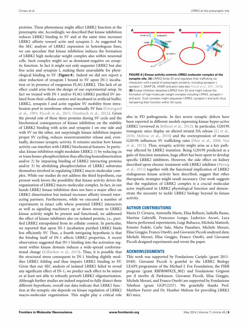
proteins. These phenomena might affect LRRK2 function at thepresynaptic site. Accordingly, we described that kinase inhibitionreduces LRRK2 binding to SV and at the same time increasesLRRK2 affinity toward actin and synapsin I. As suggested bythe SEC analysis of LRRK2 expression in heterologous lines,we can speculate that kinase inhibition induces the formationof LRRK2 high molecular weight complex also within neuronalcells. Such complex might act as dominant-negative on synap-tic function. In fact it might not only sequester LRRK2 but alsofree actin and synapsin I, making them unavailable for physi-ological binding to SV (Figure 8). Indeed we did not report aclear reduction of synapsin I bound to SV upon IN-1 incuba-tion or in presence of exogenous FLAG-LRRK2. This lack of aneffect could arise from the design of our experimental setup. Infact we treated with IN-1 and/or FLAG-LRRK2 purified SV iso-lated from their cellular context and incubated in artificial buffer.LRRK2, synapsin I and actin regulate SV mobility from intra-bouton pool to membrane where eventually SV fuse (Greengardet al., 1993; Piccoli et al., 2011; Orenbuch et al., 2012). Giventhe pivotal role of these three proteins during SV cycle and thebiochemical consequences of kinase inhibition on the stabilityof LRRK2 binding with actin and synapsin I on one side andwith SV on the other, not surprisingly kinase inhibition impairsproper SV cycling, reduces neurotransmitter release and, even-tually, decreases synaptic activity. It remains unclear how kinaseactivity can interfere with LRRK2 biochemical features. In partic-ular, kinase inhibition might modulate LRRK2: 1) by reducing cisor trans homo-phosphorylation thus affecting homodimerizationand/or 2) by impairing binding of LRRK2 interacting proteinsand/or 3) by abolishing phosphorylation of LRRK2 substratesthemselves involved in regulating LRRK2 macro-molecular com-plex. While our studies do not address the third hypothesis, ourpresent work favors the possibility that kinase activity drives theorganization of LRRK2 macro-molecular complex. In fact, in ourhands LRRK2 kinase inhibition does not have a major effect onLRRK2 dimerization but instead increases affinity toward inter-acting partners. Furthermore, while we executed a number ofexperiments in intact cells where potential LRRK2 interactorsas well as signaling machinery up or down stream to LRRK2kinase activity might be present and functional, we addressedthe effect of kinase inhibitors also on isolated protein, i.e., puri-fied LRRK2 extrapolated from its cellular context. In particular,we reported that upon IN-1 incubation purified LRRK2 bindsless efficiently SV. Thus, a fourth intriguing hypothesis is thatthe binding itself of IN-1 affects LRRK2 properties. A recentobservation suggested that IN-1 binding into the activation seg-ment within kinase domain induces a wide-spread conforma-tional change (Gillardon et al., 2013). Thus, it is possible thatthe structural stress consequent to IN-1 binding slightly mod-ifies LRRK2 folding and thus impairs LRRK2 binding to SV.Given that our SEC analysis of isolated LRRK2 failed to revealany significant effect of IN-1, we predict such effect to be minoror at least not able to robustly perturb LRRK2 oligomerization.Although further studies are indeed required to fully dissect thesedifferent hypothesis, overall our data indicate that LRRK2 func-tion at the synaptic site depends on kinase regulation of LRRK2macro-molecular organization. This might play a critical role
FIGURE 8 | Kinase activity controls LRRK2 molecular complex at the
synaptic site. (A) LRRK2 binds SV and regulates thier trafficking viainteraction with a panel of presynaptic proteins, including synapsin I,syntaxin 1, SNAP-25, VAMP, and actin (see also Piccoli et al., 2011, 2014).(B) Kinase inhibition detaches LRRK2 from SV and might induce theformation of high-molecular weight complex including LRRK2, synapsin Iand actin. Such complex might sequester LRRK2, synapsin I and actin thushampering their function within SV cycle.
also in PD pathogenesis. In fact severe synaptic defects havebeen reported in different models expressing kinase hyper-activeLRRK2 (reviewed in Belluzzi et al., 2012). In particular, G2019Stransgenic mice display an altered striatal DA release (Li et al.,2010; Melrose et al., 2010) and the overexpression of mutantG2019S influences SV trafficking rates (Shin et al., 2008; Yunet al., 2013). Thus, synaptic activity might arise as a key path-way affected by LRRK2 mutation. Being G2019S predicted as again-of-function mutation, huge effort has been spent to developspecific LRRK2 inhibitors. However, the side effect on kidneydescribed upon chronic treatment with LRRK2 inhibitor (Herziget al., 2011) together with the functional implication of LRRK2endogenous kinase activity here described, suggest that othertherapeutic strategies might result necessary. Our work suggeststhat the regulation of LRRK2 complex is a crucial molecularactor implicated in LRRK2 physiological function and demon-strate the necessity to tackle LRRK2 biology beyond its kinaseactivity.
AUTHOR CONTRIBUTIONSMaria D. Cirnaru, Antonella Marte, Elisa Belluzzi, Isabella Russo,Martina Gabrielli, Francesco Longo, Ludovico Arcuri, LucaMurru performed experiments; Luigi Bubacco, Michela Matteoli,Ernesto Fedele, Carlo Sala, Maria Passafaro, Michele Morari,Elisa Greggio, Franco Onofri, and Giovanni Piccoli analyzed data;Michele Morari, Elisa Greggio, Franco Onofri, and GiovanniPiccoli designed experiments and wrote the paper.
ACKNOWLEDGMENTSThis work was supported by Fondazione Cariplo (grant 2011-0540). Giovanni Piccoli is grateful to the LRRK2 BiologyLEAPS programme of the Michael J. Fox Foundation, the FIRBprogram (grant RBFR08F82X_002) and Fondazione Grigioniper il morbo di Parkinson. Giovanni Piccoli, Elisa Greggio,Michele Morari, and Franco Onofri are supported by FondazioneTelethon (grant GGP12237). We gratefully thanks Prof.Matthew Farrer and Dr. Heather Melrose for providing LRRK2KO mice.
Frontiers in Molecular Neuroscience www.frontiersin.org May 2014 | Volume 7 | Article 49 | 9
Cirnaru et al. LRRK2 at the presynaptic site
REFERENCESAasly, J. O., Toft, M., Fernandez-Mata, I., Kachergus, J., Hulihan, M., White, L. R.,
et al. (2005). Clinical features of LRRK2-associated Parkinson’s disease in centralNorway. Ann. Neurol. 57, 762–765. doi: 10.1002/ana.20456
Bauer, M., Kinkl, N., Meixner, A., Kremmer, E., Riemenschneider, M., Förstl,H., et al. (2009). Prevention of interferon-stimulated gene expression usingmicroRNA-designed hairpins. Gene Ther. 16, 142–147. doi: 10.1038/gt.2008.123
Belluzzi, E., Greggio, E., and Piccoli, G. (2012). Presynaptic dysfunction inParkinson’s disease: a focus on LRRK2. Biochem. Soc. Trans. 40, 1111–1116. doi:10.1042/BST20120124
Berg, D., Schweitzer, K. J., Leitner, P., Zimprich, A., Lichtner, P., Belcredi, P.,et al. (2005). Type and frequency of mutations in the LRRK2 gene in familialand sporadic Parkinson’s disease∗. Brain 128, 3000–3011. doi: 10.1093/brain/awh666
Berger, Z., Smith, K. A., and Lavoie, M. J. (2010). Membrane localization ofLRRK2 is associated with increased formation of the highly active LRRK2dimer and changes in its phosphorylation. Biochemistry 49, 5511–5523. doi:10.1021/bi100157u
Bosgraaf, L., and Van Haastert, P. J. (2003). Roc, a Ras/GTPase domain in complexproteins. Biochim. Biophys. Acta. 1643, 5–10. doi: 10.1016/j.bbamcr.2003.08.008
Brice, A. (2005). Genetics of Parkinson’s disease: LRRK2 on the rise. Brain 128,8475–8485. doi: 10.1093/brain/awh676
Civiero, L., Vancraenenbroeck, R., Belluzzi, E., Beilina, A., Lobbestael, E., Reyniers,L., et al. (2012). Biochemical characterization of highly purified leucine-richrepeat kinases 1 and 2 demonstrates formation of homodimers. PLoS ONE7:e43472. doi: 10.1371/journal.pone.0043472
Deng, J., Lewis, P. A., Greggio, E., Sluch, E., Beilina, A., and Cookson, M. R. (2008).Structure of the ROC domain from the Parkinson’s disease-associated leucine-rich repeat kinase 2 reveals a dimeric GTPase. Proc. Natl. Acad. Sci. U.S.A. 105,1499–1504. doi: 10.1073/pnas.0709098105
Deng, X., Dzamko, N., Prescott, A., Davies, P., Liu, Q., Yang, Q., et al. (2011).Characterization of a selective inhibitor of the Parkinson’s disease kinaseLRRK2. Nat. Chem. Biol. 7, 203–205. doi: 10.1038/nchembio.538
Gillardon, F., Kremmer, E., Froehlich, T., Ueffing, M., Hengerer, B., and Gloeckner,C. J. (2013). ATP-competitive LRRK2 inhibitors interfere with monoclonalantibody binding to the kinase domain of LRRK2 under native conditions. Amethod to directly monitor the active conformation of LRRK2? J. Neurosci.Methods 214, 62–68. doi: 10.1016/j.jneumeth.2012.12.015
Gloeckner, C. J., Kinkl, N., Schumacher, A., Braun, R. J., O’Neill, E., Meitinger,T., et al. (2006). The Parkinson disease causing LRRK2 mutation I2020T isassociated with increased kinase activity. Hum. Mol. Genet. 15, 223–232. doi:10.1093/hmg/ddi439
Greengard, P., Valtorta, F., Czernik, A. J., and Benfenati, F. (1993). Synaptic vesiclephosphoproteins and regulation of synaptic function. Science 259, 780–785. doi:10.1126/science.8430330
Greggio, E., Jain, S., Kingsbury, A., Bandopadhyay, R., Lewis, P., Kaganovich,A., et al. (2006). Kinase activity is required for the toxic effects ofmutant LRRK2/dardarin. Neurobiol. Dis. 23, 329–341. doi: 10.1016/j.nbd.2006.04.001
Greggio, E., Zambrano, I., Kaganovich, A., Beilina, A., Taymans, J.-M., Daniels,V., et al. (2008). The Parkinson disease-associated leucine-rich repeat Kinase 2(LRRK2) Is a dimer that undergoes intramolecular autophosphorylation. J. Biol.Chem. 283, 16906–16914. doi: 10.1074/jbc.M708718200
Guo, L., Wang, W., and Chen, S. G. (2006). Leucine-rich repeat kinase 2: rel-evance to Parkinson’s disease. Int. J. Biochem. Cell Biol. 38, 1469–1475. doi:10.1016/j.biocel.2006.02.009
Hardy, J., Cai, H., Cookson, M. R., Gwinn-Hardy, K., and Singleton, A. (2006).Genetics of Parkinson’s disease and parkinsonism. Ann. Neurol. 60, 389–398.doi: 10.1002/ana.21022
Herzig, M. C., Kolly, C., Persohn, E., Theil, D., Schweizer, T., Hafner, T., et al.(2011). LRRK2 protein levels are determined by kinase function and are cru-cial for kidney and lung homeostasis in mice. Hum. Mol. Genet. 20, 4209–4223.doi: 10.1093/hmg/ddr348
Hinkle, K. M., Yue, M., Behrouz, B., Dächsel, J. C., Lincoln, S. J., Bowles, E.E., et al. (2012). LRRK2 knockout mice have an intact dopaminergic systembut display alterations in exploratory and motor co-ordination behaviors. Mol.Neurodegener. 7:25. doi: 10.1186/1750-1326-7-25
Huttner, W. B., Schiebler, W., Greengard, P., and De Camilli, P. (1983). Synapsin I(protein I), a nerve terminal-specific phosphoprotein. III. Its association with
synaptic vesicles studied in a highly purified synaptic vesicle preparation. J. CellBiol. 96, 1374–1388. doi: 10.1083/jcb.96.5.1374
Ito, G., and Iwatsubo, T. (2012). Re-examination of the dimerization state ofleucine-rich repeat kinase 2: predominance of the monomeric form. Biochem. J.441, 987–994. doi: 10.1042/BJ20111215
Klein, C. L., Rovelli, G., Springer, W., Schall, C., Gasser, T., and Kahle, P. J. (2009).Homo- and heterodimerization of ROCO kinases: LRRK2 kinase inhibition bythe LRRK2 ROCO fragment. J. Neurochem. 111, 703–715. doi: 10.1111/j.1471-4159.2009.06358.x
Laemmli, U. K. (1970). Cleavage of structural proteins during the assembly of thehead of bacteriophage T4. Nature 227, 680–685. doi: 10.1038/227680a0
Li, X., Patel, J. C., Wang, J., Avshalumov, M. V., Nicholson, C., Buxbaum, J. D.,et al. (2010). Enhanced striatal dopamine transmission and motor performancewith LRRK2 overexpression in mice is eliminated by familial Parkinson’s diseasemutation G2019S. J. Neurosci. 30, 1788–1797. doi: 10.1523/JNEUROSCI.5604-09.2010
Luerman, G. C., Nguyen, C., Samaroo, H., Loos, P., Xi, H., Hurtado-Lorenzo, A.,et al. (2014). Phosphoproteomic evaluation of pharmacological inhibition ofleucine-rich repeat kinase 2 reveals significant off-target effects of LRRK-2-IN-1. J. Neurochem. 128, 561–576. doi: 10.1111/jnc.12483
MacLeod, D., Dowman, J., Hammond, R., Leete, T., Inoue, K., and Abeliovich,A. (2006). The familial Parkinsonism gene LRRK2 regulates neurite processmorphology. Neuron 52, 587–593. doi: 10.1016/j.neuron.2006.10.008
Marte, A., Cavallero, A., Morando, S., Uccelli, A., Raiteri, M., and Fedele, E.(2010). Alterations of glutamate release in the spinal cord of mice with exper-imental autoimmune encephalomyelitis. J. Neurochem. 115, 343–352. doi:10.1111/j.1471-4159.2010.06923.x
Marti, M., Paganini, F., Stocchi, S., Mela, F., Beani, L., Bianchi, C., et al. (2003).Plasticity of glutamatergic control of striatal acetylcholine release in experi-mental parkinsonism: opposite changes at group-II metabotropic and NMDAreceptors. J. Neurochem. 84, 792–802. doi: 10.1046/j.1471-4159.2003.01569.x
Matta, S., Van Kolen, K., da Cunha, R., van den Bogaart, G., Mandemakers,W., Miskiewicz, K., et al. (2012). LRRK2 controls an EndoA phos-phorylation cycle in synaptic endocytosis. Neuron 75, 1008–1021. doi:10.1016/j.neuron.2012.08.022
Matteoli, M., Takei, K., Perin, M. S., Sudhof, T. C., and De Camilli, P. (1992). Exo-endocytotic recycling of synaptic vesicles in developing processes of culturedhippocampal neurons. J. Cell Biol. 117, 849–861. doi: 10.1083/jcb.117.4.849
Mela, F., Marti, M., Ulazzi, L., Vaccari, E., Zucchini, S., Trapella, C., et al.(2004). Pharmacological profile of nociceptin/orphanin FQ receptors regulat-ing 5-hydroxytryptamine release in the mouse neocortex. Eur. J. Neurosci. 19,1317–1324. doi: 10.1111/j.1460-9568.2004.03220.x
Melrose, H. L., Dächsel, J. C., Behrouz, B., Lincoln, S. J., Yue, M., Hinkle, K.M., et al. (2010). Impaired dopaminergic neurotransmission and microtubule-associated protein tau alterations in human LRRK2 transgenic mice. Neurobiol.Dis. 40, 503–517. doi: 10.1016/j.nbd.2010.07.010
Messa, M., Congia, S., Defranchi, E., Valtorta, F., Fassio, A., Onofri, F., et al.(2010). Tyrosine phosphorylation of synapsin I by Src regulates synaptic-vesicletrafficking. J. Cell Sci. 123, 2256–2265. doi: 10.1242/jcs.068445
Migheli, R., Del Giudice, M. G., Spissu, Y., Sanna, G., Xiong, Y., Dawson, T.M., et al. (2013). LRRK2 affects vesicle trafficking, neurotransmitter extra-cellular level and membrane receptor localization. PLoS ONE 8:e77198. doi:10.1371/journal.pone.0077198
Moore, D. J., West, A. B., Dawson, V. L., and Dawson, T. M. (2005). Molecularpathophysiology of Parkinson’s disease. Annu. Rev. Neurosci. 28, 57–87. doi:10.1146/annurev.neuro.28.061604.135718
Nichols, R. J., Dzamko, N., Hutti, J. E., Cantley, L. C., Deak, M., Moran, J., et al.(2009). Substrate specificity and inhibitors of LRRK2, a protein kinase mutatedin Parkinson’s disease. Biochem. J. 424, 47–60. doi: 10.1042/BJ20091035
Nichols, R. J., Dzamko, N., Morrice, N. A., Campbell, D. G., Deak, M., Ordureau,A., et al. (2010). 14-3-3 binding to LRRK2 is disrupted by multiple Parkinson’sdisease-associated mutations and regulates cytoplasmic localization. Biochem. J.430, 393–404. doi: 10.1042/BJ20100483
Onofri, F., Messa, M., Matafora, V., Bonanno, G., Corradi, A., Bachi, A.,et al. (2007). Synapsin phosphorylation by SRC tyrosine kinase enhancesSRC activity in synaptic vesicles. J. Biol. Chem. 282, 15754–15767. doi:10.1074/jbc.M701051200
Orenbuch, A., Shalev, L., Marra, V., Sinai, I., Lavy, Y., Kahn, J., et al. (2012).Synapsin selectively controls the mobility of resting pool vesicles at hippocampal
Frontiers in Molecular Neuroscience www.frontiersin.org May 2014 | Volume 7 | Article 49 | 10
Cirnaru et al. LRRK2 at the presynaptic site
terminals. J. Neurosci. 32, 3969–3980. doi: 10.1523/JNEUROSCI.5058-11.2012
Paisan-Ruiz, C., Jain, S., Evans, E. W., Gilks, W. P., Simon, J., van der Brug,M., et al. (2004). Cloning of the gene containing mutations that causePARK8-linked Parkinson’s disease. Neuron 44, 595–600. doi: 10.1016/j.neuron.2004.10.023
Piccoli, G., Condliffe, S. B., Bauer, M., Giesert, F., Boldt, K., De Astis, S., et al.(2011). LRRK2 controls synaptic vesicle storage and mobilization within therecycling pool. J. Neurosci. 31, 2225–2237. doi: 10.1523/JNEUROSCI.3730-10.2011
Piccoli, G., Onofri, F., Cirnaru, M. D., Kaiser, C. J. O., Jagtap, P., Kastenmüller, A.,et al. (2014). LRRK2 binds to neuronal vesicles through protein interactionsmediated by its C-terminal WD40 domain. Mol. Cell. Biol. doi: 10.1128/MCB.00914-13. [Epub ahead of print].
Piccoli, G., Verpelli, C., Tonna, N., Romorini, S., Alessio, M., Nairn, A. C.,et al. (2007). Proteomic analysis of activity-dependent synaptic plastic-ity in hippocampal neurons. J. Proteome Res. 6, 3203–3215. doi: 10.1021/pr0701308
Reith, A. D., Bamborough, P., Jandu, K., Andreotti, D., Mensah, L., Dossang, P.,et al. (2012). GSK2578215A; a potent and highly selective 2-arylmethyloxy-5-substitutent-N-arylbenzamide LRRK2 kinase inhibitor. Bioorg. Med. Chem.Lett. 22, 5625–5629. doi: 10.1016/j.bmcl.2012.06.104
Sen, S., Webber, P. J., and West, A. B. (2009). Dependence of leucine-rich repeatkinase 2 (LRRK2) kinase activity on dimerization. J. Biol. 284, 36346–36356.doi: 10.1074/jbc.M109.025437
Sheng, Z., Zhang, S., Bustos, D., Kleinheinz, T., Le Pichon, C. E., Dominguez, S.L., et al. (2012). Ser1292 autophosphorylation is an indicator of LRRK2 kinaseactivity and contributes to the cellular effects of PD mutations. Sci. Transl. Med.4:164ra161. doi: 10.1126/scitranslmed.3004485
Shin, N., Jeong, H., Kwon, J., Heo, H. Y., Kwon, J. J., Yun, H. J., et al. (2008).LRRK2 regulates synaptic vesicle endocytosis. Exp. Cell Res. 314, 2055–2065.doi: 10.1016/j.yexcr.2008.02.015
Tong, Y., Pisani, A., Martella, G., Karouani, M., Yamaguchi, H., Pothos, E. N., et al.(2009). R1441C mutation in LRRK2 impairs dopaminergicneurotransmission
in mice. Proc. Natl. Acad. Sci. U.S.A. 106, 14622–14627. doi: 10.1073/pnas.0906334106
Verderio, C., Coco, S., Rossetto, O., Montecucco, C., and Matteoli, M. (1999).Internalization and proteolytic action of botulinum toxins in CNS neurons andastrocytes. J. Neurochem. 73, 372–379. doi: 10.1046/j.1471-4159.1999.0730372.x
West, A. B., Moore, D. J., Biskup, S., Bugayenko, A., Smith, W. W., Ross, C. A., et al.(2005). Parkinson’s disease-associated mutations in leucine-rich repeat kinase2 augment kinase activity. Proc. Natl. Acad. Sci. U.S.A. 102, 16842–16847. doi:10.1073/pnas.0507360102
Yun, H. J., Park, J., Ho, D. H., Kim, H., Kim, C.-H., Oh, H., et al. (2013). LRRK2phosphorylates Snapin and inhibits interaction of Snapin with SNAP-25. Exp.Mol. Med. 45, e36. doi: 10.1038/emm.2013.68
Zimprich, A., Biskup, S., Leitner, P., Lichtner, P., Farrer, M., Lincoln, S., et al. (2004).Mutations in LRRK2 cause autosomal-dominant parkinsonism with pleomor-phic pathology. Neuron 44, 601–607. doi: 10.1016/j.neuron.2004.11.005
Conflict of Interest Statement: The authors declare that the research was con-ducted in the absence of any commercial or financial relationships that could beconstrued as a potential conflict of interest.
Received: 11 March 2014; accepted: 09 May 2014; published online: 27 May 2014.Citation: Cirnaru MD, Marte A, Belluzzi E, Russo I, Gabrielli M, Longo F, Arcuri L,Murru L, Bubacco L, Matteoli M, Fedele E, Sala C, Passafaro M, Morari M, GreggioE, Onofri F and Piccoli G (2014) LRRK2 kinase activity regulates synaptic vesicle traf-ficking and neurotransmitter release through modulation of LRRK2 macro-molecularcomplex. Front. Mol. Neurosci. 7:49. doi: 10.3389/fnmol.2014.00049This article was submitted to the journal Frontiers in Molecular Neuroscience.Copyright © 2014 Cirnaru, Marte, Belluzzi, Russo, Gabrielli, Longo, Arcuri, Murru,Bubacco, Matteoli, Fedele, Sala, Passafaro, Morari, Greggio, Onofri and Piccoli.This is an open-access article distributed under the terms of the Creative CommonsAttribution License (CC BY). The use, distribution or reproduction in other forums ispermitted, provided the original author(s) or licensor are credited and that the originalpublication in this journal is cited, in accordance with accepted academic practice. Nouse, distribution or reproduction is permitted which does not comply with these terms.
Frontiers in Molecular Neuroscience www.frontiersin.org May 2014 | Volume 7 | Article 49 | 11

Cirnaru et al. LRRK2 at the presynaptic site
APPENDIX
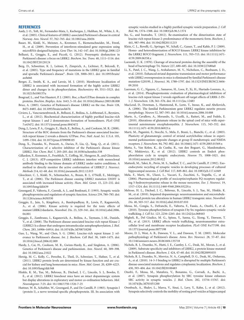
FIGURE A1 | IN-1 and GSK inhibit LRRK2 activity in vitro.
(A) Recombinant GST-LRRK2970−2527 was incubated with increasingconcentrations of LRRK2 inhibitors IN-1 or GSK in the presence of500 µM LRRKtide and 100 µM ATP (0.5 µCi 33P-ATP). Reactionswere spotted onto P81 phosphocellulose paper and LRRKtideradioactivity quantified by phosphoimaging scanner. (B) Dose-response curves and calculation of IC50 values indicates that both
inhibitors are active against LRRK2 but at different potencies (n = 6replicates, from 2 independent set of experiments). (C) Full lengthFLAG-LRRK2 purified from transfected HEK293T cells andGST-LRRK2970−2527 were resolved on SDS-PAGE and visualized viacoomassie staining. (D) Western-blotting analysis of synaptosomepurified from wild-type and LRRK2 KO mice. Synapsin I levelremains does not differ between the two genotypes.
Frontiers in Molecular Neuroscience www.frontiersin.org May 2014 | Volume 7 | Article 49 | 12