elaborating the mechanism of cell killing of a novel
TRANSCRIPT
ELABORATING THE MECHANISM OF CELL KILLING
OF A NOVEL CHEMOTHERAPEUTIC DRUG
TARGETING BREAST CANCER CELLS
by
Phat Do
Submitted in fulfillment of the
requirements for Departmental Honors in
the Department of Biology
Texas Christian University
Fort Worth, Texas
May 6, 2019
DEVELOPING CYTOTOXIC DRUGS THAT
TARGET THE ESTROGEN RECEPTOR
IN BREAST CANCER CELLS
Project Approved:
Supervising Professor: Giridhar Akkaraju, Ph.D.
Department of Biology
David Minter, Ph.D.
Department of Chemistry
Marlo Jeffries, Ph. D.
Department of Biology
ABSTRACT
Background
Breast cancer (BC) is the second most commonly diagnosed cancer among American women
after skin cancer. Traditional treatments of BC include surgery, radiation, and chemotherapy
therapy; however, these treatments are non-specific and potentially kill peripheral, healthy cells
(2). There emerges a need for more specific treatments, most notably to develop chemotherapy
agents that target a unique feature of the cancer cells. Interestingly, 70% of BC cells upregulate
estradiol-dependent pathway, a characteristic essential for rapid cell growth (3). Current BC
drugs, such as Herceptin and Tamoxifen, have targeted this pathway to preferentially kill BC
cells (4, 5). However, most women relapse within 15 years due to drug-resistance (6). Thus,
there is a need for new chemotherapeutic drugs. Our research group studies a novel estrogen-
receptor targeting drug: Est-n-Melex. This compound has the Estradiol (Est) moiety linked to a
DNA alkylating agent, Melex via a linker of varying length of methyl groups. We hypothesize
that Est-n-Melex enters the cancer cells via an interaction between the Est moiety and the
Estrogen Receptor alpha (ER-α). ER-α then dimerizes, enters the nucleus and binds to Estrogen
Response Elements on the DNA. This movement positions the Melex moiety on the DNA and
allows the transfer of a methyl group to the N-3 adenine on the DNA. In this project, we test the
hypothesized mechanism of action of our compound. Since Est-n-Melex has a DNA methylation
component (Melex) conjugated to estrogen, our hypothesis is that after the drug binds to the ER-
α in the cytosol, it translocates to the nucleus, specifically methylates the N3-region of adenine
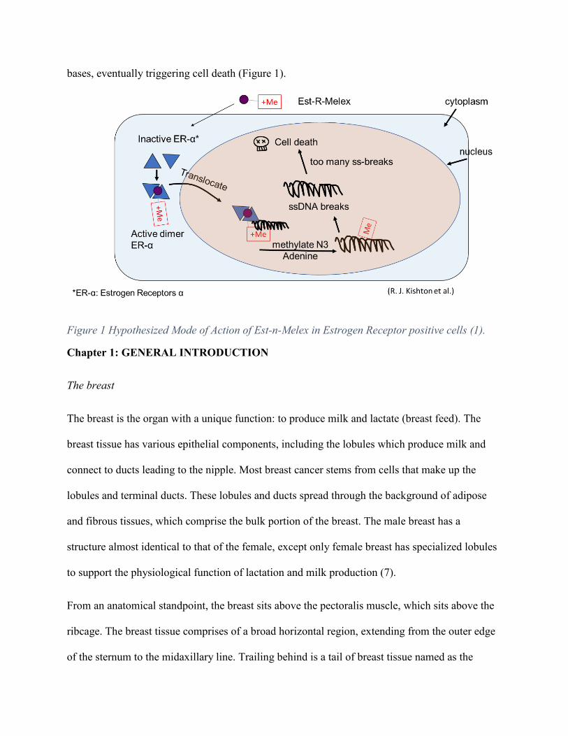
bases, eventually triggering cell death (Figure 1).
Figure 1 Hypothesized Mode of Action of Est-n-Melex in Estrogen Receptor positive cells (1).
Chapter 1: GENERAL INTRODUCTION
The breast
The breast is the organ with a unique function: to produce milk and lactate (breast feed). The
breast tissue has various epithelial components, including the lobules which produce milk and
connect to ducts leading to the nipple. Most breast cancer stems from cells that make up the
lobules and terminal ducts. These lobules and ducts spread through the background of adipose
and fibrous tissues, which comprise the bulk portion of the breast. The male breast has a
structure almost identical to that of the female, except only female breast has specialized lobules
to support the physiological function of lactation and milk production (7).
From an anatomical standpoint, the breast sits above the pectoralis muscle, which sits above the
ribcage. The breast tissue comprises of a broad horizontal region, extending from the outer edge
of the sternum to the midaxillary line. Trailing behind is a tail of breast tissue named as the

"axillary tail of Spence,” which spans directly into underarm area. This specific detail is vital
since breast cancer tissues can extend into this axillary tail, although technically, the tail
seemingly is located outside the actual breast.
Encircling the breast tissues is the fascia, a thin layer of connective tissue. Deep inside the layer
of fascia connects to the pectoralis muscle, while the layer on top of the fascia is immediately
beneath the skin. The skin tissue over the breast has similar features to the skin anywhere else in
the body, such as sweat glands and hair follicles. While conducting a breast exam, a clinician
will comprehensively study the skin as well as the breast tissue (7).
Breast cancer
Incidence, mortality, survival
Cancer is a broad term characterizing diseases in which cells undergo uncontrolled growth and
spread, often resulting in fatality. Cancer incidence has increased dramatically in recent years,
impacting various aspects of human life, including physical, mental, social, and economic
suffering (8). Cancer incidence fluctuates annually from 1 to 2 percent in developed nations, and
5% in developing countries (9). At least 7 million people are estimated to die from cancer, and
the number of new cancer cases is stipulated to rise from 10 to 15 million by 2020 (10).
Meanwhile, breast cancer remains the leading malignant neoplasm among women (11). Since
2005, the incidence rate of invasive breast cancer has stabilized among white women and rose
slightly by 0.3% every year among black women (12). In 2018, 41,400 breast cancer death
occurred. The breast cancer death rate reached the highest in 1989, then plummeted by 39% in
2015 (13). This phenomenon occurred against the background of a growing population can be
attributed to advancements in early detection (via screening and heightened awareness) and

treatment. These combined efforts are estimated to result in 322,600 fewer breast cancer deaths
per year (14).
Signs and symptoms
A lump or mass in the breast is most conspicuous symptom of a neoplasm. Less conspicuous
symptoms are systemic changes in the breast, namely thickening, swelling, distortion,
tenderness, skin irritation, redness, and nipple abnormalities or spontaneous nipple discharge.
Early breast cancer is asymptomatic and often detected via mammography screening (15) .
Risk factors
Like many other types of cancer, age is the strongest risk factor for breast cancer. Exposure of
breast tissue to reproductive hormones poses heightened risks of breast cancer as well (16).
These hormone overexposures can result from being obese or overweight, postmenopausal
hormone treatment (estrogen and progestin), physical inactivity, alcohol use, and prolonged
breast feeding (over one year). Other factors include a prolonged menstrual history (in which
menstrual periods start earlier or end later in life), bearing no children, bearing the first child
after 30 years old, having a high levels of sex hormones from birth, and frequent doses of oral
contraceptives. Familial risk factors include a family history of breast/ovarian cancer, inherited
mutations in BRCA1, BRCA2, or other breast cancer susceptibility genes, other benign breast
conditions like atypical aplasia, radiation exposure to the chest area in the young age (for
example, due to lymphoma treatment) (17). In addition, high breast tissue density and type 2
diabetes are common risk factors as well (18).
Mechanism of estradiol-induced ER-α signaling

Figure 2. Mechanism of Estradiol-induced ER-α Signaling pathway (1).
In 70% of all cases, BC growth depends on Estradiol (Est) signaling through ER-α as Est is a
mitogen for BC cells, works as a survival and anti-apoptotic factor and induces cell invasion and
migration (19). In a normal breast cell, Est moves into the cytoplasm, binds to the inactive
Estrogen Receptors alpha (ER-α) and causes it to dimerize to form an activated dimer. The
complex then functions as a transcription factor by translocating into the nucleus, binding to the
DNA and sending cell proliferation signals (Figure 2). When breast cancer cells overexpress ER-
α, the entire estradiol-response pathway is upregulated, thus increasing the rate of cell division
and risk of cancer and tumorigenesis. Aberrations in ERα, ERβ, and Progesterone (PR)
expression in tumorigenesis in the endometrial cells involve mechanisms of promoter regulation
(20). The abnormality is attributed to tumorigenesis in the endometriotic stromal cells. In these
cells, ERβ promoter is hyperactive, resulting in high ERβ expressions. ERβ downregulates ERα
expression, resulting in high ERβ-to-ERα ratios in endometriotic cells. A higher ERα-toERβ
ratio in endometriotic stromal cells causes a shift from Est inhibition to Est stimulation of

Progesterone (PR) expression in endometriotic stromal cells. This explains the proliferation of
PR resistance in women with endometriosis (20). Est is also thought to regulate the malignancy
of cancer stem-like cells (CSC) derived from the MCF7 cell line partially through Sox2. Est, in a
dose-dependent manner, produces opposite effects on proliferation, migration, colony formation,
and self-renewal capacity of CSC. In high concentration of Est, Est stimulates apoptosis and
blocks proliferation. In lower concentrations of Est, it stimulates self-renewal capacity (21).
Another study looks at Est treatment in a different angle, showing evidence that Est promotes
breast cancer cell proliferation by inducing cyclin G1 expression, thus stimulating proliferation
and cell viability (22). These discoveries can be promising for hormone combination therapy
against breast cancer tumorigenesis. Mitochondrial morphology in the Est-response pathway is
also of interest since it sheds light into understanding cancer cell proliferation or cell death.
Studies indicate that phosphorylation of dynamin-related protein 1 changes mitochondrial
morphology in MCF7 cells. ER-α assists the phosphorylation of Drp1, and subsequently results
in the mitochondrial fission and cell’s overall size reduction (23).
Early detection
To detect breast cancer tissues at the early stage, mammography is often used. Mammography is
a low-dose X-ray technique that leads to less extensive treatment and yet effectively reduces
breast cancer mortality (24). Certain shortcomings of mammography are false negative and false
positive detection. In average, around one in 10 screened women produce abnormal
mammogram, and yet, only 5% of screened women have cancer (25). Mammography also
detects in situ lesions, such as ductal carcinoma in situ, that would not progress to become
malignant, leading to over-diagnosis up to 30% of screened women (26). Nevertheless, the
American Cancer Society recommends annual mammography for at-risk women aged 40 years
and above (25). Women identified as high-risk are recommended to undergo annual magnetic
resonance imaging (MRI) as well. Among at-risk women, chemotherapeutic drugs are employed
to reduce risks of breast cancer development. Two common drugs are Tamoxifen and
Raloxifene, which are commonly prescribed to reduce breast cancer risk for at-risk women.
Although Raloxifene has a lower risk of side effects, it is more often prescribed for
postmenopausal women. Another common form of medication involves the use of Aromatase
Inhibitors (AI). Aromatase is an enzyme required for conversion of testosterone to estrogen, thus
blockers of this pathway can inhibit Est synthesis and reducing systemic Est availability (27).
AIs therefore can reduce the breast cancer risk for at-risk women. However, they are only
approved for the prevention of cancer recurrence.
Treatment
Surgery
Two most common forms of treatment include breast-conserving surgery (the surgical removal
of only the tumor and surrounding tissue) or mastectomy (surgical removal of the entire breast).
The form of surgery depends largely on the tumor characteristics, such as size, hormone receptor
status, and extent of spread, and patient preference (28).
Radiation
Most patients are recommended to have radiation in addition to breast-conserving surgery.
Studies have shown that in women having early-stage cancer (characterized as cancer that has
not spread to peripheral areas such as the skin, chest wall, or other peripheral organs), radiation
after breast-conserving surgery can result in as favorable long-term outcomes as mastectomy. In
addition, in cases of larger tumors or node-involved breast cancers, radiation is recommended for
use post-mastectomy. Women who undergoes mastectomy can decide on several breast
reconstruction options to restore breast shape, such as different type of tissue or implant used.
Reconstruction performance may occur immediately after mastectomy or delayed as a second
procedure (29).
Chemotherapy
Chemotherapy is type of cancer treatment in which drugs preferentially bind to breast cancer
cells and either prevent further cellular divisions or promote cell death. Routes of delivery are
commonly oral, musculoskeletal, or intravenous. As soon as the drugs enter the bloodstream,
they can be delivered throughout the body (systemic chemotherapy). In certain cases,
chemotherapeutic drugs may be injected directly into the cerebrospinal fluid, an organ, or a body
cavity such as the abdomen, the drug can act locally and kill cancer cells in those specific areas
(regional chemotherapy). The way the chemotherapy is given depends on the type and stage of
the cancer being treated (30). Many times, chemotherapy is conducted before the breast
cancer surgery (neoadjuvant) or post-surgery (adjuvant). Neoadjuvant chemotherapy is
conducted prior to surgery to maximize the reduction of large breast cancer mass, allowing
breast-conserving surgery. This strategy also assists physicians in figuring out the exact effect of
specific regimens on breast tumor. In contrast, adjuvant chemotherapy is done post-surgery or
post-radiation to wipe off any remaining tumor cells that surgery and radiation fails to remove.
This strategy ensures that breast cancer cells do not metastasize to peripheral parts of the body
(31).
Tamoxifen: a nonsteroidal triphenylethylene derivative. Tamoxifen became the mainstay for
ERaþ tumors since they are selective ER modulators (SERMs), which recognize and bind to ERa
and inhibit receptor to work as a transcription factor (32). This blockage prevents Est-dependent
gene expression, thus selectively down-regulating ER expression and blocking the proliferative
actions of estrogen on mammary epithelium (27). Interestingly, tamoxifen has both estrogenic
and antiestrogenic mechanism of actions, depending on the target tissue (33). It functions as an
antiestrogenic on mammary epithelium, hence is used to prevent and treat breast cancer. At the
same time, it is proestrogenic on uterine epithelium, hence causing heightening controversy
regarding its safety in cancer prevention due to the heightened incidence of endometrial
carcinoma in women consistently on tamoxifen regime (33). Studies suggest that the
antiproliferative action of tamoxifen results from the synthesis of the inductive effects of
cytokine transforming growth factor-β (TGF-β), which regulates the negative autocrine signaling
pathway (34). Moreover, immunohistochemical studies have shown that tamoxifen induces the
synthesis of TGF-β in the stromal (mesenchymal) compartment of breast cancers, suggesting a
paracrine as well as autocrine mechanism of action, independent of an interaction with the
estrogen receptor (35). However, increasing tamoxifen resistance in ERa+ patients is observed
(36). It is noted that in ERα36+ breast cancer patients, tamoxifen complementarily binds and
activates ERa36, thus increases expression of ALDH1A1 (37). As a result, it increases stemness
and promoting further metastasis. Studying the pathways of Tamoxifen resistance is useful for
therapeutic treatment plans (38).
Fulvestrant: Fulvestrant targets the Estrogen Receptor (ER) to inhibit tumor cell proliferation.
Fulvestrant acts as an antagonist to estrogen receptor, binding to ER monomers and blocking the
dimerization. As a result, activating function 1 (AF1) and AF2 become inactivated, reducing the
translocation of receptor to the nucleus and accelerating the cytosolic degradation of the ER (39).
It is also noted to physically interact with ER-α, promoting 26S proteasome-dependent
degradation and eliminating ER-α from BC cells (27). Interestingly, it is observed that
Fulvestrant has no uterotrophic side effects on the immature or ovari-ectomized rats and in a
dose-dependent manner, prevents the agonistic pathway of Est and Tamoxifen.
Natural extracts are natural compounds that have the potential to treat breast cancer proliferation.
Cinobufagin, a molecule extracted from Venunum Bufonis, is shown to cause targeted
cytotoxicity in MCF-7 cells by increasing Bax expression and reducing Bcl-2 expression, thus
inducing apoptosis and G1 phase arrest (40). Other natural extract, such as the Mexican mistletoe
Struthanthus venetus with the hydromethanolic extract (DtvHME), have antiproliferative effect
on breast cancer as well. For example, DtvHME is shown to antagonize the proliferative-
response of Est- response for uterotrophic activity of the mice. This implicated that StvHME has
the potential to act on breast cancer cells via ER-α and ER-β and can be used as the
complementary treatment against breast tumorigenesis as well (41).
Objective and Outline of this thesis
As mentioned above, aromatase inhibitors, tamoxifen, fulvestrant all commonly used to target
this Est-initiated pathway to downregulate the downstream expression. However, these drugs are
known to cause considerable side effects: AIs are clinically shown to consistently produce
musculoskeletal failures while Tamoxifen often causes endometrial cancer (27). Chronic
chemotherapy also causes BC cells to resist treatment, as one-third of women treated with
Tamoxifen for 5 years are shown to relapse within 15 years (resulting tumors will be insensitive
to Tamoxifen treatment) (42). As such, there emerges a need for new targeted chemotherapeutic
drugs.
DNA-methylating small molecules are commonly used in cancer therapy. Yet most DNA
alkylating drugs are specific for neither DNA nor cancer cells. Non-specific DNA alkylation
produces a variety of non-cytotoxic and indiscriminate DNA lesions, resulting in secondary
cancers such as leukemia (43). Non-specific uptake into the cells causes significant side-effects
like immune suppression and gastric irritation. A new strategy to overcome the therapeutic
shortcomings of DNA-methylating compounds is surprisingly simple: to link both cell-targeting
domain and DNA-binding domain to the methylating agent (1).
Since most breast carcinomas over-express the estrogen receptor alpha (ER-α), a moiety of the
drug resembling the natural ligand of ER-α, i.e. estradiol (Est), increases the drug’s affinity to
ER+ cells. This increases the concentration ratio between ER+ and ER- tissues, thus
preferentially targeting ER+ breast cancer cells. ER-α is a good target because estrogen signaling
via ER-α binding and activation is critical for the pathogenesis of breast cancer. The more
rampant presence of ER-α in breast cancer cells make it a good target for drug design.
Melex is shown binds almost exclusively to the minor DNA groove and produces over 95% of
3’-Methyl of Adenine (3-MeA) lesions (1, 44). In this case, minor DNA groove is another good
target for DNA-methylating compounds due to the high methylation specificity, resulting in high
cytotoxicity and low mutagenicity. This is desirable as compared to indiscriminate methylation
which causes side effects as mentioned above.
This design wants to neither activate nor inactivate the ER-α and DNA. Instead, it aims to bring
the already specific DNA-methylating compounds into cells overexpressing ER-α, thus more
prudentially kill breast cancer cells. This strategy takes advantage of the already well-researched
areas, i.e., estradiol (Est) signaling pathway and Melex selective methylation mechanism. To do
as such, the drug must link the two moieties (Est and MeLex) effectively to not interfere with
binding and efficacy of Est and Melex. Creating the linker requires careful deliberations
regarding the linker length between the two moieties as well as linker composition. Thereby, the
drug (Est-n-Melex) contains Est moiety conjugate to Melex via a linker of various length.
We hypothesize that Est-n-Melex enters the ER+ cancer cells more rapidly than ER- normal
cells. The Est moiety recognizes and binds to the inactive Estrogen Receptor alpha (ER-α). ER-α
then dimerizes to form an active dimer complex, which then enters the nucleus and binds to
Estrogen Response Elements on the DNA. This nuclear movement brings the drug to a proximity
with the DNA, allowing Melex to create 3-MeA lesions. This specific methylation results in
abasic sites and subsequently, single-strand breaks (SSB). High amounts of SSB ovER-αctivate
poly (ADP-ribose) polymerase (PARP) during DNA repair, costing excessive cellular ATP and
as a result, necrosis (1). In addition, co-treating the drug with PARP inhibitors can force the cell
to undergo programmed cell death, i.e., apoptosis. The proof of idea is demonstrated in
adriamycin preferentially targeting MCF-7 breast cancer cells, via activating PARP-1 activation
in a dose- and time- dependent manner and activate BRCA1, and co-treatment with a PARP-
inhibitor like 3-aminobenzamide (3AB) increases MCF-7 apoptosis (38).
Modeling the drug in three dimensions allows analysis of unintended interaction or loss of
interaction between moieties and their respective docking sites. It also allows visualization of un-
utilized / non-bonding pockets of ER-α and/or DNA to add new groups that may facilitate higher
binding affinity.
Since the drug has a moiety resembling Est, the drug is expected to bind to other ER proteins,
such as ER-β and ER-γ, as well as non-cancer ER-α expressing cells, such as brain, bone,
mammary gland, and reproductive organs. These interactions will cause side effects like
endocrine therapy or ER-α blockers such as tamoxifen or fulvestrant. If the drug is having off-
target effects, one will observe higher cell death in normal cells as compared to cancer cells.

Despite these side effects, ER-α blockers alone are insufficient since patients have high risks of
relapse several years later (27). As such, the complementation of targeted DNA-methylating
agents hold great potential in more effectively targeting and killing breast cancer cells. This
project will provide information to retain and even increase affinity and function of Est and
Melex to ER-α and DNA minor groove respectively. With this information, this study can be a
useful proof of concept for linking natural ligands to DNA-methylating small molecules to
produce tissue-specific cell death.
METHODS
Cell culture
As part of testing the drug in cell culture, MCF-7 ER+ breast cancer cells were used. MCF-7
cells were grown in DMEM (10% fetal bovine serum, 1% non-essential Amino Acids, 2mM
Glutamine, 100 units of Penicillin, and 1.7 mM Streptomycin (Sigma-Aldrich)) in a 37° C, 5%
CO2, and humidity controlled incubator. The cells were split into a new flask at around 10%
when the cells were confluent in the flask. When the cells were ready to be split, some cells
could be used in performing an experiment. 293 HEK cells (Human Embryonic Kidney
fibroblasts), which do not express the estrogen receptor, were also utilized in the study and were
grown under the same conditions.
Drug Dilution
Drugs were dissolved in DMSO (dimethyl sulfoxide) and Estradiol was dissolved in 100%
ethanol. Serial dilutions of Est-3-Melex, Melex, and Estradiol were all performed using serum
free medium (SFM).
MTT Cytotoxicity Assay
A MTT Cytotoxicity Assay was utilized for each experiment to assess percent cell death. The
assay is performed by placing around 5,000 cells in each well of a 96-well plate and the cells can
grow for 24 hours. Next, the wells were treated with increasing concentrations of Est-3-Melex,
Estradiol, or Melex, depending on the experiment. The cells were exposed to drug for 24 hours.
After this, the medium was extracted from each well and 100 µL MTT (12 mM) was added to
each well. MTT is a compound that enters the mitochondria of living cells and releases a purple
dye. Therefore, the more dye is released, the more living cells are presumed to be in that well.
MTT is dissolved in SFM. MTT remains in all wells but one (control well) for four hours. After
MTT is removed from the wells, 100 µL DMSO was then added to dissolve the MTT precipitate.
The absorbance can be quantified using a spectrophotometer at 540 nm. The relative amount of
dye released is compared to the control and the results are displayed numerically using Omega
software.
Immunofluorescence
To visualize the location of the drug within the cell, fluorescence microscopy was used. Initially,
1M HCl pre-treated coverslips were placed in each well of a six-well plate. Approximately
10,000 cells (500 µL) were placed on the coverslip in DMEM medium and the plate was placed
in the incubator for thirty minutes. After the cells are settled at the bottom, 3.5 mL of DMEM
medium was added to each well. The cells are placed in the incubator for 24 hours. Each well
was treated with drug, Estradiol, or Fulvestrant depending on the treatment. The drugs were in
each well with the cells for 24 hours in an incubator. Medium was removed from each well. Each
coverslip was washed 500 µL PBS. 500 µL paraformaldehyde was added to each well and the
plate sat in the incubator for thirty minutes. The paraformaldehyde was removed and another
wash was performed with 500 µL PBS in each well. The coverslips were then mounted onto
slides. One drop of Fluoromount was placed on the slide. The side of the coverslip with cells was
placed face down on the slide on top of the Fluoromount so that the liquid spreads across the
surface of the coverslip. The coverslip was then attached to the slide using acrylic nail polish
around the edges. After the nail polish has dried, the slide was viewed under the fluorescent
microscope using GFP wavelength.
GloMix caspase 3/7
Twenty thousand cells are seeded and incubated for 24 h, after which the medium is removed
and replaced with 50 μl fresh complete medium containing drugs of interest, such as
Staurosporine, cisplatin, Est-n-Melex. Drugs are incubated for 4.5 hrs, followed by the caspase
assay. The plate is equilibrated to room temperature, followed by the addition of 50 μl of
Caspase-Glo® 3/7 Reagent to each well (medium: caspase reagent = 1:1 ratio). One column with
medium and caspase reagent is used as blank. Wells are mixed gently using a plate shaker at
300–500 rpm for 30 seconds. The cells are incubated at room temperature for 1hr and protected
from light by covering the plate with Al foil, followed by transferring the lysate into a white-wall
multi-well plate. The luminescence of each sample is measured in a plate-reading FluoOmega
Star plate luminometer reader.
Hoechst and Est-n-NBD visualization
Eighty percent confluent MCF-7 cells were plated on a 6-well plate and was pretreated with 6.05
e-5 mM of MCF-7 overnight, followed by addition of 2.5 and 5 uM of Est-3-NBD for overnight.
The cells were washed 3 times with 1000 ul PBS in room temperature and was visualized under
confocal microscopy.
Results
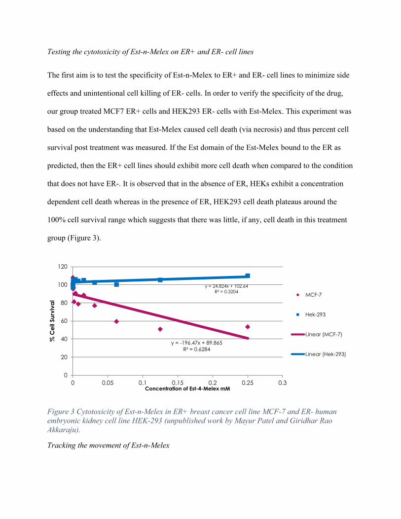
Testing the cytotoxicity of Est-n-Melex on ER+ and ER- cell lines
The first aim is to test the specificity of Est-n-Melex to ER+ and ER- cell lines to minimize side
effects and unintentional cell killing of ER- cells. In order to verify the specificity of the drug,
our group treated MCF7 ER+ cells and HEK293 ER- cells with Est-Melex. This experiment was
based on the understanding that Est-Melex caused cell death (via necrosis) and thus percent cell
survival post treatment was measured. If the Est domain of the Est-Melex bound to the ER as
predicted, then the ER+ cell lines should exhibit more cell death when compared to the condition
that does not have ER-. It is observed that in the absence of ER, HEKs exhibit a concentration
dependent cell death whereas in the presence of ER, HEK293 cell death plateaus around the
100% cell survival range which suggests that there was little, if any, cell death in this treatment
group (Figure 3).
Figure 3 Cytotoxicity of Est-n-Melex in ER+ breast cancer cell line MCF-7 and ER- human embryonic kidney cell line HEK-293 (unpublished work by Mayur Patel and Giridhar Rao Akkaraju).
Tracking the movement of Est-n-Melex
y = -196.47x + 89.865R² = 0.6284
y = 24.824x + 102.64R² = 0.3204
0
20
40
60
80
100
120
0 0.05 0.1 0.15 0.2 0.25 0.3
% C
ell S
urvi
val
Concentration of Est-4-Melex mM
MCF-7
Hek-293
Linear (MCF-7)
Linear (Hek-293)
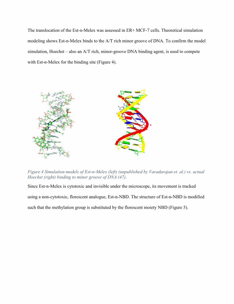
The translocation of the Est-n-Melex was assessed in ER+ MCF-7 cells. Theoretical simulation
modeling shows Est-n-Melex binds to the A/T rich minor groove of DNA. To confirm the model
simulation, Hoechst – also an A/T rich, minor-groove DNA binding agent, is used to compete
with Est-n-Melex for the binding site (Figure 4).
Figure 4 Simulation models of Est-n-Melex (left) (unpublished by Varadarajan et. al.) vs. actual Hoechst (right) binding to minor groove of DNA (47).
Since Est-n-Melex is cytotoxic and invisible under the microscope, its movement is tracked
using a non-cytotoxic, florescent analogue, Est-n-NBD. The structure of Est-n-NBD is modified
such that the methylation group is substituted by the florescent moiety NBD (Figure 5).
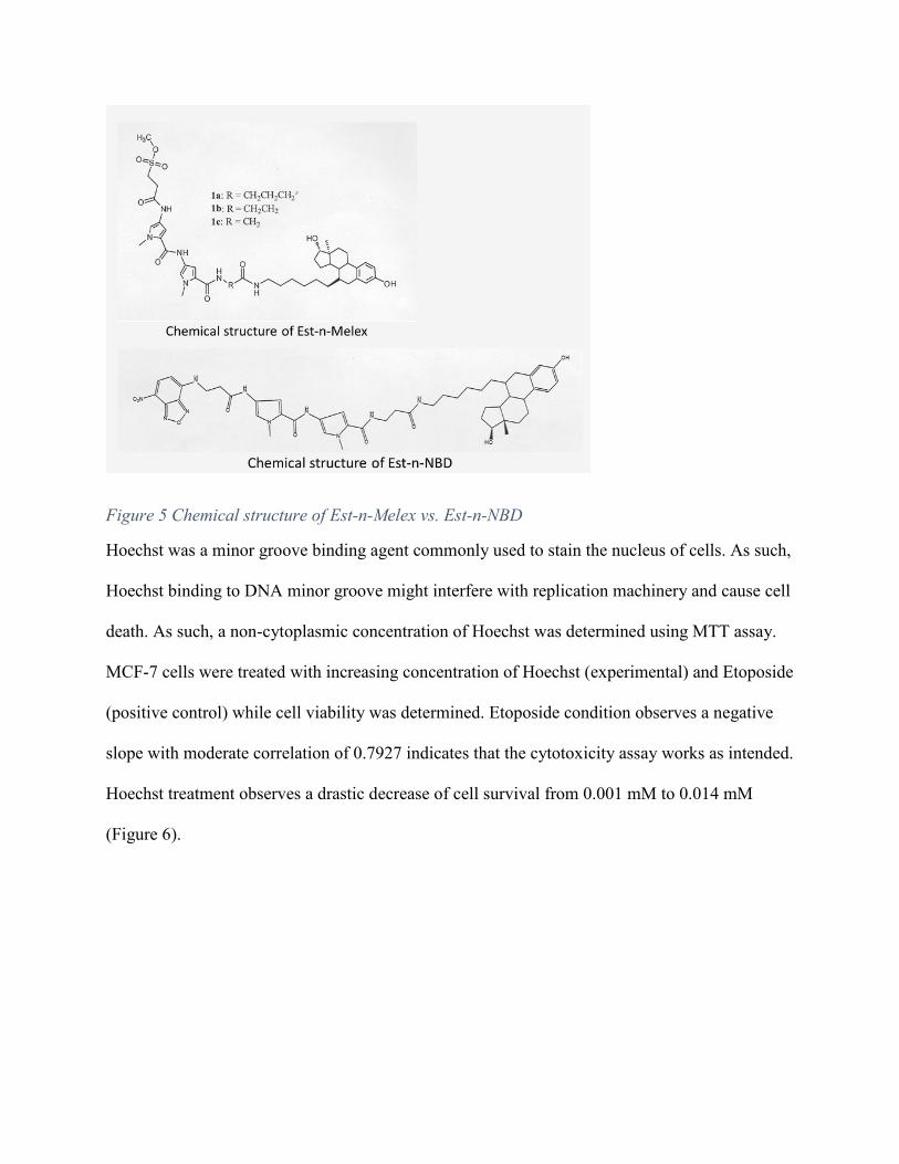
Figure 5 Chemical structure of Est-n-Melex vs. Est-n-NBD
Hoechst was a minor groove binding agent commonly used to stain the nucleus of cells. As such,
Hoechst binding to DNA minor groove might interfere with replication machinery and cause cell
death. As such, a non-cytoplasmic concentration of Hoechst was determined using MTT assay.
MCF-7 cells were treated with increasing concentration of Hoechst (experimental) and Etoposide
(positive control) while cell viability was determined. Etoposide condition observes a negative
slope with moderate correlation of 0.7927 indicates that the cytotoxicity assay works as intended.
Hoechst treatment observes a drastic decrease of cell survival from 0.001 mM to 0.014 mM
(Figure 6).
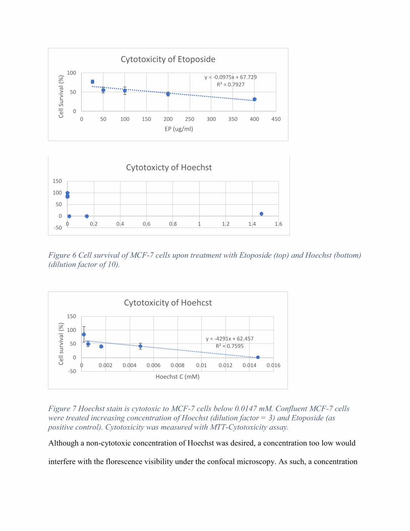
Figure 6 Cell survival of MCF-7 cells upon treatment with Etoposide (top) and Hoechst (bottom) (dilution factor of 10).
Figure 7 Hoechst stain is cytotoxic to MCF-7 cells below 0.0147 mM. Confluent MCF-7 cells were treated increasing concentration of Hoechst (dilution factor = 3) and Etoposide (as positive control). Cytotoxicity was measured with MTT-Cytotoxicity assay.
Although a non-cytotoxic concentration of Hoechst was desired, a concentration too low would
interfere with the florescence visibility under the confocal microscopy. As such, a concentration
y = -0.0975x + 67.729R² = 0.7927
0
50
100
0 50 100 150 200 250 300 350 400 450Cell
Surv
ival
(%)
EP (ug/ml)
Cytotoxicity of Etoposide
-50
0
50
100
150
0 0.2 0.4 0.6 0.8 1 1.2 1.4 1.6
Cytotoxicty of Hoechst
y = -4291x + 62.457R² = 0.7595
-50
0
50
100
150
0 0.002 0.004 0.006 0.008 0.01 0.012 0.014 0.016Cell
surv
ival
(%)
Hoechst C (mM)
Cytotoxicity of Hoehcst

of Hoechst that not only was non-cytotoxic but also provided strong florescent signals was
desired.
The range of non-cytotoxic concentration of Hoechst was further evaluated by measuring cell
death upon increasing concentration of Hoechst from the range of 0.001 to 0.014 mM (Figure 7).
Results indicated that concentration below 0.001633 was non-cytotoxic. The negative slope and
moderately strong R2 (0.7595) indicates that Hoechst was cytotoxic. But a non-cytotoxic
concentration was yet to be determined. So the cytotoxicity of Hoechst below 0.0147 mM was
studied (Figure 8).
Figure 8 Hoechst stain is not cytotoxic to MCF-7 cells below 0.001633 mM. Confluent MCF-7 cells were treated increasing concentration of Hoechst (dilution factor = 3). Cytotoxicity was measured with MTT-Cytotoxicity assay.
Results of cytotoxicity of Hoechst in concentration below 0.001633 mM shows an R2 = 0.11728
which indicates almost no correlation. This indicates that in these concentrations below 0.001633
mM, Hoechst do not play a cytotoxic role in MCF-7 cell lines. This experiment determines that a
y = -7485.9x + 87.241R² = 0.1173
0
20
40
60
80
100
120
140
0 0.0002 0.0004 0.0006 0.0008 0.001 0.0012 0.0014 0.0016 0.0018
Cell
surv
ival
(%)
Hoechst C (mM)
Cytotoxicity of Hoechst)
concentration below 0.001633 mM of Hoechst is non-cytotoxic to ER+ breast cancer cell line
MCF-7.
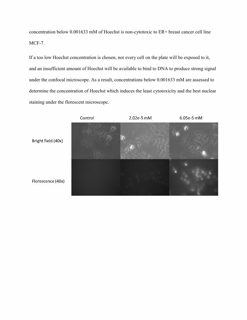
If a too low Hoechst concentration is chosen, not every cell on the plate will be exposed to it,
and an insufficient amount of Hoechst will be available to bind to DNA to produce strong signal
under the confocal microscope. As a result, concentrations below 0.001633 mM are assessed to
determine the concentration of Hoechst which induces the least cytotoxicity and the best nuclear
staining under the florescent microscope.
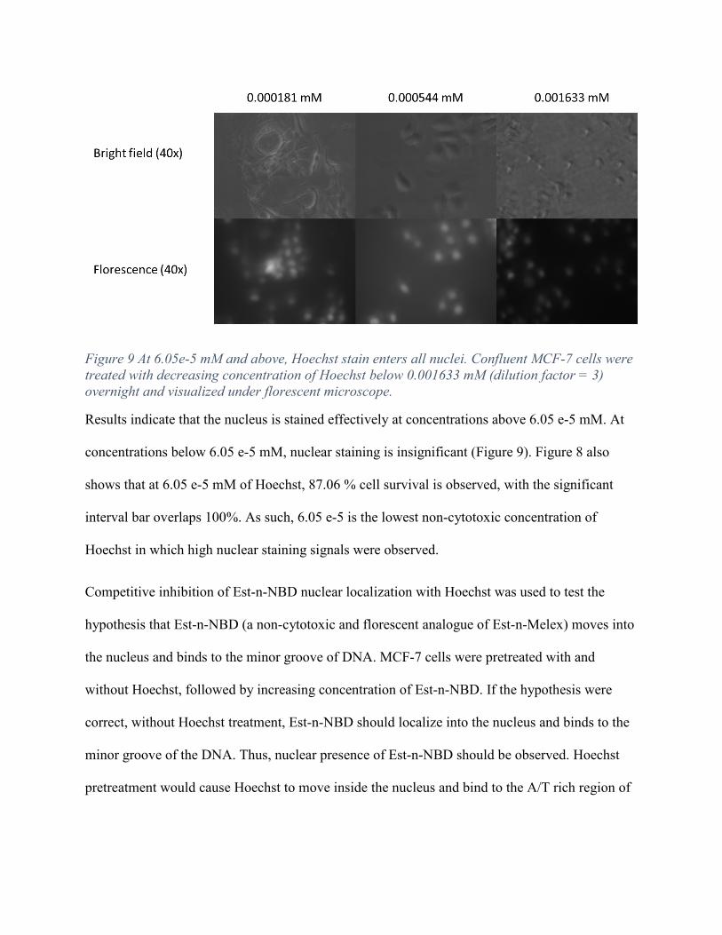
Figure 9 At 6.05e-5 mM and above, Hoechst stain enters all nuclei. Confluent MCF-7 cells were treated with decreasing concentration of Hoechst below 0.001633 mM (dilution factor = 3) overnight and visualized under florescent microscope.
Results indicate that the nucleus is stained effectively at concentrations above 6.05 e-5 mM. At
concentrations below 6.05 e-5 mM, nuclear staining is insignificant (Figure 9). Figure 8 also
shows that at 6.05 e-5 mM of Hoechst, 87.06 % cell survival is observed, with the significant
interval bar overlaps 100%. As such, 6.05 e-5 is the lowest non-cytotoxic concentration of
Hoechst in which high nuclear staining signals were observed.
Competitive inhibition of Est-n-NBD nuclear localization with Hoechst was used to test the
hypothesis that Est-n-NBD (a non-cytotoxic and florescent analogue of Est-n-Melex) moves into
the nucleus and binds to the minor groove of DNA. MCF-7 cells were pretreated with and
without Hoechst, followed by increasing concentration of Est-n-NBD. If the hypothesis were
correct, without Hoechst treatment, Est-n-NBD should localize into the nucleus and binds to the
minor groove of the DNA. Thus, nuclear presence of Est-n-NBD should be observed. Hoechst
pretreatment would cause Hoechst to move inside the nucleus and bind to the A/T rich region of

the DNA, occupying the active site of binding of subsequent Est-n-NBD. As such, Est-n-NBD
would remain cytosolic.
Figure 10 Est-n-NBD moves into the nucleus and binds to the minor groove of the DNA. MCF-7 cells are pretreated with 6.05 e-5 mM of Hoechst overnight followed by increasing concentration of 2.5 uM and 5.0 uM of Est-3-NBD. Images are captured with confocal microscopy. Composite images were created with ImageJ.
Without Hoechst treatment, Est-n-NBD was observed primarily in the nucleus. Hoechst
pretreatment overnight followed by 2.5 uM Est-n-NBD observes nuclear localization of Hoechst
but cytosolic presence of NBD. Increasing NBD concentration to 5.0 uM Est-n-NBD observes
more cytosolic presence of NBD, confirming that Hoechst and Est-n-NBD competes for the
minor groove of the DNA (Figure 10).
Tracking the localization of the target protein (Estrogen Receptors α)
Immunofluorescence study is used to track ER-α localization upon Est-n-Melex treatment. A
non-cytotoxic concentration of Est-n-Melex is determined before imaging was conducted in
order to preserve the cell morphology. Preliminary unpublished data from Mayur Patel and
Giridhar Rao indicated that Est-n-Melex is not cytotoxic (cell survival exceeds 80%) at
concentration below 15 nM (Figure 3). As such 15 nM of Est-n-Melex was chosen as the least
cytotoxic concentration for MCF-7 cells.
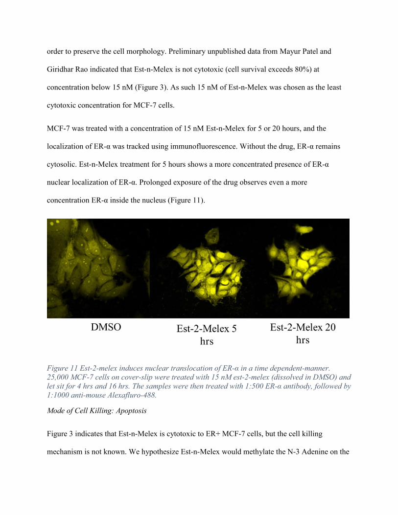
MCF-7 was treated with a concentration of 15 nM Est-n-Melex for 5 or 20 hours, and the
localization of ER-α was tracked using immunofluorescence. Without the drug, ER-α remains
cytosolic. Est-n-Melex treatment for 5 hours shows a more concentrated presence of ER-α
nuclear localization of ER-α. Prolonged exposure of the drug observes even a more
concentration ER-α inside the nucleus (Figure 11).
Figure 11 Est-2-melex induces nuclear translocation of ER-α in a time dependent-manner. 25,000 MCF-7 cells on cover-slip were treated with 15 nM est-2-melex (dissolved in DMSO) and let sit for 4 hrs and 16 hrs. The samples were then treated with 1:500 ER-α antibody, followed by 1:1000 anti-mouse Alexafluro-488.
Mode of Cell Killing: Apoptosis
Figure 3 indicates that Est-n-Melex is cytotoxic to ER+ MCF-7 cells, but the cell killing
mechanism is not known. We hypothesize Est-n-Melex would methylate the N-3 Adenine on the

A/T rich region of the minor groove of the DNA. When 3-MeA adducts are created, cells recruit
glycosylase to remove the methylated base, thus producing abasic sites. AP endonuclease are
then recruited to remove the DNA backbone of the abasic site, producing single-stranded ssDNA
breaks. We hypothesize the some ssDNA breaks cause cells to recruit PARP (Poly-ADP-Ribose
Polymerase), DNA repair enzymes that fix the DNA damage. Too many ssDNA breaks would
then over-recruit PARP, which activates pathways of cell killing, such as necrosis or apoptosis.
A common marker for apoptosis is the activation of caspases, enzymes that are cleaved and
activated when apoptosis is signaled. An increased caspase activity of caspases correlates to
apoptosis activity happening inside the cells. Caspase activity can be monitored using the
colorimetric Glomix caspase 3/7 assay. Activated caspase 3 or caspase 7 recognizes a specific
Z_DEVD sequence and cleaves the bond. The assay contains a substrate that upon cleavage of
such specific bond, produces light. The intensity of light can be captured and quantified using
luminescence plate reader.
Role of PARP in Est-n-Melex – induced DNA damage repair
We hypothesize that PARP is involved in the DNA repair mechanism induced by Est-n-Melex.
To test this hypothesis, a PARP-inhibitor 3-aminobenzamide (3AB) is co-treated with with Est-
n-Melex and apoptosis is quantified. If our hypothesis is correct, treating the cells with Est-n-
Melex should induce apoptosis and cause an upregulation of caspase 3/7 activity. Co-treating
Est-n-Melex and 3AB should prevent PARP from repair DNA damage, thus causes an
accumulation of 3-MeA adducts and upregulation of caspases.
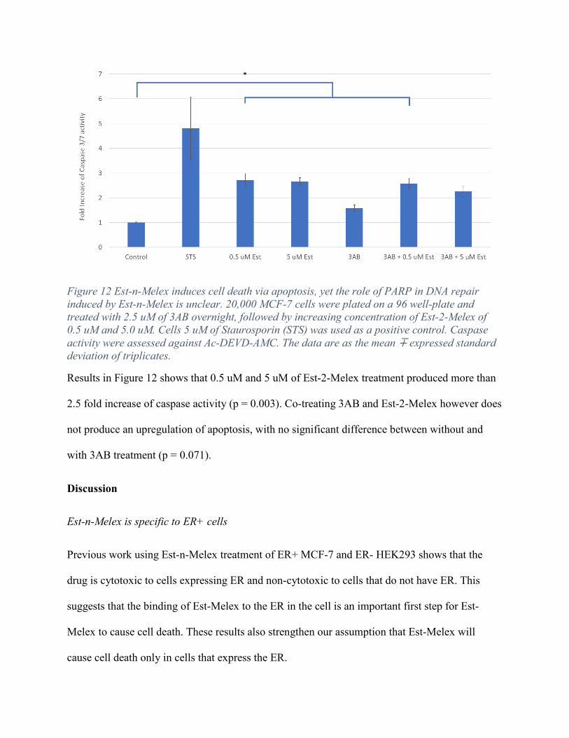
Figure 12 Est-n-Melex induces cell death via apoptosis, yet the role of PARP in DNA repair induced by Est-n-Melex is unclear. 20,000 MCF-7 cells were plated on a 96 well-plate and treated with 2.5 uM of 3AB overnight, followed by increasing concentration of Est-2-Melex of 0.5 uM and 5.0 uM. Cells 5 uM of Staurosporin (STS) was used as a positive control. Caspase activity were assessed against Ac-DEVD-AMC. The data are as the mean ∓ expressed standard deviation of triplicates.
Results in Figure 12 shows that 0.5 uM and 5 uM of Est-2-Melex treatment produced more than
2.5 fold increase of caspase activity (p = 0.003). Co-treating 3AB and Est-2-Melex however does
not produce an upregulation of apoptosis, with no significant difference between without and
with 3AB treatment (p = 0.071).
Discussion
Est-n-Melex is specific to ER+ cells
Previous work using Est-n-Melex treatment of ER+ MCF-7 and ER- HEK293 shows that the
drug is cytotoxic to cells expressing ER and non-cytotoxic to cells that do not have ER. This
suggests that the binding of Est-Melex to the ER in the cell is an important first step for Est-
Melex to cause cell death. These results also strengthen our assumption that Est-Melex will
cause cell death only in cells that express the ER.
Est-n-Melex localization and DNA binding
The absence of Hoechst, an A/T antagonist in the minor groove of the DNA, allows Est-n-NBD
to translocate into the nucleus. The presence of Hoechst reduces the ability of Melex to bind to
the minor groove (3-MeA). This positional change from nuclear concentration to cytoplasmic
concentration of Est-n-NBD, facilitated by Hoechst, suggests that Est-n-NBD does in fact bind to
minor groove A/T rich regions in the DNA. The dose-dependent cytoplasmic concentration of
Est-n-NBD shows that pretreatment of Hoechst competitively inhibits minor groove binding of
Est-n-NBD. The composite image clearly shows the dose-dependent property of Est-n-NBD in
binding to A/T rich region of the DNA.
Est-n-Melex induces ER-α nuclear localization
The presence of a non-cytotoxic concentration of Est-n-Melex allows ER-α to be activated and
move into the nucleus. This positional change from cytoplasmic concentration to nuclear
concentration, facilitated by Est-n-Melex, suggests that Est-n-Melex does cause ER-α to
translocate into the nucleus.
Identifying the mode of cell death: Apoptosis
Upregulation of caspase activity in the presence of Est-n-Melex suggests that apoptosis is
involved in the cell death mechanism of MCF-7 cells. It is also interesting to note that no dose-
dependent property of Est-n-Melex is observed as there is no significant difference in enzymatic
activity as the concentration of Est-n-Melex is increased from 0.5 µM to 5.0 µM. This means
that there might be a compensatory mechanism in place that protects rapid apoptotic activity.
The lack of a dose-dependent property may also indicate that the optimal condition for cell
killing is not found as well.
Role of PARP in DNA-repair mechanism induced by Est-n-Melex
One interesting observation is that there is no significant difference of apoptotic activity between
with and without 3AB pretreatment conditions. This means that either (1) PARP is not involved
at all in DNA repair mechanism induced by Est-n-Melex, or (2) 3AB is too weak of a PARP
inhibitor, or (3) an optimal condition for blocking PARP is yet to be found. Further research on
the optimal PARP blockage is needed to elucidate the role of PARP in DNA-repair induced by
Est-n-Melex.
Summary
In conclusion, the translocation mechanism of action of a novel chemotherapeutic compound that
can target ER+ breast cancer cells has been confirmed. This compound targets ER+ breast cancer
cells and recognizes and binds to ER-α, forming a complex that translocates into the nucleus.
Experiments are currently in progress to elaborate further the cell killing mechanism of the drug
as well as the role of poly ADP ribose polymerase (PARP) – a DNA repair enzyme – in repairing
DNA damage induced by Est-n-Melex. The successful elaboration of the translocation
mechanism of this compound demonstrates that this strategy can be used to synthesize new drugs
that can preferentially target any cell-type that over-expresses a unique receptor or enzyme.
Acknowledgements
Support of this research through grants from the Science and Engineering Research Center
SERC of Texas Christian University is gratefully acknowledged.
References
1. Kishton RJ, Miller SE, Perry H, Lynch T, Patel M, Gore VK, et al. DNA site-specific N3-
adenine methylation targeted to estrogen receptor-positive cells. Bioorganic & Medicinal
Chemistry. 2011;19(17):5093-102.
2. How Common Is Breast Cancer? [Internet].; 2019 [cited 4/20/2019]. Available from:
https://www.cancer.org/cancer/breast-cancer/about/how-common-is-breast-cancer.html.
3. Ali S, Coombes R. Estrogen Receptor Alpha in Human Breast Cancer: Occurrence and
Significance. J Mammary Gland Biol Neoplasia. 2000 Jul;5(3):271-81.
4. Subramanian A, Mokbel K. The role of Herceptin in early breast cancer. Int Semin Surg
Oncol. 2008 April 28;5:9.
5. Cuzick J, Sestak I, Cawthorn S, Hamed H, Holli K, Howell A, et al. Tamoxifen for prevention
of breast cancer: extended long-term follow-up of the IBIS-I breast cancer prevention trial.
Lancet Oncol. 2015 January 01;16(1):67-75.
6. Colleoni M, Giobbie-Hurder A. Benefits and adverse effects of endocrine therapy. Ann Oncol.
2010 October 01;21 Suppl 7:11.
7. Overview of the Breast [Internet].; 2019 [cited April 16, 2019]. Available from:
https://pathology.jhu.edu/breast/basics/overview.
8. Musarezaie A, Ghasemi TM, Esfahani HN. Investigation the quality of life and its relation
with clinical and demographic characteristics in women with breast cancer under
chemotherapy. Int J Prev Med. 2012 December 01;3(12):853-9.
9. - Does a rehabilitation program improve quality of life in breast cancer patients? - payeshj.
2010 -;- 9(- 1):61.
10. Ataollahi MR, Sharifi J, Paknahad MR, Paknahad A. Breast cancer and associated factors: a
review. J Med Life. 2015;8(Spec Iss 4):6-11.
11. American Cancer Society. Cancer Facts & Figures 2018. Atlanta: 2018.
12. Cancer Stat Facts: Female Breast Cancer [Internet]. [cited 4/20/2019]. Available from:
https://seer.cancer.gov/statfacts/html/breast.html.
13. Report: Breast Cancer Death Rates Down 39% Since 1989 [Internet].; 2017 [updated 3/10/;
cited 4/20/2019]. Available from: https://www.cancer.org/latest-news/report-breast-cancer-
death-rates-down-39-percent-since-1989.html.
14. American Cancer Society. Breast Cancer
Facts & Figures 2017-2018. Atlanta, GA: American Cancer Society Inc.; 2017.
15. Breast Cancer Signs and Symptoms [Internet].; 2017 [updated September; cited April 2019].
Available from: https://www.cancer.org/cancer/breast-cancer/about/breast-cancer-signs-and-
symptoms.html.
16. Hanf V, Hanf D. Reproduction and breast cancer risk. Breast Care (Basel). 2014 December
01;9(6):398-405.
17. Rebbeck TR. Inherited predisposition and breast cancer: modifiers of BRCA1/2-associated
breast cancer risk. Environ Mol Mutagen. 2002;39(2-3):228-34.
18. Sanderson M, O'Hara H, Foderingham N, Dupont WD, Shu XO, Peterson N, et al. Type 2
diabetes and mammographic breast density among underserved women. Cancer Causes
Control. 2015 February 01;26(2):303-9.
19. Acconcia F, Marino M. The Effects of 17beta-estradiol in Cancer are Mediated by Estrogen
Receptor Signaling at the Plasma Membrane. Front Physiol. 2011 June 30;2:30.
20. Bulun SE, Cheng YH, Pavone ME, Xue Q, Attar E, Trukhacheva E, et al. Estrogen receptor-
beta, estrogen receptor-alpha, and progesterone resistance in endometriosis. Semin Reprod
Med. 2010 January 01;28(1):36-43.
21. Guo L, Li F, Wang M, Xu Y, Wang B, Ran D, et al. 17beta-estradiol regulates the
malignancy of cancer stem-like cells derived from the MCF7 cell line partially through
Sox2. Oncol Lett. 2018 March 01;15(3):3790-5.

22. Tian JM, Ran B, Zhang CL, Yan DM, Li XH. Estrogen and progesterone promote breast
cancer cell proliferation by inducing cyclin G1 expression. Braz J Med Biol Res. 2018
January 23;51(3):1-7.
23. Oo PS, Yamaguchi Y, Sawaguchi A, Tin Htwe Kyaw M, Choijookhuu N, Noor Ali M, et al.
Estrogen Regulates Mitochondrial Morphology through Phosphorylation of Dynamin-
related Protein 1 in MCF7 Human Breast Cancer Cells. Acta Histochem Cytochem. 2018
February 27;51(1):21-31.
24. Drukteinis JS, Mooney BP, Flowers CI, Gatenby RA. Beyond mammography: new frontiers
in breast cancer screening. Am J Med. 2013 June 01;126(6):472-9.
25. American Cancer Society Guidelines for the Early Detection of Cancer [Internet].; 2018
[updated May 30,; cited 20 April, 2019]. Available from:
https://www.cancer.org/healthy/find-cancer-early/cancer-screening-guidelines/american-
cancer-society-guidelines-for-the-early-detection-of-cancer.html.
26. Morris E, Feig SA, Drexler M, Lehman C. Implications of Overdiagnosis: Impact on
Screening Mammography Practices. Popul Health Manag. 2015 September 01;18 Suppl 1:3.
27. Busonero C, Leone S, Klemm C, Acconcia F. A functional drug re-purposing screening
identifies carfilzomib as a drug preventing 17β-estradiol: ERα signaling and cell
proliferation in breast cancer cells. Molecular and Cellular Endocrinology. 2018 Jan
15,;460:229-37.
28. Sharma GN, Dave R, Sanadya J, Sharma P, Sharma KK. Various types and management of
breast cancer: an overview. J Adv Pharm Technol Res. 2010 April 01;1(2):109-26.
29. Remick J AN. Radiation Therapy, Breast Cancer, Postmastectomy. Treasure Island (FL):
StatPearls; 2018.
30. Breast Cancer Treatment (PDQ®)–Patient Version [Internet].; 2019 [updated April 12,; cited
April 16, 2019]. Available from: https://www.cancer.gov/types/breast/patient/breast-
treatment-pdq#section/_185.
31. Chatterjee A, Erban JK. Neoadjuvant therapy for treatment of breast cancer: the way
forward, or simply a convenient option for patients? Gland Surg. 2017 February
01;6(1):119-24.
32. Howell SJ, Johnston SRD, Howell A. The use of selective estrogen receptor modulators and
selective estrogen receptor down-regulators in breast cancer. Best Practice & Research
Clinical Endocrinology & Metabolism. 2004;18(1):47-66.
33. Martinkovich S, Shah D, Planey SL, Arnott JA. Selective estrogen receptor modulators:
tissue specificity and clinical utility. Clin Interv Aging. 2014 August 28;9:1437-52.
34. Oh Y, Müller HL, Ng L, Rosenfeld RG. Transforming Growth Factor- ß-induced Cell
Growth Inhibition in Human Breast Cancer Cells Is Mediated through Insulin-like Growth
Factor-binding Protein-3 Action. Journal of Biological Chemistry. 1995 June
09;270(23):13589-92.
35. Sporn MB LS, editor. Agents for Chemoprevention and Their Mechanism of Action. 6th ed.
Hamilton (ON): BC Decker; 2003.
36. Riggins RB, Schrecengost RS, Guerrero MS, Bouton AH. Pathways to tamoxifen resistance.
Cancer Lett. 2007 October 18;256(1):1-24.
37. Wang Q, Jiang J, Ying G, Xie XQ, Zhang X, Xu W, et al. Tamoxifen enhances stemness and
promotes metastasis of ERalpha36(+) breast cancer by upregulating ALDH1A1 in cancer
cells. Cell Res. 2018 March 01;28(3):336-58.
38. Wang H, Lu C, Tan Y, Xie J, Jiang J. Effect of adriamycin on BRCA1 and PARP-1
expression in MCF-7 breast cancer cells. Int J Clin Exp Pathol. 2014 August 15;7(9):5909-
15.
39. Carlson RW. The History and Mechanism of Action of Fulvestrant. Clinical Breast Cancer.
2005;6:S8.

40. Zhu L, Chen Y, Wei C, Yang X, Cheng J, Yang Z, et al. Anti-proliferative and pro-apoptotic
effects of cinobufagin on human breast cancer MCF-7 cells and its molecular mechanism.
Natural Product Research. 2018;32(4):493-7.
41. Marte Lorenzana-Jim&enez, Mar&ia Estela Avila, Alejandra Figueroa, Lorena Mendiola-
Almaraz, Gil Alfonso Magos Guerrero and Cristina Lemini. The Mexican mistletoe
Struthanthus venetus (HBK Blume) inhibits proliferation and synergizes antagonistic
actions of Tamoxifen and Fulvestrant in breast cancer MCF-7 cells. Journal of
Pharmacognosy and Phytotherapy. 2018;10(8).
42. Ao A, Morrison BJ, Wang H, Lopez JA, Reynolds BA, Lu J. Response of estrogen receptor-
positive breast cancer tumorspheres to antiestrogen treatments. PLoS One. 2011 April
14;6(4):e18810.
43. Rund D, Krichevsky S, Bar-Cohen S, Goldschmidt N, Kedmi M, Malik E, et al. Therapy-
related leukemia: clinical characteristics and analysis of new molecular risk factors in 96
adult patients. Leukemia. 2005;19:1919.
44. Zhang Y, Chen FX, Mehta P, Gold B. Groove- and sequence-selective alkylation of DNA by
sulfonate esters tethered to lexitropsins. Biochemistry (N Y ). 1993;32(31):7954-65.
45. David M. Tanenbaum, Yong Wang, Shawn P. Williams, Paul B. Sigler. Crystallographic
Comparison of the Estrogen and Progesterone Receptor's Ligand Binding Domains.
Proceedings of the National Academy of Sciences of the United States of America. 1998
May 26,;95(11):5998-6003.
46. Edwards KJ, Brown DG, Spink N, Skelly JV, Neidle S. Molecular structure of the B-DNA
dodecamer d(CGCAAATTTGCG)2. An examination of propeller twist and minor-groove
water structure at 2.2 A resolution. Journal of molecular biology. 1992 Aug
20,;226(4):1161.
47. Pjura PE, Grzeskowiak K, Dickerson RE. Binding of Hoechst 33258 to the minor groove of
B-DNA. J Mol Biol. 1987 Sep 20;197(2):257-71.
