electrical alternans in cardiac tamponade -...
TRANSCRIPT

Thorax (1975), 30, 228.
Electrical alternans in cardiac tamponadeANDREAS P. NIARCHOS
Royal Southern Hospital, Caryl Street, Liverpool 8
Niarchos, A. P. (1975). Thorax, 30, 228-233. Electrical alternans in cardiac tamponade.Of nine patients with pericardial effusion due to various causes, four developed cardiactamponade. Electrical alternans was present in all four, being total in three and ventri-cular in one. The ilternans corresponded very well with the clinical diagnosis of cardiactamponade and the radiological signs of a large pericardial effusion. In two patientsalternans was present even with heart rates below 100 per minute. Apart from theexact (1: 1) type of electrical alternans, three new types are described, a 2: 1, 3: 1, anda varying type. It is concluded that (a) electrical alternans associated with pericardialeffusion is strongly suggestive of impending or established cardiac tamponade, and (b)electrical alternans is produced when the heart is oscillating within the pericardial sac
distended by fluid with a frequency equal to one-half (exact alternans), one-third (2: 1alternans), and one-quarter (3: 1 alternans) of the heart rate.The aetiology and mechanism of electrical alternans are discussed.
Electrical alternans has been defined as analternation of the configuration of the electro-cardiographic complexes arising from the samepacemaker and being independent of periodicextracardiac phenomena (Spodick, 1962). Thiselectrocardiographic abnormality was initiallyobserved in the laboratory by Herring in 1909(Brody et al., 1973), and first reported clinicallythe year after by Lewis (1910). Other early re-ports were those of Hamburger, Katz, and Saphir(1936) and of Brody and Rossman (1937). Theliterature on the subject up to 1955 has been re-viewed by McGregor and Baskind (1955).Any electrocardiographic complex can exhibit
alternation, the P wave rarely (Kisch, 1949;Bernreiter, 1956), the QRS complex (McGregorand Baskind, 1955), the T wave alone (Dohertyand Hara, 1961; Kimura and Yoshida, 1963;Littmann, 1963; Dolara and Pozzi, 1971), or theU wave (Eyer, 1974). Finally, all main complexes(P-QRS-T) may alternate simultaneously whenthe phenomenon is called total electrical alternans(Littmann and Spodick, 1958; Brody et al., 1973).Although ventricular and total electrical alternansare not very common they are of clinical interestbecause they occur primarily in association withmassive pericardial effusion and cardiac tam-ponade (McGregor and Baskind, 1955; Colvin,1958; Littmann and Spodick, 1958; Spodick, 1962;Bashour and Cochran, 1963; Lawrence and
Cronin, 1963; Gabor, Winsberg, and Bloom, 1971;Usher and Popp, 1972; Brody et al., 1973).Two theories have been suggested to explain
the mechanism of electrical alternans. One attri-butes the alternation of the electrocardiographiccomplexes to alternating conduction within themyocardium (Spodick, 1962). The other maintainsthat the conduction within the myocardium duringalternans remains unchanged, but that a cyclicmotion of the heart within the distended peri-cardial sac accounts for the alternating electro-cardiographic pattern (Feigenbaum, Zaky, andGrabhorn, 1966; Price and Dennis, 1969; Usherand Popp, 1972; Brody et al., 1973). It is the pur-pose of this paper to report the clinical, radiologi-cal, and electrocardiographic findings in patientswith pericardial effusion and tamponade associ-ated with total and ventricular electrical alternans,in whom the confirmation of the mechanicaltheory of the genesis of alternans is based onelectrocardiographic data.
PATIENTS AND METHODS
Nine patients were studied, eight males and onefemale; their ages ranged from 36 to 72 years. Peri-cardial effusion with tamponade was diagnosed infour and pericardial effusion without tamponade infive. Clinical data are presented in Table I. All buttwo patients were treated in an intensive therapy unitunder continuous electrocardiographic monitoring.
228
on 7 July 2018 by guest. Protected by copyright.
http://thorax.bmj.com
/T
horax: first published as 10.1136/thx.30.2.228 on 1 April 1975. D
ownloaded from
Electrical alternans in cardiac tamponade
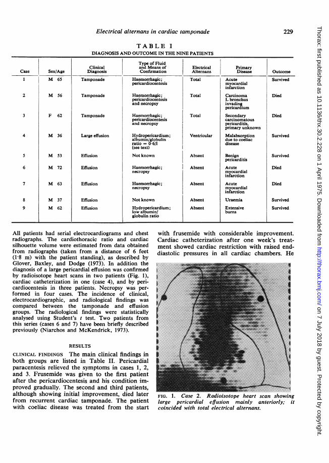
TABLE IDIAGNOSIS AND OUTCOME IN THE NINE PATIENTS
Type of FluidClinical and Means of Electrical Primary
Case Sex/Age Diagnosis Confirmation Alternans Disease Outcome
1 M 65 Tamponade Haemorrhagic; Total Acute Survivedpericardiocentesis myocardial
infarction
2 M 56 Tamponade Haemorrhagic; Total Carcinoma Diedpericardiocentesis L bronchusand necropsy invading
pericardium
3 F 62 Tamponade Haemorrhagic; Total Secondary Diedpericardiocentesis carcinomatousand necropsy pericarditis,
primary unknown
4 M 36 Large effusion Hydropericardium; Ventricular Malabsorption Survivedalbumin/globulin due to coeliacratio = 0 6/1 disease(see text) s
5 M 53 Effusion Not known Absent Benign Survivedpericarditis
6 M 72 Effusion Haemorrhagic; Absent Acute Diednecropsy myocardial
infarction
7 M 63 Effusion Haemorrhagic; Absent Acute Diednecropsy myocardial
infarction
8 M 37 Effusion Not known Absent Uraemia Survived
9 M 62 Effusion Hydropericardium; Absent Extensive Survivedlow albumin/ burnsglobulin ratio
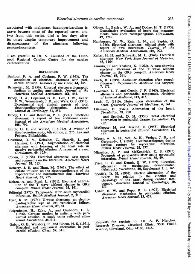
All patients had serial electrocardiograms and chestradiographs. The cardiothoracic ratio and cardiacsilhouette volume were estimated from data obtainedfrom radiographs (taken from a distance of 6 feet(1-8 m) with the patient standing), as described byGlover, Baxley, and Dodge (1973). In addition thediagnosis of a large pericardial effusion was confirmedby radioisotope heart scans in two patients (Fig. 1),cardiac catheterization in one (case 4), and by peri-cardiocentesis in three patients. Necropsy was per-formed in four cases. The incidence of clinical,electrocardiographic, and radiological findings wascompared between the tamponade and effusiongroups. The radiological findings were statisticallyanalysed using Student's t test. Two patients fromthis series (cases 6 and 7) have been briefly describedpreviously (Niarchos and McKendrick, 1973).
RESULTS
CLINICAL FINDINGS The main clinical findings inboth groups are listed in Table II. Pericardialparacentesis relieved the symptoms in cases 1, 2,and 3. Frusemide was given to the first patientafter the pericardiocentesis and his condition im-proved gradually. The second and third patients,although showing initial improvement, died laterfrom recurrent cardiac tamponade. The patientwith coeliac disease was treated from the start
with frusemide with considerable improvement.Cardiac catheterization after one week's treat-ment showed cardiac restriction with raised end-diastolic pressures in all cardiac chambers. He
FIG. 1. Case 2. Radioisotope heart scan showinglarge pericardial effusion mainly anteriorly; itcoincided with total electrical alternans.
229
on 7 July 2018 by guest. Protected by copyright.
http://thorax.bmj.com
/T
horax: first published as 10.1136/thx.30.2.228 on 1 April 1975. D
ownloaded from

A. P. Niarchos
TABLE IIMAIN CLINICAL FINDINGS IN THE NINE PATIENTS
Number of Patients
Findings Tamponade Effusion
Pulsus paradoxus 2 NilSinus arrhythmia 4 4Sinustachycardia(heartrate> 100/min) 2 4Raised JVP 4 4Hypotension (systolic BP < 100 mmHg) 4 1Dyspnoea 4 1Absent heart sounds 2 NilFaint heart sounds 2 1Pericardial friction rub 1 3Absent apex beat 4 -
was given a gluten-free diet and the pericardialeffusion disappeared radiographically when hisplasma proteins returned to normal.
ELECTROCARDIOGRAPHIC FINDINGS Sinus arrhyth-mia and generalized low voltage (that is, theamplitude of the R wave was less than 3-7 to16 6 mm in the various electrocardiographic leadsas described by Burch and Winsor (1972)) werepresent in all patients with cardiac tamponadeand in most patients with pericardial effusion.Electrical alternans, however, was present only inthe four patients with tamponade. The alternanswas total (P-QRS-T) in the first three cases (Figs2, 3, and 4) and ventricular (QRS) in the fourth.In the first case it was seen only in leads I, II, andaVF, in the second it was present in all leads,while in the third and fourth cases it was betterseen in lead V1. The alternans was not very con-stant in the fourth case. The alternating com-plexes varied in height from complex to complexeven in the same lead (Fig. 2), the difference be-ing greater in the right (V1, V2) and left (V6) chestleads. The electrical alternans disappeared incases 1, 2, and 3 after aspiration of 30, 65, and670 ml of pericardial fluid respectively (Figs 3and 4), and after treatment with frusemide in thefourth patient, but the tachycardia persisted incase 3 (Fig. 3). During reaccumulation of thepericardial effusion in cases 2 and 3 several typesof alternans were observed.The heart and alternans rate and the various
electrocardiographic types of alternans beforepericardial aspiration are shown in Table III. Aheart rate above 100 per minute was present intwo patients on three occasions, while in the restof the electrocardiograms the heart rate was below100 per minute; total alternans was present withboth fast and slow heart rates. The ratio betweenheart rate and alternans rate varied; it was always2: 1 when exact (1: 1) electrical alternans was
Lead
VI
Riqht Middlg Left....:;, _,, F
60.6bo lo0 4-0
(A11 7-0 9-0
8.0 4-0 8.0
5-0 40 50
3*5 .30 35.
Left Middle Right
45 20 3.0
FIG. 2. Case 2. Total electrical alternans (best seenin lead V0,) before pericardiocentesis; the alternansis 1: 1 (exact alternans); the numbers represent thelargest part of the QRS in millimetres, positive ornegative. See text for details.
H4eart raft o70 . 70 . 70 e 70 70 70 70. .7
Altermees rete' o 200 e 270Before pericerdioceetesis
v5 ~ ~ 7
After pericordioceeetesis
FIG. 3. Case 3. Heart rate and alternans rate ratiois nearly 3: 1 and 4: 1, but the type of total alternansis 2: 1 and 3: 1 respectively. The heart rate is 115per minute; after pericardiocentesis of 670 ml offluid the heart rate remains the same but thealternans has disappeared.
present (Figs 2 and 4), but 3 : 1 and 4: 1 when a2: 1 and 3 : 1 respectively alternans was present(Figs 3 and 5; Table IV). In addition a varyingtype of alternans was seen in case 2 soon beforethat patient's death; no constant relationshipcould be seen between the normal and alternatingcomplexes, and the altemating complexes differedgreatly from each other (Fig. 6). Conduction de-fects were not observed in the tamponade group.
230
on 7 July 2018 by guest. Protected by copyright.
http://thorax.bmj.com
/T
horax: first published as 10.1136/thx.30.2.228 on 1 April 1975. D
ownloaded from

Electrical alternans in cardiac tamponade23
TABLE LIIIHEART AND ALTERNANS RATE BEFORE AND AFTER PERICARDIOCENTESIS; VARIOUS TYPES OF ELECTRICAL
ALTERNANS ARE SHOWN
Before Pericardiocentesis AfterPericardiocentesis
Heart ~~~~~~~~~~~~ECGTypes of Electrical AlternansRate' Alternans Ratio Approx Ratio (normal/ Alternating Heart
Case (HR) Rate (AR) HR/AR Ratio HR/AR alternating QRS) Part Rate
1 94 55 1-7:1 2:1 1:1 Total 712 94 48 1-9:1 2:1 1:1 Total 832 88 21 4-3:1 4:1 3:1 Atrial2 83 27 3-0:1 3:1 2:1 Total3 115 41 2-8:1 3:1 2:1 Ventricular 1123 115 28 4-1:1 4:1 3:1 Total 1124'2 136 75 1-8:1 2:1 1:1 Ventricular 832 83 Varying Varying Varying Total
5LMarked sinus arrhythmia was present in all electrocardiograms.'No pericardiocentesis, patient treated with frusemide.
Hdeartrate. O.0 O .0 SO e 0
Before pkricdiocetesfis
After ForkeordioeeetesisFIG. 4. Case 2, lead V2 Total electrical alternans1 : 1 type (exact alternans). Heart and alternans rateis nearly 2: 1. Sinus arrhythmia is present. A fterpericardiocentesis of 65 ml of fluid the alternans hasdisappeared.
Lcod I
Letad Vs ;.. s
FIG. 5. Case 2. Recurrence of cardiac tamponade.Lead I, a 2: 1 electrical alternans is present; lead V5,3 :1 alternans is present.
Le*adVI
FIG. 6. Case 2. Varying electrical alternans.
The duration of the QRS did not vary greatly be-tween the normal and alternating complexes.
TABLE IVDURATION OF QRS (SECONDS) IN PATIENTS WITH
TAMPONADE BEFORE AND AFTER PERICARDIOCENTESIS
Before Pericardiocentesis AfterPericardio-centesis
Case Lead Normal Alternating NormalQRS QRS QRS
1 II 0-10 0-08 0062 I 0-08 0-10 -
2 V1 0-08 0.10 0-122 Vs 0-06 0-08 -
3 V1 0-08 0-08 0-084 V1 006 0-06 0-08
DISCUSSION
INCIDENCE AND AETIOLOGY OF ELECTRICAL ALTER-NANs The incidence of electrical alternans hasbeen estimated as varying between 1 in 1,212 and1 in 10,000 tracings (Hamburger et ail., 1936;Kalter and Schwartz, 1948). To date about 80cases, including the present four, have been re-ported in the literature (Brody and Rossman,1937; McGregor and Baskind, 1955; Colvin, 1958;Usher and Popp, 1972). In two-thirds of the re-ported cases the alternans was due to cardiactamponade caused by malignant haemopericar-dium or by massive pericardial effusion. Theclinical, radiological, and other laboratory findingsof the present study support the view that totalelectrical alternans is diagnostic of pericardialtamponade or massive effusion. The abnormalitydisappeared after aspiration of varying amountsof pericardial fluid, and this coincided with clinicalimprovements and reduction of the heart size onthe chest radiograph. To my knowledge, electri-cal alternans due to either cardiac tamponadeafter acute myocardial infarction or large peni-cardial effusion (hydropericardium) complicatinggluten-induced enteropathy has not previously
231
on 7 July 2018 by guest. Protected by copyright.
http://thorax.bmj.com
/T
horax: first published as 10.1136/thx.30.2.228 on 1 April 1975. D
ownloaded from

A. P. Niarchos
been described. Other causes are listed inTable V.
TABLE VAETIOLOGY OF ELECTRICAL ALTERNANS
Cardiac tamponade Malignant haemopericardiumMassive pericardial effusion due to:Tuberculous pericarditisSuppurative pericarditisIdiopathic pericarditisUraemiaAcute myocardial infarction
Constrictive pericarditisCongestive cardiac failure Rheumatic heart disease
Ischaemic heart diseaseMyocarditis
Hypertension ?Congestive cardiac failurePneumonectomyTension pneumothoraxl
'Niarchos, A. P., unpublished.
MECHANISM OF ELECTRICAL ALTERNANS The elec-
trocardiographic findings of this study do not sup-
port the aberrant conduction theory as themechanism for electrical alternans (Spodick,1962) since no conduction defects were observedand the duration of the alternating QRS com-plexes did not differ greatly from that of thenormals (Table IV). On the contrary, the presentelectrocardiographic findings lend support to thetheory which presumes exaggerated anatomicalmotion of the heart within the pericardial sac
enlarged by massive effusion. Indeed, several typesof such a cardiac motion have been demonstratedin patients with pericardial effusion and totalelectrical alternans by cineangiography (Priceand Dennis, 1969) and echocardiography (Feigen-baum et al., 1966; Usher and Popp, 1972), and byusing a laboratory model (Brody et al., 1973).With the last method both oscillatory and twistingcardiac motions with varying frequency, plane,and amplitude were observed.The variation in size of the alternating QRS
complexes as seen in the present cases can beexplained by accepting the view that the heartis oscillating within the distended pericardial sac
from left to right and vice versa (Fig. 3). Whenthe heart is close to the right chest leads V1 andV2 a large QRS (QRSr) is recorded; when in themiddle the recorded complex (QRSm) is smallbecause the heart is surrounded by fluid; and whenthe heart is close to the left chest wall the re-
corded QRS (QRSl) is larger than the QRSm,but smaller as compared with the QRSr because itis recorded from a distance. In lead V6 the oppo-site sequence of events takes place. In the middlechest leads (V3, V4, and V5) the QRSr is equal tothe QRSI because both are recorded from a more
or less equal distance from the middle. The varia-tion of the QRS size in the posterior leads only,as seen in case 1, may be explained on the samebasis but assuming that the heart is moving alongan anteroposterior plane. The observed variationin heart rate/alternans rate ratios 2: 1, 3: 1, and4: 1 (Table III) can be explained by assumingthat the frequency of the cardiac cyclic motionis one-half, one-third, and one-quarter respectivelyof the heart rate (Figs 3, 4, and 5). Likewise acardiac motion with varying frequency and planecould account for the varying alternans.The factors which probably determine the
frequency and form of the cardiac motion andhence the appearance of electrical alternans arethe heart rate, the pericardial pressure, the rateof accumulation, volume, and viscosity of thepericardial fluid, an aortic root fixed by secondarydeposits, the rigidity and configuration of thepericardial sac, and the mobility of the pendulum-like heart within the pericardial sac (Usher andPopp, 1972; Brody et al., 1973). A combination ofat least three factors is probably necessary to pro-duce the type of heart motion that is associatedwith electrical alternans, since the presence oftwo of them (Tables I and II) did not producealternans in the effusion group of patients. It hasbeen suggested that electrical alternans is presentonly when the heart rate is 100 per minute orgreater (Littman and Spodick, 1958), but this isnot confirmed in the present study as alternanswas present with heart rates below 100 per minute(Fig. 5), and it disappeared after pericardiocente-sis, although the heart rate remained above 100per minute (Fig. 3), as has been documented byothers (Usher and Popp, 1972; Cokkinos et al.,1974). The pericardial pressure probably does notplay an important role in the genesis of electricalalternans since bradyarrhythmias and not alter-nans have been reported to occur in acute cardiactamponade (Meurs et al., 1970; Editorial, 1972).
CLASSIFICATION AND PROGNOSIS It is clear fromthe present study that in addition to the well-known 1: 1 or exact alternans a 2: 1 and 3: 1 alter-nans exist, and both can be either total orventricular. Furthermore, a varying type ofelectrical alternans, characterized by markedvariation and completely irregular QRS alterna-tion, has been documented. All types of alternanswhen due to pericardial effusion should beconsidered an indication for pericardiocentesis,which may be a life-saving procedure. Theprognosis, however, of electrical alternans
232
on 7 July 2018 by guest. Protected by copyright.
http://thorax.bmj.com
/T
horax: first published as 10.1136/thx.30.2.228 on 1 April 1975. D
ownloaded from
Electrical alternans in cardiac tamponade
associated with malignant haemopericardium isgrave because most of the reported cases, andtwo from this series, died a few days afterits onset despite temporary improvement anddisappearance of the alternans followingpericardiocentesis.
I am grateful to Dr. N. Coulshed of the Liver-pool Regional Cardiac Centre for the cardiaccatheterization.
REFERENCES
Bashour, F. A. and Cochran, P. W. (1963). Theassociation of electrical alternans with peri-cardial effusion. Diseases of the Chest, 44, 146.
Bernreiter, M. (1956). Unusual electrocardiographicfindings in cardiac amyloidosis. Journal of theAmerican Medical Association, 161, 441.
Brody, D. A., Copeland, G. D., Cox, J. W., Keller,F. W., Wennemark, J. R., and Warr, 0. S. (1973).Experimental and clinical aspects of totalelectrocardiographic alternation. AmericanJournal of Cardiology, 31, 254.
Brody, J. G. and Rossman, P. L. (1937). Electricalalternans: a report of two additional cases.Journal of the American Medical Association,108, 799.
Burch, G. E. and Winsor, T. (1972). A Primer ofElectrocardiography, 6th edition, p. 276. Lea andFebiger, Philadelphia.
Cokkinos, D. V., Kelesides, C., Siaterlis, J., andHalimos, E. (1974). Augmentation of electricalalternans with lowering of the heart rate inmassive pericardial effusion. A report of a case.Circulation, 49, 1254.
Colvin, J. (1958). Electrical alternans: case reportand comments on the literature. American HeartJournal, 55, 513.
Doherty, J. E. and Hara, M. (1961). The effect ofcitrate infusion on the electrocardiogram of thehypothermic and normothermic dog. AmericanHeart Journal, 61, 225.
Dolara, A. and Pozzi, L. (1971). Electrical alterna-tion of the T wave without change in QRScomplex. British Heart Journal, 33, 161.
Editorial (1972). Tamponade after acute myocardialinfarction. British Medical Journal, 2, 310.
Eyer, K. M. (1974). U-wave alternans: an electro-cardiographic sign of left ventricular failure.American Heart Journal, 87, 41.
Feigenbaum, H., Zaky, A., and Grabhorn, L. L.(1966). Cardiac motion in patients with peri-cardial effusion. A study using reflected ultra-sound. Circulation, 34, 611.
Gabor, G. E., Winsberg, F., and Bloom, H. S. (1971).Electrical and mechanical alternation in peri-cardial effusion. Chest, 59, 341.
Glover, L., Baxley, W. A., and Dodge, H. T. (1973).Quantitative evaluation of heart size measure-ments from chest roentgenograms. Circulation,47, 1289.
Hamburger, W. W., Katz, L. N., and Saphir, 0.(1936). Electrical alternans: clinical study withreport of two necropsies. Journal of theAmerican Medical Association, 106, 902.
Kalter, H. H. and Schwartz, M. L. (1948). Electricalalternans. New York State Journal of Medicine,48, 1164.
Kimura, E. and Yoshida, K. (1963). A case showingelectrical alternans of the T wave withoutchange in the QRS complex. American HeartJournal, 65, 391.
Kisch, B. (1949). Auricular alteration after propyl-thiouracil. Experimental Medicine and Surgery,7, 173.
Lawrence, L. T. and Cronin, J. F. (1963). Electricalalternans and pericardial tamponade. Archivesof Internal Medicine, 112, 415.
Lewis, T. (1910). Notes upon alternation of theheart. Quarterly Journal of Medicine, 4, 141.
Littmann, D. (1963). Alternation of the heart.Circulation, 27, 280.and Spodick, D. H. (1958). Total electrical
alternation in pericardial disease. Circulation, 17,912.
McGregor, M. and Baskind, E. (1955). Electricalalternans in pericardial effusion. Circulation, 11,837.
Meurs, A. A. H., Vos, A. K., Verhey, J. B., andGerbrandy, J. (1970). Electrocardiogram duringcardiac rupture by myocardial infarction.British Heart Journal, 32, 232.
Niarchos, A. P. and McKendrick, C. S. (1973).Prognosis of pericarditis after acute myocardialinfarction. British Heart Journal, 35, 49.
Price, E. C. and Dennis, E. W. (1969). Electricalalternans: its mechanism demonstrated.(Abstract.) Circulation, 40, Supplement 3, p. 165.
Spodick, D. H. (1962). Electric alternation of theheart: its relation to the kinetics andphysiology of the heart during cardiac tam-ponade. American Journal of Cardiology, 10,155.
Usher, B. W. and Popp, R. L. (1972). Electricalalternans: mechanism in pericardial effusion.American Heart Journal, 83, 459.
Requests for reprints to: Dr. A. P. Niarchos,Research Division, Cleveland Clinic, 9500 EuclidAvenue, Cleveland, Ohio 44106, USA.
233
on 7 July 2018 by guest. Protected by copyright.
http://thorax.bmj.com
/T
horax: first published as 10.1136/thx.30.2.228 on 1 April 1975. D
ownloaded from