electromagnetic radiation causes cancer: the implications
TRANSCRIPT
Electromagnetic Radiation causes cancer: the implications for breast
cancer.
Dr Neil Cherry O.N.Z.M. Associate Professor* of Environmental Health
World Conference on Breast Cancer Ottawa, Canada, 26-31 July 1999
© Dr Neil Cherry 2002-2005
Human Sciences Department P.O. Box 84
Lincoln University Canterbury, New Zealand
* Associate Professor N.Z. = Full Professor U.S.
O.N.Z.M: Royal honour: Officer of the New Zealand Order of Merit

1
Electromagnetic Radiation causes cancer: the implications for breast cancer.
World Conference on Breast Cancer Ottawa, Canada, 26-31 July 1999
Dr Neil Cherry O.N.Z.M.
Lincoln University Canterbury, New Zealand
Abstract Breast cancer is a serious problem for women and also a risk for men. In assessing the risk of breast cancer associated with exposure to electromagnetic fields and radiation (EMF & EMR) this review approaches the problem primarily from both the whole-body point of view. Our minds control many body functions through the central nervous system and through mediating a wide range of hormones. This includes melatonin, which is a highly potent free radical scavenger. Hence melatonin protects cells from cancer and it strengthens the immune system. EMR is shown to influence the brain, reduce the output of key hormones (e.g. Melatonin and Thyrotropin), and to impair the immune system. Thus EMR is carcinogenic. Alteration of cellular calcium ions is a well-established biological effect of EMR exposure. Calcium ion influx is associated with the survival of damaged cells, and thus increases cancer risk. Calcium ion efflux is associated with enhanced cell death (apoptosis) of damaged cells, and hence enhances neurodegenerative diseases. Calcium ion efflux is also related to impairment of the immune system, and to alteration of reaction times and brain EEG rhythms. German research has proven that human brains detect and use the Schumann Resonances (SR) for timing synchronization. Altering the intensity and frequency of the SR changes human reaction times and circadian rhythms. A large body of research shows that there is an optimal intensity of SR, with increases and decreases in natural EMR being associated with a wide range of adverse neurological and cardiac health effects, and breast cancer. This research proves that sensitive and vulnerable human beings are made ill and can die when the natural EMR changes. The mean ELF intensity level of the SR is about 0.1 pW/cm2. This is 2 billion times lower than internationally recommended public health guidelines for ELF exposure. Cell line and animal exposure experiments, and epidemiological studies of populations who are occupationally and residentially exposed to EMR reveal significant hormonal, neurological, cardiac and cancer effects. A series of laboratory experiments involving breast cancer prone rats exposed to 50/60Hz exposure down to 0.1µT EMF, produced dose response relationships with the size and number of mammary tumors, with melatonin reduction and with reduced T lymphocytes. They also show a significant increase in proto oncogene activity. Occupational and residential studies of human populations show significant increases in breast cancer across the EMR spectrum, especially for pre-menopausal women and for positive estrogen receptor breast cancer. Since cell phones pose a very high risk it is recommended that all EMR exposures, especially cell phone exposure, be minimized.

2
1. Cancer development and the role of EMR Electromagnetic radiation (EMR) across the spectrum, from extremely low frequency (ELF, 50/60 Hz) to radiofrequency and microwave (RF/MW) causes breast cancer. This conclusion is contrary to "official" and industry authorities but it is based on sound scientific research. 1.1 Cancer is a multi-stage process: Cancer is the result of a cell's acquiring multiple genetic abnormalities. In addition to the conversion of proto oncogenes into oncogenes, cancer production often involves the loss of a functional tumor suppression gene. About half of all human cancers are associated with loss of the p53 tumor suppressor gene. The p53 gene product has complex effects, including the stimulation of DNA repair mechanisms, but, most critically, it somehow controls the transition from the G1 to S phase of the cell cycle and thus is involved in the prevention of uncontrolled cell division, Elliott and Elliot (1996). Cancer development is understood to be a multi-stage process that involves initiation, promotion and progression. The time taken to progress from the initial cell damage to a malignant tumor can take many years and involves many processes, Figure 1
Figure 1: A multi-stage model of cancer involving initiation, promotion and progression.
Initiation results from a single exposure to a carcinogen that damages the nuclear DNA. Promotion involves multiple exposures to agents that do not damage DNA directly. Promotion leads to conversion from benign to malignant tumors, with progression increasing the degree of malignancy, from Adey (1992).
The greater the frequency and intensity of genetic damage the greater the risk of the cell being transformed into a neoplastic (cancer) cell. Genetic damage increases if more damaging events occur, with more free radicals for example, or if the repair mechanisms are inhibited. "Repair" includes the destruction and elimination of damaged cells so that they can't produce neoplastic progeny. Within the cell cycle damaged DNA can be detected which usually leads to programmed cell death (apoptosis). Suppression of the p53 gene, conversion of the proto oncogenes into oncogenes and elevated concentrations of intracellular calcium ions can all make the survival of a damaged cell more likely. Hence factors which cause any of these factors to occur are carcinogenic. The genes are contained in the DNA and are incorporated into the chromosomes later in the cell cycle. Hence DNA strand breaks and chromosome aberrations are associated with enhanced apoptosis and increased cancer, depending on the circumstances of the
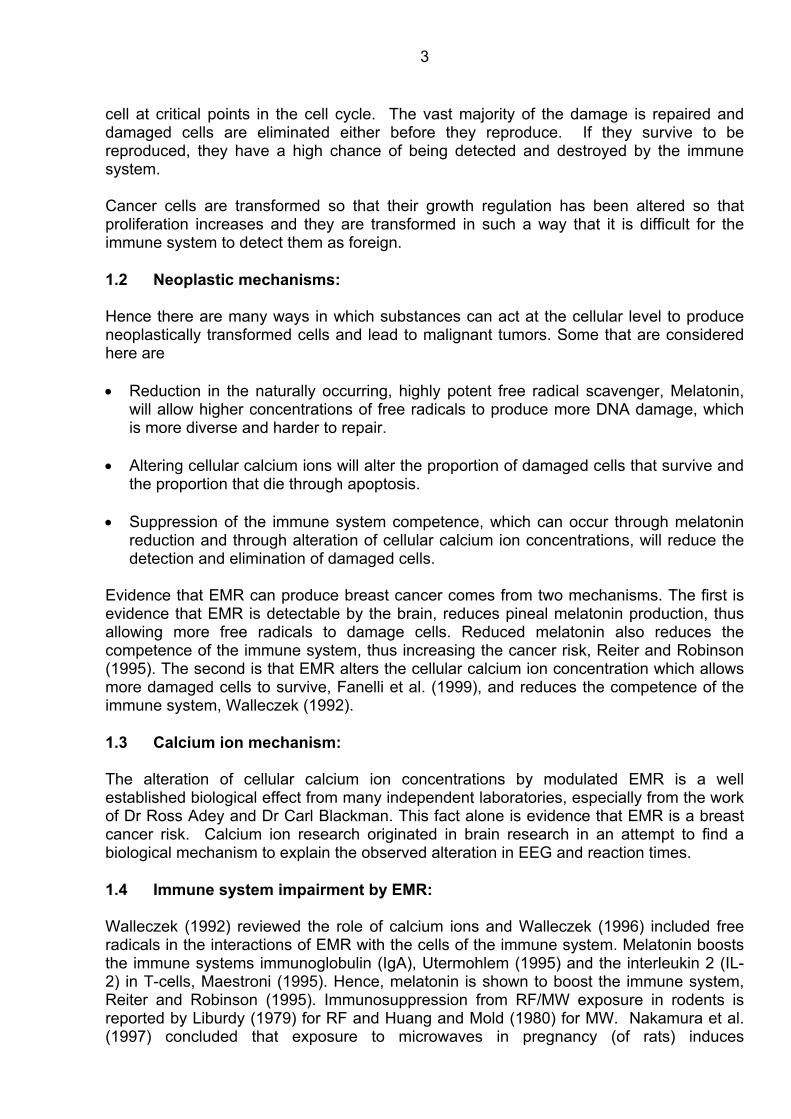
3
cell at critical points in the cell cycle. The vast majority of the damage is repaired and damaged cells are eliminated either before they reproduce. If they survive to be reproduced, they have a high chance of being detected and destroyed by the immune system. Cancer cells are transformed so that their growth regulation has been altered so that proliferation increases and they are transformed in such a way that it is difficult for the immune system to detect them as foreign. 1.2 Neoplastic mechanisms: Hence there are many ways in which substances can act at the cellular level to produce neoplastically transformed cells and lead to malignant tumors. Some that are considered here are • Reduction in the naturally occurring, highly potent free radical scavenger, Melatonin,
will allow higher concentrations of free radicals to produce more DNA damage, which is more diverse and harder to repair.
• Altering cellular calcium ions will alter the proportion of damaged cells that survive and
the proportion that die through apoptosis. • Suppression of the immune system competence, which can occur through melatonin
reduction and through alteration of cellular calcium ion concentrations, will reduce the detection and elimination of damaged cells.
Evidence that EMR can produce breast cancer comes from two mechanisms. The first is evidence that EMR is detectable by the brain, reduces pineal melatonin production, thus allowing more free radicals to damage cells. Reduced melatonin also reduces the competence of the immune system, thus increasing the cancer risk, Reiter and Robinson (1995). The second is that EMR alters the cellular calcium ion concentration which allows more damaged cells to survive, Fanelli et al. (1999), and reduces the competence of the immune system, Walleczek (1992). 1.3 Calcium ion mechanism: The alteration of cellular calcium ion concentrations by modulated EMR is a well established biological effect from many independent laboratories, especially from the work of Dr Ross Adey and Dr Carl Blackman. This fact alone is evidence that EMR is a breast cancer risk. Calcium ion research originated in brain research in an attempt to find a biological mechanism to explain the observed alteration in EEG and reaction times. 1.4 Immune system impairment by EMR: Walleczek (1992) reviewed the role of calcium ions and Walleczek (1996) included free radicals in the interactions of EMR with the cells of the immune system. Melatonin boosts the immune systems immunoglobulin (IgA), Utermohlem (1995) and the interleukin 2 (IL-2) in T-cells, Maestroni (1995). Hence, melatonin is shown to boost the immune system, Reiter and Robinson (1995). Immunosuppression from RF/MW exposure in rodents is reported by Liburdy (1979) for RF and Huang and Mold (1980) for MW. Nakamura et al. (1997) concluded that exposure to microwaves in pregnancy (of rats) induces

4
immunosuppression through a reduction in NK cells and activation of the hypothalmic-pituitary-adrenal axis. Quan et al. (1992) shows that compared to conventional heating of human breast milk, microwave heating significantly reduces the IgA for E. Coli bacteria. A series of papers on Polish workers in TV transmission and radar stations shows significant alteration of human immune system factors, including increased IgG and IgA and decreased lymphocytes and T8 cells, Moszczynski et al. (1999). On the other hand Dmoch and Moszczynski (1998) found for TV exposed workers, increased IgG, IgA, lymphocytes and T8 cells, and decreases natural killer cells (NK) and a lower T-helper/T-suppressor ratio. This concluding that different combinations of RF/MW signals produced different immune system responses. For more that 25 years several villages in the western region of Latvia have been subjected to pulse EMR from the Skrunda radar station. A group of 108 residents were randomly chosen from exposed villages and 61 from 2 non-exposed villages and their immune system factors were assayed. Bruvere at al. (1998) concluded that "immune system of residents exposed for 21 years to pulse radio-frequency electromagnetic radiation is affected." They showed elevated IgA levels a significantly impaired ability of peripheral blood leukocytes to produce interferons. At distances of 10 km or more from the radar, the mean exposure level was in the order of 1 pW/cm2. 1.5 Brain and hormone mechanisms: Our brain is a bioelectromagnetic organ that regulates the neural and neuroendocrine systems. There are three primary systems, the Dopamine, Opiate and Melatonin/Serotonin Systems. These systems are not independent but interact with each other. All have been shown to be affected by low intensity EMR, Frey (1995). The Dopamine and Opiate systems interact with the pineal system, mediating melatonin and regulating the immune system. Hence the brain and its systems form a central part of this review. Key evidence shows that EMR alters the brain electrical and biochemical activity, which leads directly through the neuroendocrine system to reduced melatonin and impairment of the immune systems, and therefore to higher cancer rates, including breast cancer. 1.6 Summary of Evidence and Conclusions: In this paper I will concentrate on the more direct evidence of human sensitively to EMR and the genetic damage and cancer increase which results from such exposures. The sequence of evidence that I will present will show that: • Our brains are exquisitely sensitive to naturally occurring EMR. • Melatonin is reduced in animals and humans under a wide range of exposures to EMR. • Cellular genetic damage to DNA and chromosomes has been documented in several
independent laboratories from ELF exposure to RF/MW exposure. • Chronic exposure of rodents to RF/MW radiation significantly increases chromosome
damage and cancer rates.

5
• Animals and humans are shown to have significantly impaired immune systems. • Military radio and radar radiation exposures, increases the rates of cancer in many
body organs. • Occupational and residential exposure across the spectrum is observed to increase
many cancers, most frequently, leukaemia and brain cancer. • EMR is shown to cause cancer. • EMR is shown to increase breast cancer rates in exposed populations. • Cell phone radiation poses a very strong breast cancer risk. 2. Human brains detect and react to natural electromagnetic signals 2.1 Schumann Resonances and Geomagnetic Activity Global lightning from thousands of thunder storms, radiates around the world at the speed of light, trapped in a resonant cavity created between the ionosphere and the earth. This resonance spectrum, called the Schumann Resonances (SR), has a fundamental frequency of 7.8 Hz because in 1 second light travels around the earth 7.8 times. Secondary resonance peaks occur at 14.1, 20.3, 26.4 and 32.5 Hz. As the earth rotates in its daily cycle the base of the ionosphere is higher during the night and lower during the day giving a localized daily modulation of the intensity of the Schumann Resonances, Figure 2. The Q-value of the cavity varies with the height of the ionosphere as it goes through the sunspot cycle and periods of Quiet Sun and Solar Storms. Hence all of these activities are reflected in changes to the intensity of the primary frequencies of the Schumann Resonances. Polk (1982) summarizes the electric field intensity range of the 8 - 21 Hz band as 0.06 to 0.34 mV/m/Hz-1/2, or 0.003 to 0.114pW/cm2. Figure 3 shows a 2 second segment of a recording of Schumann Resonances, taken by M.I.T. in June 1960, Balser and Wagner (1960). The whole 2 day record had an RMS amplitude of 0.55mV/m, which converts to 0.08pW/cm2. This is well within the range cited by Polk (1982).
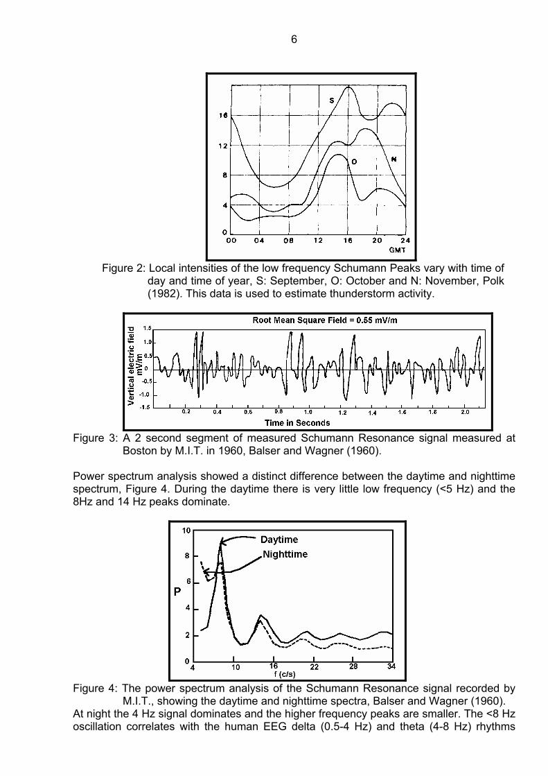
6
Figure 2: Local intensities of the low frequency Schumann Peaks vary with time of
day and time of year, S: September, O: October and N: November, Polk (1982). This data is used to estimate thunderstorm activity.
Figure 3: A 2 second segment of measured Schumann Resonance signal measured at
Boston by M.I.T. in 1960, Balser and Wagner (1960). Power spectrum analysis showed a distinct difference between the daytime and nighttime spectrum, Figure 4. During the daytime there is very little low frequency (<5 Hz) and the 8Hz and 14 Hz peaks dominate.
Figure 4: The power spectrum analysis of the Schumann Resonance signal recorded by
M.I.T., showing the daytime and nighttime spectra, Balser and Wagner (1960). At night the 4 Hz signal dominates and the higher frequency peaks are smaller. The <8 Hz oscillation correlates with the human EEG delta (0.5-4 Hz) and theta (4-8 Hz) rhythms

7
which is also dominant at night, while the human alpha (8-13 Hz) and beta (13-30 Hz) rhythms dominate during the daytime. This relationship and the close correspondence of the higher frequency peaks of both the EEG and the Schumann Resonances, Figure 5, strongly suggests that our brains have evolved to use the Schumann Resonances as environmental time-cues or Zeitgebers.
Figure 5: Comparison of the frequency spectra of the daytime human EEG and Schumann Resonances.
There is a slight diurnal variation in the frequency of the peaks. Polk (1982) gives an example of the 8Hz peak which has a maximum of about 7.9 Hz in the late morning (local time) and a minimum of about 7.3 Hz around 8 to 10 pm. 2.2 Human Reaction times and the EEG: If the Schumann Resonances are Zeitgebers that changes in the intensity of the Schumann Spectrum could well change human reaction times and circadian rhythms. Reaction times were investigated by König (1974), using the reaction times of about 50,000 people, 4,500 per half hour, who were tested at a display at the Munich Transport Exhibition in 1953, Figure 6.

8
Figure 6: Schumann resonance (8-14 Hz) speeds reaction times, König (1974).
Schumann Resonance intensity was highly correlated with speeding up the human reaction time, and 3 Hz sferics signals with slowing it down, Figure 7.
Figure 7: 3 Hz signals slow reaction times, König (1974).
To support the probable Zeitgeber biological mechanism König (1974) presents a comparison between the Schumann Resonance and the human EEG alpha rhythm and the 3 Hz sferics and the EEG delta rhythm, Figure 8.
9
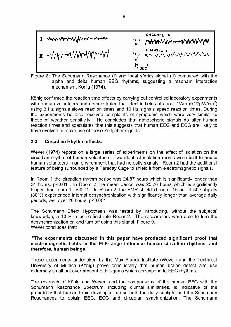
Figure 8: The Schumann Resonance (I) and local sferics signal (II) compared with the
alpha and delta human EEG rhythms, suggesting a resonant interaction mechanism, König (1974).
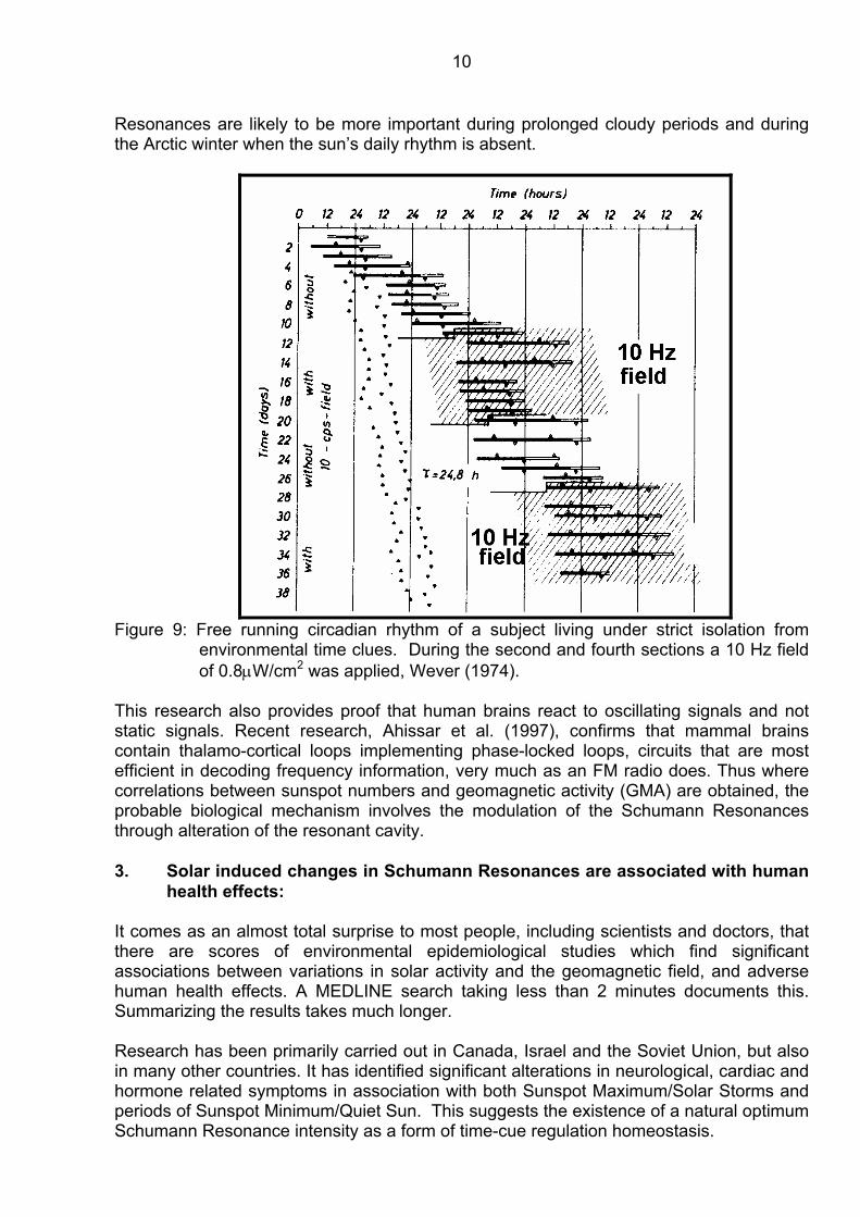
König confirmed the reaction time effects by carrying out controlled laboratory experiments with human volunteers and demonstrated that electric fields of about 1V/m (0.27µW/cm2) using 3 Hz signals slows reaction times and 10 Hz signals speed reaction times. During the experiments he also received complaints of symptoms which were very similar to those of weather sensitivity. He concludes that atmospheric signals do alter human reaction times and speculates that this suggests that human EEG and ECG are likely to have evolved to make use of these Zeitgeber signals. 2.3 Circadian Rhythm effects: Wever (1974) reports on a large series of experiments on the effect of isolation on the circadian rhythm of human volunteers. Two identical isolation rooms were built to house human volunteers in an environment that had no daily signals. Room 2 had the additional feature of being surrounded by a Faraday Cage to shield it from electromagnetic signals. In Room 1 the circadian rhythm period was 24.87 hours which is significantly longer than 24 hours, p<0.01 . In Room 2 the mean period was 25.26 hours which is significantly longer than room 1, p<0.01. In Room 2, the EMR shielded room, 15 out of 50 subjects (30%) experienced internal desynchronization with significantly longer than average daily periods, well over 26 hours, p<0.001 . The Schumann Effect Hypothesis was tested by introducing, without the subjects’ knowledge, a 10 Hz electric field into Room 2. The researchers were able to turn the desynchronization on and turn off using this signal, Figure 9. Wever concludes that: "The experiments discussed in this paper have produced significant proof that electromagnetic fields in the ELF-range influence human circadian rhythms, and therefore, human beings.” These experiments undertaken by the Max Planck Institute (Wever) and the Technical University of Munich (König) prove conclusively that human brains detect and use extremely small but ever present ELF signals which correspond to EEG rhythms. The research of König and Wever, and the comparisons of the human EEG with the Schumann Resonance Spectrum, including diurnal similarities, is indicative of the probability that human brain developed to use both the daily sunlight and the Schumann Resonances to obtain EEG, ECG and circadian synchronization. The Schumann
10
Resonances are likely to be more important during prolonged cloudy periods and during the Arctic winter when the sun’s daily rhythm is absent.
Figure 9: Free running circadian rhythm of a subject living under strict isolation from
environmental time clues. During the second and fourth sections a 10 Hz field of 0.8µW/cm2 was applied, Wever (1974).
This research also provides proof that human brains react to oscillating signals and not static signals. Recent research, Ahissar et al. (1997), confirms that mammal brains contain thalamo-cortical loops implementing phase-locked loops, circuits that are most efficient in decoding frequency information, very much as an FM radio does. Thus where correlations between sunspot numbers and geomagnetic activity (GMA) are obtained, the probable biological mechanism involves the modulation of the Schumann Resonances through alteration of the resonant cavity. 3. Solar induced changes in Schumann Resonances are associated with human
health effects: It comes as an almost total surprise to most people, including scientists and doctors, that there are scores of environmental epidemiological studies which find significant associations between variations in solar activity and the geomagnetic field, and adverse human health effects. A MEDLINE search taking less than 2 minutes documents this. Summarizing the results takes much longer. Research has been primarily carried out in Canada, Israel and the Soviet Union, but also in many other countries. It has identified significant alterations in neurological, cardiac and hormone related symptoms in association with both Sunspot Maximum/Solar Storms and periods of Sunspot Minimum/Quiet Sun. This suggests the existence of a natural optimum Schumann Resonance intensity as a form of time-cue regulation homeostasis.
11
Optimal Geomagnetic Field (GMF) variations for optimal brain function dynamics were identified by Belisheva et al. (1995). Svanidze et al. (1994) showed that both increased and decreased GMA retard the activity of the ciliary apparatus (cell movement in fluids) in the cells of new born rats. 3.1 Neurological effects 3.1.1 High GMA: Tambiev, Medvedev and Egorova (1995) found a significant correlation between GMF and memory and attention. EEG activity was positively correlated and EEG synchronization and negatively correlated with GMA, Belov, Kanunikov and Kiselev (1998). This was confirmed by Selitskii, Karlov and Sorokina (1999) who, by artificially reducing GMA, were able to increase the synchronization of alpha-rhythm EEG and generalized slow-wave discharges in epileptics. This shows that increased GMA produces de-synchronization of the EEG alpha-rhythm. Urgent hospitalization for mental disorders and suicide rise significantly with solar activity, Oraevskii et al. (1998). Psychiatric admissions increase with solar radio flux activity (p<0.05) and sudden magnetic storms (p<0.01) and decrease with GMA (p<0.05), Raps, Stoupel and Shimshoni (1991). Admission for Depression increases significantly after solar storms, Kay (1994). Migraine attack frequency increases with GMA, De Matteis et al. (1994). 3.1.2 Low GMA: • Highly anxious pilots have greater anxiety on days of quiet sun, Usenko (1992) and
aviation accidents are positively correlated with solar storms, Komarov et al. (1998). • Epileptic seizures and dizziness are correlated with GMA, Stoupel, Martfel and
Rotenberg (1991), Renton and Persinger (1998), and Rajaram and Mitra (1981). Persinger (1995) identified a significant correlation between GMA and sudden epileptic death.
• Conesa (1995, 1997) associated high GMA with isolated sleep paralysis, and low GMA
with vivid dreaming. • Serial Crime in Moscow increased 3 days after a low solar activity period. Avdonina
and Samovichev (1995). St Pierre and Persinger (1998) found that aggressive biting in rats was positively correlated with GMA.
• Tunyi and Tesarova (1991) found that suicide, sports injuries, fatal work injuries,
alcoholism are more prevalent during periods of low solar activity, 3.2 Cardiac Effects:

12
During periods of Active Sun and increased GMA the following statistically significant effects have been observed: • Increased blood coagulation and platelet aggregation, Pikin, Gurfinkel and Oraevskii
(1998). • Red blood cell aggregation and slowing of capillary flow in patients with Ischemic heart
diseases and an increase in arterial blood pressure, Ghione et al. (1998) • Cardiac Arrhythmia in children, Markariv (1998). • Heart Attack, Villoresi et. al. (1998), Sudden cardiovascular death, Sitar (1990) Quiet Sun periods are significantly associated with: • Sudden death from cardiac arrhythmia, especially paroxymal atrial fibrillation, and
stroke, Stoupel (1993) and Stoupel et al. (1995a). Stoupel, Martfel and Rotenberg (1994)
• Ischaemic Heart Disease and suicide for ages >70 years. Stoupel et al. (1995b). Stoupel, Martfel and Rotenberg (1994) conclude that these results are consistent with previous studies showing increased heart electrical instability during periods of lowest geomagnetic activity. 3.3 Hormone effects Seasonal daylength high stability and low geomagnetic activity were correlated with peak melatonin levels, and vice versa, Bergiannaki, Paparrigopoulos and Stefanis (1996). Thyroxine highly correlated with GMA supports the melatonin reduction hypothesis, O’Connor and Persinger (1996). Serotonin increases with low magnetic field, Babych (1996). Reiter and Robinson report a relationship between infants with small pineal glands and Sudden Infant Death Syndrome (SIDS) based on the work of Dr Larry Sparks with p<0.0001 (Sparks pers. Comm.) and Sparks and Hunsaker (1988). Hence O’Connor and Persinger (1997), tested the hypothesis of melatonin reduction from GMA and identified a significant correlation (r=0.90) between SIDS and GMA. Increased GMA decreases melatonin and increases serotonin, whereas decreased GMA increases melatonin and decreases serotonin. 3.4 Other effects: Zaitseva and Pudovkin (1995) found over a 30 year period that mortality rate went up (r=0.78) and birth rate went down (r=-0.67) in association with a geomagnetic index (Kp). Stoupel et al. (1993) found that Intraocular pressure in relation to four levels of daily GMA was lowest on stormy days and highest on quiet solar days. 3.5 Breast Cancer

13
Riabykh and Bodrova (1992) found a significant correlation between sunspot activity and pre-cancerous breast disease. 3.6 Summary and Conclusion: These is a large body of research identifying adverse human health effects for both high and low solar and geomagnetic activity, particularly for neurological and cardiac illness and death associated with arrhythmia and the loss of timing control homeostasis. This confirms that human beings have evolved to use a mean intensity of a timing signal, the Schumann Resonances. When the intensity of these signals is altered by changing the Q-value of the resonant cavity from day to night, with Solar Storms, period of Quiet Sun, Sunspot Maximum and Minimum, then adverse health effects are seen in large populations amongst those who are significantly compromised already. Thus it is established that human beings detect and are sensitive to changes in, extremely small, low frequency, naturally occurring electromagnetic signals, and hence to extremely low levels of non-ionizing electromagnetic radiation. 4. The higher the RF/MW carrier frequency the greater the induced field in
tissue: A vital biophysical principle is that low frequency carriers penetrate tissue far less, with far faster absorption than higher frequency carriers. This is shown in the figure below for calcium ion efflux, a well established biological effect of ELF modulated EMR.
Figure 10: Calcium ion efflux from a 16 Hz ELF field of 56 V/m produces a
significant influx with a tissue gradient of 10-7 V/cm. The upper graph is for a 56 V/m (0.8mW/cm2) 147 MHz carrier which produces a tissue gradient of 10-1V/cm, Adey (1981).
Adey (1981) shows that the 147 MHz carrier produces a tissue gradient a million times higher than the ELF signal. From the basic information about how the dielectric constant of tissue carries with carrier frequency, from Schwan and Foster (1980), Viganti and Giuliani (1997) calculated the relative current which will be produced in muscle tissue for a unit power density input, as a function of carrier frequency, Figure 11.
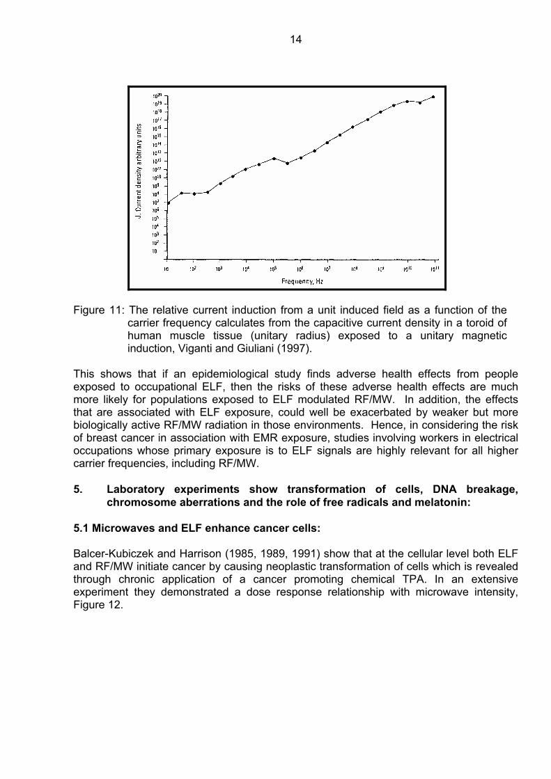
14
Figure 11: The relative current induction from a unit induced field as a function of the carrier frequency calculates from the capacitive current density in a toroid of human muscle tissue (unitary radius) exposed to a unitary magnetic induction, Viganti and Giuliani (1997).
This shows that if an epidemiological study finds adverse health effects from people exposed to occupational ELF, then the risks of these adverse health effects are much more likely for populations exposed to ELF modulated RF/MW. In addition, the effects that are associated with ELF exposure, could well be exacerbated by weaker but more biologically active RF/MW radiation in those environments. Hence, in considering the risk of breast cancer in association with EMR exposure, studies involving workers in electrical occupations whose primary exposure is to ELF signals are highly relevant for all higher carrier frequencies, including RF/MW.
5. Laboratory experiments show transformation of cells, DNA breakage,
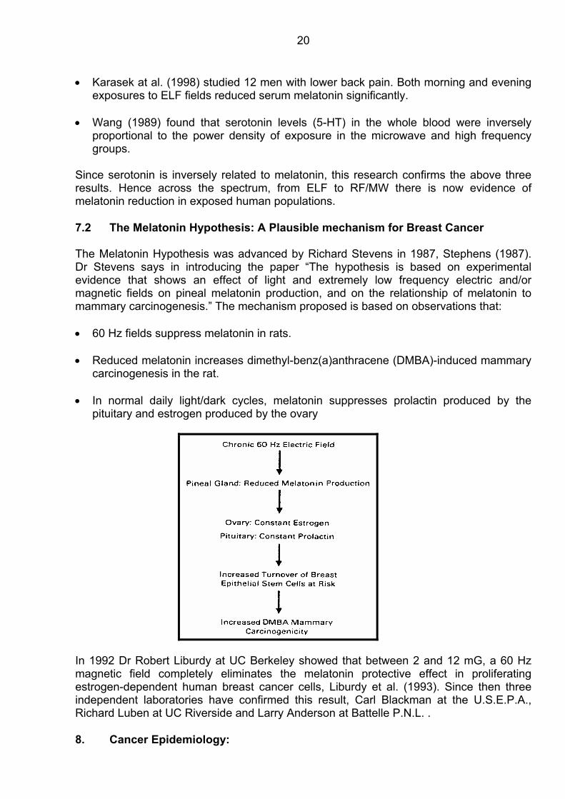
chromosome aberrations and the role of free radicals and melatonin: 5.1 Microwaves and ELF enhance cancer cells: Balcer-Kubiczek and Harrison (1985, 1989, 1991) show that at the cellular level both ELF and RF/MW initiate cancer by causing neoplastic transformation of cells which is revealed through chronic application of a cancer promoting chemical TPA. In an extensive experiment they demonstrated a dose response relationship with microwave intensity, Figure 12.

15
Figure 12: Dose response relationship for induction of neoplastic transformation in
C3H/10T1/2 cells by a 24 h exposure to 2.45 GHz microwaves at a specific absorption rate indicated on the abscissa, with or without TPA post-treatment for 8 weeks, Balcer-Kubiczek and Harrison (1991).
Figure 13: Induction of neoplastic transformation in C3H/10T1/2 cells by a 24 h
exposure to a 60Hz, 200µT magnetic field and a 0.5 Gy Xray, with or without TPA post-treatment for 8 weeks, Balcer-Kubiczek and Harrison (1985).
5.2 DNA Strand Breaks: Lai and Singh (1995, 1996 and 1997) have shown that RF/MW causes single and double-strand DNA breaks in a dose response manner. Their latest paper has shown that free radicals are involved and that melatonin removes the DNA damage. Two other laboratories have also shown that EMR in the form of cell phone signals also breaks DNA, Verschaeve et al. (1994) and Phillips et al. (1998). Four independent laboratories have published papers reporting that 50/60 Hz signals also cause DNA strand breaks, Lai and Singh (1997), Svedenstal et al. (1998), Phillips et al. (1998) and Ahuja et al. (1997). 5.3 Chromosome Aberrations: As long ago as 1959 Heller and Teixeira-Pinto used a 5 minute exposure to a pulsed 27 MHz RF signal to produce controlled breakage of chromosomes in garlic roots. They state

16
that they found chromosome aberrations that mimicked those produced by ionizing radiation and c-mitogenic substances. They say:
“this led us to believe that this force might be used as a powerful and controlled mutagenic agent”.
They conclude:
“The effects noted [of pulsed RF radiation] mimicked those produced by ionizing radiation and c-mitotic substances.”
Since 1959 we have known that RF radiation “mimicked ionizing radiation” in its mutagenic effects. Subsequent research continually re-affirms this and extends the results as more sophisticated techniques become available. Chromosome damage in RF/MW exposed animals cells has been reported by Yao (1978 & 1982), Alam et al. (1978), Antipenko et al. (1984), Garaj-Vrhovac et al. (1991), Koveshnikova and Antipenko (1991a and b) and Timchenko and Ianchevskaia (1995). Chromosome damage in RF/MW human tissues and cells has been documented by Garaj-Vrhovac et al. (1990b and 1992), Maes et al. (1993) and in human beings by, Jacobson (1969), and Garaj-Vrhovac et al. (1993). Nordensson et al. (1994) found significant chromosome aberrations in human amniotic cells after intermittent exposure to 30 µT 50 Hz fields. Garaj-Vrhovac et al. (1993) analyzed the structural chromosome aberrations in a group of radar station personnel who were engaged in repairing radar devices a couple of days earlier. This follows the authors’ repeated observations that RF/MW does cause chromosome aberrations. This paper is sought to identify the chromosome repair recovery rate after radar has damaged chromosomes in living human beings. Initial chromosome damage varied from 3 % to 33 %. Subjects were monitored for 30 weeks of follow-up. In every case the number of chromosome aberrations declined over time. Figure 14 shows the recovery curve for subject 5 who had an initial breakage rate of 33 %.
Figure 14: The time-dependent decrease in the number of chromosomal aberrations
for subjects with extremely high number of chromosomal impairments, Garaj-Vrhovac et al. (1993).

17
The authors conclude that after 30 weeks persons with a large number of chromosomal aberrations show a decrease from 33 % to 3.5 %, with unstable aberrations still present. They also state:
“According to other authors, consequences of these mutations are related to neoplasms. Out of 29 epidemiological studies published in the last decade, 22 suggest a relationship between various neoplasms and exposure to electromagnetic fields, Szmigielski (1991).”
Maes et al. (1996) found increased chromosome damage in human blood sample which were jointly exposed to the DNA damaging agent mitomycin C (MMC) and the RF/MW radiation from a antenna of a GSM cell phone base station. 5.4 Chromosome aberrations lead to cancer: Links between chromosome aberrations and enhanced incidence of cancer is provided by the following four papers: Ciccone et al. (1993) conducted a case control study of 50 acute myeloid leukaemias (AML), 17 chronic myeloid leukaemias (CML), 19 myelodysplastic syndrome (MDS) and 246 controls. The chromosome aberrations were assayed according to the international System for Human Cytogenetic Nomenclature. Chromosome aberrations were not associated with chemical exposures (OR=1.0), but a non-statistically significant excess was noted in association to electromagnetic fields (OR=2.1). Hagmar et al. (1994) found through studying a large group of Scandinavian people. Those who had a history for a higher rate of chromosome aberration had a significant higher incidence of cancer. Hence, evidence of EMR producing increased DNA damage and chromosome aberrations shows that EMR does increase the incidence of cancer. Goldsmith (1995, 1998) reported the health effects of the staff and dependents of the U.S. Embassy in Moscow who where chronically exposed to pulsed MW radiation from a radar. They had increased levels of chromosome damage and elevated rates of many adult cancers and the children had a significant higher rate of leukaemia (RR=5.0) Vijayalaxmi et al. (1997 and 1998) report the results of mice chronically exposed to 2450 MHz RF/MW radiation. They had statistically significantly more chromosome damage in peripheral blood and bone marrow, and a 42 % increased incidence of tumours in the exposed group compared to the control group. Thus we have well documented biological mechanisms and frequently repeated observations that show that ELF and RF/MW can damage cells in such a way that cancer can result. This has been directly shown at the cellular level, in animals and in people. 6. Animal experiments show reductions in melatonin and increases in cancer: Chronic exposure of rats to an ELF electric field significantly reduces pineal melatonin levels, Wilson et al. (1986).

18
Figure 15: Pineal melatonin (top) and NAT activity (bottom) in groups of rats
exposed to an electric field for 1 to 4 weeks, compared to sham exposed rats, Wilson et al. (1986).
Three rodent experiments exposing the animals to non-thermal RF/MW radiation over periods of many months show significant increases in cancer. Chou et al. (1992) exposed 100 mice to a 0.4 W/kg radar signal for 25 months and compared tumor rates in 100 sham exposed mice. In the exposed group, 18 had malignant tumors compared to 5 of the sham exposed mice, RR = 3.6 (95%CI: 1.34-9.70). Repacholi et al. (1997) used 200 mice that were genetically engineered to be prone to develop lymphomas in their immune systems. One hundred mice were exposed to the far field of a Motorola cell phone signal in the mean range 0.005 to 0.58W/kg. At the end of 20 months exposure, 43 of the exposed mice had lymphomas and 22% of the sham exposed mice had lymphomas, OR = 2.4 (95%CI: 1.3-4.5, p=0.006). Vijayalaxmi et al. (1997) exposed mice who were prone to get mammary tumors to RF/MW radiation for 18 months at 1 W/kg. They were testing for the genotoxicity of RF/MW by looking for chromosome damage and tumor increase. They found a highly significant 12.5% increase in chromosome aberrations in the exposed verses sham exposed mice, and a 42 % increase in tumors (12/62 exposed compared to 8/58 sham exposed). 7. Melatonin reduction causes increases in cancer including breast cancer: 7.1 Melatonin studies Melatonin is a natural neurohormone produced by the pineal gland from serotonin. Melatonin passes through cell membranes and enters every cell in the body. It is a very potent free radical scavenger that removes naturally and unnaturally produced free

19
radicals. Free radicals which are known to be able to damage the DNA. Hence melatonin reduction by EMR is a probable mechanism showing that EMR from ELF to RF/MW is carcinogenic, Reiter (1994). Reduced melatonin is also associated with sleep difficulties, which have been identified with RF/MW exposure in a number of studies. Naturally occurring melatonin levels vary a great deal from person to person, from season to season, from day to day, during the day and over our life times.
Figure 16: Nocturnal melatonin levels that are typical over human
life span, Reiter and Robinson (1996).
There are people with higher than average and lower than average melatonin. This is a key way in which people are very different, and not all show the same effects to a given external stimulus or substance. Reduced melatonin may improve the situation for high melatonin people at some stages of their life. However, in populations of people, a large proportion will have lower than optimal melatonin and a reduction will cause increased health problems, including sleep, fatigue and cancer. After puberty, the older we get the lower our melatonin levels become, hence we are more and more prone to sleep difficulties, fatigue and cancer. Four recent human occupational studies have found significant reductions in melatonin and one an increase in serotonin, with EMR exposure, including ELF radiofrequency and microwave exposure, and cell phone use. • Burch et al. (1999) studied 142 male utility workers and found that those who worked in
low light situations experience a progressive decrease in melatonin in response to 60 Hz fields, while those in bright light showed no such change.
• Burch et al. (1997) showed that electrical workers who were exposed to above average
60 Hz fields, and who used cell phones, had significantly reduced daytime melatonin, (p<0.04).
• Wood et al. (1998) showed a half hour delay in the nocturnal peak and a marginally
significant reduction in the nocturnal peak in 30 human subjects exposed to 50 Hz fields.
20
• Karasek at al. (1998) studied 12 men with lower back pain. Both morning and evening exposures to ELF fields reduced serum melatonin significantly.
• Wang (1989) found that serotonin levels (5-HT) in the whole blood were inversely
proportional to the power density of exposure in the microwave and high frequency groups.
Since serotonin is inversely related to melatonin, this research confirms the above three results. Hence across the spectrum, from ELF to RF/MW there is now evidence of melatonin reduction in exposed human populations. 7.2 The Melatonin Hypothesis: A Plausible mechanism for Breast Cancer The Melatonin Hypothesis was advanced by Richard Stevens in 1987, Stephens (1987). Dr Stevens says in introducing the paper “The hypothesis is based on experimental evidence that shows an effect of light and extremely low frequency electric and/or magnetic fields on pineal melatonin production, and on the relationship of melatonin to mammary carcinogenesis.” The mechanism proposed is based on observations that: • 60 Hz fields suppress melatonin in rats. • Reduced melatonin increases dimethyl-benz(a)anthracene (DMBA)-induced mammary
carcinogenesis in the rat. • In normal daily light/dark cycles, melatonin suppresses prolactin produced by the
pituitary and estrogen produced by the ovary
In 1992 Dr Robert Liburdy at UC Berkeley showed that between 2 and 12 mG, a 60 Hz magnetic field completely eliminates the melatonin protective effect in proliferating estrogen-dependent human breast cancer cells, Liburdy et al. (1993). Since then three independent laboratories have confirmed this result, Carl Blackman at the U.S.E.P.A., Richard Luben at UC Riverside and Larry Anderson at Battelle P.N.L. . 8. Cancer Epidemiology:

21
8.1 Military cancer studies Two large military studies are relevant, the Korean War Study, Robinette et al. (1980) and the Polish Military Study, Szmigielski (1996). 8.1.1 Korean War Study: Robinette et al. mixed the low exposure and high exposure groups by including Aviation Electricians Mates (AE) in the operations group (low exposure) when they should have been in the repair and maintenance (high exposure) group. However, they did carry out a job matrix exposure assessment which ranked Electronics Technicians (ET) as low exposure, Fire Control Technicians (FT) and moderate to high exposure, and Aviation Electronics Technicians (AT) has high exposure. Thus a low vs high exposure dichotomization can be obtained by comparing ET with AT, Table 1. There are elevated rate ratios for every death category except motor death, and the following reach statistical significance for p<0.05: All deaths, Accidental death, All disease, Malignant Neoplasms, Lymphatic and Hematopoietic cancer and digestive system disease. The Korean War study is often described as confirm that there are no effects and that its results are inconsistent with the Polish Military Study. Table 1 shows the first statement is not true and the data in Section 8.1.2 shows the second is not true. Table 1: Mortality Incidence per 1000 and Risk Ratio (AT/ET) as an indication of
the high exposure (AT) to low exposure (ET) difference. Low High Risk Ratio 95 % CI Causes of Death All Deaths 33.7 60.5 1.79 1.52 - 2.12 Accidental Death 13.5 29.6 2.20 1.72 - 2.82 Motor Vehicle Death 6.3 6.1 0.97 0.60 - 1.59 Suicide, Homicide, Trauma 4.4 6.1 1.38 0.83 - 2.29 Suicide 3.4 2.7 0.80 0.39 - 1.63 All Diseases 15.2 23.5 1.55 1.19 - 2.01 Malignant Neoplasms 5.0 8.2 1.66 1.06 - 2.60 Digestive and Peritoneum 1.1 1.2 1.07 0.35 - 3.21 Respiratory 1.2 2.1 1.75 0.72 - 4.25 Eye, Brain, CNS (FT/ET) 0.4 0.9 2.40 0.57 - 10.03 Skin 0.2 0.6 2.66 0.45 - 15.94 Lymphatic and Hematopoietic 1.4 3.1 2.22 1.02 - 4.81 Circulatory System Disease 7.6 9.5 1.24 0.83 - 1.85 Digestive System Disease 0.8 2.7 3.27 1.35 - 7.89 Other Diseases 1.6 2.7 1.71 0.78 - 3.74 8.1.2 Polish Military Study:
22
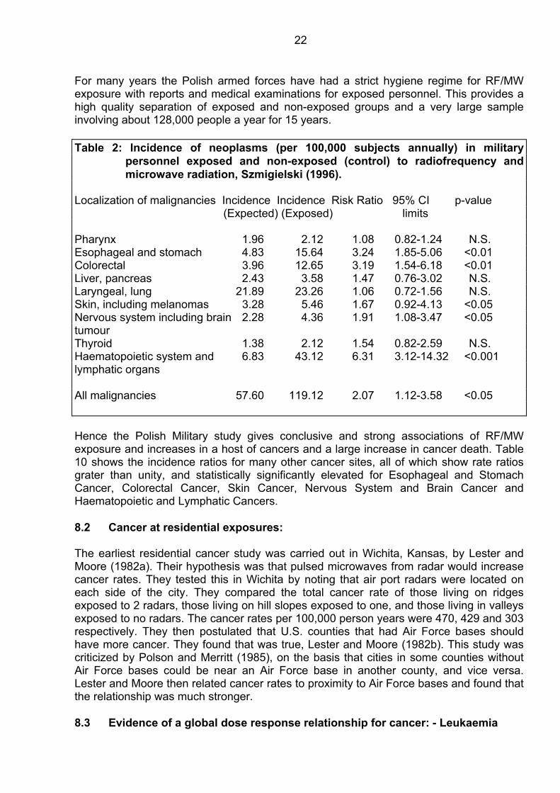
For many years the Polish armed forces have had a strict hygiene regime for RF/MW exposure with reports and medical examinations for exposed personnel. This provides a high quality separation of exposed and non-exposed groups and a very large sample involving about 128,000 people a year for 15 years. Table 2: Incidence of neoplasms (per 100,000 subjects annually) in military
personnel exposed and non-exposed (control) to radiofrequency and microwave radiation, Szmigielski (1996).
Localization of malignancies Incidence Incidence Risk Ratio 95% CI p-value (Expected) (Exposed) limits Pharynx 1.96 2.12 1.08 0.82-1.24 N.S. Esophageal and stomach 4.83 15.64 3.24 1.85-5.06 <0.01 Colorectal 3.96 12.65 3.19 1.54-6.18 <0.01 Liver, pancreas 2.43 3.58 1.47 0.76-3.02 N.S. Laryngeal, lung 21.89 23.26 1.06 0.72-1.56 N.S. Skin, including melanomas 3.28 5.46 1.67 0.92-4.13 <0.05 Nervous system including brain 2.28 4.36 1.91 1.08-3.47 <0.05 tumour Thyroid 1.38 2.12 1.54 0.82-2.59 N.S. Haematopoietic system and 6.83 43.12 6.31 3.12-14.32 <0.001 lymphatic organs All malignancies 57.60 119.12 2.07 1.12-3.58 <0.05 Hence the Polish Military study gives conclusive and strong associations of RF/MW exposure and increases in a host of cancers and a large increase in cancer death. Table 10 shows the incidence ratios for many other cancer sites, all of which show rate ratios grater than unity, and statistically significantly elevated for Esophageal and Stomach Cancer, Colorectal Cancer, Skin Cancer, Nervous System and Brain Cancer and Haematopoietic and Lymphatic Cancers. 8.2 Cancer at residential exposures: The earliest residential cancer study was carried out in Wichita, Kansas, by Lester and Moore (1982a). Their hypothesis was that pulsed microwaves from radar would increase cancer rates. They tested this in Wichita by noting that air port radars were located on each side of the city. They compared the total cancer rate of those living on ridges exposed to 2 radars, those living on hill slopes exposed to one, and those living in valleys exposed to no radars. The cancer rates per 100,000 person years were 470, 429 and 303 respectively. They then postulated that U.S. counties that had Air Force bases should have more cancer. They found that was true, Lester and Moore (1982b). This study was criticized by Polson and Merritt (1985), on the basis that cities in some counties without Air Force bases could be near an Air Force base in another county, and vice versa. Lester and Moore then related cancer rates to proximity to Air Force bases and found that the relationship was much stronger. 8.3 Evidence of a global dose response relationship for cancer: - Leukaemia

23
Table 3: A summary of epidemiological studies involving adult leukaemia mortality or incidence, ranked by probable RF/MW exposure category.
Study Reference Exposure Leukaemia Risk 95% Confidence Category Type Ratio Interval Polish Military Szmigielski (1996) High ALL 5.75 1.22-18.16 (Mortality) CML 13.90 6.72-22.12 CLL 3.68 1.45-5.18 AML 8.62 3.54-13.67 All Leuk. 6.31 3.12-14.32 Amateur Radio Milham (1988) Moderate AML 1.79 1.03-2.85 (Mortality) UK TV/FM Dolk et al. (1997a) Low/Mod All Leuk. 1.62 0.56-4.66 (Incidence) Rate 2-3km ring UK TV/FM Dolk et al. (1997b) Low/Mod All Leuk. 1.15 0.91-1.45 (Incidence) Peak rate 2-3km ring North Sydney Hocking et al.(1996) Low All Leuk. 1.17 0.96-1.43 TV/FM towers ALL+CLL 1.39 1.00-1.92 (Mortality) AML+CML 1.01 0.82-1.24 Other Leuk1.57 1.01-2.46 UK TV/FM Dolk et al. (1997b) Low All Leuk. 1.03 1.00-1.07 (Incidence) (Mean from all cases) Note: ALL : Acute Lymphatic Leukemia; CLL: Chronic Lymphatic Leukaemia; AML
Acute Myeloid Leukaemia; CML: Chronic Myeloid Leukaemia; and All Leuk.: All Adult Leukaemias.
8.3 Brain tumors: I have identified 65 epidemiological studies that associate EMR with increased brain tumor incidence. I give some examples that show how studies cover the EMR spectrum: • ELF [Tornqvist et al. (1986), Tomenus et al. (1986), Savitiz and Loomis (1995)]. • Electrical occupations [Floderous et al. (1993), Mack, Preston-Martin and Peters
(1991), Speers et al. (1988), Miller et al. (1996)); Dosemeci and Blair (1994)]. • Computer programmers [Beall et al. (1996)] • Industrial RF/MW [Thomas et al. (1987), Tornqvist et al. (1991)]. • Military RF/MW exposure [Grayson (1996), Szmigielski (1996), Robinette et al. (1980)]. • Cell phones [Hardell et al. (1999)].

24
Twenty studies show statistically significant increases in brain tumor and 4 show significant dose response relationships. Given the strong data from the 23 studies published up to 1998 it was not surprising that Dr Hardell and his colleagues found that cell phones increased the incidence of a particular type of brain tumor. 9. Breast Cancer Studies It is abundantly clear that EMR causes significant increases in cancer in exposed human populations, even at residential ELF and RF/MW exposure levels found in most cities, especially in proximity to broadcast towers such as radio, TV and cell site base station towers. Residential ELF exposures from electric blankets and other appliances are also implicated. 9.1 ELF enhances breast cancer in rats: Modern technology is carrying out a giant experiment on us by exposing us to a complex soup of EMR. Professor Loscher and this team at the School of Veterinary Medicine in Hannover, Germany, have varied out a progressive series of controlled experiments on rats. These experiments prove that 50/60Hz EMR increases the number and size of tumors in rats that have had mammary tumors initiated by a chemical, DMBA. They observed: • Significant reductions in melatonin down to 0.3 to 1µT, in a dose response manner; • Significant alteration of T-Cell proliferation in a dose response manner; • A significant increase in ODC (ornithine decarboxylase) in mammary cells and spleen,
and • A significant increase in malignant mammary tumor size and number in a dose
response manner. Loscher et al. (1993, 1994), Loscher and Mevissen (1994, 1995) Mevissen et al. (1993, 1995, 1996a, 1996b, 1998) and Baum et al. (1995). 9.2 Human occupational studies find increased breast cancer: Human epidemiological studies have identified significant increases of breast cancer in both males and females exposed to EMR. Exposures cover the spectrum from 50/60 Hz, electrical and electronic occupational exposure and RF/MW exposures. Male breast cancer is quite rare and female breast cancer is quite common. There is evidence that the more breast tissue we have the greater the breast cancer risk might be. Hence if studies find elevated male breast cancer, as did Demers et al. (1991), then female breast cancer risk from EMR exposure must be significantly higher. The following two tables summarize the epidemiological studies involving EMR exposure, the first for male breast cancer and these second for female breast cancer. Table 4: Male breast cancer associated with EMR exposure. Group SIR/RR/OR 95%CI(p-value) Reference

25
Electrical Occupations OR = 1.8 1.0-3.7 Demers et al. (1991) Electricians, telephone OR = 6.0 1.7-21 “ linemen, electric power workers. Radio communication OR = 2.9 0.8-10 “ workers. Electronic workers Increased Risk Guenel et al. (1993) Swedish Railway workers RR = 4.9 1.6-11.8 Floderus, Tornqvist and Stenhund (1994) There are only three male breast cancer studies available and they are limited by small numbers, because of the rarity of male breast cancer and the small size of the groups studied. Hence it is of interest that three groups studied achieved statistical significance and relatively high Odds and Risk Ratios. Table 6 shows the greater number of studies on breast cancer incidence in relation to a wider range of EMR than the male studies. A large number of EMR exposed groups of women show significantly higher rates of breast cancer. Limited exposure measurements limit the ability to obtain dose response relationships. The one study which had moderately good exposure estimates, Coogan et al. (1996), even though the numbers in the exposure class (computer equipment operators) was small, the high exposure group had a significantly elevated risk and the trend approached significance, p=0.06. 9.4 Breast cancer conclusions from epidemiology: Studies have shown significant increases and male and female breast cancer from exposure to EMR from ELF to RF/MW, as with leukaemia and brain tumor. There is a tendency for higher rates in pre-menopausal women and those with estrogen-receptor-positive breast cancer (ER+), and for black women. Because electromagnetic fields are ubiquitous, we are exposed to them at home, work and walking along roads, there is no unexposed population. Work place exposures are mixed and so no pure high exposure and low exposure groups are available. This is a dilutionary effect that reduces the relative risk significantly. Hence all epidemiological results are significant under-estimates of the breast cancer risk.

26
Table 5: Female Breast cancer associated with EMR exposure Group SIR/RR/OR 95%CI/(p-value) Reference Radio-telegraph SIR=1.5 Tynes et al. (1996) operators Electrical Engineers OR = 1.73 0.92-3.29 Loomis, Savitz and Ananth (1994) Electrical technicians OR = 1.28 0.79-2.07 “ Telephone installers OR = 2.12 1.17-4.02 “ repairers, line work Electrical Workers OR = 1.38 1.04-1.89 “ Radiofrequency EMR Low Exp. White OR = 1.15 p<0.05 Cantor et al. (1995) High Exp. White OR = 1.14 p<0.05 “ Low Exp. Black OR = 1.23 p<0.05 “ High Exp. Black OR = 1.34 p<0.05 “ High Exposure ELF OR = 1.43 0.99-2.09 Coogan et al. (1996) Pre-menopausal OR = 1.98 1.04-3.78 “ Post-menopausal OR = 1.38 0.82-2.17 “ Computer equipment OR =1.80 1.04-3.12 [Trend p = 0.06] operators, high Exp. Electric Blankets, heavy RR = 1.43 0.94-2.17 Vena et al. (1994) use, pre-menopausal All women OR = 1.45 1.08-1.94 " > 2 years of use OR = 1.60 1.15-2.22 " > 5 years of use OR = 1.56 1.09-2.25 " Positive Estrogen receptor RR = 1.12 0.78-1.43 Gammon et al. (1998) aged 45 - 55 years. Powerline, Sweden > 0.2 µT, men RR = 2.1 0.3-14.1 Feychting et al. (1998) >0.2 µT, women < 50 yr RR = 1.8 0.7-4.4 “ >0.01 µT, women with RR = 1.6 0.6-4.1 “ + estrogen receptor >0.01 µT, women with RR = 7.4 1.0-178.1 “ + estrogen receptor, aged < 60 years Table 5: Cont'd:

27
Occupational Exposure, Norway Total Sample, 2 methods: Cumulative Hr of Work RR = 1.14 1.1-1.19 Kliukiene et al. (1999) Job Matrix estimate RR = 1.08 1.01-1.16 " Aged <50yrs (Hours) RR = 1.20 1.11-1.29 " Aged 50 yrs (Matrix) RR = 1.12 0.98-1.28 " Occupational Exposure, Sweden Recent reading >0.25µT Total Sample RR = 1.0 0.6-1.7 Forssen et al. (2000) Aged < 50 yrs RR = 1.5 0.6-3.5 " Aged<50yr, ER+ RR = 3.2 0.5-18.9 " There is consistent and significant increases in Breast Cancer in EMR exposed populations of women and men. Increased risk is under-estimated by the given Relative Risks. Women under 50 years of age have more EMR induced breast cancer than other women in the same age range. When women are older than 50 the background rate of breast cancer increases, reducing the relative differences in the EMR exposed group. EMR is especially active in initiating and/or accelerating estrogen receptor positive breast cancer. 10. Cell phone pose a serious breast cancer risk: Cell phones pose the highest risk factor to people today because they are held so close to our heads and bodies that they produce far higher intensities of radiation exposure that in the high military exposure situations. Analogue cell phones emit an analogue modulated RF/MW signal similar to an FM radio or TV signal. The digital cell phones radiate a pulse RF/MW signal similar to a radar. Such signals have been shown to: • Alter brain activity, including EEG and reaction times, memory loss, headaches, fatigue
and concentration problems, dizziness (the Microwave Syndrome), • Impair sleep and learning • Increase Alzheimer's Disease • Alter blood pressure and heart rhythm (heart rate variability) • Impair the immune system • Reduce melatonin • Break DNA strands, damage chromosomes, alter gene transcription activity, and • Increase the incidence of many types of cancer, including leukaemia, brain tumor,
testicular cancer and breast cancer.

28
For years the cell phone companies and government authorities have assured us that cell phone are perfectly safe. They state that the particular set of radiation parameter associated with cell phones are not the same as any other radio signal and therefore earlier research does not apply. They also mount biased review teams who falsely dismiss any results that indicate adverse biological and health effects. Now we have government and industry funded research that shows that cell phone radiation causes the following symptoms: • Alters brain activity including EEG, Von Klitzing (1995), sleep and EEG, Mann and
Roschkle (1996) and human reaction times, Preece et al. (1999). • Causes memory loss, concentration difficulties, fatigue, and headache, in a dose
response manner, (Mild et al. (1998)). • Reduces the pituitary production of Thyrotropin (Thyroid Stimulating Hormone, TSH):
Figure 17: A significant reduction in Thyrotropin (Thyroid Stimulating Hormone)
during cell phone use, de Seze et al. (1998). • Increases blood pressure, Braune et al. (1998). • Reduces melatonin, Burch et al. (1997). • Breaks DNA strands (Verschaeve at al. (1994), Phillips et al. (1998)). • Produces an up to three-fold increase in chromosome aberrations in a dose response
manner from all cell phones tested, Tice, Hook and McRee, reported in Microwave News, April/May 1999.
• Doubles c-fos gene activity (a proto oncogene) for analogue phones and increases it
by 41 % for digital phones, Goswami et al. (1999), altered c-jun gene, Ivaschuk et al. (1997).
• Doubles the cancer in mice, Repacholi et al. (1997). • Increases human brain tumor rate by 2.5 times (Hardell et al. (1999)).
29
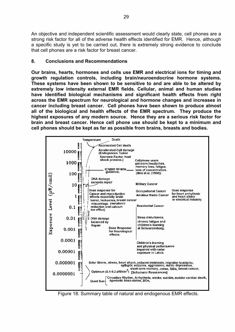
An objective and independent scientific assessment would clearly state, cell phones are a strong risk factor for all of the adverse health effects identified for EMR. Hence, although a specific study is yet to be carried out, there is extremely strong evidence to conclude that cell phones are a risk factor for breast cancer. 8. Conclusions and Recommendations Our brains, hearts, hormones and cells use EMR and electrical ions for timing and growth regulation controls, including brain/neuroendocrine hormone systems. These systems have been shown to be sensitive to and are able to be altered by extremely low intensity external EMR fields. Cellular, animal and human studies have identified biological mechanisms and significant health effects from right across the EMR spectrum for neurological and hormone changes and increases in cancer including breast cancer. Cell phones have been shown to produce almost all of the biological and health effects of the EMR spectrum. They produce the highest exposures of any modern source. Hence they are a serious risk factor for brain and breast cancer. Hence cell phone use should be kept to a minimum and cell phones should be kept as far as possible from brains, breasts and bodies.
Figure 18: Summary table of natural and endogenous EMR effects.

30
All industrial and residential EMR exposures should be kept extremely low, including placing cell sites far from residents, schools, hospitals and workplaces. Radio and TV towers should be in remote locations. World governments should adopt public health standards based on epidemiological studies and not on industry positions. Adverse health effects have been shown down to about 1 pW/cm2. The median measure exposure in 15 U.S. cities in 1980 was 5 nW/cm2, Tell and Mantiply (1980). Allowable exposure levels should be brought down by 20,000 from 200 µW/cm2 to 10 nW/cm2, 0.1µT for 50/60 Hz fields, and lifetime exposures be kept as low as possible. References: Adey, W.R., 1981: “Tissue interactions with non-ionizing electromagnetic fields”. Physiological Reviews, 61:
435-514. Adey, W.R., 1992: "ELF magnetic field and promotion of cancer: experimental studies". In "Interaction
mechanisms of low-level electromagnetic fields in living systems", Bengt Norden and Claes Ramel, Eds. Oxford University Press., pp23-46.
Ahuja, Y.R., Bhargava, A., Sircar, S., Rizwani, W., Lima, S., Devadas. A.B. and Bhargava, S.C. 1997:
"Comet assay to evaluate DNA damage caused by magnetic fields". Proceedings of the International Conference on Electromagnetic Interference and Compatibility, December, 1997, Hyderabad, India, pp. 273-276.
Ahissar, E., Haidarliu, S. and Zacksenhouse, M., 1997: "Decoding temporally encoded sensory input by
cortical oscillations and thalamic phase comparators". Proc. Natl. Acad. Sci.(USA), 94(21): 11633-11638.
Alam, M.T., Barthakur, N., Lambert, N.G. and Kasatiya, S.S., 1978: "Microwave radiation induced
chromosomal aberrations in corneal epithelium of Chinese hamsters". Canadian J. Genet. Cytol. 20(1): 23-30.
Antipenko, E.N., Timchenko, O.I., Volkova, T.M. and Fedorova, A.A., 1984: "Effect of thytoxine in the G0
stage of the cell cycle on the yield of chromosome aberrations in rat hepatocytes and human lymphocytes after X-radiation", Radiobiologiia, 24(2): 233-236.
Antipencko, E.N., Koveshnikova, I.V. and Timchenko, O.I., 1984: "Effect of microwaves of nonthermal
intensity on the number of aberrant hepatocytes in rats". Radiobiologiia, 24(3): 403-405. Avdonina, E.N., and Smaovichev, E.G., 1995: "Some heliogeophysical characteristics of a series of
especially dangerous crimes". Biofizika, 40(5): 1060-1063. Babych, V.I., 1996: "Serotonin metabolism under the action of a low geomagnetic field". [In Ukrainian].
Fiziol Zh., 42(1-2): 79-82. Balcer-Kubiczek, E.K. and Harrison, G.H., 1985: “Evidence for microwave carcinogenesis”.
Carcinogenesis, 6: 859-864. Balser, M. and Wagner, C.A., 1960: "Observations of earth-ionosphere cavity resonances". Nature, 188
(Nov 19), 638-641.

31
Baum, A., Mevissen, M., Kamino, K., Mokr, U. and Loscher, W., 1995: "A histopathological study on alterations in DMBA-induced mammary carcinogenesis in rats with 50 Hz, 100µT magnetic field exposure". Carcinogenesis, 16(1):119-125.
Beall, C., Delzell, E., Cole, P., and Brill, I., 1996: “Brain Tumors among electronics Industry Workers”.
Epidemiology, 7(2): 125-130. Belisheva, N.K., Popov, A.N., Petukhova, N.V., Pavlova, L.P., Osipov, K.S., Tkachenko, S.E. and Varanova,
T.I., 1995: "Qualitative and quantitative assessment of exposure to geomagnetic field variations on the functional status of the human brain". [In Russian]. Biofizika, 40(5):1005-1012.
Belov, D.R., Kanunikov, I.E. and Kiselev, B.V., 1998: "Dependence of human EEG synchronization on the
geomagnetic activity on the day of experiment". Ross Fiziol Zh Im I M Sechenova, Aug; 84(8): 761-774.
Bergiannaki Joff, Paparrigopoulos, T.J., and Stefanis, C.N., 1996: "Seasonal pattern of melatonin excretion
in humans: relationship to daylength variation rate and geomagnetic field fluctuations". Experimenta, 52(3): 253-258.
Braune, S., Wrocklage, C., Raczek, J., Gailius, T. and Lucking, C.H., 1998: "Resting blood pressure
increase during exposure to a radio-frequency electromagnetic field". The Lancet, 351(June 20): 1857-1858.
Bruvere, R., Feldmane, G., Heisele, O., Volrate, A. and Balodis, V., 1998: "Several immune system
functions of the residents from territories exposed to pulse radio-frequency radiation". Tenth Annual Conference of the International Society for Environmental Epidemiology, Boston, Ma, August 15-18, 1998. Reporeted in Epidemiology, 9(4): p S109.
Burch, J.B., Reif, J.S., Yost, M.G., Keefe, T.J. and Pitrat, C.A., 1998: "Nocturnal excretion of a urinary
melatonin metabolite among electric utility workers". Scand. J. Work Env. Health, 24(3): 183-189. Burch, J.B., Reif, J.S., Yost, M.G., Keefe, T.J. and Pitrat, C.A., 1999: "Reduced excretion of a melatonin
metabolite in workers exposed to 60 Hz magnetic fields". Am. J. Epidemiology, 150(1): 27-36. Cantor, K.P.. Stewart, P.A., Briton, L.A. and Dosemeci, M., 1995: "Occupational exposures and female
breast cancer mortality in the United States". J. Occup. and Environ. Medicine, 37(3): 336-348. Chou, C-K., Guy, A.W., Kunz, L.L., Johnson, R.B., Crowley, J.J. and Krupp, J.H., 1992: “Long-term low level
microwave irradiation of rats”. Bioelectromagnetics, 13: 469-496. Ciccone, G., Mirabelli, D., Levis, A., Gavarotti, P., Rege-Cambrin, G., Davico, L., and Vineis, P., 1993:
“Myeloid leukemias and myelodysplastic syndromes: chemical exposure, histologic subtype and cytogenetics in a case-control study”. Cancer Genetics & Cytogenetics 1993 Jul 15;68(2):135-9.
Conesa, J., 1997: "Isolated sleep paralysis, vivid dreams and geomagnetic influences: II." Percept. Mot.
Skills, 85(2): 579-584. Conesa, J., 1995: "Relationship between isolated sleep paralysis and geomagnetic influences: a case
study". Percept. Mot. Skills, 80(3 Pt2): 1263-1273. Coogan, P.F., Clapp, R.W., Newcomb, P.A., Wenzl, T.B., Bogdan, G., Mittendorf, R., Baron, J.A. and
Longnecker, M.P., 1996: “Occupational exposure to 60-hertz magnetic field and the risk of breast cancer in women”. Epidemiology, 7(5):459-464.
De Matteis, G., Vellante, M., Marrelli, A., Villante, U., Santalucia, P., Tuzi, P. and Prencipe, M, 1994:
"Geomagnetic Activity, humidity, temperature and headache: is there any correlation?". Headache, 34(1): 41-43.
Demers, P.A., Thomas, D.B., Rosenblatt, K.A., Jimenez, L.M., Mc Tiernan, A, Stalsberg, H., Stemhagen,
A., Thompson, W.D., McCrea, M.G., Satariano, W., Austin, D.F., Isacson, P., Greenberg, R.S., Key, C., Kolonel, L.N., and West, D.W., 1991: “Occupational exposure to electromagnetic fields and breast cancer in men”., Am. J. Epidemiology, 134 (4): 340-347.

32
De Seze, R., Fabbro-Peray, P. and Miro, L., 1998: "GSM radiocellular telephones do not disturb the
secretion of antepituitary hormones in humans". Bioelectromagnetics, 19: 271-278. Dmoch, A. and Moszczynski, P., 1998: "Levels of immunoglobulin and subpopulations of T lymphocytes
and NK cells in men occupationally exposed to microwave radiation in frequencies 6-12 GHz". [In Polish]. Med. Pr. 49(1): 45-49.
Dolk, H., Shaddick, G., Walls, P., Grundy, C., Thakrar, B., Kleinschmidt, I. and Elliott, P., 1997a: “Cancer
incidence near radio and television transmitters in Great Britain, I - Sutton-Colfield transmitter”. American J. of Epidemiology, 145(1):1-9.
Dolk, H., Elliott, P., Shaddick, G., Walls, P., Grundy, C., and Thakrar, B.,1997b: “Cancer incidence near
radio and television transmitters in Great Britain, II All high power transmitters”. American J. of Epidemiology, 145(1):10-17.
Dosemeci, M. and Blair, A., 1994: “Occupational Cancer Mortality Among Women Employed in the
Telephone Industry”. Journal of Occupational Medicine, 36 (11): 1204-1209. Elliott, W.H. and Elliott, D.C., 1996: ""Biochemistry and molecular biology". Publ. Oxford University Press,
Oxford. Fanelli, C., Coppola, S., Barone, R., Colussi, C., Gualandi, G., Volpe, P. and Ghibelli, L., 1999: "Magnetic
fields increase cell survival by inhibiting apoptosis via modulation of Ca2+ influx". FASEB J. 13: 95-102.
Feychting, M., Forssen, U., Rutqvist, L.E., and Ahlbom, A., 1998: “ Magnetic fields and breast cancer in
Swedish adults residing near high-voltage power lines”. Epidemiology, 9(4): 392-397. Frey, A.H., 1995: "An integration of the data on mechanisms with particular reference to cancer". In "On the
nature of electromagnetic field interactions with biological systems" pp 9-28, Publ. R.G. Landes Company, Austin, Texas.
Floderus, B., Tornqvist, S. and Stenhund, C., 1994: “Incidence of selected cancers in Swedish railway
workers, 1961-79”. Cancer causes control, 5(2): 189-94. Forssen, U.M., Feychting, M., Rutqvist, L.E., Floderus, B. and Ahlbom, A., 2000: "Occupational and
residential magnetic field exposure and breast cancer in females". Epidemiology, 11 (1): 24-29.
Gammon, M.D., Schoenberg, J.B., Britton, J.A., Kelsey, J.L., Stanford, J.L., Malone, K.E., et al., 1998: “Electric blanket use and breast cancer among younger women:. Am. J. Epid. 148(6): 556-563.
Garaj-Vrhovac, V., Fucic, A, and Horvat, D., 1990: "Comparison of chromosome aberration and micro-
nucleus induction in human lymphocytes after occupational exposure to vinyl chloride monomer and microwave radiation"., Periodicum Biologorum, Vol 92, No.4, pp 411-416.
Garaj-Vrhovac, V., Horvat, D., and Koren, Z., 1990: ”The effect of microwave radiation on the cell genome”.
Mutation Research, 243: 87-93. Garaj-Vrhovac, V., Horvat, D., and Koren, Z., 1991: ”The relationship between colony-forming ability,
chromosome aberrations and incidence of micronuclei in V79 Chinese Hamster cells exposed to microwave radiation”. Mutation Research, 263: 143-149.
Garaj-Vrhovac, V., Fucic, A, and Horvat, D., 1992: The correlation between the frequency of micronuclei
and specific aberrations in human lymphocytes exposed to microwave radiation in vitro”. Mutation Research, 281: 181-186.
Garaj-Vrhovac, V., and Fucic, A., 1993: “The rate of elimination of chromosomal aberrations after
accidental exposure to microwave radiation”. Bioelectrochemistry and Bioenergetics, 30:319-325. Ghione, S., Mezzasalma, L., Del Seppia, C. and Papi, F., 1998: "Do geomagnetic disturbances of solar
origin affect arterial blood pressure ?". J. Hum Hypertension, 12(11): 749-754.

33
Goldsmith, J.R., 1992: “Incorporation of epidemiological findings into radiation protection standards Public”.
Health Rev 1991/92; 19: 19-34. Goldsmith, J.R., 1995: “Epidemiological Evidence of Radiofrequency (Microwave) Effects in Military,
Broadcasting and Occupational Studies”. International Journal of Occupational and Environmental Health, 1:47-57.
Goldsmith, J.R., 1998: “Epidemiologic evidence relevant to radar (microwave) effects”. Environmental
Health Perspectives, 105 (Suppl 6), Dec 1997, pp 1579-1587. Goswami. P.C., Albee, L.D., Parsian, A.J., Baty, J.D., Moros, E.G., Pickard, W.F., Roti Roti, J.L. and Hunt,
C.R., 1999: "Proto-oncogene mRNA levels and activities of multiple transcription factors in C3H 10T1/2 murine embryonic fibroblasts exposed to 835.62 and 847.74 MHz cellular phone communication frequency radiation". Radiation Research, 151(3): 300-309.
Grayson, J.K., 1996: “Radiation Exposure, Socioeconomic Status, and Brain Tumour Risk in the US Air
Force: A nested Case-Control Study”. American J. of Epidemiology, 143 (5), 480-486. Guenel, P., Raskmark, P., Anderson, J.B. and Lynge, E., 1993: “Incidence of cancer in persons with
occupatinal exposure to electromagnetic fields in Denmark”. Br. J. Ind. Med., 50(8): 758-764. Hagmar, L., Brogger, A., Hansteen, I.L., et al. (1994): “Cancer risk in humans predicted by increased levels
of chromosomal aberrations in lymphocytes: Nordic Study Group on the health risk of chromosome damage”. Cancer Research, 54: 2919-2922.
Hardell, L., Nasman, A., Pahlson, A., Hallquist, A. and Hansson Mild, K., 1999: "Use of cellular telephones
and the risk fro brain tumours: A case-control study". Int. J. Oncology. 15(1): 113-116. Heller, J.H., and Teixeira-Pinto, A.A., 1959: “A new physical method of creating chromosome aberrations”.
Nature, Vol 183, No. 4665, March 28, 1959, pp 905-906. Hocking, B., Gordon, I.R., Grain, H.L., and Hatfield, G.E., 1996: “Cancer incidence and mortality and
proximity to TV towers”. Medical Journal of Australia, Vol 165, 2/16 December, pp 601-605. Huang, A. T-F. and Mold, N.G., 1980: "Immunologic and hematopoietic alterations by 2450 MHz
electromagnetic radiation". Bioelectromagnetics, 1: 77-87. Ivaschuk, O.I., Jones, R.A., Ishida-Jones, T., Haggren, W., Adey, W.R. and Phillips, J.L., 1997: "Exposure
of nerve growth factor-treated PC12 rat pheochromacytoma cells to a modulated radiofrequency field at 836.55 MHz: effects on c-jun and c-fos expression". Bioelectromagnetics, 18(3): 223-229.
Jacobson, C.E., 1969: "Progress report on SCC 31732, 4 February 1969", The George Washington
University. Subcontract to: Lilienfeld, A.M., Tonascia, J., Tonascia, S., Libauer, C.A., and Cauthen, G.M., 1978: “Foreign service health status study - evaluation of health status of foreign service and other employees from selected eastern European posts”. Final Report (Contract number 6025-619073) to the U.S. Department of State, July 31, 1978.
Karasek, M., Woldanska-Okonska, M., Czernicki, J., Zylinska, K. and Swietoslawski, J., 1998: "Chronic
exposure to 2.9 mT, 40 Hz magnetic field reduces melatonin concentrations in humans". J. Pineal Rese., 25(4): 240-244.
Kay, R.W., 1994: "Geomagnetic Storms: association with incidence of depression as measured by hospital
admission". Br. J. Psychiatry, 164(3): 403-409. Kliukiene, T., Tynes, T., Martinsen, J.I., Blaasaas, K,G., Andersen, A., 1999: "Incidence of breast cancer in
a Norwegian cohort of women with potential workplace exposure to 50 Hz magnetic fields". Am. J. Ind. Med. 36(1): 147-154.

34
König, H.L., 1974: “Behavioural changes in human subjects associated with ELF electric fields”. In “ELF and VLF electromagnetic field effects”, M.A. Persinger Ed, Publ. Plenum Press, New York.
Komarov, F.I., Oraevskii, V.N., Sizov, Iu.P., Tsirul'nik, L.B., Kanoidi, Kh.D., Ushakov, I.B., Shalimov, P.M.,
Kimlyk, M.V., and Glukhov,.D.V., 1998: "Heliogeophysical factors and aviation accidents". Biofizika, 43(4): 742-745.
Koveshnikov, I.V. and Antipenko, E.N., 1991b: "Quantitative patterns in thecytogenetic action of
microwaves". Radioliologiia, 31(1): 147-149. Koveshnikov, I.V. and Antipenko, E.N., 1991a: "The participation of thyroid hormones in modifying the
mutagenic effect of microwaves". Radioliologiia, 31(1): 149-151. Lai, H. and Singh, N.P., 1995: “Acute low-intensity microwave exposure increases DNA single-strand
breaks in rat brain cells”. Bioelectromagnetics, Vol 16, pp 207-210, 1995. Lai, H. and Singh, N.P., 1996: “Single- and double-strand DNA breaks in rat brain cells after acute
exposure to radiofrequency electromagnetic radiation”. Int. J. Radiation Biology, 69 (4): 513-521. Lai, H., and Singh, N.P., 1997: “Melatonin and a Spin-trap compound Block Radiofrequency
Electromagnetic Radiation-Induced DNA Strand Breaks in Rat Brain Cells”. Bioelectromagnetics, 18: 446-454.
Lai, H. and Singh, N.P., 1997: "Acute exposure to a 60-Hz magnetic field increases DNA strand breaks in rat
brain cells". Bioelectromagnetics, 18:156-165. Lester, J.R., and Moore, D.F., 1982a: “Cancer incidence and electromagnetic radiation”. Journal of
Bioelectricity, 1(1): 59-76. Lester, J.R., and Moore, D.F., 1982b: “Cancer mortality and air force bases”. Journal of Bioelectricity, 1(1):
77-82. Lester, J.R., and Moore, D.F., 1985: “Reply to: Cancer mortality and air force bases, a reevaluation”.
Journal of Bioelectricity, 4(1): 129-131. Liburdy, R., 1979: "Radio frequency alters the immune system: Modulation of T- and B-lymphocyte levels
and cell-mediated immunocompetence by hyperthermic radiation". Radiation Research, 77: 34-46.
Liburdy, R.P., Sloma, T.R., and Yaswen, P., 1993: “ELF magnetic fields, breast cancer and melatonin: 60
Hz fields block melatonin’s oncostatic action on ER+ breast cancer cell proliferation”. Journal of Pineal Research, 14 (2): 89-97.
Lilienfeld, A.M., Tonascia, J., Tonascia, S., Libauer, C.A., and Cauthen, G.M., 1978: “Foreign service health
status study - evaluation of health status of foreign service and other employees from selected eastern European posts”. Final Report (Contract number 6025-619073) to the U.S. Department of State, July 31, 1978.
Lin, R.S., Dischinger, P.C., Conde, J., and Farrell, K,P., 1985: “Occupational exposure to electromagnetic
fields and brain tumors”. J. Occup. Med., 21:474-480. Loomis, D.P., Savitiz, D.A. and Ananth, C.V., 1994: “Breast cancer mortality among female electrical
workers in the United States”. J. National Cancer Institute, 86(12): 921-925. Loscher, M., Mevissen, M., Lehmacher, W. and Stamm, A., 1993: "Tumor promotion in a breast cancer
model by exposure to a weak alternating magnetic field". Cancer Letters, 71(1-3): 75-81. Loscher, W. and Mevissen, M., 1994: "Animal studies on the role of 50/60-Hertz magnetic fields in
carcinogenesis". Life Sciences, 54(21): 1531-1543. Loscher, W., Wahnschaffe, U., Mevissen, M., Lerchl, A, and Stamm, A., 1994: "Effects of weak alternating
magnetic fields on nocturnal melatonin production and mammary carcinogenesis in rats". Oncology, 51(3): 288-295.

35
Loscher, W. and Mevissen, M., 1995: "Linear relationship between flux density and tumor co-promoting
effect of prolonged magnetic field exposure in a breast cancer model". Cancer Letters, 96(2): 175-180.
Maes, A., Verschaeve, L., Arroyo, A., De Wagter, C. and Vercruyssen, L., 1993: "In vitro cytogenetic effects
of 2450 MHz waves on human peripheral blood lymphocytes". Bioelectromagnetics, 14: 495-501. Maes, A., Collier, M., Slaets, D., and Verschaeve, L., 1996: “954 MHz Microwaves enhance mutagenic
properties of Mitomycin C”. Environmental and Molecular Mutagenesis, 28: 26-30. Maestroni, G.J., 1995: "T-Helper-2 lymphocytes as peripheral target of melatonin signaling", Journal of
Pineal Research, 18: 84-89. Makarov, L.M., 1998: "Role of geomagnetic field in development of biorhythm profile of venticular
arrhythmia onset". Klin. Med. (Mosk), 76(6):31-35. Mann, K., and Roschkle, J., 1996: “Effects of pulsed high-frequency electromagnetic fields on human
sleep”. Neuropsychobiology, 33: 41-47. Mevissen, M, Kietzmann, M. and Loscher, W., 1995: "In vivo exposure of rats to a weak alternating
magnetic field increases ornithine decarboxylase activity in the mammary gland by a similar extent as the carcinogen DMBA". Cancer Letters, 90(2): 207-214.
Mevissen, M, Lerchl, A., Szamel, M. and Loscher, W., 1996: "Exposure of DMBA-treated female rats in a
50-Hz, 50 microTesla magnetic field: effects on mammary tumor growth, melatonin levels, and T lymphocyte activation". Carcinogenesis, 17(5): 903-910.
Mevissen, M, Lerchl, A. and Loscher, W., 1996: "Study on pineal function and DMBA-induced breast cancer
formation in rats during exposure to a 100 mG, 50 Hz magnetic field". J. Toxicol. Environ. Health, 48(2): 169-185.
Mevissen, M, Haussler, M., Szamel, M. Emmendorffer, A., Thun-Battersby, S. and Loscher, W., 1998:
"Complex effects of long-term 50 Hz magnetic field exposure in vivo on immune functions in female Sprague-Dawley rats depend on duration of exposure". Bioelectromagnetics, 19(40: 259-270.
Moszczynski, P, Lisiewicz, J., Dmoch, A., Zambinski, Z., Bergier, L., Rucinska, M. and Sasiadek, U., 1999:
"The effect of various occupational exposures to microwave radiation on the concentrations of immunogobulins and T lymphocyte subsets". [In Polish]. Wiad. Lek, 52(1-2): 30-34.
Nakamura, H., Seto, T., Nagase, H., Yoshida, M., Dan, S. and Ogina, K., 1997: "Effects of exposure to
microwaves on cellular immunity and placental steroids in pregnant rats". Occupational and Environmental Medicine. 54(9): 676-680.
Nordenson, I., Hansson Mild, K., Andersson, G., and Sandstron, M., 1994: "Chromosomal aberrations in
human amniotic cells after intermittent exposure to fifty Hertz magnetic fields", Bioelectromagnetics, 15: 293-301.
O'Connor, R.P. and Persinger, M.A., 1997: "Geophysical variables and behavior LXXXII. Strong association
between sudden infant death syndrome and increments of global geomagnetic activity - possible support for the melatonin hypothesis". Percept. Mot. Skills, 84(2): 395-402.
O'Connor, R.P. and Persinger, M.A., 1996: Increases in geomagnetic activity associated with increases in
thyroxine levels in a single patient: implications for melatonin levels". International Journal of Neuroscience, 88(3-4): 243-247.
Oraevskii, V.N., Kuleshova, V.P., Gurfinkel', Iu.F., Guseva, A.V., and Rapoport, S.I., 1998: "Medico-
biological effect of natural electromagnetic variations". [In Russian], Biofizika, 43(5): 844-888.
36
Phillips, J.L., Campbell-Beachler, M., Ivaschuk, 0., Ishida-Jones, T. and Baggnen, W., 1998: "Exposure of molt-4 lymphoblastoid cells to a 1 G sinusoidal magnetic field at 60-Hz". In: 1998 Annual Review of Research on Biological Effects of Electric and Magnetic Fields from the Generation, Delivery, and Use of Electricity; W/L Associates, Ltd, Frederick, MD.
Phillips, J.L., Ivaschuk, O., Ishida-jones, T., Jones, R.A., Campbell-Beachler, M., and Baggren, W., 1998:
"DNA Damage in Molt-4 Tlymphoblastoid cells exposed to cellular telephone radiofrequency fields in vitro". Bioelectrochem. Bioenerg. 45: 103-110.
Pikin, D.A., Gurginkel', Iu.I. and Oraevskii, V.N., 1998: "Effect of geomagnetic disturbances on the blood
coagulation system in patients with ischemic heart disease and prospects for correction medication". [In Russian]. Biofizika, 43(4): 617-622.
Polk, C., 1982: "Schumann Resonances". In CRC Handbook of Atmospherics, Vol 1, pp 111-177, Polson, P. and Merritt, J.H., 1985: "Cancer mortality and Air Force bases, A reevaluation". J. of
Bioelectricity, 4: 121-127. Preston-Martin, S., Henderson, B.E., and Peters, J.M., 1982: "Descriptive epidemiology of nervous system
neoplasms in Los Angeles County". Ann. N.Y. Acad. Sci. 381:202-208. Preston-Martin, S., Mack, W., and Henderson, B.E., 1989: "Risk factors for gliomas and meningiomas in
males Los Angeles County". Cancer Research, Vol 49, p 6137. Riabykh, T.P. and Bodrova, N.B., 1992: "Correlation of the probability of occurrence of breast neoplasms
with solar activity". Biofizika, 37(4): 710-715. Rajaram, M. and Mitra, S., 1981: "Correlation between convulsive seizure and geomagnetic activity".
Neurosci. Lett., 24(2): 187-191. Raps, A., Stoupel, E., and Shimshoni, M., 1991: "Solar activity and admissions of psychiatric inpatients,
relations and possible implications of seasonality". Isr. J. Psychiatry Relation. Sci., 28(2): 50-59. Reiter RJ., 1994: “Melatonin suppression by static and extremely low frequency electromagnetic fields:
relationship to the reported increased incidence of cancer.” Reviews on Environmental Health. 10(3-4):171-86, 1994.
Reiter, R.J. and Robinson, J, 1995: "Melatonin: Your body's natural wonder drug". Publ. Bantam Books,
New York. Renton, C.M. and Persinger, M.A., 1997: "Elevations of complex partial epileptic-like experiences during
increased geomagnetic activity for women reporting "pre-menstral syndrome". Percept. Mot. Skills, 86(1):240-242.
Repacholi, M.H., Basten, A., Gebski, V., Noonan, D., Finnie, JH., and Harris, A.W., 1997: “Lymphomas in
Eµ-Pim1 Transgenic Mice Exposed to Pulsed 900 MHz Electromagnetic Fields”. Radiation Research, 147:631-640.
Robinette, C.D., Silverman, C. and Jablon, S., 1980: “Effects upon health of occupational exposure to
microwave radiation (radar)”. American Journal of Epidemiology, 112(1):39-53, 1980. Sagripanti, J. and Swicord, M.L., 1976: DNA structural changes caused by microwave radiation. Int. J. of
Rad. Bio., %(1), pp 47-50, 1986. Sarkar, S., Sher, A., and Behari, J., 1994: “Effect of low power microwave on the mouse genome: A direct
DNA analysis”. Mutation Research, 320: 141-147. Schlehofer, B., Kunze, S., Sachsenheimer, W., Blettner, M., Niehoff, D., and Wahrendorf, J., 1990:
“Occupational risk factors for brain tumors: results from a population-bsed case-control study in Germany”. Cancer Causes and Control, 1(3): 209-215.

37
Schwan, H.P. and Foster, K.R., 1980: “RF-field interactions with biological systems: electrical properties and biophysical mechanisms”. Proc. of the IEEE, 68(1): 104-113.
Selitskii, G.V., Karlov, V.A. and Sorokina, N.D., 1999: "The influence of hypogeomagnetic field on the
bioelectric activity of the brain in epilepsy". Zh. Nevrol. Psikhiatr. Im. S.S. Korsakova., 99(4): 48-50.
Selvin, S., Schulman, J. and Merrill, D.W. 1992: “Distance and risk measurements for the analysis of spatial
data: a case study of childhood cancers”. Soc. Sci. Med., 34(7):769-777. Shandala, M.G., Dumanskii, U.D., Rudnev, M.I., Ershova, L.K., and Los I.P., 1979: “Study of Non-ionising
Microwave Radiation Effects on the Central Nervous System and Behavior Reactions”. Environmental Health Perspectives, 30:115-121.
Sitar, J., 1990: "The causality of lunar changes on cardiovascular mortality". [In Czech.] Cas. Lek. Cesk.
129(45):1425-1430. Sparks, L.D., and Hunsaker, J.C., 1991: "Increased ALZ-50- Reactive neurons in the brains of SIDS infants:
An indicator of greater neuronal death "". Journal of Child Neurology, 6: 123-127. Speers, M.A., Dobbins, J.G., and Miller, V.S., 1988: “Occupational exposures and brain cancer mortality: a
preliminary study of East Texas Residents”. American Journal of Industrial Medicine, 13:629-638. Stevens, R.G., Davis, S., Thomas, D.B., Anderson, L.E. and Wilson, B.W., 1992: “Electric power, pineal
function and the risk of breast cancer”. FASEB J. 6: 853-860. Stoupel, E., Hod, M., Shimshoni, M., Friedman, S., Ovadia, J. and Keith, L., 1990: "monthly cosmic actgivity
and pregnancy induced hypertesnsion". Clin. Exp. Obstet. Gynecol., 17(1): 7-12. Stoupel, E., Martfel, J. and Rotenberg, Z., 1991: "Admissions of patients with epileptic seizures (E) and
dizziness (D) related to geomagnetic and solar activity levels: differences in female and male patients". Med. Hypothesis, 36(4): 384-388.
Stoupel, E., 1993: "Sudden cardiac deaths and ventricular extrasystoles on days of four levels of
geomagnetic activity". J. Basic Physiol. Pharmacol., 4(4): 357-366. Stoupel, E., Goldenfeld, M., Shimshoni, M. and Siegel, R., 1993: "Intraocular pressure (IOP) in relation to
four levels of daily geomagnetic and extreme yearly solar activity". Int. J. Biometeorology, 37(1): 42-45.
Stoupel, E., Martfel, J.N., and Rotenberg, Z., 1994: "Paroxysmal atrial fibrulation and stroke (cerebrovasular
accidents) in males and females above and below the age 65 on days of different geomagnetic activity levels". J. Basic. Clin. Physiol. Pharmacol., 5(3-4): 315-329.
Stoupel, E., Petrauskiene, J., Kalediene, R., Abramson, E. and Sulkes, J., 1995: "Clinical cosmobiology: the
Lithuanian study 1990-1992". Int. J. Biometeorology, 38(4): 204-208. Stoupel, E., Abramson, E., Sulkes, J., Martfel, J., Handelman, M., Shimshoni, M., Zadka, P. and Gadday,
U., 1995: "Relationship between suicide and myocardial infarction with regard to changing physical environmental conditions". Int. J. Biometeorology, 38(4): 199-203.
Stoupel, E., Petrauskiene, J., Kalediene, R., Domarkiene, S., Abramson, E. and Sulkes, J., 1996:
"Distribution of deaths from ischemic heart disease and stroke. Environmental and aging influences in men and women". J. Basic. Clinical Physiol. Pharmacol., 7(4): 303-319.
St Pierre, L. and Persinger, M.A., 1998: "Geophysical variables and behavior: LXXXIV. Quantitative
increases in group aggression in male epileptic rats during increases in geomagnetic activity". Percept. Mot. Skills, 86(3 Pt2):1392-1394.
Svanidze, I.K., Sandodze, V.Ia., Didimova, E.V., Chkhikvadze, T.I., Portnoi, V.N. and Razdol'skii, A.S.,
1994: "Effect of hypo- and hyper magnetic fields on the motor activity of the ciliary apparatus of the ependymal cells". [In Russian]. Radiats. Biol. Radioecol., 34(1): 100-104.
38
Svedenstal, B. M., Johanson, K.L., Mattsson, M.0. and Paulson, L.E., 1998: "DNA damage, ODC activities
and cell kinetics in CBA mice exposed to magnetic fields generated by transmission lines". In: 1999 Annual Review of Research on Biological Effects of Electric and Magnetic Fields from the Generation, Delivery, and Use of Electricity; WIL Associates, Ltd, Frederick, MD.
Szmigielski, S., 1991: International Science Meeting, Belgrade, 8-11 April, 1991, p34. Szmigielski, S., 1996: “Cancer morbidity in subjects occupationally exposed to high frequency
(radiofrequency and microwave) electromagnetic radiation”. Science of the Total Environment, Vol 180, 1996, pp 9-17.
Tambiev, A.E., Medvedev, S.D. and Egorova, E.V., 1995: "The effect of geomagnetic disturbances on the
functions of attention and memory". [In Russian]. Aviakosm. Ekolog. Med., 29(3): 43-45. Tell, R.A. and Mantiply, E.D., 1980: "Population exposure to VHF and UHF broadcast radiation in the
United States". Proc. IEEE, 68(1): 4-12. Thomas, T.L., Stolley, P.D., Stemhagen, A., Fontham, E.T.H., Bleecker, M.L., Stewart, P.A., and Hoover,
R.N., 1987: "Brain tumor mortality risk among men with electrical and electronic jobs: A case-control study". J. Nat. Canc. Inst., Vol 79, No.2, pp 233-238., August 1987.
Timchenko, O.I., and Ianchevskaia, N.V., 1995: “The cytogenetic action of electromagnetic fields in the
short-wave range”. Psychopharmacology Series, Jul-Aug;(7-8):37-9. Tornqvist, S., Knave, B., Ahlbom, A., and Persson, T, 1991: “Incidence of leukaemia and brain tumours in
some ‘electrical occupations”. British Journal of Industrial Medicine, 48: 597-603. Tunyi, I. And Tesarova, O., 1991: "Suicide and geomagnetic activity". [In Slovak]. Soud. Lek. 36(1-2): 1-11. Tynes, T., Hannevik, M., Anderson, A., Vistnes, A.I. and Haldorsen, T., 1996: “Incidence of breast cancer in
Norewegian female radio and telegraph operators”. Cancer causes Control., 7(2): 197-204. Usenko, G.A., 1992: "Psychosomatic status and the quality of the piloting in fliers during geomagnetic
disturbances". Aviakosm. Ekolog. Med., 26(4): 23-27. Utermohlen, V., 1995: A Cornell University unpublished study cited by Reiter and Robinson (1995). Vena, J.E., Freudenheim, J.L., Marshall, J.R., Laughlin, R., Swanson, M. and Graham, S., 1994: “Risk of
premenopausal breast cancer and use of electric blankets”. Am. J. Epid., 140(11): 974-979. Verschaeve, L., Slaets, D., Van Gorp, U., Maes, A. and Vankerkom, i., 1994: "In vitro and in vivo genetic
effects of microwaves from mobile telephone frequencies in human and rat peripheral blood lymphocytes". Proceedings of Cost 244 Meetings on Mobile Communication and Extremely Low Frequency Field : Instrumentation and Measurements in Bioelectromagnetics Research. Edited by: D. Simunic. pp. 74-83.
Vignati, M. and Giuliani, L., 1997: "Radiofrequency exposure near high-voltage lines", Environmental Health
Perspectives, 105 (Suppl. 6): 1569-1573. Vijayalaxmi, Frei, M.R., Dusch, S.J., Guel, V., Meltz, M.L. and Jauchem, R., 1997: “Frequency of micronuclei
in the peripheral blood and bone marrow of cancer-prone mice chronically exposed to 2450 MHz radiofrequency radiation”. Radiation Research, 147: 495-500.
Vijayalaxmi, Frei, M.R., Dusch, S.J., Guel, V., Meltz, M.L. and Jauchem, R., 1998: Correction of an error in
calculation in the article “Frequency of micronuclei in the peripheral blood and bone marrow of cancer-prone mice chronically exposed to 2450 MHz radiofrequency radiation”. Radiation Research, 149: 308.
Villoresi, G., Ptitsyna, N.G., Tiasto, M.I. and Iucci, N., 1998: "Myocardial infarct and geomagnetic
disturbances: analysis of data on morbidity and mortality". [In Russian]. Biofizika, 43(4): 623-632.

39
Von Klitzing, L., 1995: “Low frequency pulsed electromagnetic fields influence EEG of man”. Physica Medica XI (2) April-June 1995, pp77-80.
Wever, R., 1974: “ELF-effects on Human Circadian Rhythms”, pp 101-144 in “ELF and VLF
Electromagnetic Field Effects”, Ed. M.A. Persinger, Publ. Plenum Press, New York. Wang, S.G., 1989: "5-HT contents change in peripheral blood of workers exposed to microwave and high
frequency radiation". [In Chinese]. Chung Hua Yu Fang I Hsueh Tsa Chih., 23(4): 207-210. Walleczek, J., 1992: "Electromagnetic field effects on cells of the immune system: the role of calcium
signaling". FASEB J., 6: 3177-3185. Walleczek, J., 1996: "Immune cell interactions with extremely low frequency magnetic fields: experimental
verification and free radical mechanisms". In "On the nature of electromagnetic field interactions with biological systems" pp 168-179, Publ. R.G. Landes Company, Austin, Texas.
Wilson, B.W., Chess, E.K., and Anderson, L.E., 1986: “60 Hz electric field effects on pineal melatonin
rhythms: time course and onset of recovery”. Bioelectromagnetics, 7: 239-242. Wolff, M.S., Collman, G.W., Barrett, J.C. and Huff, J., 1996: “Breast cancer and environmental risk factors:
epidemiological and experimental finding”. Annu. Rev. Pharmacol Toxicol., 36: 574-596. Wood, A.W., Armstrong, S.M., Sait, M.L., Devine, L. and Martin, M.J., 1998: "Changes in human plasma
melatonin profiles in response to 50 Hz magnetic field exposure". J. Pineal Research, 25(2): 116-127.
Yao, K.T., 1978: "Cytological effects of microwave radiation in Chinese Hampster cells in vitro". J. Hered.,
69(6): 409-412. Yao, K.T., 1982: "Cytogenetic consequences of microwave irradiation on mammalian cells incubated in
vitro". J. Hered., 73: 133-138. Zaitseva, S.A. and Pudovkin, M.I., 1995: "Effect of solar and geomagnetic activity on population dynamics
among residents of Russia". [In Russian]. Biofizika, 40(4): 861-864.