electrophysiological studies of face processing in ... · john towler a & martin eimer a ......
TRANSCRIPT

This article was downloaded by: [Birkbeck College]On: 16 October 2012, At: 03:20Publisher: Psychology PressInforma Ltd Registered in England and Wales Registered Number: 1072954 Registered office:Mortimer House, 37-41 Mortimer Street, London W1T 3JH, UK
Cognitive NeuropsychologyPublication details, including instructions for authors and subscriptioninformation:http://www.tandfonline.com/loi/pcgn20
Electrophysiological studies of faceprocessing in developmental prosopagnosia:Neuropsychological and neurodevelopmentalperspectivesJohn Towler a & Martin Eimer aa Department of Psychological Sciences, Birkbeck College, University ofLondon, UK
Version of record first published: 16 Oct 2012.
To cite this article: John Towler & Martin Eimer (2012): Electrophysiological studies of face processingin developmental prosopagnosia: Neuropsychological and neurodevelopmental perspectives, CognitiveNeuropsychology, DOI:10.1080/02643294.2012.716757
To link to this article: http://dx.doi.org/10.1080/02643294.2012.716757
PLEASE SCROLL DOWN FOR ARTICLE
Full terms and conditions of use: http://www.tandfonline.com/page/terms-and-conditions
This article may be used for research, teaching, and private study purposes. Any substantialor systematic reproduction, redistribution, reselling, loan, sub-licensing, systematic supply, ordistribution in any form to anyone is expressly forbidden.
The publisher does not give any warranty express or implied or make any representation that thecontents will be complete or accurate or up to date. The accuracy of any instructions, formulae,and drug doses should be independently verified with primary sources. The publisher shall notbe liable for any loss, actions, claims, proceedings, demand, or costs or damages whatsoever orhowsoever caused arising directly or indirectly in connection with or arising out of the use of thismaterial.

Electrophysiological studies of face processing indevelopmental prosopagnosia: Neuropsychological and
neurodevelopmental perspectives
John Towler and Martin Eimer
Department of Psychological Sciences, Birkbeck College, University of London, UK
People with developmental prosopagnosia (DP) show severe face-recognition deficits that typicallyemerge during childhood without history of neurological damage. We review findings from recentevent-related brain potential (ERP) studies of face perception and face recognition in DP. Thegeneric face-sensitivity of the N170 component is present in most DPs, suggesting rapid category-selective streaming of facial information. In contrast, DPs show atypical N170 face inversioneffects, indicative of impaired structural encoding, specifically for upright faces. In line with neurode-velopmental accounts of DP, these effects are similar to those observed for other developmental dis-orders, as well as for younger children and older adults. Identity-sensitive ERP components (N250,P600f) triggered during successful face recognition are similar for DPs and control participants, indi-cating that the same mechanisms are active in both groups. The presence of covert face-recognitioneffects for the N250 component suggests that visual face memory and semantic memory canbecome disconnected in some individuals with DP. The implications of these results for neuropsycho-logical and neurodevelopmental perspectives on DP are discussed.
Keywords: Face processing; Face recognition; Face perception; Prosopagnosia; Event-related brainpotentials; Visual cognition.
Prosopagnosia is a severe deficit of visual facerecognition in the absence of low-level visualimpairments and intellectual disability. Individualswho have this condition may also experience diffi-culties in processing other aspects of faces, such asexpressions of emotion, or may even show moregeneral deficits in object recognition. Traditionally,
prosopagnosia was regarded as a relatively rare con-sequence of acquired head injury or stroke(Bodamer, 1947). However, another type of proso-pagnosia of a developmental origin without anyapparent brain damage has recently been found tobe much more common than prosopagnosiaacquired in adulthood (AP). Current estimates of
Correspondence should be addressed to Martin Eimer, Department of Psychological Sciences, Birkbeck College, University of
London, Malet Street, London WC1E 7HX, UK. (E-mail: [email protected]).
This research was supported by a grant from the Economic and Social Sciences Research Council (ESRC), UK. Our thanks to
Angela Gosling, Joanna Parketny, Friederike Zimmermann, and Brad Duchaine for their contributions to the work reviewed in this
paper, to Margot Taylor for providing us with the data for Figure 3, and to Annette Karmiloff-Smith for comments on an earlier
version of the manuscript.
# 2012 Psychology Press, an imprint of the Taylor & Francis Group, an Informa business 1http://www.psypress.com/cogneuropsychology http://dx.doi.org/10.1080/02643294.2012.716757
COGNITIVE NEUROPSYCHOLOGY, iFirst, 1–27
Dow
nloa
ded
by [
Bir
kbec
k C
olle
ge]
at 0
3:20
16
Oct
ober
201
2

the prevalence of developmental prosopagnosia (DP)range from 1.9% to 2.5% in different ethnic popu-lations (Kennerknecht et al., 2006; Kennerknecht,Pluempe, & Welling, 2008), and both family andtwin studies suggest a strong genetic contributionfor such face-recognition impairments (Duchaine,Germine, & Nakayama, 2007a; Lee, Duchaine,Nakayama, & Wilson, 2010; Wilmer et al., 2010;Zhu et al., 2010).
Because DP has only recently become the focus ofsystematic research, relatively little is known aboutits developmental origin and trajectory, the natureof the perceptual and cognitive processes that areaffected, and its neural basis. The aim of this paperis to review recent research that has employedbrain activity measures such as functional magneticresonance imaging (fMRI), magnetoencephalogra-phy (MEG), and electroencephalography (EEG)to identify neural correlates of developmentalprosopagnosia, and to understand the nature of theface-processing deficits in individuals with DP.The main focus of this review will be on recent find-ings from event-related brain potential (ERP)studies that investigated and compared the temporaldynamics of face perception and face recognition inparticipants with DP and in control participants.On a more theoretical level, we will discusswhether the neuropsychological model that is appro-priate when interpreting the face-processing impair-ments in patients with acquired prosopagnosia is alsosufficient for an understanding of developmentalprosopagnosia, or whether additional neurodevelop-mental observations and considerations need to betaken into account.
The first section contrasts traditional neuropsy-chological and neurodevelopmental approaches tothe study of cognitive function and dysfunctionin general, and face processing in particular, andraises the possibility that a neurodevelopmentalperspective might be particularly useful for thestudy of DP and its neural and functional basis.After a brief summary of insights into the func-tional architecture of face processing and its dis-ruption in AP and DP in the second section, thetwo main review sections will discuss results fromrecent ERP studies into impairments of face per-ception (third section) and face recognition
(fourth section) in participants with DP. Thefinal section presents a summary of the main find-ings and their implications for neuropsychologicaland neurodevelopmental perspectives on DP.
ACQUIRED AND DEVELOPMENTALPROSOPAGNOSIA:NEUROPSYCHOLOGICAL VERSUSNEURODEVELOPMENTALAPPROACHES
Studies of patients with acquired lesions to cir-cumscribed brain areas have traditionally beencrucial for identifying and localizing specific cog-nitive functions in the intact adult brain(Shallice, 1988). In particular, the discovery ofdouble dissociations of normal and impaired func-tions in patients with distinct brain lesions is apowerful indicator of functionally specialized cog-nitive modules. This classic neuropsychologicalapproach has also been successfully applied to thestudy of face processing. Insights from patientswith AP have been instrumental in elucidatingthe processes underlying face perception and rec-ognition in individuals without face-processingimpairments (Bruce and Young, 1986; Ellis andYoung, 1996; Young, 1992). Importantly, onceneuropsychologically inspired cognitive models offace processing have been developed, they canthen also be used to interpret and categorize indi-vidual patterns of deficits in patients with AP, bylinking them to selective impairments of function-ally defined stages of face perception orrecognition.
The neuropsychological literature reveals thatdeficits observed for individual patients can beremarkably specific across and even within cognitivedomains (e.g., Dehaene, Piazza, Pinel, & Cohen,2003; Happe, Brownell, & Winner, 1999; Mahon& Caramazza, 2009). For example, there are rela-tively rare cases of patients with “pure” AP whoshow specific face-recognition impairments in theabsence of more general deficits in the recognitionof non-face objects (e.g., Busigny, Joubert,Felician, Ceccaldi, & Rossion, 2010), and evenrarer cases of patients who show the reverse pattern
2 COGNITIVE NEUROPSYCHOLOGY, 2012, 00 (0)
TOWLER AND EIMER
Dow
nloa
ded
by [
Bir
kbec
k C
olle
ge]
at 0
3:20
16
Oct
ober
201
2

of intact face processing with impaired object recog-nition (Moscovitch, Winocur, & Behrmann, 1997).The existence of these patients appears to providestrong evidence for the domain-specificity and mod-ularity of a dedicated face-processing system in theadult brain (e.g., Kanwisher, 2000; Kanwisher &Yovel, 2006; but see also Gauthier, Behrmann, &Tarr, 1999; Tarr & Gauthier, 2000). In addition tolending general support to modular accounts, neu-ropsychological studies of intact and impaired faceprocessing have shaped the development of func-tional models of human face-processing system.Bruce and Young (1986) used evidence from casestudies of AP to motivate a hierarchical system offace processing where different types of informationconveyed by faces (identity, emotional expression,direction of gaze, etc.) are extracted independentlyand in parallel. An initial perceptual “structuralencoding” stage constructs view-dependent andexpression-independent descriptions. The recog-nition of an individual face involves matching these“structural codes” with visual descriptions of a fam-iliar face that are stored as face-recognition units(FRUs) in visual memory. Activation of FRUsresults in the subsequent retrieval of person-specificsemantic and episodic representations, via modality-unspecific person identity nodes (PINs). Accordingto Bruce and Young (1986), the analysis of morevariable aspects of faces, such as gaze and emotionalexpression, takes place in parallel with and indepen-dent of the processes that result in face recognition.This hypothesis is supported by double dissociationsbetween the recognition of identity and emotionalexpression across different patients with AP(Calder, Keane, Manes, Antoun, & Young, 2000;Calder et al., 1996; Young, Newcombe, de Haan,Small, & Hay, 1993; for a critical review, seeCalder & Young, 2005).
Further neuropsychological support for themodel proposed by Bruce and Young (1986)comes from patients with AP who are able tomatch unfamiliar faces successfully, but areunable to recognize previously familiar faces,along with patients who show the reverse pattern(Malone, Morris, Kay, & Levin, 1982; Younget al., 1993; but see Duchaine and Weidenfeld,2003, for a critical review), suggesting
dissociations between disruptions of structuralencoding and impaired access to stored FRUs invisual memory. The case of patient ME (deHaan, Young, & Newcombe, 1991), who canmake familiarity decisions about familiar facesbut is unable to retrieve specific semantic or episo-dic information about these individuals, suggestsintact FRUs and a deficit in the subsequentaccess to person-specific semantic informationvia PINs. Furthermore, the existence of patientswith anomia who can recognize faces and givesome semantic details, but are unable to namethem (Flude, Ellis, & Kay, 1989; although seeBurton & Bruce, 1992), points towards a separablestage of name retrieval. Many distinctions pro-posed by Bruce and Young (1986) have stood thetest of time, and additional modelling and neurop-sychological research have expanded this model(Ellis & Young, 1990; Young, 1992; Young &Burton, 1999). More recent advances have comefrom mappings from cognitive/functional descrip-tions of the face-processing architecture ontoneural systems which implement these functions(Calder & Young, 2005; Haxby, Hoffman, &Gobbini, 2000). In summary, the neuropsycholo-gical approach has provided considerable supportfor the modular nature of human face processing,and has inspired most current models of face per-ception and recognition. In the context of thisapproach, face perception or face-recognition def-icits observed in patients with AP are interpretedin terms of focal impairments of a module (orseries of interconnected modules) within an other-wise intact face-processing system.
The cognitive neuropsychological framework isappropriate for the study of acquired prosopagno-sia, which results when brain damage disrupts apreviously normally functioning face-processingsystem. However, it is not at all clear whetherthe rules of the game change when investigatingthe pattern of abilities and impairments that areobserved in developmental disorders. In fact,numerous authors have challenged the view thatcognitive neuropsychological methods and theassociated concept of intact and damagedmodules are always appropriate for the study ofdevelopment and its disorders (Bishop, 1997;
COGNITIVE NEUROPSYCHOLOGY, 2012, 00 (0) 3
ERP STUDIES OF FACE PROCESSING IN DP
Dow
nloa
ded
by [
Bir
kbec
k C
olle
ge]
at 0
3:20
16
Oct
ober
201
2
Karmiloff-Smith, 1998; Karmiloff-Smith, Scerif,& Ansari, 2003; Thomas & Karmiloff-Smith,2002; Young, 2011). A critical argument in thisdebate is that neurodevelopmental disorders mayviolate the core assumption of the double dis-sociation logic, that observed performance deficitsreflect specific dysfunctions of separable processingmodules within an otherwise normally organizedand functioning system (e.g., Caramazza, 1986;Shallice, 1988). In individuals with neurodevelop-mental disorders, the possibility needs to be takeninto account that the cognitive system as a whole,or major parts of the cognitive system such as themechanisms responsible for visual recognition,may have never achieved the architecture that isobserved in the typical adult cases. In otherwords, it is far from obvious that developmentaldisorders will produce a cognitive system that isnormal except for the absence of specific modularcomponent processes. It is also not clear that theabsence of such processes will only have localeffects within a given domain, and no impact onother cognitive processes or the interactionsbetween them. Even a relatively selective deficitsuch as DP may not be completely analogous toacquired disorders: a lifetime of learning to com-pensate for this deficit might result in a cognitiveor neural architecture where systems are recruitedfor functions that they would not normally fulfilin the typical case.
Neurodevelopmental or neuroconstructivistapproaches to the study of developmental dis-orders emphasize the role of interactions betweenexternal input and internal cognitive architectureduring ontogenesis in transforming the highlyinterconnected brain in infancy into the relativelymodular and specialized brain mechanisms thatare often observed in adulthood (Oliver,Johnson, Karmiloff-Smith, & Pennington, 2000;Westermann et al., 2007; Mareschal et al., 2007).These approaches go beyond the common dichot-omy between domain-specific and domain-generalprocesses by emphasizing the role of domain-relevant mechanisms which may become progress-ively specialized and modularized over the courseof development (Karmiloff-Smith, 1998). In thiscontext, the interpretation of developmental
disorders and their neural and cognitive effectsdiffers from the standard neuropsychologicalmodel: instead of reflecting damage to modularsubsystems within an otherwise intact cognitivearchitecture, they are interpreted as the conse-quence of atypical trajectories of neural and cogni-tive development. Such deviations from typicaldevelopmental pathways may be traced back toearly perturbations of relatively low-level processes(e.g., Brown et al., 2003), and result in neural andcognitive systems that operate in a qualitativelydifferent fashion than the systems observed intypical adults (Karmiloff-Smith, 1992, 2009).
Neurodevelopmental and neuropsychologicalmodular accounts offer different perspectives onthe nature of developmental disorders. From aneuropsychological view, such disorders are oftenviewed as the result of damage to innately specifiedcognitive modules (e.g., grammar or face pro-cessors) which are domain-specific from birth(Kanwisher & Yovel, 2006; McKone, Crookes,& Kanwisher, 2009). In contrast, neurodevelop-mental accounts point to low-level properties ofvisual stimuli and response preferences in thedeveloping visual system as the initial source ofprocessing biases towards certain stimulus cat-egories such as faces, which can result in the emer-gence of category-specific cognitive systems in thecourse of development (e.g., Johnson, Grossman,& Cohen Kadosh, 2009). For example, dis-sociations between individuals with Williams syn-drome (WS) and specific language impairment(SLI) have often been used to support claims ofan innately prespecified “syntax modules” (e.g.,Pinker, 1999) or face-processing systems, becauseWS individuals have spared language and face pro-cessing skills in spite of often severe general cogni-tive and visuo-spatial impairments, whereas SLIindividuals have impaired language abilities along-side otherwise intact perceptual and cognitiveskills. However, the hypothesis that language andface processing are spared and operate normallyin individuals with WS (Bellugi, Lichtenberger,Jones, Lai, & St George, 2000) has been chal-lenged by demonstrating atypical developmentaltrajectories and cognitive mechanisms in thesedomains (Donnai & Karmiloff-Smith, 2000;
4 COGNITIVE NEUROPSYCHOLOGY, 2012, 00 (0)
TOWLER AND EIMER
Dow
nloa
ded
by [
Bir
kbec
k C
olle
ge]
at 0
3:20
16
Oct
ober
201
2
Karmiloff-Smith et al., 2003, 2004; Laing et al.,2002). Even though performance in behaviouraltests of face processing is often in the normalrange, closer scrutiny reveals that individualswith WS show an atypical reliance on feature-based information to support this performance.They are less efficient at detecting configuralchanges in upright faces, and are less sensitive toface inversion than controls (Deruelle, Mancini,Livet, Casse-Perrot, & de Schonen, 1999).Accordingly, typical behavioural face inversioneffects emerge only weakly in individuals withWS over development (Karmiloff-Smith et al.,2004), or not at all (Annaz, Karmiloff-Smith,Johnson, & Thomas, 2009).
These considerations may have importantimplications for the study of developmental proso-pagnosia. Individuals with DP show severe impair-ments of face recognition that often emerge in earlychildhood, and are assumed to result from a failureto develop normally functioning face-processingmechanisms (e.g., Duchaine, 2011). Even thoughsome adult DPs show remarkably face-specific def-icits (Duchaine, Yovel, Butterworth, & Nakayama,2006), the question remains as to whetherimpaired face processing in DP can be fullyaccounted for by a modular neuropsychologicalapproach, or whether a neurodevelopmental per-spective might provide additional useful insights.Should we always interpret the pattern of face per-ception and face-recognition impairments in adultindividuals with DP as evidence for localized dys-functions of specific modular subprocesses withinan otherwise intact face-processing system? Orare these impairments at least sometimes theresult of qualitative differences in face processingbetween DPs and unimpaired individuals thatcan be traced back to atypical developmentaltrajectories?
In the remaining sections of this paper, we willdiscuss these questions on the basis of a selectivereview of recent neuroscientific studies of themechanism of face perception and recognition inDP. After a brief summary of fMRI studies ofimpaired and intact face processing, we will focuson recent ERP experiments that have investigatedthe temporal dynamics of face perception and face
recognition in individuals with DP, and comparedit to the mechanisms of face perception and recog-nition in people with intact face processing.
NEUROIMAGING STUDIES OF APAND DP
The core regions involved in face processing, asidentified with fMRI measures, include the occipi-tal face area (OFA) in the inferior occipital gyrus(see Pitcher, Duchaine, Walsh, Yovel, andKanwisher, 2011a, for review), the fusiform facearea (FFA) in the middle fusiform gyrus(Kanwisher, McDermott, & Chun, 1997;Kanwisher & Yovel, 2006), and posterior parts ofthe superior temporal sulcus (pSTS; Haxby et al.,2000). Structural MRI studies of patients withAP have revealed that the posterior part of thiscore network is typically damaged, either bilater-ally or in the right hemisphere, with lesionsoften centred in occipito-temporal areas (e.g.,Barton, 2008). Findings from fMRI studies ofpatients with AP are somewhat less clear. Forexample, there are patients who have lesions tobilateral or right inferior occipital regions includ-ing the OFA and therefore no face-sensitiveOFA activity, but still show activation of theFFA and pSTS (Rossion et al., 2003; Steeveset al., 2006). This suggests that the architectureof the core face-processing system may not bestrictly linear (e.g., Haxby et al., 2000), and thatthere is direct visual input to higher-level face-pro-cessing areas that bypass more posterior corticalregions such as the OFA. Prosopagnosia can alsoresult from damage to anterior regions of temporalcortex that are not implicated in the core face-pro-cessing system, although the symptoms associatedwith damage to these regions appear to be morememory-related and less perceptual than the defi-cits found as a consequence of occipito-temporallesions (Barton, 2008; Kanwisher & Barton,2011). In line with the neuropsychologicalmodel, these neuroimaging studies demonstratethat acquired prosopagnosia is often the result ofdamage to relatively focal regions of cortex thatare involved in distinct aspects of face processing.
COGNITIVE NEUROPSYCHOLOGY, 2012, 00 (0) 5
ERP STUDIES OF FACE PROCESSING IN DP
Dow
nloa
ded
by [
Bir
kbec
k C
olle
ge]
at 0
3:20
16
Oct
ober
201
2

In AP, lesions typically involve one or more of thecore regions that implement relatively early visualstages of face perception (Barton, 2008). Suchlesions will have obvious knock-on effects onlater processing stages that depend on the avail-ability of intact visual face representations (Bruce& Young, 1986).
Despite similar behavioural deficits, the neuroi-maging evidence is less clear-cut in developmentalprosopagnosia. Although DPs do not have anyobvious brain damage, there is now some evidencefor structural and white-matter connectivitydifferences compared to individuals with typicalface processing (e.g., Behrmann, Avidan,Marotta, & Kimchi, 2007; Garrido et al., 2009;Thomas et al., 2008). Findings from fMRIstudies of individuals with DP remain inconclu-sive. Evidence that the core face-processing archi-tecture may be intact in DP comes from the case ofYT (Hasson, Avidan, Deouell, Bentin, & Malach,2003), who showed normal face-selectiveresponses in the left and right FFA and the rightOFA, despite severe behavioural difficulties inrecognizing known individuals. Similarly, fourother DPs also showed normal category-sensitiveactivity in the FFA and in other ventral occipito-temporal areas to faces, relative to buildings andother objects (Avidan, Hasson, Malach, &Behrmann, 2005). A recent investigation foundrelatively subtle functional differences in the faceselectivity of fusiform gyrus between DPs and con-trols (Furl, Garrido, Dolan, Driver, & Duchaine,2011), but there were no group differences inface-specific repetition suppression effects inthese regions. If face-selective regions identifiedby fMRI can be mapped onto cognitively definedface-processing stages (Calder & Young, 2005),such findings appear to rule out the strong viewthat in analogy to AP, developmental prosopagno-sia is caused by the elimination of one or moreparts of the core face-processing system.However, and in contrast to findings of apparentresidual normality of face-specific brain activityin DP, there are also reports of other individualswith DP who do not show any face-selective acti-vation of the core face network (Bentin, DeGutis,D’Esposito, & Robertson, 2007), or show no
face-selective responses at all (Duchaine, 2011;Duchaine et al., 2006). In summary, the fMRIstudies to date do not afford any clear-cut infer-ences with respect to the architecture of the face-processing system in DP, or with regard to thequestion of whether the standard neuropsycholo-gical approach can provide an appropriateaccount of the face-processing deficits found inindividuals with DP.
While functional neuroimaging studies caninform about the location and connectivity ofbrain regions involved in face processing, andhow these are affected in AP and DP, they lackcrucial information about the time-course of faceperception and recognition, as it unfolds in thefirst few hundred milliseconds after a specificface is encountered. EEG and MEG can providethis fine-grained temporal information, which isimportant to gain insights into the architectureof human face processing in the typical case, andto contrast these insights with observations frompatients with AP and participants with DP. Inthe next two sections, we will review results fromrecent studies that measured ERP correlates offace perception and face recognition in develop-mental prosopagnosia. We will also discuss theimplications of these findings for our understand-ing of the mechanisms that are responsible for DP,against the background of both neuropsychologi-cal and neurodevelopmental accounts of develop-mental disorders.
IMPAIRMENTS OF FACEPERCEPTION IN DEVELOPMENTALPROSOPAGNOSIA: EVIDENCEFROM THE N170 COMPONENT
A well-established early ERP marker of face-sen-sitive cortical processing is the N170 component(Bentin, Allison, Puce, Perez, & McCarthy,1996; Eimer, 2011; Rossion and Jacques, 2011).The N170 is measured as an enhanced negativityto faces as compared to non-face control stimulithat is triggered over lateral occipito-temporalelectrodes between 150 and 200 ms after stimulusonset, and is often larger over the right
6 COGNITIVE NEUROPSYCHOLOGY, 2012, 00 (0)
TOWLER AND EIMER
Dow
nloa
ded
by [
Bir
kbec
k C
olle
ge]
at 0
3:20
16
Oct
ober
201
2

hemisphere. N170 components have been linkedto the perceptual categorization of a stimulus as aface (e.g., George, Jemel, Fiori, Chaby, &Renault, 2005; Rossion and Jacques, 2008). Thefact that the N170 is sensitive to face parts andfacial configurations (Bentin et al., 1996; Zion-Golumbic & Bentin, 2007; Eimer, Gosling,Nicholas, & Kiss, 2011), but not to the familiarityof a face (Bentin & Deouell, 2000; Eimer, 2000a;Rossion et al., 1999; Schweinberger, Pickering,Jentzsch, Burton, & Kaufmann, 2002), stronglysuggests that this component is associated withthe perceptual aspects of face encoding thatprecede subsequent stages involved in face recog-nition (Bruce & Young, 1986). The face-sensitiveN170 is usually accompanied by an enhanced posi-tivity to faces, which is maximal at midline elec-trode Cz (Botzel & Grusser, 1989; Jeffreys,1989). This vertex positive potential (VPP) andthe N170 component are closely associated andare often assumed to reflect the same underlyingface-sensitive generator processes (e.g., Joyce &Rossion 2005).
Importantly, the N170 is strongly affected byface inversion. Relative to upright faces, invertedfaces trigger larger and delayed N170 components(e.g., Bentin et al., 1996; Eimer, 2000b; Rossionet al., 2000; Itier, Alain, Sedore, & McIntosh,2007). The factors that produce N170 face inver-sion effects are not yet fully understood (e.g.,Sadeh & Yovel, 2010). Some authors initiallysuggested that the enhancement of the N170 toinverted faces is linked to an inversion-induceddisruption of face-specific configural processingand the resulting increased processing demandsimposed by these faces (e.g., Rossion et al.,1999). However, the observation that activity inthe face-selective fusiform and occipital faceareas is actually reduced for inverted as comparedto upright faces (Yovel & Kanwisher, 2005)appears inconsistent with this proposal.Alternatively, N170 amplitudes may be larger forinverted faces because they activate additionalneural populations, such as eye-selective neurons(Itier et al., 2007) or object-selective cells (e.g.,Rossion et al., 2000), which are not activated byupright faces. In support of this latter hypothesis,
fMRI studies have demonstrated increased acti-vation of object-selective regions when viewinginverted as compared to upright faces (e.g.,Haxby et al., 1999), ERP investigations haveshown distinct neural responses for uprightversus inverted faces (Sadeh & Yovel, 2010,Eimer et al., 2010; Rossion & Jacques, 2008),and rTMS experiments have found that stimulat-ing object-selective areas impairs the processing ofinverted by not upright faces (Pitcher, Walsh, &Duchaine, 2011b).
What have studies that measured the N170component in individuals with AP or DP revealedabout the nature of their face-processing impair-ments? The standard neuropsychological modelmakes clear predictions: if prosopagnosia were pri-marily or exclusively a deficit of early perceptualstages of face processing (apperceptive prosopag-nosia; De Renzi, Faglioni, Grossi, & Nichelli,1991), there should be no differential N170response to faces as compared to non-face objectsin individuals with AP or DP. In contrast, if theorigin of face-recognition deficits in AP and/orDP were primarily post-perceptual, such as deficitsof long-term face memory, or disconnected linksbetween face perception and face memory (associ-ative prosopagnosia; De Renzi et al., 1991), theface-sensitivity of the N170 component shouldbe preserved, and atypical ERP responses to facesshould emerge only at longer latencies.
Results from those few studies that measuredN170 components in patients with AP suggestclose links between the structural and functionalintegrity of one or more components of the coreface-processing system and the presence orabsence of the N170. One early study from ourlab found no differential N170 response to facesversus houses for patient PHD who has diffusecortical damage including a focal left temporo-parietal lesion (Eimer & McCarthy, 1999),suggesting that his face-recognition problems aredue to a disruption of early face-selective percep-tual processing stages. More recently, Dalrympleet al. (2011) reported that the presence orabsence of a face-selective N170 component infive patients with AP depended on whether theirlesions included posterior face-sensitive regions
COGNITIVE NEUROPSYCHOLOGY, 2012, 00 (0) 7
ERP STUDIES OF FACE PROCESSING IN DP
Dow
nloa
ded
by [
Bir
kbec
k C
olle
ge]
at 0
3:20
16
Oct
ober
201
2
(fusiform and occipital face areas, posteriorsuperior temporal sulcus). Additional cases alsoshow that the N170 is not always completelyeliminated in AP: Alonso-Prieto, Caharel,Henson, and Rossion (2011) found a preservedright-hemisphere N170 component over theright but not left hemisphere for prosopagnosicpatient PS, whose lesions include the left fusiformand right occipital face areas (see also Bobes et al.,2004 for a preserved N170 in another patient withAP). Even fewer studies have measured inversion-induced N170 modulations in AP. Patient PHDshowed no N170 amplitude difference betweenupright and inverted faces (Eimer & McCarthy,1999), consistent with the absence of any differen-tial N170 modulation to faces versus houses in thispatient, and further supporting an early perceptuallocus of his face-processing deficit. In contrast,Alonso-Prieto et al. (2011) found that the N170was enhanced in response to inverted versusupright faces in patient PS, suggesting that herprosopagnosic impairment may be located at alater post-perceptual stage of face processing.
Other ERP studies have investigated whetherthe face-sensitive N170 component is present orabsent in individuals with DP. However, no clearpattern has emerged from these studies. Bentin,Deouell, and Soroker (1999) measured ERPs tofaces and non-face objects for developmental pro-sopagnosic YT, whose core face-processingnetwork appears to be largely intact (Hassonet al., 2003; see “Neuroimaging studies of APand DP” above). The right-hemisphere N170component was larger to faces than non-faces,but this difference was significantly smaller thanthe N170 modulations observed in 12 control par-ticipants. In another study, no statistically reliableN170 differences between faces and houses werefound in two participants with DP (Kress andDaum, 2003; see also Bentin et al., 2007, forsimilar findings). Harris, Duchaine, & Nakayama(2005) reported that three out of five DPs didnot show the magnetic counterpart of the N170(M170). Two participants were tested with EEGin the same study, and one of them showed aface-sensitive N170. Righart and De Gelder(2007) found enhanced N170 amplitudes in
response to faces versus non-face objects for twoDPs, but not for the other two DPs that weretested. Minnebusch et al. (2007) also tested fourDPs and found a face-sensitive N170 componentfor three of them. Overall, the observation thatsome participants with DP do not show thetypical N170 amplitude differences between facesand non-face objects may point towards a deficitin the early perceptual structural encoding offaces, or apperceptive prosopagnosia. However,the fact that other DPs showed a typical face-sensitive N170 component despite severe face-recognition problems strongly suggests that thisis by no means uniformly the case in DP, andthat the neural and functional basis of DP can behighly variable across individuals.
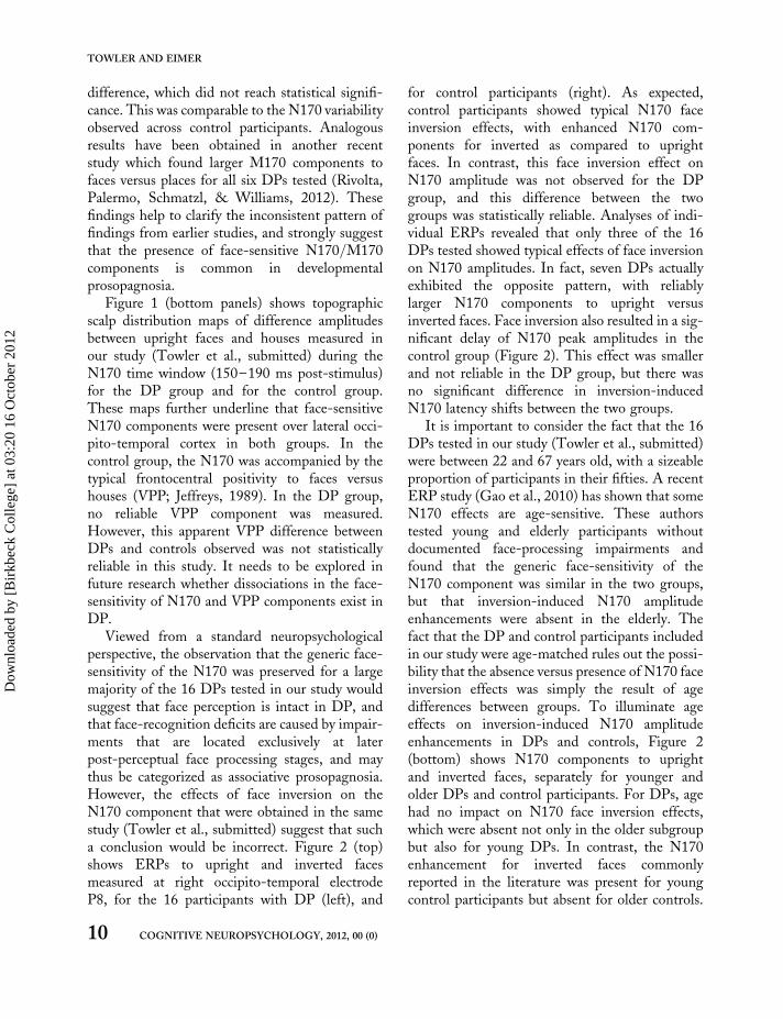
To gain more systematic insights into the prop-erties of the face-sensitive N170 component indevelopmental prosopagnosia, we recently testeda much larger sample of 16 participants with DP(Towler, Gosling, Duchaine, & Eimer, sub-mitted). They were presented with photographsof upright or inverted neutral or fearful faces andupright houses, and had to detect and respond tothe immediate repetition of an image that wasshown on the preceding trial (one-back task; seeEimer & Holmes, 2002). In addition, a group of16 control participants with intact face-recog-nition abilities was tested. As can be seen inFigure 1 (top right), the control group showedthe typical face-sensitivity of the component.N170 amplitudes at right occipito-temporal elec-trode P8 were enhanced to upright neutral facesas compared to upright houses. Importantly, theresults for the 16 participants with DP were verysimilar (Figure 1, top left): N170 amplitudeswere again larger for faces versus houses, and thesize of this effect did not differ statisticallybetween the group of DPs and the controlgroup. To check whether a face-sensitive N170component was present or absent for individualDPs, we ran non-parametric bootstrap analyses(as described by Di Nocera & Ferlazzo, 2000)for individual ERP datasets. These analysesrevealed that 12 of the 16 DPs tested had reliablylarger N170 amplitudes to faces relative to houses,and two others showed the same typical amplitude
8 COGNITIVE NEUROPSYCHOLOGY, 2012, 00 (0)
TOWLER AND EIMER
Dow
nloa
ded
by [
Bir
kbec
k C
olle
ge]
at 0
3:20
16
Oct
ober
201
2

Figure 1. Top panels: Grand-averaged ERPs elicited by upright neutral faces and upright houses at right occipito-temporal electrode P8 in the
300 ms interval after stimulus onset. ERPs are shown separately for a group of 16 DPs (left) and for a control group of 16 participants without
DP (right). Bottom panels: Topographic maps showing the scalp distribution of ERP difference amplitudes (upright neutral faces minus
upright houses) in the N170 time window (150–190 ms post-stimulus), for the DP group (left) and the control group (right). Enhanced
N170 amplitudes to faces versus houses are triggered in a similar fashion in both groups, but the VPP component is only present in the
control group. Data from Towler et al. (submitted).
COGNITIVE NEUROPSYCHOLOGY, 2012, 00 (0) 9
ERP STUDIES OF FACE PROCESSING IN DP
Dow
nloa
ded
by [
Bir
kbec
k C
olle
ge]
at 0
3:20
16
Oct
ober
201
2
difference, which did not reach statistical signifi-cance. This was comparable to the N170 variabilityobserved across control participants. Analogousresults have been obtained in another recentstudy which found larger M170 components tofaces versus places for all six DPs tested (Rivolta,Palermo, Schmatzl, & Williams, 2012). Thesefindings help to clarify the inconsistent pattern offindings from earlier studies, and strongly suggestthat the presence of face-sensitive N170/M170components is common in developmentalprosopagnosia.
Figure 1 (bottom panels) shows topographicscalp distribution maps of difference amplitudesbetween upright faces and houses measured inour study (Towler et al., submitted) during theN170 time window (150–190 ms post-stimulus)for the DP group and for the control group.These maps further underline that face-sensitiveN170 components were present over lateral occi-pito-temporal cortex in both groups. In thecontrol group, the N170 was accompanied by thetypical frontocentral positivity to faces versushouses (VPP; Jeffreys, 1989). In the DP group,no reliable VPP component was measured.However, this apparent VPP difference betweenDPs and controls observed was not statisticallyreliable in this study. It needs to be explored infuture research whether dissociations in the face-sensitivity of N170 and VPP components exist inDP.
Viewed from a standard neuropsychologicalperspective, the observation that the generic face-sensitivity of the N170 was preserved for a largemajority of the 16 DPs tested in our study wouldsuggest that face perception is intact in DP, andthat face-recognition deficits are caused by impair-ments that are located exclusively at laterpost-perceptual face processing stages, and maythus be categorized as associative prosopagnosia.However, the effects of face inversion on theN170 component that were obtained in the samestudy (Towler et al., submitted) suggest that sucha conclusion would be incorrect. Figure 2 (top)shows ERPs to upright and inverted facesmeasured at right occipito-temporal electrodeP8, for the 16 participants with DP (left), and
for control participants (right). As expected,control participants showed typical N170 faceinversion effects, with enhanced N170 com-ponents for inverted as compared to uprightfaces. In contrast, this face inversion effect onN170 amplitude was not observed for the DPgroup, and this difference between the twogroups was statistically reliable. Analyses of indi-vidual ERPs revealed that only three of the 16DPs tested showed typical effects of face inversionon N170 amplitudes. In fact, seven DPs actuallyexhibited the opposite pattern, with reliablylarger N170 components to upright versusinverted faces. Face inversion also resulted in a sig-nificant delay of N170 peak amplitudes in thecontrol group (Figure 2). This effect was smallerand not reliable in the DP group, but there wasno significant difference in inversion-inducedN170 latency shifts between the two groups.
It is important to consider the fact that the 16DPs tested in our study (Towler et al., submitted)were between 22 and 67 years old, with a sizeableproportion of participants in their fifties. A recentERP study (Gao et al., 2010) has shown that someN170 effects are age-sensitive. These authorstested young and elderly participants withoutdocumented face-processing impairments andfound that the generic face-sensitivity of theN170 component was similar in the two groups,but that inversion-induced N170 amplitudeenhancements were absent in the elderly. Thefact that the DP and control participants includedin our study were age-matched rules out the possi-bility that the absence versus presence of N170 faceinversion effects was simply the result of agedifferences between groups. To illuminate ageeffects on inversion-induced N170 amplitudeenhancements in DPs and controls, Figure 2(bottom) shows N170 components to uprightand inverted faces, separately for younger andolder DPs and control participants. For DPs, agehad no impact on N170 face inversion effects,which were absent not only in the older subgroupbut also for young DPs. In contrast, the N170enhancement for inverted faces commonlyreported in the literature was present for youngcontrol participants but absent for older controls.
10 COGNITIVE NEUROPSYCHOLOGY, 2012, 00 (0)
TOWLER AND EIMER
Dow
nloa
ded
by [
Bir
kbec
k C
olle
ge]
at 0
3:20
16
Oct
ober
201
2

Figure 2. Grand-averaged ERPs elicited by upright faces and inverted faces at right occipito-temporal electrode P8 in the 300 ms
interval after stimulus onset. Top panel: ERP waveforms measured across all 16 DPs, and all 16 control participants without
DP. The control group (right) shows typical face inversion effects on N170 amplitude. In the DP group (left), this effect is
absent. Middle and bottom panels: ERPs shown separately for subgroups of younger and older DPs, and younger and older
controls. Inversion-induced N170 amplitude enhancements were only present for younger control participants. Data from Towler
et al. (submitted).
COGNITIVE NEUROPSYCHOLOGY, 2012, 00 (0) 11
ERP STUDIES OF FACE PROCESSING IN DP
Dow
nloa
ded
by [
Bir
kbec
k C
olle
ge]
at 0
3:20
16
Oct
ober
201
2

These observations are similar to the resultsreported by Gao et al. (2010), except for the factthat their old participants were between 61 and85 years old, whereas the age range of our oldercontrols was 38–65 years. This age bracket israrely tested in ERP research, and our resultssuggest that deviations from the standard patternof N170 results typically found with young adultsmight already emerge in middle age. More gener-ally, these observations underline the criticalimportance of age matching for research on DPand other developmental disorders, in particularwhen employing measures that are sensitive tochanges in underlying neural systems.
Evidence for atypical N170 face inversioneffects in DP has been found in a previoussingle-case study (De Gelder & Stekelenburg,2005), and for three of four participants withDP in another study (Righart & De Gelder,2007). However, these findings were not substan-tiated by statistical analyses of individual ERPs,nor were DPs and controls matched by age, ascontrols in their early twenties were comparedto DPs in their forties. The fact that such atypicaleffects were observed in our study for a muchlarger sample of DPs provides potentially impor-tant new evidence for specific perceptual face-processing impairments in DP that can emergewithin 150 ms of stimulus onset. At first glance,the dissociation between the intact N170 face-sensitivity and the presence of atypical N170face inversion effects in DP is puzzling. Becausethe N170 component is associated with the per-ceptual sensory encoding of face stimuli, standardneuropsychological accounts would assume thatfocal lesions affecting this face-processing stageshould be reflected by the absence of all face-specific N170 modulations, including both itssensitivity to faces versus non-face objects andits sensitivity to face inversion. On the otherhand, if the face-sensitivity of the N170 is gener-ally preserved in DP, this would indicate that theperceptual structural encoding of faces is unim-paired. Therefore, no atypical face inversioneffects should be observed for the N170. Theresults observed in our study demonstrate thatthis line of reasoning is not correct.
It is important to note that the N170 com-ponent is not uniquely associated with a singleaspect of face processing, but instead reflects dis-tinct neural sources that appear to be linked todifferent face-sensitive brain mechanisms (e.g.,Eimer et al., 2010; Rossion & Jacques, 2008,2011; Sadeh & Yovel, 2010). For example, it hasbeen demonstrated that the N170 is sensitive tospecific face components, such as the eyes, evenwhen these are presented in isolation (Bentinet al., 1996; Itier et al., 2007). The preservedface-sensitivity of the N170 that was observedfor most DPs in our study may be a reflection oftheir ability to detect and encode individual faceparts. However, the N170 is not just a marker ofpart-based face processing, but is also sensitive toother aspects of face perception, such as the con-figural or holistic analysis of face stimuli (e.g.,Eimer et al., 2011). This is demonstrated by obser-vations that N170 components are triggered inresponse to schematic faces versus non-faces(Sagiv & Bentin, 2001) as well as to Mooneyfaces (Latinus & Taylor, 2006; Eimer et al.,2011). Because configural/holistic face-processingstages are assumed to be selectively tuned toupright faces, the absence of typical N170 faceinversion effects in DP suggests impairments atthis level: unlike control participants, DPs mayuse similar neural systems to process bothupright faces and inverted faces.
Overall, the observation that individuals withDP do not differ from individuals with normalface processing in terms of the generic face-sensi-tivity of the N170 component, but show atypicalN170 face inversion effects, cannot be easily inter-preted in terms of a simple neuropsychologicalmodel that postulates intact or damaged modulesin the face processing system which are linked tothe presence of absence of face-selective ERPcomponents. Can an alternative neurodevelop-mental approach provide a satisfactory explanationof these findings? To answer this question, it isuseful to compare the pattern of N170 modu-lations observed in DP in response to uprightfaces, inverted faces, and non-face objects withresults found in investigations of other develop-mental disorders. In addition, it should be
12 COGNITIVE NEUROPSYCHOLOGY, 2012, 00 (0)
TOWLER AND EIMER
Dow
nloa
ded
by [
Bir
kbec
k C
olle
ge]
at 0
3:20
16
Oct
ober
201
2
informative to take account of observations fromstudies that have investigated the typical develop-ment of the N170 across infancy and childhood.
Interestingly, the absence of typical N170 faceinversion effects does not appear to be restrictedto DP, but can also be found in individuals withother types of developmental disorders. In astudy of 32 high-functioning participants withautism spectrum disorder (ASD), Webb et al.(2012) found normal face-sensitivity of theN170 to faces versus houses, but a reduction ofinversion-induced N170 amplitude modulationsrelative to the effects observed for control partici-pants. Further evidence for links between atypicaleffects of face inversion on the N170 componenthave been reported by Grice et al. (2001). Theseauthors recorded ERPs in response to uprightand inverted faces from eight individuals withASD, eight individuals with WS, and eightcontrol participants. Controls showed the typicalpattern of enhanced and delayed N170 com-ponents to inverted versus upright faces. For indi-viduals with ASD, inversion-induced modulationsof N170 amplitudes were much reduced andeffects on N170 latency absent. For WS individ-uals, N170 amplitude enhancements to invertedfaces were completely absent, even though theusual latency delay was observed.
The similarity between the atypical N170 faceinversion effects observed for individuals withASD or WS and the atypical effects found for indi-viduals with DP is intriguing, and may point toshared underlying deficits in face-processingacross these three different developmental dis-orders. Webb et al. (2012) attributed the reductionof face inversion effects on N170 amplitudes in par-ticipants with ASD to a strong bias towards thepart-based processing of both upright and invertedfaces in this group. Along similar lines, Grice et al.(2001) refer to deficits in the integration of singlefacial features into a global-configural face rep-resentation as one core impairment of individualswith WS. An analogous account in terms ofimpaired configural face processing, and theresulting tendency to analyse both inverted andupright faces in a part-based fashion mightexplain the observed atypical face inversion effects
in individuals with DP. Any such deficit in percep-tual structural encoding processes that are selec-tively tuned to upright faces should result inimpaired structural representations of familiarfaces both perceptually and in visual memory,which could be responsible for some of the severeand persistent problems with face recognition thatare reported by individuals with DP.
According to a neurodevelopmental approach,further insights into the nature of the atypicalN170 face inversion effects observed in individualswith DP might also be obtained by studying thedevelopmental trajectory of the N170 componentin typically developing individuals. DifferentialERP responses to faces as compared to non-faceobjects have been found in young children andeven for six-month-old infants (De Haan,Pascalis, & Johnson, 2002; De Haan, Johnson, &Halit, 2003; Halit et al., 2003). Because the“infant N170” initially emerges about 100 mslater than the N170 in adults, it is also referredto as N290 component (De Haan et al., 2003).From the age of four years onwards, the latencyof the face-sensitive N170 component appears toresemble the latency typically observed in adultparticipants. In fact, the N170 appears to beremarkably stable in terms of its face-sensitivity,amplitude, and scalp topography between theages of four and 17 years (Kuefner, de Heering,Jacques, Palmero-Soler, & Rossion, 2010). Thesefindings suggest that the face-sensitive perceptualprocesses that are responsible for differentialN170 responses to faces versus non-face objectsdevelop rapidly, are already present in a relativelyadult-like form from early childhood, and do notchange substantially throughout development.These insights from developmental studies are rel-evant for interpreting the apparently normal face-sensitivity of the N170 component in individualswith DP. If the neural processes that producethis face-sensitivity are established early in thedevelopment of face processing, the presence oftypical N170 responses to faces versus non-faceobjects in individual DPs may point to develop-mental deficits that originate at a somewhat laterstage. In this context, the absence of age-appropri-ate face-sensitive N170 components in individual
COGNITIVE NEUROPSYCHOLOGY, 2012, 00 (0) 13
ERP STUDIES OF FACE PROCESSING IN DP
Dow
nloa
ded
by [
Bir
kbec
k C
olle
ge]
at 0
3:20
16
Oct
ober
201
2
DPs would be particularly interesting, as it wouldsuggest that their face-processing impairmentsmight have been present since early infancy, oreven from birth (Behrmann & Avidan, 2005).
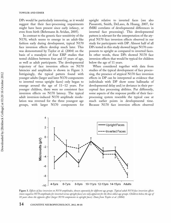
In contrast to the generic face-sensitivity of theN170, which seems to emerge in an adult-likefashion early during development, typical N170face inversion effects develop much later. Thiswas demonstrated by Taylor et al. (2004) on thebasis of a reanalysis of four ERP studies thattested children between four and 15 years of age,as well as adult participants. The developmentaltrajectory of face inversion effects on N170latencies and amplitudes is shown in Figure 3.Intriguingly, the typical pattern found withyounger adults (larger and later N170 componentsto inverted versus upright faces) only began toemerge around the age of 11–12 years. Foryounger children, there were no consistent faceinversion effects on N170 latency. The typicaladult inversion-induced N170 amplitude modu-lation was reversed for the three youngest agegroups, with larger N170 components for
upright relative to inverted faces (see alsoPassarotti, Smith, DeLano, & Huang, 2007, forfMRI correlates of developmental differences ininverted face processing). This developmentalpattern is relevant for the interpretation of the aty-pical N170 face inversion effects observed in ourstudy for participants with DP. Almost half of allDPs tested in this study showed larger N170 com-ponents to upright as compared to inverted faces.In other words, these DPs showed N170 faceinversion effects that would be typical for childrenbelow the age of 11 years.
When considered together with data fromstudies of the typical development of face proces-sing, the presence of atypical N170 face inversioneffects in DP can be interpreted as evidence thatindividuals with DP show some hallmarks ofdevelopmental delay and/or deviance in their per-ceptual face processing abilities. Put differently,some aspects of the response profile of their face-processing system resemble the typical case atmuch earlier points in developmental time.Because N170 face inversion effects observed
Figure 3. Effects of face inversion on N170 amplitudes, shown separately for different age groups. Typical adult N170 face inversion effects
(more negative N170 amplitudes for inverted versus upright faces) are only apparent for the three oldest age groups. Children below the age of
10 years show the opposite effect (larger N170 components to upright faces). Data from Taylor et al. (2004).
14 COGNITIVE NEUROPSYCHOLOGY, 2012, 00 (0)
TOWLER AND EIMER
Dow
nloa
ded
by [
Bir
kbec
k C
olle
ge]
at 0
3:20
16
Oct
ober
201
2
during typical development show a systematic tra-jectory towards the patterns observed in adult-hood, they could provide valuable markers forthe emerging tuning of face perception to canoni-cally oriented upright faces. In this context, thepresence of atypical N170 face inversion effectsin DP may provide important clues with respectto the point in the development of face perceptionwhere selective processing deficits first emerge.Such an interpretation of N170 face inversioneffects also needs to take account of the differencesobserved between younger and older control par-ticipants (Figure 2). We will return to this pointin the final section.
EXPLICIT AND COVERT FACERECOGNITION INDEVELOPMENTALPROSOPAGNOSIA: EVIDENCEFROM THE N250 COMPONENT
As discussed in the previous section, the N170component is linked to early perceptual stages offace processing that precede the explicit recog-nition and identification of individual faces.Because DPs are specifically impaired in theirability to recognize or identify familiar faces,they may also show atypical patterns of ERP com-ponents that are more directly associated with facerecognition. In typical adult participants, ERPmarkers of identity-related face processing firstemerge at post-stimulus latencies of about 250ms. An enhanced occipito-temporal negativity iselicited in response to the faces of personallyknown or famous individuals, as well as to therepeated presentation of a previously unfamiliarface (e.g., Schweinberger, Pfutze, & Sommer,1995; Tanaka, Curran, Porterfield, & Collins,2006; Gosling & Eimer, 2011). This N250 com-ponent is assumed to reflect the activation of astored representation of a specific face in visualmemory that is triggered when there is a matchwith a currently presented face. It is typically fol-lowed by a broadly distributed sustained positivity(P600f) to familiar faces (Eimer, 2000a; Bentin &Deouell, 2000). The P600f has been linked to later
stages of face recognition, such as name retrieval oraccess to semantic information about a specificindividual (e.g., Gosling & Eimer, 2011).
Because ERP research into the neural basis ofAP and DP has so far focused on the N170 com-ponent, little is known about how ERP com-ponents associated with face recognition such asthe N250 or P600f are affected in prosopagnosia.In one study of face recognition in AP (Eimer,2000a), ERPs to famous and non-famous faceswere measured for patient PHD. As reportedearlier, this patient did not show any N170 face-selectivity (Eimer & McCarthy, 1999), demon-strating damage to early perceptual stages of faceprocessing. PHD did also not show any evidencefor identity-sensitive ERP components at longerlatencies. This would be predicted by neuropsy-chologically inspired models of adult face proces-sing (e.g., Bruce & Young, 1986), as deficits atthe level of structural encoding should have detri-mental knock-on effects on later stages of face rec-ognition and identification.
A recent study conducted in our lab (Eimer,Gosling, & Duchaine, 2012) was designed tospecifically investigate ERP markers of face recog-nition for a group of 12 participants with DP.They watched sequentially presented photographsof famous and non-famous faces, and had to cat-egorize them in terms of their identity. Each facecould be classified as definitely known, familiar,unfamiliar, or definitely unknown. Famous faceswere those of actors, politicians, musicians, sportspersonalities, and other celebrities well known inthe UK. Controls correctly classified more than80% of these famous faces as known or familiar(Gosling & Eimer, 2011), whereas DPs recog-nized less than 30% of the same famous faces.Figure 4 (top left) shows ERPs measured at rightoccipito-temporal electrode P8 to famous faceson those relatively few trials where DPs correctlyclassified these faces as known or familiar, and tonon-famous faces that were correctly judged tobe unfamiliar/unknown. This figure also includesa topographical map of ERP difference amplitudesbetween these famous and non-famous faces in theN250 time window (230–400 ms post-stimulus).For comparison, analogous ERP waveforms and
COGNITIVE NEUROPSYCHOLOGY, 2012, 00 (0) 15
ERP STUDIES OF FACE PROCESSING IN DP
Dow
nloa
ded
by [
Bir
kbec
k C
olle
ge]
at 0
3:20
16
Oct
ober
201
2
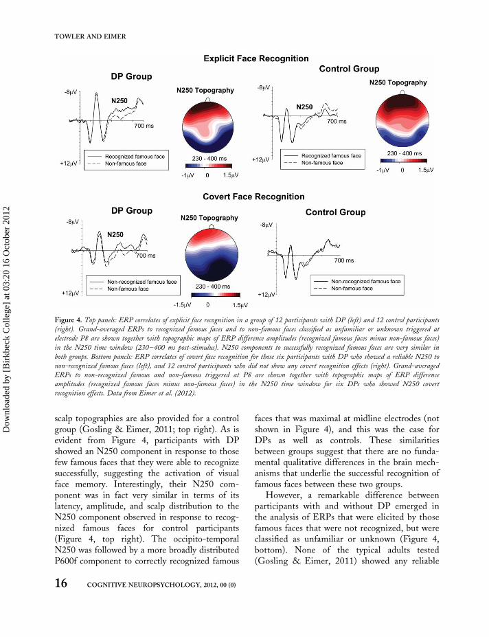
scalp topographies are also provided for a controlgroup (Gosling & Eimer, 2011; top right). As isevident from Figure 4, participants with DPshowed an N250 component in response to thosefew famous faces that they were able to recognizesuccessfully, suggesting the activation of visualface memory. Interestingly, their N250 com-ponent was in fact very similar in terms of itslatency, amplitude, and scalp distribution to theN250 component observed in response to recog-nized famous faces for control participants(Figure 4, top right). The occipito-temporalN250 was followed by a more broadly distributedP600f component to correctly recognized famous
faces that was maximal at midline electrodes (notshown in Figure 4), and this was the case forDPs as well as controls. These similaritiesbetween groups suggest that there are no funda-mental qualitative differences in the brain mech-anisms that underlie the successful recognition offamous faces between these two groups.
However, a remarkable difference betweenparticipants with and without DP emerged inthe analysis of ERPs that were elicited by thosefamous faces that were not recognized, but wereclassified as unfamiliar or unknown (Figure 4,bottom). None of the typical adults tested(Gosling & Eimer, 2011) showed any reliable
Figure 4. Top panels: ERP correlates of explicit face recognition in a group of 12 participants with DP (left) and 12 control participants
(right). Grand-averaged ERPs to recognized famous faces and to non-famous faces classified as unfamiliar or unknown triggered at
electrode P8 are shown together with topographic maps of ERP difference amplitudes (recognized famous faces minus non-famous faces)
in the N250 time window (230–400 ms post-stimulus). N250 components to successfully recognized famous faces are very similar in
both groups. Bottom panels: ERP correlates of covert face recognition for those six participants with DP who showed a reliable N250 to
non-recognized famous faces (left), and 12 control participants who did not show any covert recognition effects (right). Grand-averaged
ERPs to non-recognized famous and non-famous triggered at P8 are shown together with topographic maps of ERP difference
amplitudes (recognized famous faces minus non-famous faces) in the N250 time window for six DPs who showed N250 covert
recognition effects. Data from Eimer et al. (2012).
16 COGNITIVE NEUROPSYCHOLOGY, 2012, 00 (0)
TOWLER AND EIMER
Dow
nloa
ded
by [
Bir
kbec
k C
olle
ge]
at 0
3:20
16
Oct
ober
201
2

ERP differences between non-recognized famousfaces and non-famous faces that were correctlyclassified as unfamiliar or unknown. In otherwords, there was no electrophysiological evidencefor covert face recognition (Diamond, Valentine,Mayes, & Sandel, 1994) in these participants. Incontrast, a clear and statistically reliable N250component was observed in response to non-recognized famous faces for six of the 12 DPstested. Moreover, the scalp topography of thiscomponent (Figure 4, bottom left) was similarto the topography observed for explicitly recog-nized faces. Even though these six DPs classifiedthe majority of famous faces as unfamiliar orunknown, N250 components clearly distin-guished between these famous faces and objec-tively non-famous faces. These results suggestthat many famous faces that were not recognizedby participants with DP did activate stored visualrepresentations of familiar faces. They also indi-cate that the activation of these visual represen-tations is not sufficient for overt facerecognition, in line with current functionalmodels of normal face processing (Bruce andYoung, 1986; Burton et al., 1990). Consciousrecognition may require that faces are also pro-cessed at subsequent post-perceptual semanticstages, which are associated with the laterP600f component. Interestingly, non-recognizedfamous faces did not trigger a P600f component,even for those DPs who showed covert recog-nition effects for the N250 component, whichfurther substantiates the link between the P600fand conscious face recognition.
The identification of the N250 component asan electrophysiological marker of covert face rec-ognition in DP extends previous behavioural find-ings by Avidan and Behrmann (2008) and Rivolta,Palermo, Schmalzl, & Coltheart (2012a). Overall,these observations can be readily interpreted by astandard neuropsychological account. The pres-ence of an N250 component to non-recognizedfamous faces in some individuals with DP indi-cates that these faces were able to successfully acti-vate corresponding representations in visualmemory, even though there was no overt recog-nition. Together with the fact that the P600f
component was absent for the same faces, thisobservation points towards a relatively late locusof face-recognition impairments for these DPs.Functionally, their deficit appears to be caused bya disruption of the links between stored visual rep-resentations of familiar individuals and semantic orepisodic representations in long-term memory. Inother words, their profile could resemble that ofpatients with acquired associative prosopagnosia(De Renzi et al., 1991). In contrast, for thoseother six DPs who showed no N250 componentto non-recognized famous faces, the core face pro-cessing deficit may be located at an earlier stage,such as the impaired structural descriptions offaces, and/or poor representations of familiarfaces in visual memory (e.g., Schweinberger &Burton, 2003). In this sense, they could besimilar to patients with acquired apperceptive pro-sopagnosia (De Renzi et al., 1991).
Overall, the pattern of identity-sensitive ERPcomponents observed for participants with DPappears to provide evidence for considerableresidual normality of face recognition. Asexpected, individuals with DP were much lesslikely than controls to recognize images offamous faces. However, on those relatively infre-quent occasions where face recognition was suc-cessful, it was accompanied by the same patternof N250 and P600f components that was observedduring explicit face recognition in participantswith normal face recognition, suggesting qualitat-ively similar neural processes in the two groups(Eimer et al., 2012). This is in line with fMRIresults discussed previously, which demonstrate asubstantial degree of functionality in the visualareas responsible for face recognition, at least insome DPs (e.g., Avidan et al., 2005). From anadult neuropsychological perspective, the presenceof N250 components as markers of covert face rec-ognition in some participants with DP, and theabsence of such covert recognition effects inothers, may reflect apperceptive and associativesubtypes of DP; that is, selective deficits at rela-tively early or late stages, or severed linksbetween specific subprocesses, within an otherwiseintact face-processing system. This interpretationis in line with functional imaging studies
COGNITIVE NEUROPSYCHOLOGY, 2012, 00 (0) 17
ERP STUDIES OF FACE PROCESSING IN DP
Dow
nloa
ded
by [
Bir
kbec
k C
olle
ge]
at 0
3:20
16
Oct
ober
201
2
showing that disconnection between the coreventral occipito-temporal regions and theextended face network is implicated in face-recog-nition difficulties in DP (e.g., Avidan &Behrmann, 2009; Thomas et al., 2009).
How does the apparent residual normality offace recognition in DP relate to the atypicalpattern of N170 effects observed for participantswith DP? The N170 results discussed in“Impairments of face perception in developmentalprosopagnosia” above suggest that aspects of thestructural encoding of faces are atypical in DP,while the findings reported in this section showthat DPs show an apparently typical pattern ofN250 and P600f components during successfulface recognition. This difference may be moreapparent than real. There is now some evidencethat N250 components are not exclusively associ-ated with face recognition, but are instead amore general marker of successful matchesbetween visual inputs and object representationsstored in visual memory. For example, Scott,Tanaka, Sheinberg, and Curran (2006, 2008)observed N250 components during categorizationtasks that required a within-class discrimination ofobjects (in particular objects of expertise),suggesting that the N250 is a generic reflectionof a match between incoming visual informationand visual memory. Its presence in individualswith DP indicates that although atypical face pro-cessing at early perceptual stages results inimpaired visual face representations, a successfulmatch with visual face memory is still possible onsome occasions, and that the matching processitself operates normally.
SUMMARY AND CONCLUSIONS
This review of research into the neural basis offace-processing deficits in DP, with its specificemphasis on recent ERP studies, had two objec-tives. The first aim was to discuss the implicationsof this research with respect to which face proces-sing mechanisms are affected in individuals withDP. Another more theoretical aim was to considerwhether a standard neuropsychological model of
impairments of face-selective modules provides asatisfactory account of developmental prosopagno-sia, or whether a wider neurodevelopmental per-spective is also required.
In acquired prosopagnosia, the face-sensitiveN170 component is often absent, in particularwhen lesions include core face-processingsystems. Consistent with the neuropsychologicalmodel, this suggests close links between selectiveimpairments in the structural encoding of facesand the structural integrity of posterior brainareas involved in face perception. In contrast, asdiscussed above, the face-sensitivity of the N170component is present for most individuals withdevelopmental prosopagnosia. This result addsan important temporal dimension to observationsfrom MRI studies, because it demonstrates thatthe initial face-sensitive activation of these areasoccurs rapidly in participants with DP; that is,within approximately 150 ms after stimulusonset. In this respect, face processing in DPappears to follow a similar time-course to face pro-cessing in typical individuals. However, the pres-ence of a face-sensitive N170 component in mostDPs does not imply that their face perceptionmechanisms operate in exactly the same fashionas in controls. There were reliable differencesbetween DPs and control participants withrespect to N170 enhancements caused by faceinversion. At the group level, these effects werepresent for controls and absent for DPs. It isimportant to note that the presence of inversion-induced N170 amplitude enhancements was age-dependent in the control group, as it was presentfor young but absent for older controls.However, the fact that these effects were absentin DPs regardless of their age provides clear evi-dence that the processing of upright versusinverted faces is atypical in DP.
This dissociation between typical N170 face-sensitivity and atypical N170 face inversion effectsin DP is difficult to reconcile with the dichotomyof a perceptual structural encoding module that iseither intact or damaged. A different perspectivefor the interpretation of these findings is offeredby an alternative neurodevelopmental account.The presence of atypical N170 face inversion
18 COGNITIVE NEUROPSYCHOLOGY, 2012, 00 (0)
TOWLER AND EIMER
Dow
nloa
ded
by [
Bir
kbec
k C
olle
ge]
at 0
3:20
16
Oct
ober
201
2

effects does not appear to be restricted to DP, buthas been observed also for other developmental dis-orders such as ASD and WS (Grice et al., 2001;Webb et al., 2012). In addition, the absence ofinversion-induced N170 amplitude modulationsfor participants with DP is reminiscent of thepattern observed for typically developing childrenbelow the age of 11 years (Taylor et al., 2004),and apparently also for older adults without face-processing impairments (Gao et al., 2010). In con-trast, the generic face-sensitivity of the N170,which is largely preserved in DP, is alreadypresent at a much earlier stage of the typical devel-opment of the face-processing system (Kuefneret al., 2010). If the trajectory of the N170 com-ponent across development reflects the gradualtuning of face perception mechanisms towards thetypical young adult state, the pattern of N170effects observed for individuals with DP suggeststhat their face perception has never reached thisend state, because its development was delayed orhas deviated from this typical trajectory at a specificpoint during developmental time.
As discussed above, the presence of inversion-induced N170 amplitude enhancements is oftenregarded as evidence that only inverted and notupright faces are able to recruit object-selectiveneurons (e.g., Rossion et al., 2000). In this context,the absence of N170 differences between uprightand inverted faces can be interpreted as evidencefor an enhanced capacity of upright faces to activateobject-selective areas, or, more generally, for areduction in the face-specificity of visual processing.The observation that N170 face inversion effects areabsent in young children and older adults, as well asin individuals with DP or other developmental dis-orders, may be an important pointer towards lessface-selective functional specialisation withinventral visual areas in these groups. In support ofthis hypothesis, it has been shown that activationin face-selective regions becomes progressivelymore specialized through childhood into adulthood(Golarai et al., 2007), and that these face-selectiveregions become less differentiated and specializedwith age (Park et al., 2004; Goh, Suzuki, & Park,2010). Furthermore, individuals with ASD showreduced or atypical neural specialization for faces
(e.g. Schultz et al., 2000; Pierce, Muller, Ambrose,Allen, & Courchesne, 2001). Atypical N170 faceinversion effects and reduced levels of face-specificfunctional specialization could both be linked to areduction in the connectivity of the right inferior-occipital fasciculus, which has been observed for asmall group of DPs (Thomas et al., 2009), and isalso associated with an age-related decline of faceperception (Thomas et al., 2008). It should benoted that individuals with WS do not show areduction in face selectivity, but instead have largerFFAs than matched controls, again demonstratingan atypical neural specialization for faces (Golaraiet al., 2010).
While the presence of atypical N170 face inver-sion effects points to observable differences in theneurodevelopmental history of the face-processingarchitecture in individuals with and without DP,the pattern of identity-sensitive ERP componentsdescribed above provides evidence for functionallyand neurally similar face-recognition mechanismsin both groups. N250 and P600f components inresponse to successfully recognized famous faceswere very similar for a group of DPs and forcontrol participants (Eimer et al., 2012), suggestingthat the same neural processes were active duringface recognition in the two groups. In particular,the presence of an apparently normal N250 com-ponent in individuals with DP (see Figure 4)demonstrates that at least on some occasions,stored representations of familiar faces in visualmemory were successfully activated by incomingperceptual face representations. The fact that DPswere much less likely than controls to recognizeitems from the same set of famous faces may bedue to the impaired structural encoding of currentlypresented faces, the poor quality of stored visualrepresentations of familiar faces (see Gruter,Gruter, Bell, & Carbon, 2009, for impairments ofvisual imagery in DP), or a combination of thetwo factors. In addition, it appears that for asubset of DPs, face-recognition impairments arethe result of severed links between stored face rep-resentations in visual memory and semantic or epi-sodic memory.
We wish to conclude this review with one meth-odological and one more general theoretical point.
COGNITIVE NEUROPSYCHOLOGY, 2012, 00 (0) 19
ERP STUDIES OF FACE PROCESSING IN DP
Dow
nloa
ded
by [
Bir
kbec
k C
olle
ge]
at 0
3:20
16
Oct
ober
201
2
As regards methodology, neuroscientific researchinto acquired or developmental impairments of cog-nitive processing and their neural basis has oftenbeen conducted via single case studies or studieswith very small sample sizes. This is also the casefor most behavioural, ERP, and fMRI studies ofDP. This approach is clearly problematic wheninvestigating deficits that show considerable hetero-geneity across individuals, as is likely to be the casefor DP. Even though all DPs show severely impairedvisual face recognition, the functional and neuralbasis of this deficit may be very different in differentindividuals, and this may account for the fact thatmany of the studies reviewed earlier have producedinconsistent or inconclusive results. For thisreason, the use of larger sample sizes is one obviousrequirement for future research into DP and itsneural basis. In addition, research needs to focusnot just on generic differences in face processingbetween larger groups of DPs and control partici-pants, but also on possible differences in the func-tional and neural architectures that implement faceprocessing across individuals with DP, as well ason age-related differences in DPs and control par-ticipants. These requirements necessitate a com-bined approach, where the analysis of differencesbetween DPs and control participants at the grouplevel is ideally complemented by the additionalanalysis of behavioural and neural measures at theindividual level. Non-parametric bootstrap analyses(e.g., Di Nocera & Ferlazzo, 2000) provide auseful tool to ascertain the reliability of the effectsof specific experimental manipulations for individualparticipants. As discussed above, this combined caseseries approach may lead to new insights into distinctsubtypes of DP and their neural basis.
The final theoretical point goes back to theimportant difference between neuropsychologicaland neurodevelopmental accounts of the originsof DP, as highlighted in the first section of thisreview. Proponents of an innate modular view offace processing and its developmental trajectory(e.g., Duchaine et al., 2006; McKone,Kanwisher, & Duchaine, 2007) argue that coredeficits may be located at a relatively highdomain-specific level of cortical processing.Individuals with DP fail to develop a normally
operating face-processing system, while otheraspects of visual perception and cognitiondevelop independently and normally, resulting inintact visual processing in non-face domains. Incontrast, advocates of a neurodevelopmental per-spective (e.g., Thomas & Karmiloff-Smith,2002) propose that the deficit responsible fordevelopmental disorders such as DP will beexplained by lower-level mechanisms which aredomain-relevant, and that these effects willcascade into domain-specific processes over devel-opmental time (Karmiloff-Smith, 1998). As a con-sequence, the resulting impairments are notconfined to a single innate module, but should beat least subtly evident across domains that rely onshared mechanisms for typical development.Furthermore, neurodevelopmental approaches toface processing (e.g., Johnson et al., 2009) expli-citly acknowledge the possibility that the face-specificity of different brain regions can bothincrease and decrease in the course of development(see Joseph, Gathers, & Bhatt, 2011, for evidence).This is very much consistent with our suggestionthat a reduced or otherwise atypical level of func-tional specialization for faces in ventral visualcortex could be a core feature of DP and otherdevelopmental disorders.
Although there is ample evidence of protracteddevelopmental change in face processing (e.g.,Joseph et al., 2011; Taylor et al., 2004), othershave recently argued that the role of experiencein the development of face recognition may havebeen overestimated (e.g., McKone, Crookes,Jeffery, & Dilks, in press). Future research intothe development of DP will be relevant for thisdebate, and this research is likely to profit fromtaking both neuropsychological and neurodevelop-mental perspectives into account. The fact thatadult face processing is to some extent modular(Busigny et al., 2010; Moscovitch, Winocur, &Behrmann, 1997) does not imply that it developsindependently from other aspects of visual cogni-tion, because there may be important contributionsfrom lower-level domain-relevant mechanismsduring development. Such mechanisms couldinclude local biases in visual attention, theabsence of attentional biases towards face-like
20 COGNITIVE NEUROPSYCHOLOGY, 2012, 00 (0)
TOWLER AND EIMER
Dow
nloa
ded
by [
Bir
kbec
k C
olle
ge]
at 0
3:20
16
Oct
ober
201
2

stimuli or the eye region, difficulties in rapidlybinding together spatially separate componentsof visual objects, or impairments of automaticwithin-category object individuation processes.The atypical operation of such processes mayhave a disproportionate impact during sensitiveperiods of early development when higher-leveldomain-specific visual processes are becomingestablished (e.g., Le Grand, Mondloch, Maurer,& Brent, 2004). These cognitive and other low-level neural biases could contribute, either bythemselves or in combination, to the impairedface recognition that we observe in adult DPs. Incontrast to control participants, some individualswith DP perform better in response to local ascompared to global elements of hierarchicalNavon stimuli (Behrmann & Avidan, 2005;Avidan, Tanzer, & Behrmann, 2011; but seeDuchaine, Yovel, & Nakayama, 2007b, for differ-ent results), suffer less interference when proces-sing aligned composite faces (Palermo et al.,2011), and often show no or reduced behaviouralface inversion effects (e.g., Avidan et al., 2011).Interestingly, some DPs show a negative corre-lation between their local bias on a non-faceNavon task and global interference on the compo-site face task (Avidan et al., 2011), which wouldnot be predicted by innately modular accounts oftheir face processing deficits. Overall, such find-ings suggest that many individuals with DPshow a bias towards the local part-based proces-sing of visual objects. If such a bias emergesearly, it could affect the typical development of aconfigural or holistic mode of face processingthat is tuned to canonically oriented uprightfaces. A consequence of such a low-level butdomain-relevant atypical bias of visual processingis that individuals with DP will never developthe special mechanisms used for upright face rec-ognition and will consequently analyse bothupright and inverted faces in a similar fashion,and thus are impaired in tasks that depend onthe configural or holistic processing of uprightfaces.
While the ERP results discussed in this articleare consistent with both modular and neurodeve-lopmental accounts of DP, they highlight the
utility of an integrated approach whereby theprocess of typical and atypical development is con-sidered alongside adult and neuropsychologicalevidence in the understanding of neurodevelop-mental disorders. Even though research into thefunctional, neural and genetic basis of DP hasonly begun, one fact that has already becomeevident is the heterogeneity of this condition.The debate between proponents of modular neu-ropsychological and neuroconstructivist accountsof developmental prosopagnosia is certain tocontinue.
Manuscript received 18 November 2011
Revised manuscript received 6 July 2012
Revised manuscript accepted 13 July 2012
First published online 16 October 2012
REFERENCES
Alonso-Prieto, E., Caharel, S., Henson, R. N., &Rossion, B. (2011). Early (N170/M170) face-sensi-tivity despite right lateral occipital brain damage inacquired prosopagnosia. Frontiers in Human
Neuroscience, 5, 138.Annaz, D., Karmiloff-Smith, A., Johnson, M. H., &
Thomas, M. S. C. (2009). A cross-syndrome studyof the development of holistic face recognition inchildren with autism, Down syndrome, andWilliams syndrome. Journal of Experimental Child
Psychology, 102, 456–486.Avidan, G., & Behrmann, M. (2008). Implicit famili-
arity processing in congenital prosopagnosia.Journal of Neuropsychology, 2, 141–164.
Avidan, G., & Behrmann, M. (2009). Functional MRIreveals compromised neural integrity of the face pro-cessing network in congenital prosopagnosia. Current
Biology, 19, 1146–1150.Avidan, G., Hasson, U., Malach, R., & Behrmann, M.
(2005). Detailed exploration of face related proces-sing in congenital prosopagnosia: 2. Functional neu-roimaging findings. Journal of Cognitive Neuroscience,17, 1150–1167.
Avidan, G., Tanzer, M., & Behrmann, M. (2011).Impaired holistic processing in congenital prosopag-nosia. Neuropsychologia, 49, 2541–2552.
Barton, J. J. S. (2008). Structure and function inacquired prosopagnosia: Lessons from a series of 10
COGNITIVE NEUROPSYCHOLOGY, 2012, 00 (0) 21
ERP STUDIES OF FACE PROCESSING IN DP
Dow
nloa
ded
by [
Bir
kbec
k C
olle
ge]
at 0
3:20
16
Oct
ober
201
2

patients with brain damage. Journal of
Neuropsychology, 2, 197–225.Behrmann, M., & Avidan, G. (2005). Congenital pro-
sopagnosia: Face blind from birth. Trends in
Cognitive Sciences, 9, 180–187.Behrmann, M., Avidan, G., Gao, F., & Black, S. (2007).
Structural imaging reveals anatomical alterations ininferotemporal cortex in congenital prosopagnosia.Cerebral Cortex, 17, 2354–2363.
Behrmann, M., Avidan, G., Marotta, J. J., & Kimchi, R.(2005). Detailed exploration of face-related proces-sing in congenital prosopagnosia: 1. Behavioral find-ings. Journal of Cognitive Neuroscience, 17,
1130–1149.Bellugi, U., Lichtenberger, L., Jones, W., Lai, Z., &
St George, M. (2000). The neurocognitive profileof Williams syndrome: A complex pattern ofstrengths and weaknesses. Journal of Cognitive
Neuroscience, 12, 7–29.Bentin, S., Allison, T., Puce, A., Perez, E., &
McCarthy, G. (1996). Electrophysiological studiesof face perception in humans. Journal of Cognitive
Neuroscience, 8, 551–565.Bentin, S., DeGutis, J. M., D’Esposito, M., &
Robertson, L. C. (2007). Too many trees to see theforest: Performance, ERP and fMRI manifestationsof integrative congenital prosopagnosia. Journal of
Cognitive Neuroscience, 19, 132–146.Bentin, S., & Deouell, L. Y. (2000). Structural encoding
and identification in face processing: ERP evidencefor separate mechanisms. Cognitive Neuropsychology,17, 35–54.
Bentin, S., Deouell, L. Y, & Soroker, N. (1999).Selective visual streaming in face recognition:Evidence from developmental prosopagnosia.NeuroReport, 10, 823–827.
Bishop, D. V. M. (1997). Cognitive neuropsychologyand developmental disorders: Uncomfortable bedfel-lows. Quarterly Journal of Experimental Psychology:
Human Experimental Psychology, 50, 899–923.Bobes, M., Lopera, F., Dias Comas, L., Galan, L.,
Carbonell, F., Bringas, M. L., & Valdes-Sosa, M.(2004). Brain potentials reflect residual face proces-sing in a case of prosopagnosia. Cognitive
Neuropsychology, 21, 691–718.Bodamer, J. (1947). Die Prosop-Agnosie. (Die Agnosie
des Physiognomieerkennens). Archiv fur Psychiatrie
und Nervenkrankheiten, 179, 6–53.Botzel, K., & Grusser, O. J. (1989). Electric brain
potentials evoked by pictures of faces and non-faces:
A search for face-specific EEG-potentials.Experimental Brain Research, 77, 349–360.
Brown, J., Johnson, M. H., Paterson, S., Gilmore, R.,Longhi, E., & Karmiloff-Smith, A. (2003). Spatialrepresentation and attention in toddlers withWilliams syndrome and Down syndrome.Neuropsychologia, 41, 1037–1046.
Bruce, V., & Young, A. (1986). Understanding face rec-ognition. British Journal of Psychology, 77, 305–327.
Burton, A. M., & Bruce, V. (1992). I recognise your facebut I can’t remember your name: A simple expla-nation? British Journal of Psychology, 83, 45–60.
Burton, A. M., Bruce, V., & Johnston, R. A. (1990).Understanding face recognition with an interactiveactivation model. British Journal of Psychology, 81,
361–380.Busigny, T., Joubert, S., Felician, O., Ceccaldi, M., &
Rossion, B. (2010). Holistic perception of the indi-vidual face is specific and necessary: Evidence froman extensive case study of acquired prosopagnosia.Neuropsychologia, 48, 4057–4092.
Calder, A. J., Keane, J., Manes, F., Antoun, N., &Young, A. W. (2000). Impaired recognition andexperience of disgust following brain injury. Nature
Neuroscience, 3, 1077–1078.Calder, A. J., & Young, A. W. (2005). Understanding
the recognition of facial identity and facialexpression. Nature Reviews Neuroscience, 6, 641–651.
Calder, A. J., Young, A. W., Rowland, D., Perrett, D. I.,Hodges, J. R., & Etcoff, N. L. (1996). Facialemotion recognition after bilateral amygdaladamage: Differentially severe impairment of fear.Cognitive Neuropsychology, 13, 699–745.
Caramazza, A. (1986). On drawing inferences about thestructure of normal cognitive systems from the analy-sis of patterns of impaired performance: The case forsingle-patient studies. Brain and Cognition, 5, 41–66.
Dalrymple, K., Oruc, I., Duchaine, B., Pancaroglu, R.,Fox, C. J., Iaria, G., . . . Barton, J. J. (2011). The neu-roanatomic basis of the face-selective N170 inacquired prosopagnosia, a combined ERP/fMRIstudy. Neuropsychologia, 49, 2553–2563.
De Gelder, B., & Stekelenburg, J. J. (2005). Naso-tem-poral asymmetry for the N170 for processing faces innormal viewers but not in developmental prosopag-nosia. Neuroscience Letters, 376, 40–45.
De Haan, M., Johnson, M. H., & Halit, H. (2003).Development of face-sensitive event-related poten-tials during infancy: A review. International Journal
of Psychophysiology, 51, 45–58.
22 COGNITIVE NEUROPSYCHOLOGY, 2012, 00 (0)
TOWLER AND EIMER
Dow
nloa
ded
by [
Bir
kbec
k C
olle
ge]
at 0
3:20
16
Oct
ober
201
2
De Haan, M., Pascalis, O., & Johnson, M. H. (2002).Specialization of neural mechanisms underlyingface recognition in human infants. Journal of
Cognitive Neuroscience, 14, 199–209.De Haan, E. H. F., Young, A. W., & Newcombe, F.
(1991). A dissociation between the sense of famili-arity and access to semantic information concerningfamiliar people. European Journal of Cognitive
Psychology, 3, 51–67.Dehaene, S., Piazza, M., Pinel, P., & Cohen, L. (2003).
Three parietal circuits for number processing.Cognitive Neuropsychology, 20, 487–506.
De Renzi, E., Faglioni, P., Grossi, D., & Nichelli, P.(1991). Apperceptive and associative forms of proso-pagnosia. Cortex, 27, 213–221.
Deruelle, C., Mancini, J., Livet, M. O., Casse-Perrot,C., & de Schonen, S. (1999). Configural and localprocessing of faces in children with Williams syn-drome. Brain and Cognition, 41, 276–98.
Di Nocera, F., & Ferlazzo, F. (2000). Resamplingapproach to statistical inference: Bootstrappingfrom event-related potentials data. Behaviour
Research Methods, Instruments & Computers, 32,
111–119.Diamond, B. J., Valentine, T., Mayes, A. R., &
Sandel, M. E. (1994). Evidence of covert recog-nition in a prosopagnosic patient. Cortex, 30,
377–393.Donnai, D., & Karmiloff-Smith, A. (2000). Williams
syndrome: From genotype through to the cognitivephenotype. American Journal of Medical Genetics, 97,
164–171.Duchaine, B. (2011). Developmental prosopagnosia:
Cognitive, neural, and developmental investigations.In A. J. Calder et al. (Eds.), The Oxford handbook of
face perception (pp. 821–838). Oxford, UK: OxfordUniversity Press.
Duchaine, B., Germine, L., & Nakayama, K. (2007a).Family resemblance: Ten family members with pro-sopagnosia and within-class object agnosia. Cognitive
Neuropsychology, 24, 419–430.Duchaine, B., & Weidenfeld, A. (2003). An evaluation
of two commonly used tests of unfamiliar face recog-nition. Neuropsychologia, 41, 713–720.
Duchaine, B., Yovel, G., Butterworth, E., & Nakayama,K. (2006). Prosopagnosia as an impairment to face-specific mechanisms: Elimination of the alternativeexplanations in a developmental case. Cognitive
Neuropsychology, 23, 714–747.Duchaine, B., Yovel, G., & Nakayama, K. (2007b). No
global processing deficit in the Navon task in 14
developmental prosopagnosics. Social Cognitive and
Affective Neuroscience, 2, 104–113.Eimer, M. (2000a). Event-related brain potentials dis-
tinguish processing stages involved in face perceptionand recognition. Clinical Neurophysiology, 111,
694–705.Eimer, M. (2000b). Effects of face inversion on the
structural encoding and recognition of faces:Evidence from event-related brain potentials.Cognitive Brain Research, 10, 145–158.
Eimer, M. (2011). The face-sensitive N170 componentof the event-related brain potential. In A. J. Calderet al. (Eds.) , The Oxford handbook of face perception
(pp. 329–344). Oxford, UK: Oxford UniversityPress.
Eimer, M., Gosling, A., & Duchaine, B. (2012).Electrophysiological markers of covert face recog-nition in developmental prosopagnosia. Brain, 135,
524–554.Eimer, M., Gosling, A., Nicholas, S., & Kiss, M.
(2011). The N170 component and its links to config-ural face processing: A rapid neural adaptation study.Brain Research, 1376, 76–87.
Eimer, M., & Holmes, A. (2002). An ERP study onthe time course of emotional face processing.NeuroReport, 13, 427–431.
Eimer, M., Kiss, M., & Nicholas, S. (2010). Responseprofile of the face-sensitive N170 component: Arapid adaptation study. Cerebral Cortex, 20,
2442–2452.Eimer, M., & McCarthy, R. (1999). Prosopagnosia and
structural encoding of faces: Evidence from event-related potentials. NeuroReport, 10, 255–259.
Ellis, H. D., & Young, A. W. (1990). Accounting fordelusional misidentifications. British Journal of
Psychiatry, 157, 239–248.Ellis, A. W., & Young, A. W. (1996). Human cognitive
neuropsychology: A textbook with readings. Hove, UK:Psychology Press.
Flude, B. M., Ellis, A. W., & Kay, J. (1989). Face pro-cessing and name retrieval in an anomic aphasic:Names are stored separately from semantic infor-mation about familiar people. Brain and Cognition,11, 60–72.
Furl, N., Garrido, L., Dolan, R. J., Driver, J., &Duchaine, B. (2011). Fusiform gyrus face selectivityrelates to individual differences in facial recognitionability. Journal of Cognitive Neuroscience, 23,
1723–1740.Gao, L., Xu, J., Zhang, B. W., Zhao, L., Harel, A., &
Bentin, S. (2010). Aging effects on early-stage face
COGNITIVE NEUROPSYCHOLOGY, 2012, 00 (0) 23
ERP STUDIES OF FACE PROCESSING IN DP
Dow
nloa
ded
by [
Bir
kbec
k C
olle
ge]
at 0
3:20
16
Oct
ober
201
2
perception: An ERP study. Psychophysiology, 46,
970–983.Garrido, L., Furl, N., Draganski, B., Weiskopf, N.,
Stevens, J., Tan, G. C-Y., . . . Duchaine, B. (2009).VBM reveals reduced grey matter volume in the tem-poral cortex of developmental prosopagnosics. Brain,132, 3443–3455.
Gauthier, I., Behrmann, M., & Tarr, M. J. (1999). Canface recognition really be dissociated from object rec-ognition? Journal of Cognitive Neuroscience, 11,
349–370.George, N., Jemel, B., Fiori, N., Chaby, L., & Renault,
B. (2005). Electrophysiological correlates of facialdecision: Insights from upright and upside-downMooney-face perception. Cognitive Brain Research,24, 663–673.
Goh, J. O., Suzuki, A., & Park, D. C. (2010). Reducedneural selectivity increases fMRI adaptation with ageduring face discrimination. NeuroImage, 51,
336–344.Golarai, G., Ghahremani, D. G., Whitfield-Gabrieli,
W., Reiss, A., Eberhardt, J. L., Gabrieli, J. D. E.,& Grill-Spector, K. (2007). Differential develop-ment of high-level visual cortex correlates with cat-egory-specific recognition memory. Nature
Neuroscience, 10, 512–522.Golarai, G., Hong, S., Haas, B. W., Galaburda, A. M.,
Mills, D. L., Bellugi, U., . . . Reiss, A. L. (2010). Thefusiform face area is enlarged in Williams syndrome.Journal of Neuroscience, 30, 6700–6712.
Gosling, A., & Eimer, M. (2011). An event-relatedbrain potential study of explicit face recognition.Neuropsychologia, 49, 2736–2745.
Grice, S., Spratling, M. W., Karmiloff-Smith, A., Halit,H., Csibra, G., de Haan, M., & Johnson, M. H.(2001). Disordered visual processing and oscillatorybrain activity in autism and Williams syndrome.NeuroReport, 12, 2697–2700.
Gruter, T., Gruter, M., Bell, V., & Carbon, C. C.(2009). Visual mental imagery in congenital proso-pagnosia. Neuroscience Letters, 453, 135–140.
Halit, H., de Haan, M., & Johnson, M. H. (2003).Cortical specialisation for face processing: Face-sen-sitive event-related potential components in 3- and12-month-old infants. NeuroImage, 19, 1180–1193.
Happe, F. G. E., Brownell, H., & Winner, E. (1999).Acquired ‘theory of mind’ impairments followingstroke. Cognition, 70, 211–240.
Harris, A., Duchaine, B., & Nakayama, K. (2005).Normal and abnormal face selectivity of the M170
response in developmental prosopagnosics.Neuropsychologia, 43, 2125–2136.
Hasson, U., Avidan, G., Deouell, L. Y., Bentin, S., &Malach, R. (2003). Face-selective activation in acongenital prosopagnosic subject. Journal of
Cognitive Neuroscience, 15, 419–431.Haxby, J. V., Hoffman, E. A., & Gobbini, M. I. (2000).
The distributed human neural system for face per-ception. Trends in Cognitive Sciences, 4, 223–233.
Haxby, J. V., Ungerleider, L. G., Clark, V. P., Schouten,J. L., Hoffman, E. A., & Martin, A. (1999). Theeffect of face inversion on activity in human neuralsystems for face and object perception. Neuron, 22,
189–199.Itier, R. J., Alain, C., Sedore, K., & McIntosh, A. R.
(2007). Early face processing specificity: It’s in theeyes!. Journal of Cognitive Neuroscience, 19,
1815–1826.Jeffreys, D. A. (1989). A face-responsive potential
recorded from the human scalp. Experimental Brain
Research, 78, 193–202.Johnson, M. H., Grossman, T., & Cohen Kadosh, K.
(2009). Mapping functional brain development:Building a social brain through interactive specializ-ation. Developmental Psychology, 45, 151–159.
Joseph, J. E., Gathers, A. D., & Bhatt, R. S. (2011).Progressive and regressive developmental changesin neural substrates for face processing: Testingspecific predictions of the interactive specializationaccount. Developmental Science, 14, 227–241.
Joyce, C., & Rossion, B. (2005). The face-sensitiveN170 and VPP components manifest the samebrain processes: The effect of reference electrodesite. Clinical Neurophysiology, 116, 2613–2631.
Kanwisher, N. (2000). Domain specificity in face per-ception. Nature Neuroscience, 3, 759–763.
Kanwisher, N., & Barton, J. J. S. (2011). The functionalarchitecture of the face system: Integrating evidencefrom fMRI and patient studies. In A. J. Calder (Ed.),The Oxford handbook of face perception (pp. 111–129).Oxford, UK: Oxford University Press.
Kanwisher, N., McDermott, J., & Chun, M. M. (1997).The fusiform face area: A module in human extra-striate cortex specialized for face perception. Journal
of Neuroscience, 17, 4302–4311.Kanwisher, N., & Yovel, G. (2006). The fusiform face
area: A cortical region specialized for the perceptionof faces. Philosophical Transactions of the Royal Society
of London, Series B: Biological Sciences, 361,
2109–2128.
24 COGNITIVE NEUROPSYCHOLOGY, 2012, 00 (0)
TOWLER AND EIMER
Dow
nloa
ded
by [
Bir
kbec
k C
olle
ge]
at 0
3:20
16
Oct
ober
201
2
Karmiloff-Smith, A. (1992). Beyond modularity: A devel-
opmental approach to cognitive science. Cambridge,MA: MIT Press.
Karmiloff-Smith, A. (1998). Development itself is thekey to understanding developmental disorders.Trends in Cognitive Sciences, 2, 389–398.
Karmiloff-Smith, A. (2009). Nativism versus neurocon-structivism: Rethinking the study of developmentaldisorders. Developmental Psychology, 45, 56–63.
Karmiloff-Smith, A., Scerif, G., & Ansari, D. (2003).Double dissociations in developmental disorders?Theoretically misconceived, empirically dubious.Cortex, 39, 161–163.
Karmiloff-Smith, A., Thomas, M., Annaz, D.,Humphreys, K., Ewing, S., Brace, N., . . .
Campbell, R. (2004). Exploring the Williams syn-drome face processing debate: The importance ofbuilding developmental trajectories. Journal of Child
Psychology and Psychiatry, 45, 1258–1274.Kennerknecht, I., Grueter, T., Welling, B., Wentzek,
S., Horst, J., Edwards, S., & Grueter, M. (2006).First report of prevalence of nonsyndromic heredi-tary prosopagnosia (HPA). American Journal of
Medical Genetics, A140, 1617–1622.Kennerknecht, I., Pluempe, N., & Welling, B. (2008).
Congenital prosopagnosia: A common hereditarycognitive dysfunction in humans. Frontiers in
Bioscience, 13, 3150–3158.Kress, T., & Daum, I. (2003). Event-related potentials
reflect impaired face recognition in patients withcongenital prosopagnosia. Neuroscience Letters, 352,
133–136.Kuefner, D., de Heering, A., Jacques, C., Palmero-
Soler, E., & Rossion, B. (2010). Early visuallyevoked electrophysiological responses over thehuman brain (P1, N170) show stable patterns offace-sensitivity from 4 years to adulthood. Frontiers
in Human Neuroscience, 3, 67.Laing, E., Butterworth, G., Ansari, D., Gsodl, M.,
Longhi, E., Panagiotaki, G., et al. (2002). Atypicaldevelopment of language and social communicationin toddlers with Williams syndrome. Developmental
Science, 5, 233–246.Latinus, M., & Taylor, M. J. (2006). Face processing
stages: impact of difficulty and separation of effects.Brain Research, 1123, 179–187.
Lee, Y., Duchaine, B., Nakayama, K., & Wilson, H.(2010). Three cases of developmental prosopagnosiafrom one family: Detailed neuropsychological andpsychophysical investigation of face processing.Cortex, 46, 949–964.
Le Grand, R., Mondloch, C. J., Maurer, D., & Brent,H. P. (2004). Impairment in holistic face processingfollowing early visual deprivation. Psychological
Science, 15, 762–768.Mahon, B. Z., & Caramazza, A. (2009). Concepts and
categories: A cognitive neuropsychological perspec-tive. Annual Review of Psychology, 60, 27–51.
Malone, D. R., Morris, H. H., Kay, M. C., & Levin, H.S. (1982). Prosopagnosia: A double dissociationbetween the recognition of familiar and unfamiliarfaces. Journal of Neurology, Neurosurgery &
Psychiatry, 45, 820–822.Mareschal, D., Johnson, M. H., Sirois, S., Spratling, M.
W., Thomas, M. S. C., & Westermann, G. (2007).Neuroconstructivism: Vol. I. How the brain constructs
cognition. Oxford, UK: Oxford University Press.McKone, E., Crookes, K., Jeffery, L., & Dilks, D. D. (in
press). A critical review of the development of facerecognition: Experience is less important than pre-viously believed. Cognitive Neuropsychology,Advance online publication. Retrieved June 30,2012. doi: 10.1080/02643294.2012.660138
McKone, E., Crookes, K., & Kanwisher, N. (2009). Thecognitive and neural development of face recognitionin humans. In M. S. Gazzaniga (Ed.), The cognitive
neurosciences (4th ed., pp. 467–482). Cambridge,MA: MIT Press.
McKone, E., Kanwisher, N., & Duchaine, B. C. (2007).Can generic expertise explain special processing forfaces? Trends in Cognitive Sciences, 11, 8–15.
Minnebusch, D. A., Suchan, B., Ramon, M., & Daum,I. (2007). Event-related potentials reflect heterogen-eity of developmental prosopagnosia. European
Journal of Neuroscience, 25, 2234–2247.Moscovitch, M., Winocur, G., & Behrmann, M.
(1997). What is special about face recognition?Nineteen experiments on a person with visualobject agnosia and dyslexia but normal face recog-nition. Journal of Cognitive Neuroscience, 9, 555–604.
Oliver, A., Johnson, M. H., Karmiloff-Smith, A., &Pennington, B. (2000). Deviations in the emergenceof representations: A neuroconstructivist frameworkfor analysing developmental disorders.Developmental Science, 3, 1–23.
Palermo, R., Willis, M. L., Rivolta, D., McKone, E.,Wilson, C. E., & Calder, A. J. (2011). Impaired hol-istic coding of facial expression and facial identity incongenital prosopagnosia. Neuropsychologia, 49,
1226–1235.Park, D. C., Polk, T. A., Park, R., Minear, M., Savage,
A., & Smith, M. R. (2004). Aging reduces neural
COGNITIVE NEUROPSYCHOLOGY, 2012, 00 (0) 25
ERP STUDIES OF FACE PROCESSING IN DP
Dow
nloa
ded
by [
Bir
kbec
k C
olle
ge]
at 0
3:20
16
Oct
ober
201
2

specialization in ventral visual cortex. Proceedings of
the National Academy of Sciences of the United States
of America, 101, 13091–13095.Passarotti, A. M., Smith, J., DeLano, M., & Huang, J.
(2007). Developmental differences in the neuralbases of the face inversion effect show progressivetuning of face-selective regions to the upright orien-tation. NeuroImage, 34, 1708–1722.
Pierce, K., Muller, R. A., Ambrose, J., Allen, G., &Courchesne, E. (2001). Face processing occursoutside the fusiform ‘face area’ in autism: Evidencefrom functional MRI. Brain, 124, 2059–2073.
Pinker, S. (1999). Words and rules. London: Weidenfeld& Nicolson.
Pitcher, D., Duchaine, B., Walsh, V., Yovel, G., &Kanwisher, N. (2011). The role of lateral occipitalface and object areas in the face inversion effect.Neuropsychologia, 49, 3448–3453.
Pitcher, D., Walsh, V., & Duchaine, B. (2011). The roleof the occipital face area in the cortical face proces-sing network. Experimental Brain Research, 209,
481–493.Righart, R., & De Gelder, B. (2007). Impaired
face and body perception in developmental proso-pagnosia. Proceedings of the National Academy of
Sciences of the United States of America, 104,
17234–17238.Rivolta, D., Palermo, R., Schmalzl, L., & Coltheart, M.
(2012a). Covert face recognition in congenital proso-pagnosia: A group study. Cortex, 48, 344–352.
Rivolta, D., Palermo, R., Schmatzl, L., & Williams, M.A. (2012b). Investigating the features of the M170 incongenital prosopagnosia. Frontiers in Human
Neurocience, 6, 45.Rossion, B., Caldara, R., Seghier, M., Schuller, A.-M.,
Lazeyras, M., & Mayer, E. (2003). A network ofoccipito-temporal face-sensitive areas besides theright middle fusiform gyrus is necessary for normalface processing. Brain, 126, 2381–2395.
Rossion, B., Delvenne, J. F., Debatisse, D., Goffaux, V.,Bruyer, R., Crommelinck, M., et al. (1999). Spatio-temporal localization of the face inversion effect: Anevent-related potentials study. Biological Psychology,50, 173–189.
Rossion, B., Gauthier, I., Tarr, M. J., Despland, P.,Bruyer, R., Linotte, S., & Crommelinck, M.(2000). The N170 occipito-temporal component isdelayed and enhanced to inverted faces but not toinverted objects: An electrophysiological account offace-specific processes in the human brain.NeuroReport, 11, 69–74.
Rossion, B., & Jacques, C. (2008). Does physicalinterstimulus variance account for early electro-physiological face sensitive responses in the humanbrain? Ten lessons on the N170. NeuroImage, 39,
1959–1979.Rossion, B., & Jacques, C. (2011). The N170:
Understanding the time-course of face perceptionin the human bra. In S. Luck & E. Kappenman(Eds.), The Oxford handbook of ERP components
(pp. 115–142). Oxford, UK: Oxford UniversityPress.
Sadeh, B., & Yovel, G. (2010). Why is the N170enhanced for inverted faces? An ERP competitionexperiment. NeuroImage, 53, 782–789.
Sagiv, N., & Bentin, S. (2001). Structural encoding ofhuman and schematic faces: Holistic and part-based processes. Journal of Cognitive Neuroscience,13, 937–951.
Schultz, R. T., Gauthier, I., Klin, A., Fulbright, R. K.,Anderson, A. W., Volkmar, F., . . . Gore, J. C.(2000). Abnormal ventral temporal cortical activityduring face discrimination among individuals withautism and Asperger syndrome. Archives of General
Psychiatry, 57, 331–340.Schweinberger, S. R., & Burton, A. M. (2003). Covert
recognition and the neural system for face processing.Cortex, 39, 9–30.
Schweinberger, S. R., Pfutze, E. M., & Sommer, W.(1995). Repetition priming and associative primingof face recognition: Evidence from event-relatedpotentials. Journal of Experimental Psychology:
Learning, Memory and Cognition, 21, 722–736.Schweinberger, S. R., Pickering, E. C., Jentzsch, I.,
Burton, A. M., & Kaufmann, J. M. (2002). Event-related brain potential evidence for a response ofinferior temporal cortex to familiar face repetitions.Cognitive Brain Research, 14, 398–409.
Scott, L. S., Tanaka, J. W., Sheinberg, D. L., & Curran,T. (2006). A reevaluation of the electrophysiologicalcorrelates of expert object processing. Journal of
Cognitive Neuroscience, 18, 1453–1465.Scott, L. S., Tanaka, J. W., Sheinberg, D. L., & Curran,
T. (2008). The contributions of category experienceand learning to perceptual expertise: A behaviouraland neurophysiological study. Brain Research, 1210,
204–215.Shallice, T. (1988). From neuropsychology to mental struc-
ture. Cambridge, UK: Cambridge University Press.Steeves, J. K., Culham, J. C., Duchaine, B. C., Pratesi,
C. C., Valyear, K. F., Schindler, I., . . . Goodale,M. A. (2006). The fusiform face area is not sufficient
26 COGNITIVE NEUROPSYCHOLOGY, 2012, 00 (0)
TOWLER AND EIMER
Dow
nloa
ded
by [
Bir
kbec
k C
olle
ge]
at 0
3:20
16
Oct
ober
201
2
for face recognition: Evidence from a patient withdense prosopagnosia and no occipital face area.Neuropsychologia, 44, 594–609.
Tanaka, J. W., Curran, T., Porterfield, A. L., & Collins,D. (2006). Activation of preexisting and acquiredface representations: The N250 event-related poten-tial as an index of face familiarity. Journal of Cognitive
Neuroscience, 18, 1488–1497.Tarr, M. J., & Gauthier, I. (2000). FFA: A flexible fusi-
form area for subordinate-level visual processingautomated by expertise. Nature Neuroscience, 3,
764–769.Taylor, M. J., Batty, M., & Itier, R. J. (2004). The faces
of development: A review of early face processingover childhood. Journal of Cognitive Neuroscience,16, 1426–1442.
Thomas, M. S. C., & Karmiloff-Smith, A. (2002). Aredevelopmental disorders like cases of adult braindamage? Implications from connectionist modeling.Behavioral and Brain Sciences, 25, 727–780.
Thomas, C., Moya, L., Avidan, G., Humphreys, K.,Jung, K. J., Peterson, M. A., & Behrmann, M.(2008). Reduction in white matter connectivity,revealed by diffusion tensor imaging, may accountfor age-related changes in face perception. Journal
of Cognitive Neuroscience, 20, 268–284.Thomas, C., Avidan, G., Humphreys, K., Jung, K.-J.,
Gao, F., & Behrmann, M. (2009). Reduced struc-tural connectivity in ventral visual cortex in congeni-tal prosopagnosia. Nature Neuroscience, 12, 29–31.
Towler, J., Gosling, A., Duchaine, B., & Eimer, M.(submitted for publication). The face-sensitive N170
component in developmental prosopagnosia.Webb, S. J., Merkle, K., Murias, M., Richards, T.,
Aylward, E., & Dawson, G. (2012). ERP responsesdifferentiate inverted but not upright face processing
in adults with ASD. Social Cognitive and Affective
Neuroscience, 7, 578–587.Westermann, G., Mareschal, D., Johnson, M. H.,
Sirois, S., Spratling, M. W., & Thomas, M. S. C.(2007). Neuroconstructivism. Developmental Science,10, 75–83.
Wilmer, J. B., Germine, L., Chabris, C. F., Chatterjee,G., Williams, M., Loken, E., . . . Duchaine, B.(2010). Human face recognition ability is highly heri-table. Proceedings of the National Academy of Sciences of
te United States of America, 107, 5238–5241.Young, A. W. (1992). Face recognition impairments.
Philosophical Transactions of the Royal Society of
London, Series B: Biological Sciences, 335, 47–54.Young, A. W. (2011). Disorders of face perception. In
A. J. Calder et al. (Eds.) , The Oxford handbook of
face perception (pp. 77–91). Oxford, UK: OxfordUniversity Press.
Young, A. W., & Burton, A. M. (1999). Simulating facerecognition: Implications for modelling cognition.Cognitive Neuropsychology, 16, 1–48.
Young, A. W., Newcombe, F., de Haan, E.H. F., Small,M., & Hay, D. C. (1993). Face perception after braininjury: Selective impairments affecting identity andexpression. Brain, 116, 941–959.
Yovel, G., & Kanwisher, N. (2005). The neural basis ofthe behavioral face-inversion effect. Current Biology,15, 2256–2262.
Zhu, Q., Song, Y., Hu, S., Li, X., Tian, M., Zhen, Z., . . .Liu, J. (2010). Heritability of the specific cognitiveability of face perception. Current Biology, 20,
137–142.Zion-Golumbic, E., & Bentin, S. (2007). Dissociated
neural mechanisms for face detection and configuralencoding: Evidence from N170 and induced gamma-band oscillation effects. Cerebral Cortex, 17, 1741–1749.
COGNITIVE NEUROPSYCHOLOGY, 2012, 00 (0) 27
ERP STUDIES OF FACE PROCESSING IN DP
Dow
nloa
ded
by [
Bir
kbec
k C
olle
ge]
at 0
3:20
16
Oct
ober
201
2