electrophysiology in epilepsy - isetonline.org · electrophysiology in epilepsy thomas hoeppner,...
TRANSCRIPT

Electrophysiology in Epilepsy
Thomas Hoeppner, PHD [email protected]
ISET November 8, 2014 Part1 of 2

Conflict of Interest
The presenter declares that there is no conflict of interest with the material presented in this educational session.

Objectives:
• Describe the role of electrophysiology (esp. EEG) in the diagnosis of epilepsy and in guiding the treatment of epileptic activity : YOUR ROLE IS CRITICAL
- Identify epileptic spikes and epileptic seizures in the
EEG and differentiate common artifacts - Describe the use of EEG, electrocorticography, evoked
potentials and cortical stimulation in the surgical treatment of medically intractable epilepsy
• Describe some of the developing technologies:
- Automatic spike & seizure detection - Artifact reduction - Trending

Outline • CASES • Epilepsy definition • Differential Diagnosis • EEG – Electroencephalogram
– Epileptic Spike – Electrographic seizure – Video EEG (VEEG)
• Artifacts: EKG, eye movements, muscle • Surgical treatment of medically intractable epilepsy
– Electrocorticogram (ECOG), Functional Mapping (Cortical Stimulation, SSEPs)
• Developing technologies: – automatic spike & seizure detection – artifact reduction – trending

Case 1 • A young women complains of sudden feelings of terror that strike
without warning. These episodes can occur at any time, even during sleep – Suspecting these are panic attacks, she is treated with anxiolytics and antidepressants.
These help, but the episodes continue
• Referred to a neurologist who finds a normal neurological exam and orders an EEG – The EEG shows right temporal spikes and sharp waves, a diagnosis of epilepsy is made,
she is treated with an antiepileptic drug and there is a dramatic decrease in the number of episodes
A single routine EEG will contain interictal epileptiform discharges (spikes or sharp waves) in 30-50% of patients with epilepsy. (a normal EEG does not exclude epilepsy, but other diseases should be considered)
By the 4th routine EEG about 90% of the patients with epilepsy are detected
The location and pattern of epileptic spikes suggests the type of epilepsy, the treatment and the prognosis

Case 1: Epilepsy
EEG shows right temporal spikes and sharp waves
- note also EKG artifact

Case 2 • An 85 year old man has episodes in which he becomes unresponsive for
a few seconds, sometimes he falls and shakes during these episodes
– The routine EEG is normal.
– The EKG shows a variable heart rate, sometimes down to 30 beats/minute
• Diagnosis: Syncope/bradycardia
– A cardiac pacemaker is implanted and there are no further episodes

Case 2: Syncope
• Arrhythmia: esp. bradycardia, tachycardia – May be detected with an electrocardiogram
(sometimes an EKG/ECG channel on EEG) • Bradycardia (less that 50 or 60 beats per minute (bpm)
– May require long term EKG-Holter monitor/ Loop
• Especially common in the elderly – most common cause of elderly patients brought to
the ED with a suspicion of seizures

Bradycardia, apnea, then deep breath
Note slowed heart rate during EEG recording

ECG: Bradycardia

Bradycardia/breathing and EEG changes may be tightly correlated

Patient with bradycardia may also have seizures that are not related to the bradycardia
VEEG shows left temporal seizure,
- note no change in EKG

Case 3 • A 40 year old woman has intermittent episodes in which her eyes roll up,
she becomes still and then she rotates her shoulders back and forth
– The routine EEG is normal. She is started on an antiepileptic drug, but the episodes continue.
– She has a 3 day VEEG during which she has 5 of her typical episodes; EEG during the episodes remains normal, but is partially obscured by muscle, eye blink and movement artifact
• Diagnosis: Psychogenic non-epileptogenic seizures (PNES)
– Psychotherapy is initiated
– The antiepileptic drug is gradually withdrawn

Case 3: Psychogenic Non-Epileptic Seizure (PNES)

Case 3: Psychogenic Non-Epileptic Seizure (PNES)

Psychogenic Non-Epileptic Seizure (PNES)
• Normal EEG during events – EEG may be obscured by movement artifact
• Movement and EEG do not generally show the evolution seen in seizures
• Often shows non-anatomic distribution of behavioral seizure • Often prolonged • Don’t respond to anticonvulsants

Psychogenic Non-Epileptic Seizure (PNES)
• Note that many patients with epilepsy also have PNES
– In this case, it may be dangerous to withdraw antiepileptic drugs
• Both the epilepsy and the underlying cause of the PNES must be dealt with

Case 4 • A 60 year old woman is brought into the emergency department
breathing, but unresponsive
– In the Intensive Care Unit the EEG shows almost continuous epileptiform activity, even though there are no obvious movements
– Diagnosis: Non-convulsive status epilepticus, treatment with antiepileptic drugs dramatically improves the EEG

Case 4: Non-Convulsive Status Epilepticus (NCSE)
EEG shows almost continuous epileptiform activity

Case 4: Non-Convulsive Status Epilepticus (NCSE)
EEG seizure may be poorly organized

Non-Convulsive Status Epilepticus (NCSE)
Continuous epileptiform activity may produce additional damage to the brain
Antiepileptogenic treatment may provide protection from further brain damage
Patients in the ICU with altered mental status are the most rapidly growing patient population for whom EEG monitoring is being utilized
8% of a consecutive series of patients referred for EEG because of coma had NCSE (Non-convulsive status epilepticus) Towne et al Neurology 2000;54:340-5
Recognition of the epileptogenic patterns in this patient population poses additional challenges, because the background activity may be very abnormal and the seizure patterns may be poorly organized


Epilepsy Seizures and epilepsy are not the same.
• An epileptic seizure is a transient occurrence of signs and/or symptoms due to abnormal excessive or synchronous neuronal activity in the brain.
• Epilepsy is a disease characterized by an enduring predisposition to generate epileptic seizures and by the neurobiological, cognitive, psychological, and social consequences of this condition.
Translation: a seizure is an event and epilepsy is the disease involving recurrent unprovoked seizures.
ILAE, 2014

Not all spells are epileptic seizures
• Syncope (e.g., cardiac arrhythmia, vasovagal syncope, dysautonomia)
• Metabolic conditions (e.g., hypoglycemia, hyponatremia)
• Migraine (e.g., migrainous aura, migraine equivalent)
• Vascular conditions (e.g., transient ischemic attacks)
• Sleep disorders (e.g., cataplexy, narcolepsy, night terror)
• Movement disorders (e.g., paroxysmal dyskinesia)
• Gastrointestinal conditions (e.g., esophageal reflux in neonates and infants)
• Psychiatric conditions (e.g., conversion, panic attacks, breath-holding spells, malingering, secondary gain)
Medscape, 2014

Disease states often misdiagnosed as epilepsy: 1
• Convulsive syncope:
Decreased cardiac output causes reduced cerebral perfusion with loss of consciousness and convulsive motor activity.
Scheepers et al reported that cardiovascular disease was the most common diagnosis among patients whose conditions were initially misdiagnosed as epilepsy.
Using a comprehensive battery of cardiovascular tests in a population of patients diagnosed with epilepsy, Zaidi et al reported alternative diagnoses in 41%.
• Psychogenic nonepileptic attacks (PNES):
These are the most common misdiagnosed conditions at epilepsy monitoring units, comprising more than 90% of misdiagnosed adult cases and more than 50% of cases in children.
Medscape, 2014

Disease states often misdiagnosed as epilepsy: 2
• Transient ischemic attack
• Transient global amnesia
• Paroxysmal vertigo
• Migraine
• Sleep disorders, parasomnias, non–rapid-eye movement (REM) parasomnias (e.g., night terrors, sleepwalking, and confusional arousals), REM behavior disorder, cataplexy (part of the narcolepsy tetrad, consisting of an abrupt loss of tone), hypnic jerks (i.e., benign myoclonic jerks)
• Paroxysmal movement disorders, including acute dystonic reactions, hemifacial spasms, and nonepileptic myoclonus
Medscape, 2014

Not all spells are epileptic seizures
• The wrong diagnosis of epilepsy is common; 20-30% of cases seen at epilepsy centers are misdiagnosed.
• The annual cost of misdiagnosis of nonepileptic spells as epileptic seizures has been reported to be between $650 million and $4 billion.
• The different diagnoses have very different treatments.
Medscape, 2014
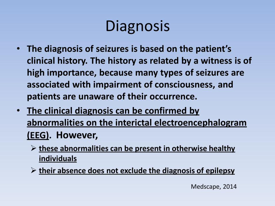
Diagnosis • The diagnosis of seizures is based on the patient’s
clinical history. The history as related by a witness is of high importance, because many types of seizures are associated with impairment of consciousness, and patients are unaware of their occurrence.
• The clinical diagnosis can be confirmed by abnormalities on the interictal electroencephalogram
(EEG). However, these abnormalities can be present in otherwise healthy
individuals
their absence does not exclude the diagnosis of epilepsy
Medscape, 2014

Diagnosis
• Two studies are often recommended after a seizure: Neuroimaging evaluation (e.g., brain
magnetic resonance imaging [MRI], head computed tomography [CT] scanning). a CT scan is often obtained in the emergency
department to exclude an obvious structural lesion, but an MRI is indicated if the patient continues to have seizures.
Electroencephalography (EEG).
Medscape, 2014

Operational (Practical) Definition of Epilepsy
A person is considered to have epilepsy if they meet any of the following conditions.
At least two unprovoked (or reflex) seizures occurring greater than 24 hours apart. One unprovoked (or reflex) seizure and a probability of
further seizures similar to the general recurrence risk (at least 60%) after two unprovoked seizures, occurring over the next 10 years.
Diagnosis of an epilepsy syndrome • Epilepsy is considered to be resolved for individuals who had an
age-dependent epilepsy syndrome but are now past the applicable age or those who have remained seizure-free for the last 10 years, with no seizure medicines for the last 5 years.
ILAE, 2014

EEG – Electroencephalogram
• Epileptic spikes and sharp waves
• Electrographic seizures

EEG – Electroencephalogram
• Interictal (between seizures)epileptiform activity:
– Epileptic spikes (20 - 70 msec)
– Sharp waves (70 - 200 msec)
• often followed by a slow wave (150-350 msec)
• Together these are simply referred to as Spikes

EEG – Electroencephalogram • Ictal (seizure) events: Electrographic seizures:
Typically seen as repetitive spikes or sharp waves or rhythmic sinusoidal waves, sometimes electrodecremental – May not be seen in brief EEG recordings
– Best observed with long term, continuous VEEG, especially in patients withdrawn from antiepileptic medication

Epileptic spikes – right temporal
with EKG artifact

with focal slowing
Epileptic spikes - left frontal

Epileptic seizure Types of seizures include
Generalized
– 3/sec spike and slow wave
• bilateral polyspike and slow wave
– Tonic-clonic
– Tonic
• electrodecremental response
Focal
– Simple partial
– Complex partial

Left temporal Right temporal
Left frontal Generalized (absence)
EEG during a seizure: 4 patients
an electrical storm in the brain

sz XLEvent staring
eyes roll up
moves LT leg
looks down and resumes playing
XLSpike
08:58:30.000 08:58:34.000 08:58:38.000 08:58:42.000 08:58:46.000 08:58:50.000
Fp1-F7
F7-T3
T3-T5
T5-O1
Fp2-F8
F8-T4
T4-T6
T6-O2
Fp1-F3
F3-C3
C3-P3
P3-O1
Fp2-F4
F4-C4
C4-P4
P4-O2
Comment
Time
605 uV
605 uV
605 uV
605 uV
605 uV
605 uV
605 uV
605 uV
605 uV
605 uV
605 uV
605 uV
605 uV
605 uV
605 uV
605 uV
Primary generalized (Absence) seizure, 3/sec spike and wave Defined treatment, Good prognosis

Slower spike and wave- More guarded prognosis

Right temporal lobe seizure, sphenoidal electrodes may have a high yield

nd4
PT 3- CPS#4: left frontotemporal

Sz 1 onset: right temporal

PT 10- SIMPLE PARTIAL SEIZURE

2-1-30
PT 2- GENERALIZED TONIC CLONIC SEIZURE (GTC)
#1-see later detection (may be difficult to identify in EEG with EMG)

Rb1 10mm/sec
PT 2- GENERALIZED TONIC CLONIC SEIZURE (GTC)
#1- detected

Medically intractable epilepsy
Seizures that are not controlled by
antiepileptic drugs may be controlled
by surgical removal of the brain tissue
generating the seizures.

Medically intractable epilepsy
Pre-surgical workup
• MRI to identify structural abnormality • Identify the site of epileptic activity with scalp EEG/ VEEG to correlate behavioral seizure with EEG abnormality.
•Long term monitoring (LTM)-usually 3-5 days • This may be adequate to guide surgical resection of the epileptogenic area …………………………………………………………………. • Targeting may be guided in part by functional neuroimaging
• Positron Emission Tomography (PET) • Single Positron Emission Computed Tomography (SPECT) • Subtracted Ictal Spect Coregistered to MRI (SISCOM)
Targeting may be markedly enhanced by source localization of the magnetoencephalogram (MEG) or EEG on a volume rendering of the brain from a structural MRI

Medically intractable epilepsy
Pre-surgical workup
• If necessary to more precisely localize the seizure onset site/or if the seizure onset site appears to be close to eloquent cortex, electrodes may be implanted on the brain (subdurally) over and around the suspected epileptogenic focus
- Electrocorticogram (ECOG) to more precisely localize the epileptic abnormality
Evoked Potentials for locating sensory cortex Cortical stimulation to localize eloquent cortex (language, sensory, motor)
Note that fMRI and MEG may also be used to for functional localization of sensory, motor and language cortex