elemhhh - dtic.mil · bility of "miscoding" was explored, i.e., miscoding by a cell free...
TRANSCRIPT

AD-AL12 944 CALIFORNIA UNIV SA FRANCISCO SCHOOL Or MEDICINE F/6 6/15NARCOT IC TOLERANCE AND DEPENDENCE MECHANISM: A NEUROLOGICAL COR--ETC(U)MAY 77 H H LOH DAOA17-73-C-3006
UNCLASSIFIED NI
ELEmhhh

iiiii ,_.o5Ll
I125 112.08
iiii jjjQ 1.6 =
MICROCOPY RESOLUTION TEST CHART
NAflONAL BUREAU Of SrANOARDS 1963-A
NARCOTIC TOLERANCE AND DEPENDENCE MECHANISM:A NEUROLOGICAL CORRELATE
FINAL PROGRESS REPORT
(1972 - 1976)
May 1977
by
Horace H. Loh, Ph. D.
Supported by4
US Army Medical Research and Development CommandFort Detrick, Frederick, Maryland 21701
Contract No. DADA 17-73-C-3006
University of California School of MedicineSan Francisco, California 94143
411
Approved for public release; distribution unlimited
The findings in this report are not to be constzuedas an official Department of the Army position unlessso designated by other authorized documents.Cl-
QDTICELECTE
APR 6O

SECURITY CLASSIPICATION OP TMIS PAGE (Wht., Data Entered)
REPORT DOCUMENTATION PAGE RED ISTRUCTIOSBEFORE COMPLETING FORM
r'VEPORT NUMBER VT ACCESSION NO 3. RCCIPINTS CATALOG NUMBER
4. TITE (rod Cubotle) tL TyPe O REPORT & PZRIOO COVERED
Narcotic Tolerance and Dependence Mechanism: Final ReportA Neurological Correlate 1977 .- 1976. PERFPORMING ONG. REPORT NUMBER
7. AUTNOR(s') '. CONTRACT OR GRANT NUMNER(a)
Horace H. Loh, Ph. D. DADA 17-73-C-3006
S. PERFORMING ORGANIZATION NAME AND ADORESS i0. PROGRAM ELEMENT. PROJECT, TASK
University of California School of Medicine AREA & WORK UNIT NUMBERS
62110A 3A062110A833.00.001San Francisco, California 94143 62758A 3A762758A833.00.001
I. CONTROLLING OFFICE NAMIE AND ADDRESS 12. REPORT DATE
US ArmyMedical Research and Development Command May 1977Fort Detrick, Frederick, Maryland 21701 13. NUMBEROF PAGES
6514. MONITORING AGENCY N AME & A0ORESS(I differnot hom Controllind Office) 1S. SECURITY CLASS. (of this report)
UnclassifiedDEC. 1ECLASSII CA TION/DOWN GRACINGSCHEOULE
14. DISTRIBUTION STATEMENT (at ths Report)
Approved for public release; distribution unlimited
17. DISTRIBUTION STATEMENT (of the ab t ct enteed In Block 20, It djifeent fIat Repmt)
IS. SUPPI.EMENTARY NOTES
1S. KEY WOROS (Contine o. revee. aidEl II neeeary and idenifyL by block number)
Narcotic tolerance Narcotic receptorsMorphine CerebrosidesNarcotic dependence Opiate agonistEtorphine Opiate antagonistNaloxone
26, ASITRAC? rCmtham reverse *14w ff nemlwgem a" Idau.afr by block mumbe)
rDO I JARPS 147n EDITION Of I NOV 6S IS OBSOLETE
SECURITY CLASSIFICA ,ON OF TrtS PAGE (WrNn DOt& Entred)

TABLE OF CONTENTS
Page
Introduction 1
1l,"4 he Role of Cerebral Protein Synthesis in OpiateTolerance Mechanism
12. Mechanisms of. Morphine-increased Protein Synthesis
(Quantitative) 4
The Capacity of the CFPS for Protein Synthesis 6
Gel Electrophoresis) 6
The Mechanism of Morphine on Translation' 20
3. The Role of Gene Expression on Morphine ToleranceDevelopment -_ 21
Electrophoresis of the Phosphorylated ChromatinProteins 33
4. The Role of Membrane Acidic Lipids in Opiate ReceptorBinding 38
-Narcotic Binding Studies * 40,/
Supported in Full or in Part by DADAI7-73-C-3006, 60
£oeession Yo• t l3?is GRA&I
SDTIC TAB 0* hU=nnounced [3
Justitiatio
~BYDistribution/
tAvailability CodesAvail and/o
Dist Special

Introduction
During the period of support from U.S. Army Contract DADA17-73-C-
3006, the research proposal detailed in our original application was
successfully carried out and subsequently resulted in a substantial
number of important publications. Previous quarterly and annual
reports have contained detailed summaries of our work. Additional
experimental data may be found in the enclosed reprints of publications.
This Final Report contains summaries of several pieces of work supported
by the above named contract as well as the significance and possible
practical application of the resulting information.
1. The Role of Cerebral Protein Synthesis in Opiate Tolerance Mechanism
Based on indirect evidence obtained in our own laboratories as well
as by others, it has been concluded that development of tolerance to and
physical dependence on narcotics is related to cerebral protein synthesis.
In our studies of morphine effects on protein synthesis in mouse brain,
we have found that mice tolerant to morphine have a higher rate of protein
synthesis. The mechanism of this phenomenon is unknown. Our studies
showed that morphine binds to brain free polysomes both in vitro and in
vivo. Furthermore, morphine stabilizes them from breaking down. These
results suggested that these effects of morphine on brain free polysomes
may be related to the mechanism of morphine increased protein synthesis
in tolerant mice. Furthermore, the mechanism of morphine stabilization
of brain polysomes is at least partially related to its inhibitory action
*on RNase, the enzyme responsible for the breakdown of m-RNA. The possi-
bility that morphine effects polysomes integrally via the drug's action
on Mg++ concentration was investigated. However, our findings indicated
- ----U" " .

-2-
that morphine did not alter the cellular Mg++ concentration.
The effect of acute and chronic morphine treatment on the synthesis
and turnover of 3H-leucine labeled protein and 14 C-choine labeled phos-
phatidylcholine was also studied in discrete regions of the rat brain.
Our results showed that chronic morphine treatment increased the 3H-
protein turnover in the microsomal fraction. However, acute morphine
treatment increased 1 4C-phosphatidylcholine synthesis in the hypothala-
mus (95%) and diencephalon (285%) and 3H-protein synthesis in the
hypothalamus (55%) and caudate nucleus (285%).
As stated in the specific aims of our original proposal, the
effect of morphine on individual proteins which play an important role
in regulation of brain functions will be studied. In this regard, we
have examined in detail the effect of acute and chronic treatment of
morphine on adenyl cyclase, tyrosine hydroxylase, and tryptophan hydroxy-
lase. High doses of morphine stimulated the activity of adenyl cyclase
but did not show any direct action on the other two enzymes. A detailed
description and discussion of these studies has been published.
Another aim of our original proposal was to test any possible
qualitative changes in protein synthesis due to "miscoding" of m-RNA
after morphine-polysome interaction. This means that narcotic tolerance
may be related to a change in the quality of cerebral proteins. In
this case, narcotics need not alter the rate of turnover of brain pro-
teins, but the tolerance development still could be blocked by inhibitors
of protein synthesis. The fact that morphine binds to polysome presents
*the possibility that morphine could interact with the m-RNA which may,
in turn, alter the coding process. The result of this "miscoding" would
be to produce a "different" protein(s).
w ___________

3
These studies were initiated in collaboration with Dr. James Meyerhoff
and his associates at WRAIR. In the first phase of this work, the possi-
bility of "miscoding" was explored, i.e., miscoding by a cell free protein
synthetic system utilizing a synthetic m-RNA with known coding properties
(e.g., poly U codes for phenylalanine).
Radiolabeled proteins were prepared in vitro in the presence and
absence of morphine. A typical experiment would determine the relative
r, tio of 14C-Phe to 3H-Leu as well as other 3H amino acids in the newly
formed polypeptides utilizing poly U as the synthetic messenger. This
study's significance lies in its ability to indicate whether or not
morphine may induce a misreading of the genetic code causing qualitative
differences in the normal brain protein.
Double labeling experiments for both in vivo and in vitro studies
were used to test the possibility of "miscoding." 1 4C-Phenylalanine
(or another amino acid) was injected into control mice and 3H-phenylalanine
into morphinized mice (or vice versa). The mice were sacrificed, the
brains removed and dissected into various regions. The brains (or discrete
areas) from control and morphinized mice were combined and subjected to
column chromatography. The various eluted fractions were examined for
3H and 14C radioactivity. The elution patterns of 3H and 14C-proteins
will furnish information to determine whether different proteins are
formed in the brain of morphine tolerant mice. A similar type of
experiment was carried out in vitro by using a cell-free protein
synthesis system. 14C-Labeled proteins were prepared in vitro by a
polysome-pH 5 enzyme system in the absence of morphine while 3H-labeled
protein was made in the presence of morphine. The soluble proteins
I .
-4-
(3H and 1 4C) produced will be combined and fractionated with column
chromatography. The fractions were assayed for 3H and 14C radio-
activity.
Similar types of approaches have been employed in our laboratory
using a cell-free protein synthesis system. In these studies, 1 4C-
labeled proteins were prepared in vitro by a polysome-pH 5 enzyme
system in the absence of morphine while 3H-labeled proteins were made
in the presence of morphine. The newly formed soluble proteins (3H
and 1 4C) were combined and fractionated with various chromatographic
techniques. The fractions were assayed for 3H and 1 4C-radioactivity.
Similarly, polysome and pH 5 enzymes isolated from morphine treated
(both acute and tolerant) mice were also studied. The preliminary
results of these studies follow.
2. Mechanisms of Morphine-increased Protein Synthesis (Quantitative)
In an attempt to localize the site of action of morphine in regard
to protein synthesis, we have intensively studied the interaction of
morphine with polysomes (as described in previous progress reports).
The fact that morphine inhibits endonuclease which is responsible for
the breakdown of m-RNA may, in turn, be related to the action of morphine
on polysome stability. Preliminary results do not support a drug-
induced ionic deficient environment as the mechanism responsible for
morphine induced polysome stability. RNA synthesis in tolerant mice
was studied further since preliminary results using 14C-orotic acid
indicated that morphine tolerant mice possess an increased rate of
nuclear RNA synthesis, especially in the heavy subunit. Moreover,
we have examined the effect of acute and chronic morphinization on the
~~i1
-5-
following processes in a cell-free system.
a. The activation of amino acids (activating enzymes)
b. The transfer of activated amino acids to t-RNA (synthesis of
aminoacyl-t-RNA and the transferase)
c. The transfer reaction (the transfer of 14C-amino acid from
carrier RNA to ribosomal protein), and
d. The chain initiation and release of peptides from the ribosome
To study the effect of morphine on the fidelity of translation, we
found that when morphine was added to the CFPS (10- 3 mol to 10-6 mol,
it was ineffective at any of the concentrations studied. Poly U produced
a maximal increase of phenylalanine incorporation into protein at 250 ug
poly U per mg of PL protein. This is demonstrated in Fig. A-a. There
was no significant difference in the incorporation of leucine (A-b) or
lysine (A-c) when poly U was added to the incubation medium. Morphine
alone at 10-4 mol had no effect on the incorporation of these amino acids
into protein. In addition, morphine has no effect on the poly U directed
incorporation of phenylalanine into protein. Nor does morphine plus poly
U cause any significant increase or decrease in the incorporation of
leucine, lysine or an amino acid mixture (A-d) into protein. This is also
true of several other amino acids studied in a similar paradigm.
The lack of morphine effect is apparent irrespective of the source
of PL and pH 5 enzyme. In Fig. A, the source was untreated control mice.
Fig. B depicts virtually identical results in a CFPS isolated from morphine
tolerant mice. The addition of exogenous morphine at 10- 4 mol has no
effect on the incorporation of phenylalanine or leucine relative to control.
250 ug of poly U produced the expected increase in phenylalanine incorpora-

6 --
tion and neither increased nor decreased leucine incorporation. Similar
studies with PL and pH 5 enzyme from placebo treated mice were identical
with these.
The Capacity of the CFPS for Protein Synthesis
A comparison of PL and pH 5 enzyme from morphine tolerant and placebo
mice in a cross-over paradigm (Fig. C) established significant differences
between the different fractions to incorporate amino acids into protein.
In this series of experiments, all parameters of the CFPS were constant
with the exception of the source of PL and pH 5 enzyme. The specific
activity of the homologous fraction of PL and pH 5 from morphine tolerant
mice is more than twice that of the homologous PL and pH 5 from placeb
based upon equivalent protein content. In the case where heterologous
PL and pH 5 were cross-incubated, the protein synthetic capacity of the
system remained significantly elevated relative to placebo control. The
purpose of the cross-over study was to ascertain if the increase in protein
synthesis was due to a factor in the PL fraction or pH 5 enzyme fraction.
It is apparent both fractions contribute a part to the total increase in
labeled protein. The PL fraction is responsible for the major portion of
the difference, but the results are more than additive at 60 minutes.
Gel Electrophoresis
An obvious question arising from these data is whether the increase
in radiolabelled protein is due to a general quantitative change in the
specific activity of newly formed protein, or to a qualitative change in
one or a few species of protein. The total TCA precipitable fraction
v as solubilized and electrophoresed on acrylamide disc gels from a 60 min

-7-
incubation. No attempt was made to isolate the newly synthesized protein.
The Coomassie Blue stain of such gels reflected no visual differences in
the protein pattern between morphine tolerant and placebo treated CFPS.
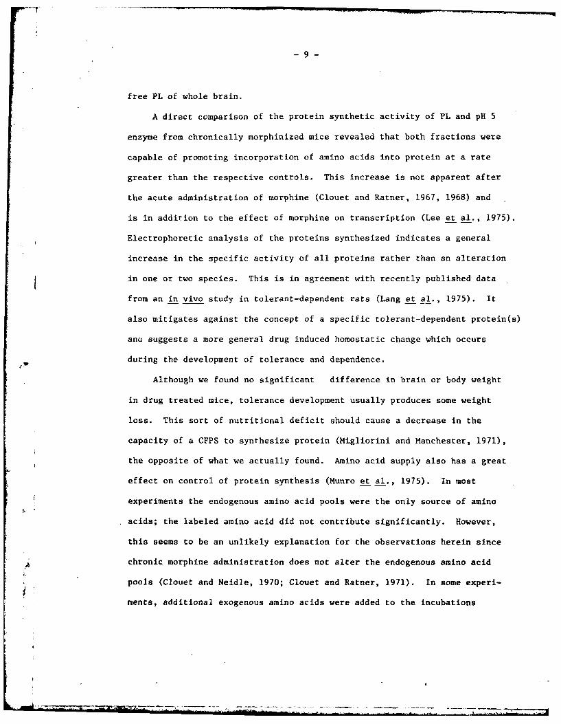
'The diagram on the abscissa of Fig. D is representative. The majority
of the proteins stained by the dye are due to PL and pH 5 protein, not
newly synthesized protein. The gels were cut in 3 mm sections and dpm
per section plotted over the diagram of the gel. It can be seen from
Fig. D that there is a general quantitative increase in dpm per section
along the entire length of the gel which contained protein from a morphine
tolerant group.
This experiment would not accurately demonstrate small qualitative
changes in specific proteins due to artifacts introduced in cutting the
different gels, electrophoresis, etc. Thus, we utilized a dual label
system to increase the accuracy. In these studies, the marker amino acid
was either L-(3H)-lysine or L-(1 4C)-lysine. We were then able to co-
electrophorese TCA precipitates from two different incubations on a single
gel. Fig. E is representative of these studies. In E-a and E-b, the
homologous PL-pH 5 enzyme from morphine tolerant and placebo mice were
incubated with 3H and 1 4C lysine as indicated. Fractions were co-electro-
phoresed and the activity per section as a percent of the total in the
gel was plotted. The TCA precipitates from four separate incubations
were combined and co-electrophoresed in triplicate to minimize technical
error. Fig. E-a serves as the morphine tolerant control and is the gel
with the poorest pattern of reproducibility in the triplicate sample.
If all proteins were identical and there was no technical error, the
curves would superimpose, since the only difference in the samples in
fthe label of the marker amino acid. Fig. E-b is the placebo control.
-8-
In Fig. E-c and E-d we coelectrophoresed TCA precipitates from incubations
with morphine tolerant and placebo PL and pH 5 enzymes. A comparison of
these graphs with control shows no evidence of a specific qualitative
change in the newly synthesized protein which could account for the major
differences in Fig. C.
Evidence from the studies cited on alternation of opiate tolerance
development by inhibitors of protein synthesis provided the impetus to
undertake the present study. The initial stages were designed to examine
the teleologically attractive hypothesis that a tolerance-dependence
protein might be responsible for the observed pharmacologic action of
prolonged morphine administration. Other data indicating that morphine
binds to and stabilized PL (Stolman and Loh, 1975) suggested the possi-.
bility that opiates could induce errors in translation of m-RNA resulting
in aberrant proteins, a situation analogous to streptomycin dependence
in certain bacteria. The present data do not substantiate any morphine
induced ambiguity in translation of m-RNA.
When the drug was added in vitro to a CFPS isolated from untreated,
placebo implanted or morphine implanted mice (i.e., tolerant-dependent)
it had no effects on the incorporation of several labeled amino acids
into protein. The effect of adding exogenous synthetic m-RNA, polyll,
was also as expected in all cases. It produced an increased incorporation
of Phe and no other amino acid. When morphine was added in conjunction
with polyll there was no detectable increase in the incorporation of any
of the amino acids examined other than Phe. 'Thus, within the limits of
detection in this particular system, we are unable to demonstrate any
morphine induced ambiguity of translation in the proteins synthesized by
- .
-9-
free PL of whole brain.
A direct comparison of the protein synthetic activity of PL and pH 5
enzyme from chronically morphinized mice revealed that both fractions were
capable of promoting incorporation of amino acids into protein at a rate
greater than the respective controls. This increase is not apparent after
the acute administration of morphine (Clouet and Ratner, 1967, 1968) and
is in addition to the effect of morphine on transcription (Lee et al., 1975).
Electrophoretic analysis of the proteins synthesized indicates a general
increase in the specific activity of all proteins rather than an alteration
in one or two species. This is in agreement with recently published data
from an in vivo study in tolerant-dependent rats (Lang et al., 1975). It
also mitigates against the concept of a specific tolerant-dependent protein(s)
and suggests a more general drug induced homostatic change which occurs
during the development of tolerance and dependence.
Although we found no significant difference in brain or body weight
in drug treated mice, tolerance development usually produces some weight
loss. This sort of nutritional deficit should cause a decrease in the
capacity of a CFPS to synthesize protein (Migliorini and Manchester, 1971),
the opposite of what we actually found. Amino acid supply also has a great
effect on control of protein synthesis (Munro et al., 1975). In most
experiments the endogenous amino acid pools were the only source of amino
acids; the labeled amino acid did not contribute significantly. However,
this seems to be an unlikely explanation for the observations herein since
Achronic morphine administration does not alter the endogenous amino acid
pools (Clouet and Neidle, 1970; Clouet and Ratner, 1971). In some experi-
ments, additional exogenous amino acids were added to the incubations

- 10 -
without qualitative effects. Moreover, this could not be the sole
explanation since in the crossover studies (Fig. C), the PL fraction
from tolerant dependent mice, which does not contain the amino acids
pool, was more active than the pH 5 fraction.
This suggests that something occurs during development of tolerance
to morphine which affects a compound present in both the PL and pH 5
fraction or that multiple events are occurring. Both the amino acyl
ligases and initiation factors are present in the PL and pH 5 fractions
and either may be a primary control factor in protein synthesis (Pain
and Clemens, 1973). We are in the process of analyzing these macromole-
cules in tolerant-dependent mouse. Morphine may also exert an effect
through a more complex mechanism. There is ample evidence that ethanol,
another central depressant to which tolerance and physical Jependence
develops, produces a decrease in brain protein synthesis and that this
is due to an effect on several of the different aspects of the synthetic
process (Noble and Tewari, 1975).
Finally, it may not be necessary to postulate an effect on one or
several of the macromolecules directly involved in protein synthesis to
explain the increase seen after tolerance development. Morphine interacts
with cerebroside sulfate in vitro in such a way that many of the criteria
for a morphine receptor complex are fulfilled (Loh et al., 1974). Morphine
also may interact with several other lipids (Cho et al., 1976). Lipids
are known to be an integral component of the protein synthetic matrix i.1
vivo and extraction and reconstitution of lipid in vitro decreases and
increases respectively cell free protein synthesis (Hradec, 1975). It

- 11 -
has been shown that the amino acyl ligases which are usually isolated
as soluble enzymes may, in fact, exist as a super molecule complex in
close association with the PL in vive. Under very mold conditions, these
complexes have been obtained and characterized (Bandyopadhyay:and
Deutscher, 1971; Vennegoor and Bloemendal, 1972). The lipids have been
extracted and characterized (Bandyopadhyay and Deutscher, 1973). It is
postulated that the lipid serves as a medium to orient the various compo-
nents of the protein synthetic matrix. Morphine may act during tolerance
development to increase the effectiveness of this matrix to synthesize
protein.
1!'
-12-
FIGURE LEGENDS A - E:
Fig. A - Effect of morphine and poly U on the ability of the CFPS to
incorporate amino acids into protein. Incubation medium consists of:
ATP generating system; buffered salts pH 7.6, PL:pH 5 enzyme (1:4) iso-
lated from naive mice; radiolabeled amino acid; in addition - contains
10- 4 mol morphine; - 250 ug poly U; - 10-4 mol morphine and 250 ug
poly U. Each point is the mean of 4 determinations. Fig. A-a - L-(3H)
phenylalanine, Fig. A-b - L-(1 4C)-leucine, Fig. A-c - L-(3 H) phenylalanine,
Fig. A-b - L-(1 4C)-leucine, Fig. A-c - L-(3H)-lysine, Fig. A-d - L-(3H)
amino acid mixture. Results are expressed as dpm of amino acid incor-
porated into protein per mg of PL protein.
Fig. B - Effect of morphine and poly U on the ability of a CFSP isolated
from chronically morphine tolerant morphinized mice to incorporate amino
acids into protein. Incubation medium consisted of: ATP generating
system; buffered salts pH 7.6, PL:pH 5 (1:4) isolated from morphine
tolerant mice; radiolabeled amino acid. In addition, contains 10-4
mol morphine, - 250 ug poly U, - i0 4 mol morphine plus 250 ug
poly U. Each point is the mean of 4 determinations. Fig. B-a - L-(3H)
phenylalanine, Fig. B-b - L-( 14 C) leucine. Results are expressed as dpm
of amino acid incorporated into protein per mg PL protein.
Fig. C - A comparison of the ability of the PL and pH 5 enzyme from
morphine tolerant and placebo mice to incorporate L-(3H)-phenylalanine
and L-(1 4C) leucine. - PL and pH 5 enzyme from morphine tolerant
mice; - FL from morphine tolerant pH 5 from placebo; - L from placebo

13 -
pH 5 from morphine tolerant; 0 - PL and pH 5 from placebo. Each point
is the mean of 4 determinations. Results are expressed as dpm of amino
acid incorporated into protein per mg of PL protein. The ratio of PL:
pH 5 enzyme was 1:4. All points are significantly different from control,
at 30 and 60 min, p> .01.
Fig. D - Protein pattern seen after acrylamide gel electrophoresis of the
TCA precipitated incubation medium. The Coomassie Blue staining pattern
is depicted along the abscissa and dpm per 3 mm gel section superimposed.
is PL and pH 5 enzyme (1:4) from morphine tolerant mice; M is PL and
pH 5 enzyme (1:4) from placebo mice. All other conditions were identical.
The protein fractions electrophoresed were combined aliquots from 4 dif-
ferent incubations. There were no visually detectable differences in the
Coomassie Blue stain between tolerant and placebo. The amount of labeled
amino acid incorporated into TCA precipitable fraction was consistently
greater in the morphine tolerant group.
Fig. E - Representative patterns of radioactivity from dual labeled gels.
Two gractions were electrophoresed on each gel. In the fraction designated
by the closed circle, * , the marker amino acid was L-( 3H) lysine, by the
open circle, 0 , L-(14 C) lysine. In E-b, the PL and pH 5 enzyme were from
morphine tolerant mice in both cases, thus the only difference is the ito-
topic label of the lysine. Similarly, in E-a, both PL and pH 5 are from
placebo. Therefore, E-a and E-b are controls for comparison with E-c and
E-d in which the PL and pH 5 are from different sources as indicated. A
qualitative change. in protein synthesis would be indicated by a difference
between * and 0 within a single gel section in E-c and E-d, which is
greater than any difference seen within a single section in E-a or E-b.

- 14 -
We conclude there are no major qualitative differences between the proteins
synthesized in vitro by PL - pH 5 from morphine tolerant and placebo mice.
ft

-15-
Figure A
A Controlsio-4 M morphine 15-*250 ug poly U
60- oMorphine +poly u lys
-phe 10-
40-
20 A- 5
15- 15u AA mixture
100
10 10-
5- 5-
10 30 50 10 30 60-

-16-
Figure B
LO 0O
0
oeD
-0 0
'*1r

Figure C
a
60 phe m 0 0
PLMMC 4P P
pH5 P M M~ c
40- l eu
00
2 I0 A
10 30 60 10 30 60
-18 -
Figure D
15 *Morphine
uPlacebo0
0-
5
515 25

-19-
Figure E
LC Uf
0 0 0 0
o LC) 0 it)
LC) NCN l
x> U V) :E
0 A* 0
0Q 10 oW-o 0 *L0
mill&

- 20 -
The Mechanism of Morphine on Translation
Since the last report on this project, experiments were begun to
determine at which level of the translation process opiates exert an
effect. There is very little information available in the literature
on the isolation, purification and study of the various enzymes and nucleic
acids necessary for translation'in brain tissue. A good deal of time
was spent making modifications of techniques suitable for preparations of
these factors from other tissues or prokaryotic cells. There has been
considerable success in these efforts and we now have stockpiled several
hundred milligrams of pure, highly active amino acyl ligase from mouse and
rat brain. We also have purified brain soluble RNA from these same animals.
Smaller amounts of pure stripped ribosomes are made on an "as needed" basis.
The initiation factors I, II, II and peptidyl transferase have been pre-
pared in a crude form and need further purification before 7tudies can be
undertaken. It would be premature to comment on the results of these
enzymatic studies at this time. The optimal conditions of enzyme, substrate,
isoacceptor tRNA, Mg++ and ATP are different for each amino acid. Current
efforts are dedicated to determining the optimal parameters for each amino
acid before comparisons are made between control and drug treated groups.
Other research support is being sought to enable completion of the final
stage of this research.
A!

- 21 -
3. The Role of Gene Expression on Morphine Tolerance Development
The synthesis of ribonucleic acid (RNA) in E. coli is inhibited in
the presence of levorphanol, a morphine analog. The synthesis of RNA
in HeLa cells is also inhibited in the presence of levorphanol and
levallorphan. Becke et al have shown that the most intense and con-
sistent inhibitory effect of levorphanol is upon ribosomal RNA synthesis,
although the non-ribosomal RNA is also inhibited. Recent reports have
shown that the inhibition of ribonucleic acid synthesis antagonizes the
development of morphine-induced analgesic tolerance. The authors con-
cluded that tolerance development to the analgesic effect of morphine
in animals can be reduced or prevented by several drugs which, in dif-
ferent ways, inhibit RNA synthesis and/or protein in the brain. However,
the difficulty of interpreting experiments in which drug treatment results
in severe disturbance of essential aspects of cell metabolism should also
be emphasized.
In attempts to study RNA metabolism in chronic morphine treated
animals, Datta and Antopol reported that the chronic administration of
morphine produced dose-dependent decreases in uridine and thymidine in-
corporating abilities of liver and brain homogenates and of their
subcellular fractions, such as nuclei, mitochondria and microsomes.
Castles qt al indicated in their study that newly formed RNA from brains
of tolerant rats was not lost as rapidly as RNA from brains of non-
tolerant rats.
In our studies, attempts were made to determine the chromatin
template activity in tbe brains of tolerant and non-tolerant mice in
order to establish if the changes in RNA metabolism during tolerance
development were due to the alleviation of chromatin template activity.

- 22 -
Our preliminary experiments indicated that the rates of DNA dependent
RNA polymerase from E. coli K-12 were linear up to 2 hours when mouse
brain chromatin was used as DNA template. The UTP-H 3 incorporation into
RNA was proportional to the amount of DNA added up to 30 ug. In routine
assay, less than 20 ug of DNA was used. Tolerant chromatin showed higher
template activity than non-tolerant one. The rate from tolerant chromatin
was linear up to at least 50 ug DNA, whereas the non-tolerant chromatin
began to level off above 30 ug DNA. Table 1 shows that the chronic treat-
ment of morphine resulted in increased specific activities of brain chroma-
tin which served as DNA template. Omission of chromatin or exogenous
RNA polymerase from the reaction mixture showed negligible incorporation,
thus the difference cannot be accounted for by the RNA polymerase contami-
nation in isolated chromatin. Additions of morphine yp to 0.1 mM into
the reaction medium showed no significant change in the rates of UTP-H3
incorporation.
However, when the chromatins were washed excessively, the chromatin
template activity from non-tolerant animals increased almost 2-fold. The
tolerant chromatin activity remained essentially the same (Table 2).
Therefore, the template activity in tolerant animals after excessive
washing was about the same or slightly lower than placebo activity.
If there is an inhibitor(s) present in the nuclei which has been
removed during the wash, one would expect the inhibitor to inhibit the
chromatin dependent UTP incorporation if added back to the reaction

- 23 -
medium. The supernatant collected from the seven steps of excessive wash
was lyophilized thoroughly to remove all the salts. The dialysis cellulose
bag has a 3000 molecular weight cutoff. This means that molecules bigger
than 3,000 would be retained in the bag; otherwise they would go into the
dialysate. The concentrated and dialyzed solution was then added back to
the reaction mixture. The results show that if the wash contains an in-
hibitor, the inhibitor must have a molecular weight of less than 3,000
because there is no inhibition of UTP incorporation in the presence of the
concentrate.
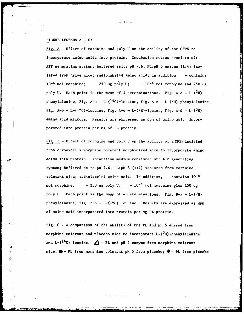
Table 3 shows the result when the histones were removed from chromatins
with H2 SO4 extraction. The histone-free chromatins showed approximately a
6 to 10-fold increase in their template activities. However, excessively
washed chromatin from placebo treated animals still shows higher activity
than the regularly washed placebo chromatin. Apparently, the difference
in their template activity cannot be due to the difference in the histones.
The data described above show that when the mice are rendered tolerant
to morphine with morphine pellet implantation, the rate of chromatin-
dependent UTP incorporation is increased. The increase is not due to the
presence of more DNA since the specific activities are calculated based
on per mg DNA. Omission of chromatin or RNA polymerase showed negligible
incorporation. Removal of the histone increases the rate of incorporation
by 6 to 10-fold. However, the DNA protein complex (free of histone) obtained
from tolerant animals still showi higher template activity than the placebo
control. Hodgson et al reported that DNA devoid of all proteins from
either placebo or tolerant rats gave identical rate of UTP incorporation.
Therefore, the data suggested that the non-histone protein which complexed

- 24 -
with DNA may have been altered during the morphine tolerance development.
This was supported by the results shown in Table 4.
During the preparation of chromatin, if one washed the nuclei and
chromatin excessively, i.e., 10 times more volume for each step of wash
than the regular preparation, one observes a 2-fold increase in placebo
chromatin-directed UTP incorporation, whereas the tolerant chromatin
remains the same. Some components which control the activity of DNA
dependent UTP incorporation must have been removed during the excessive
wash. The chromatin activity from tolerant animals remains the same
regardless of the washing procedure indicating that the controlling factor
is either not present or is very labile in tolerant animals. However,
the concentrated and dialyzed wash showed no inhibitory activity when
added back to the reaction medium. Therefore, if there is a regulator
which was removed, it must be smaller than 3000 m.w. Further investi-
gation is being carried out to identify the possible regulator.
*1 A

TABLE 1
EFFECT OF CHRONIC MORPHINE TREATMENT ON CHROIATINTEMPLATE ACTIVITY ISOLATED FROM f:OUSE
BRA I N
SpecifiS Activity i S.E.UTP-H' incorporation
Treatment nmoles/mg DNA
Non-Tolerant 41.96 * 3.38 (9)*
Tolerant 65.55 * 7.87 (9)*
, Number of preparations performed. P<O.Ol.
TABLE 2
THE EFFECT OF EXCESSIVE WASHING ON CHRO"ATINACTIVITY I; DIRECTINS UTP-H 3 INCORPORATION INTO RNA
Specific Activity * S.E.UTP-H 3 Incorporation
Treatment nmoles/mg DNA
t4j
Regular Wash
Non-Tolerant 47.75 ± 2.60 (4 )ab
Tolerant 80.75 * 15.70 (4)a
Excessive Wash
Non-Tolerant 84.50 * 7.93 (4)b
Tolerant 72.12 * 8.73 (4)
a. p /.0.05
b. p 0.01
A
;2 C
TABLE 3
COMPARISON OF CHR01.3ATIt TEMPLATE ACTIVITIES AFTERTHE REMDVAL OF HISTCIES
Specific ActivityUTP-H3 Incorporat-ion
Chromatin (Histone Bound) nmoles/mg DNA
Non-Tol erant
Regular Wash 47.6
Excessive Wash 66.6
Tolerant
Regular Wash 73.3
Excessive Wash 53.6
Chromatin (Histone Free) £
i
Non-Tolerant
Regular Wash 282
Excessive Wash 458
Tolerant
Regular Wash 380
Excessive I-lash 360

- 27 -
Table 4
COMPARISON OF CHROMATIN * TEMPLATE ACTIVITIESAFTER THE REMOVAL OF ACIDIC PROTEINS
Specific Activity * S.E.UTP-H 3 Incorporation
Treatment nmoles/ing DNA
Non-Tolerant 62.25 1 2.75
Tolerant 82.90 + 0.7
Non-Tolerant(-acidic proteins) 101.50 * 7.6
Tolerant(-acidic proteins) 55.50 + 6.6
• The chromatins %,ere prepared according to the regular washing
procedure described in the Methods.
A
p
-28-
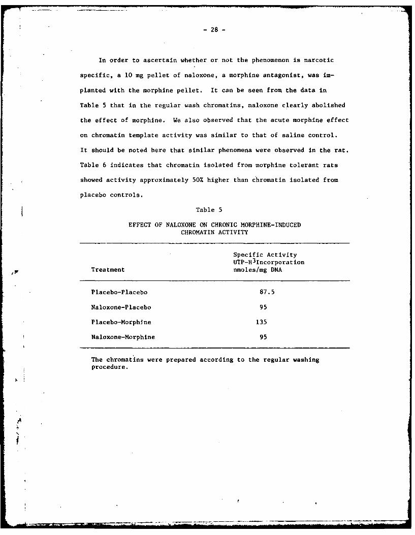
In order to ascertain whether or not the phenomenon is narcotic
specific, a 10 mg pellet of naloxone, a morphine antagonist, was im-
planted with the morphine pellet. It can be seen from the data in
Table 5 that in the regular wash chromatins, naloxone clearly abolished
the effect of morphine. We also observed that the acute morphine effect
on chromatin template activity was similar to that of saline control.
It should be noted here that similar phenomena were observed in the rat.
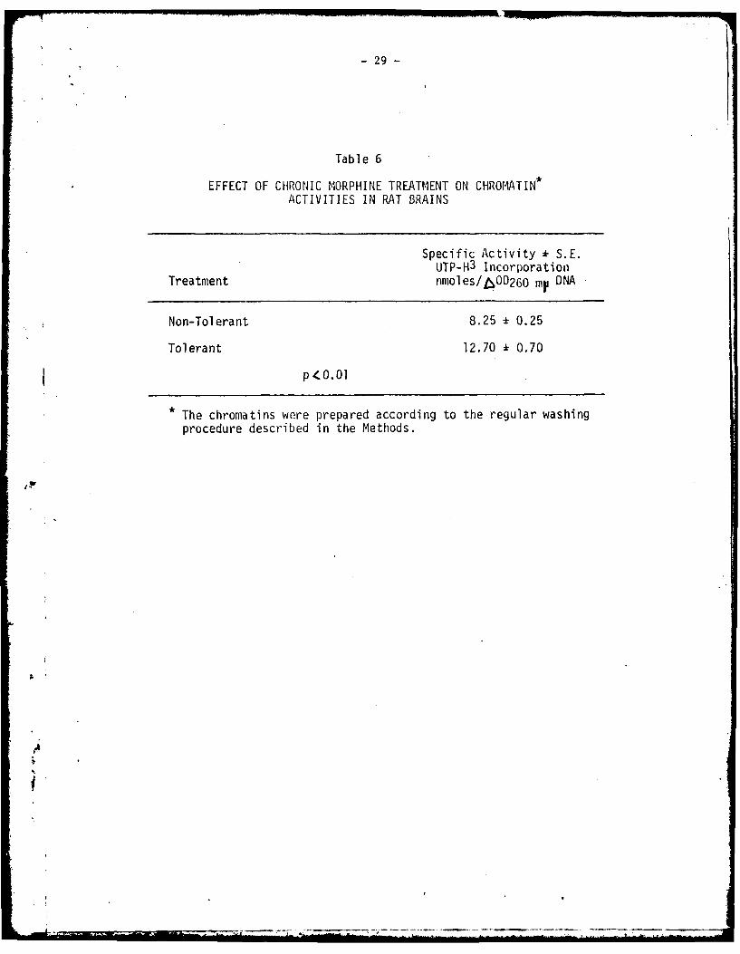
Table 6 indicates that chromatin isolated from morphine tolerant rats
showed activity approximately 50% higher than chromatin isolated from
placebo controls.
Table 5
EFFECT OF NALOXONE ON CHRONIC MORPHINE-INDUCEDCHROMATIN ACTIVITY
Specific ActivityUTP-H 31ncorporation
Treatment nmoles/mg DNA
Placebo-Placebo 87.5
Naloxone-Placebo 95
Placebo-Morphine 135
Naloxone-Morphine 95
The chromatins were prepared according to the regular washingprocedure.
A
- 29 -
Table 6
EFFECT OF CHRONIC MORPHINE TREATMENT ON CHROMATIN*ACTIVITIES IN RAT BRAINS
Specific Activity k S.E.UTP-H 3 Incorporation
Treatment nmoles/6OD260 my DNA
Non-Tolerant 8.25 - 0.25
Tolerant 12.70 1 0.70
p(O.O1
• The chromatins were prepared according to the regular washing
procedure described in the Methods.
-30-
These studies showed that regulation of brain protein synthesis was
altered following chronic morphine treatment. Moreover, one possible
site of action is the gene. Also, our work indicated that the non-
histone proteins are responsible for the mechanism of the morphine-
induced changes in gene expression. The non-histone proteins in cell
nuclei have been implicated in the regulation of DNA template activity
in chromatin and the heterogeneity of these proteins makes it difficult
to study each protein with respect to its function and regulation. We
chose to study the phosphorylation of nuclear proteins, since the phos-
phorylation of the nuclear proteins, especially non-histone proteins,
has been suggested as a means of positive gene regulation. The enzyme
responsible for protein phosphorylation, i.e., protein kinase, was also
measured.
As shown in Fig. 1, we found the activity of protein kinase isolated
from mouse brain chromatin to be linear up to 10 min. The zero time
count was always less than 10% of the total counts and has been subtracted.
The pH optimum of the enzyme was quite broad; in the range tested 9Ph 6.0
to 8.0), there was no obvious peak. Thus, in routine assay, pH 6.5 was
used. The W-( 32p)ATP incorporation was linear with increasing concentra-
tion of ATP up to 5 uM.
The enzyme may use its endogenous chromatin proteins as substrate.
Additional histone, 40 ug/0.2 ml did not increase the amount of !-(32P)ATP
incorporation. However, additional casein (40 tjq/0.2 ml) resulted in
50-60% more phosphorylation. The enzyme was active when chromatin was
freshly prepared; however, within a couple of days of storate at -100,

-31-
more than 80% of the activity was lost. Thus, assay of the protein kinase
activity immediately after chromatin preparation is important.
Table 7 shows that the protein kinase activity from chromatin isolated
from morphine-induced tolerant mice was 65% higher than that of the control
group. Addition of morphine sulfate (10-4 to 10-7 M) or cAI4P (10-5 to
10-7 M) in vitro had no significant effect on protein phosphorylation
(Table 8).
Fig. 1
P~otr1. j~Table 7
l ii oo i. ki i mot mic tretIenIon ch1ronid-
fill ~ ~ ~ m pfo l hIlcil))f11W
l1)\IiiC % *'~ I tu i .\cII c~c~ Ib ii li t liat 54 ) 4 3.47 IV~it m.1ills coi k IIimlg~ 1111 I l mmIloimumm111II t o lclm \\'I, Mm I'IVciIII
* addccI. ill, 1 1 7 . %II Iif, Jill %AIIs,' . s IItill I .Pill- 10 l ca-Ciut IcutIot I41Plte& hti11ler ucmc ancI md Il i att k:mc % .I 't- lir 1111C m I4 (l)~ :I) 23 1 4 1
M W. NI lcI p IIIII)CC Ill 1.lC IlIll',1 i iilti)Ci of C\

-32-
Table 8
I tiC,] "I H biiil' .1od~ \Nil'o1 IitiIilI1i)IIl o~iI 4.l4illatil p101vil '
III).1I4rA 4o iii .4 c ,lI.c iiN 4
(.MI,' O i spil' I!Ll. 1 0
(,ill I ,I I ii'Iii iilic III \I 6'4 X .15I
(. litl I c\ \1 1. 11) NI i's 1
Fig. 2 Fig.
Fig.
1kolero n t250(i
'5 [O~eil~it6
200
IQ, ea
* 000
Placetio ulp
Time ot iricub~o ,l 1415vi 'i10 5
.11111"vi f polmlk Ii'mpola d " jplo -Slices
4 11,1C tIdd lilllil 1144 Ai, I4 ICII4 ll 1 kl' N I' V~ ic
SITS 4.1i loh tii 1 L1 i.4lolplli.47 Ii 1474 lil11i
41711 4. 1 I 141a i i, cI' G o 4.V4 li i at (o It 1ill4%4

- 33 -
In order to determine whether or not the increase in phosphorylation
of chromatin protein isolated from tolerant animals was due to a decrease
in phosphoprotein phosphatase activity., the cold ATP dilution procedure
was used. Fig. 2 shows that dephospl'oi,.ation was evident in both the
placebo and the tolerant groups. At 20 min, dephosphorylation was
identical for both preparations. At 60 min, in chromatin isolated from
the placebo and the tolerant groups, 31 and 18% of the 32P were lost,
respectively. However, in the absence of cold ATP, chromatin isolated
from tolerant animals increased phosphorylation by 56% at 60 min, whereas
in chromatin isolated from placebo, phosphorylation increased less than
20%. This result indicated that protein kinase was responsible for the
increase of phosphorylation in chromatin protein isolated from tolerant
animals.
Electrophoresis of the Phosphorylated Chromatin Proteins
Nuclear chromatin comprises a heterogeneous mixture of proteins
differing in molecular weight, amino acid composition and degree of
phosphorylation. The heterogeneity of molecular sizes is indicated
by the differences in electrophoretic mobility in SDS-polyacrylamide gel.
The degree of phosphorylation can also be measured in the gel with 32P-
labeled chromatins.
Fig. 3 reveals the complicated banding pattern of chromatin protein
subunits. There are at least 30 different multiple polypeptide bands
ranging in molecular weight from 15,000 to 200,000 daltons calculated
from proteins with known molecular weights. The most prominent bands
A,Vafter staining with Coomassie Brilliant Blue were the histone proteins.
It has been reported that two of the histone proteins were located in
the middle of the gel (slices 19-22) and two more were observed in the

- 34 -
lower part of the gel (slices 30-36). The specific activity of 32p_
labeling was low in the histone protein-rich area. In agreement with
the findings of Richwood et al., this indicated that histone proteins
were a rather weak substrate for this protein kinase reaction. The
other bands of the gels were non-histone protein bands. The high molecular
weight region (slices 1-5) represents a series of very finely separated
high molecular weight protein subunits. Although the bands were rather
light after staining with Coomassie Blue, it was quite clear that they
are highly phosphorylated (Fig. 3). The degree of phosphorylation in
this region was about 74% higher in the tolerant group than in the control
group (Table 9), although the electrophoretic patterns between those two
groups were similar.
Since there was no visible protein 3taining, the 3 2p-labeling was
quite significant in slices 38-46 (molecular weight about 7000 daltons).
The intensity of phosphorylation was about the same between chromatins
isolated from tolerant animals and controls. However, the band moved
slightly faster in the tolerant group (Fig. 4). The shifting in mobility
is a true phenomenon, since we have repeated it more than 5 times in 5
different nuclei preparations and found it to be reproducible each time.
When morphine sulfate (1 mM) was added during phosphorylation in vitro
of chromatin proteins isolated frbm control or tolerant animals, the
electrophoretic pattern of this shift was unaffected.
Cyclic AMP did not stimulate the phosphorylation of chromatin protein
in vitro. Electrophoretic patterns of the chromatin proteins phosphory-
lated with or without the presence of cAMP (5 uM) were similar, except in
slices 38-46. Fig. 5 shows that this peak shifted when phosphorylated in

-35-
the presence of cAMP. This shift was observed in both types of chromatins.
The data presented demonstrate the apparent protein kinase activity in
oligodendroglial-rich chromatin. Since it is unclear whether or not the
protein kinase(s) is a separate protein(s) or the kinase activity is
inherent in the phosphoproteins themselves, we decided to refer to the
activity as "apparent protein kinase activity." The addition of caseini
has been shown to increase 3 2p-labeling in TCA-precipitable materials.
Since casein is not a natural component of brain tissue, the significance
of the stimulation of phosphorylation is unclear. There is no obvious
111116 1 4 ,fi , I ,
Table 9*42..
Fig. 4 -
Fig. 5/ K
A Tnier in t c 4MP y
MI I , ( ilt HcI' II L IN 3111 1.111,1 lo 3 I il II II33 II il3ill flIC:
dI %ni lal I lCii3d c- 1 opion i,. itLI Ic 0.- II
3 , C ' 1 ' .14 4 i6
S'I ce ,
lilt .th~c3 it ,I pIC, 1lkv 0, t, ANII' (5 , 1 l 'Nit te-
lXiL .111dI 'I,, ii.11.,*C c, c c d In MIltlNd
(ie t. l l I , it .c

- 36 -
optimum pH for this enzyme activity indicaLing that several enzymes
may be responsible for the phosphorylation. Kish and Kleinsmith
have reported the separation of protein kinase activity into 12 distinct
enzyme fractions in beef liver chromatin. This possibility of multiple
enzymes would also have to be carefully examined in oligodendroglial-
rich chromatin preparation. Unlike the histone kinase, this protein
kinase activity was very labile; 80% of the activity was lost within only
a few days of storage. This presents a serious problem in further puri-
fication of the enzymes. A search for methods to stabilize the activity
is in progress.
Chronic morphine treatment resulted in an increase in total apparent
protein kinase activity. It was evident in cold ATP dilution experiments
that the increase in phosphorylation of chromatin in tolerant animals
was, indeed, due to "apparent protein kinase activity." Compared to the
placebo group, although the turnover rate of 32P was slower in chromatin
protein isolated from tolerant animals, the phosphorylation rate in the
absence of cold ATP was considerably higher at all time periods. These
results suggest that this may be at least partially due to the increase
in protein kinase activity.
The phosphorylation of proteins occurred mainly in the acidic protein
region of chromatin proteins. Electrophoresis of SDS-acrylamide gel
revealed that chronic morphine treatment increased the 3 2P incorporation
in this area. It is not known if the gel slices still contain protein
kinase activity. Furthermore, the ability of a one-dimensional dodecyl-
sulfate-electrophoresis system to demonstrate the true complexity of
ichromatin non-histone proteins is limited, therefore requiring more detailed

- 3.
work to elucidate this high molecular weight region. In slices 39-45,
where there was no visible protein stain, significant phosphate labeling
was observed. It is interesting to note that this band was similar
in position to the one reported by MacGillivray and Richwood. Chronic
morphine trtdtment shifted this band slightly; it is not known if this
was the result of a change in peptide molecular weight.
It has been suggested that phosphorylation of chromatin proteins
would normally increase chromatin template activity. Therefore, our
observation may represent one of the positive controls of gene expression.
The addition of morphine sulfate in vitro had no effect on 32p-labeling
of protein, indicating that morphine did not directly interfere with
protein phosphorylation. Therefore, the effect of chronic morphine
treatment in vivo may be via some other mechanism, indirectly affecting
protein phosphorylation of chromatin proteins.
In summary, we have demonstrated apparent protein kinase activity in
oligodendroglial-rich chromatin. The activity was unaffected by cAMP
or morphine sulfate in vivo. Chronic morphine treatment resulted in
increased phosphorylation which may be due to protein kinase activity
rather than to a decrease of phosphoprotein phosphatase activity.
The increase was located primarily in high molecular weight regions
of the SDS gel. As we reported previously, oligodendroglial-rich template
activity increased in chronic morphine treated animals; the increase in
phosphorylation of non-histone proteins observed in this study may be
related Further studies are in progress to elucidate the phosphorylation
reaction and its relationship to chromatin template activities in different
types of nuclei.

- 38 -
4. The Role of Membrane Acidic Lipids in Opiate Receptor Binding
There have been numerous attempts to elicit the mechanism of action
of morphine and its surrogates in analgesia, tolerance and physical
dependence. As an initial approach to studying selective narcotic-
receptor interaction, the distribution and binding characteristics
of the active D(-) enantiomorph in the brain have been compared either
with the inactive isomer or in the presence of an antagonist. However,
these attempts to demonstrate selective binding by pharmacologically
active narcotics were unsuccessful beccuse of the lack of suitable tools
for the purpose. Recently, Goldstein et al, using levorphanol and dex-
trorphan, elaborated a procedure for demonstrating stereospecific binding
and reported that mouse brain contains fraction that binds opiates stereo-
specifically. Subsequently, several groups of investigators using an
antagonist and various narcotic agonists with high radioactivity, also
have demonstrated stereospecific binding of narcotics with varying degrees
of affinity for certain brain areas. The primary binding site has been
identified to be in the membrane of nerve terminals at certain brain
regions.
In our laboratory, we have been interested in endogenous substances
in the brain which might interact with morphine agonists and antagonists
in a stereospecific manner. Based on molecular modelp, it was found that
parts of the structures of several glycolipids appear to exhibit structural
complementariness to morphine and several neurotransmitters we well (Cho
et al, unpublished). Indeed, the structures of cerebrosides, cerebroside
sulfates and gangliosides appeared to fulfill the requisites of the

- 40 - PREGLUD.AG
analgetic receptor postulated by Beckett and Casy and by Portoghese.
Moreover, these glycolipids are optically active and capable of stereo-
specific interaction with narcotics, and since cerebrosides, cerebroside
sulfates and gangliosides are located in the nerve membrane and nerve
endings of the central nervous system, an examination of these substances
for stereospecific binding to opiates was initiated.
Our preliminary studies indicated that high affinity stereospecific
binding to narcotic compounds was exhibited by cerebrosides. While
these studies were in progress, Lowney et al announced the isolation
from mouse brain of a partially purified opiate receptor which they
reported to be a proteolipid. Our further investigations revealed that
the narcotic binding properties exhibited by cerebroside sulfate, separated
from our commercial source of cerebroside, were similar to those of the
reported opiate receptor. Inasmuch as the cerebroside sulfate which we
purified contained no protein contaminants, it became incumbent upon us
to compare in a more definitive fashion the characteristics of cerebroside
sulfate and the mouse brain opiate receptor. Subsequently, we reported
narcotic stereospecific binding to cerebrosides and provided evidence that
one of the cerebrosides, cerebroside sulfate (or sulfatide), is possibly
identical to the purified mouse brain narcotic receptor. We also provided
explanations for the apparent proteo-like behavior of this mouse brain
opiate receptor.
Narcotic Binding Studies
Stereospecific binding of H3-etorphine at 2 x 10⁻⁸ M concentration
and H 3-naloxone at 2 x 10- 7 M concentration to cerebroside were studied
under conditions as shown in Table 1 according to a modification of the
method of Goldstein et al.
.ii

- 41 -
For fractionation and estimation of cerebroside sulfate and its levorphanol
complex, the method of Soto et al was used. To isolate the opiate receptor,
10 brains (about 5 g) were homogenized at room temperature in 100 ml of a
mixture of chloroform-methanol (C-M 2:1 v/v) and processed in the manner
described by Lowney et al who essentially adapted the procedures of Folch
et al. The extract was filtered, washed once with 0.2 vol of distilled
water, and chilled at 50C. 280 ml of cold diethyl ether was added to the
cold CHC1 3 solution (70 ml). After 1 hr, the mixture was centrifuged
at 50C for 10 min at 8,000 x g. The precipitate was dissolved in 5 ml of
C-M 2:1 and applied to a Sephadex LH-20 column pre-equilibrated with
chroroform. The column was eluted as described previously. Aliquots
(0.5 ml) of the separated fractions were dried and determiAed for
cerebroside (C) by the phenol sulfuric acid methods or by a more specific
procedure for cerebroside sulfate (CS). The absorbance of each fraction at
280 nm was also determined. Various fractions were qualitatively examined
by application on precoated silica gel sheets and the chromatograms
were developed by a solvent system of C:M:H 20 (35:15:2, by volume). Spots
were visualized on separate plates with iodine vapor and with fluorescamine
spray. Qualitative analysis for proteins and amino acids in opiate
receptor fractions were carried out after dansylation and subsequent
separation on a mini polyamide thin layer plate as described by Neuhoff
et al.
H3-Etorphine and H3-naloxone binding to cerebrosides were prevented
significantly by levorphanol but not by its enantiomer, dextrorphan.
As shown in Table 1, the stereospecific binding of H 3-etorphine at 1000-
fold excess of levorphanol plus dextrorphan was 17% of total binding.
. . . . . .. . .. - r . . . . . ... . ... .I . . . . . .. .i I : . . . . ... . i. . .. . . .. . . . . .. . . .

- 42 -
TABLE 1
STER:OSPECIFIC BIN DING OF (n3 )-rALOXuo:lLAHiD (H3 )-ETORPIIIIE TO CEREBROSIDES
(u 3 )-ETORIPIIlNE (1I3 )-NALOXONECO:CENTRATIOrN 2 X 10⁻⁸ fIl 2 X 10⁻⁷ ,.i
UNLABELED DRUGS (m) (CPl ± SE) (CPul - SE)
NONE 5500 ± 70 5720 , 110
DEXTRORPHAN (D) 2 X 1O- 5 5470 * 59 5670 * 82
LEVORPHANOL (L) 2 X lO- 5 4551 G 39 3988 * 173
FIORPHINE (N) 2 X 10- 5 4302 i* 23 3726 * 51
ETORPHINE (E) 4 X 10⁸ 4080 * 20 3894 ±-108
IfI STEREOSPECIFIC BINDING
D-L 909 (17%) 1682 (30%)
D-M 1168 (21%) 1944 (34%)
D-E 1390 (25%) 1776 (31%)
1 ing cerebroside .ias mixed with unlabeled drug and incubated for 2hours. Labeled drugs were added and stereospcific binding deter-mined as described in Nethods.

- 43 -
For H3-naloxone, binding at 100-fold excess of levorphanol plus dextror-
phan was 30% of total binding. Similar inhibitions in H3-etorphine and
H3-naloxone binding were observed in the presence of morphine and etor-.
phine.
The binding of etorphine to 1.0 mgm of cerebroside was not saturable
over the range. 2-240 nM of H3-etorphine. As shown in Fig. 1, four step-
wise increases in binding were observed with increasing concentrations
of H3-etorphine. Additionally, the etorphine binding with increasing
cerebrosides was nearly linear over the range of 0.2-0.5 mg of cere-
broside, virtually constant over the range 1.0-3.0 mg of cerebroside,
after which the binding slowly increased.
The concentration of dextrorphan, levorphanol, etorphine and morphine
FIG, 1
6 BINDING CURVE FOR (H )-ETCtPHINE TO CEREBROSiDE
14 //
.a12
10
C'6
4-
/2
10 20 30 40 50 W 70 '0 53 0 120 140 60 160 2013 220 240
A. Hs-Elorp e , MolOr x 0'9
SH3-Eto1Phin . Billirlfl to Cerehrosides. 1 mg of cerebroside
liposomn was itrK:uhc tod at 370C for I hr vith iitreasing con-centrations of 113 -etorphine (3.4 Ci/rM) in 5O ji.i sodiumphosphate buffer.
-44-
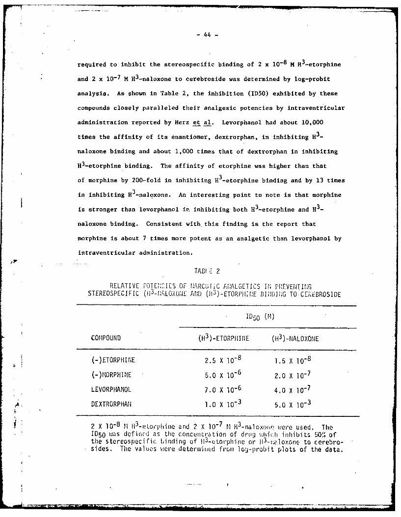
required to inhibit the stereospecif ic binding of 2 x 10⁻⁸ H H3-etorphine
and 2 x 10⁻⁷ M 113-naloxone to cerebroside was determined by log-probit
analysis. As-shown in Table 2, the inhibition (ID50) exhibited by these
compounds closely paralleled their analgesic potencies by intraventricular
administration reported by Herz et al. Levorphanol had about 10,000
times the affinity of its enantiomer, dextrorphan, in inhibiting H3-
naloxone binding and about 1,000 times that of dextrorphan in inhibiting
H3-etorphine binding. The affinity of etorphine was higher than that
of morphine by 200-fold in inhibiting H3_etorphine binding and by 13 times
in inhibiting H3 _naloxone. An interesting point to note is that morphine
is stronger than levorphanol in inhibiting both H3-etorphine and H3-
naloxone binding. Consistent with this finding is the report that
morphine is about 7 times more potent as an analgetic than levorphanol by
intraventricular administration.
TABI C-- 2
RELATIVE FOJENCILS OF NARCO[LC At!ALGETICS IrN PREVENJTING
STEREOSPECIFIC (113-11ALOX0OJE ANDO (113)-ETORPInE BINDm];,G TO CET EBROSIDE
1050 (MI~)
COMPOUND (H3) -ETORP11ItE (H3 )-NALOXONE
(-)ETORPHHJE 2.5 X 10⁻⁸ 15X1-
(-).IORPHINE 5.0 X 10⁻⁶ 2.0 X 10⁻⁷
LEVORPHANOL 7.0 X 10⁻⁶ 4.0 X 1-
ADEXTRORPHAN 1.0 X 10⁻³ 5.0 X 10⁻³
2 X 10⁻⁸ 1. II3-e toji'phe and 2 X 10⁻⁷ 1. H13-naloxojir %-,ere used. TheID50 was defincd cis the concentration of drig wh1iCh inhibits 50'* Ofthe stereospecific binding of 113-tol-phinoe or jI3-r1;lOoone to cerebro-sides. The valueos were determined fromi lo'j-probit plots of the data.

- 45 -
The elution of the commercial preparation of cerebroside previously
applied to the Sephadex column resulted in 3 major peaks. The first and
second peaks (fractions 27 and 42) were identified as two cerebrosides
by thin layer chromatography. The third peak (fraction 67) was identi-
fied as cerebroside sulfate. Determination of gaiactose in these
fractions indicated that the commercial cerebroside preparation contained
about 95% cerebroside and 10% cerebroside sulfate. It appeared to us that
the cerebroside sulfate present in the chloroform:methanol (1:1) eluate
was eluted in the same fraction as the proteolipid opiate receptor which
Lowney et al obtained from mouse brain.
To test the possibility that CS might be the opiate receptor,
levorphanol complexes of CS were prepared from commercial CS and sub-
jected to chromatographic analysis on Sephadex LH-20 in the same
manner described for the opiate receptor. Application and elution
of these complexes on the column resulted in a shift of the elution
pattern corresponding to that reported for the proteolipid opiate
receptor. Purified cerebroside sulfate was eluted in fractions 67-72
(Fig. 2a) while the levorphanol complexes were eluted in fractions 17 -
24 (Fig. 2b). The fraction with a peak around 41 corresponded to free
levorphanol. Over 90% of the total binding capacity of levorphanol was
due to the CS. The pattern of the elution curve for CS and its levor-
phanol complex was very similar to that of the opiate receptor. The
mouse brain opiate receptor was reported to be aluted around fraction 21.
On separating the opiate receptor as described by Lowney et al
and subjecting it to thin layer chromatography and color development with
iodine, two major spots were established to be CS (hydroxylated and non-
hydroxylated) and one minor spot at the origin was not identified.
4 F

-46-
Flu'.. 2 (a~b)
Ao
- - ~ -.-----..-- -~--~- ~-- - -~ - ---.---.- LO
0 5 0 I 20 2 30 3 43 45 53 55 E is 70 m5 60
FRACTION
E1 ut ion flehavica' oil Sephadex U-20 of:
B - ceobroside sulfate levorphanol co !ylex.Time 0')o pa Earound fraction 19 corrson, t hcoup1 ex; the peak around 42 corresponds to free1 evorphanol . -denotes cordbru!Jide sul fate.6 - 6 denotes Ilevorphanal . The arrovis indicatethe fra(ction ill Which successive elutin~g mixturesf ir st uie r~je.

- 47 -
When another thin layer plate was sprayed with fluorescamine, the opiate
receptor fraction exhibited three very faint spots (Rf 0.24, 0.1 and
0) while purified CS did not show any spots.
Since the purifed narcotic receptor was reported to be proteolipid
in nature, we examined it and the cerebrosides as well for protein content
by the fluorescamine procedure. With the opiate receptor fraction, about
20 nmoles of amine per gram tissue was obtained and, after acid hydrolysis,
it was increased to 508 nmoles. The latter value is almost identical
to that reported for the opiate receptor isolated by Lowney et al. When
100 nmoles hexose equivalent of the opiate receptor fraction and of com-
mercial cerebroside were subjected to acid hydrolysis and estimated for
fluorescamine fluorescene, the difference in amino nitrogen before and after
acid hydrolysis for the two substances was almost identical, being 98
nmoles for the opiate receptor fraction and 110 nmoles for the pure cere-
brosides (Table 3). Thin layer chromatography analysis of both acid
hydrolyzates showed that only sphingosine spots were present.
A search for proteins and amino acids was conducted also in the
opiate receptor fraction before and after acid hydrolysis by thin layer
chromatography using the dansylation procedure on polyamide plates.
With this method, which is sensitive to 5 ng of amino acids, no evidence
for the presence of protein or amino acids could be detected in the
separated fractions either before or after hydrolysis.
Generally speaking, stereoskecific binding of morphine-like compounds
is necessary but not sufficient requirement for identification of any
substance as the rec.eptor. Since many natural substances are optically
active, they could interact stereosRecifically with the opiate enantiomers

- 48 -
TAILE 3
AM1INO HI1F '6C[H co: TE:; It THE NiOUSE BLR.'AIN OPIATEF1.CLPTOR ANL) IH PURIFIED CEREtR(.SII)E
Amino Hitrogen (nmoles)Hexose Equivalent Before After
Preparation r4oles Hydrolysis Hydrolysis Difference
Opiate Receptor 100 10 108 98
Cerebroside 100 0 100 100
An aliquot of the opiate receptor fractions and cerebrosides correspondingto 62 nmoles of hexose ;:ere dried and the residue was hydrolyzed in asealed tube with 0.5 ml of 6N HCI at 1000 C for 1 hr. The hydrolyzate wasneutralized with 0.5 ,fl of 6;2 11,Ol and aliquots (0.2 r;l ) Iere mixed ith1.3 ml of 0.2 H Na borate buffer pli 9.0. The solution w;as agitated rigor-
ously while adding 0.5 il fluorescamine soluLion (15 !;ij %' in acetone). Thefluorescence was excited at 390 mp and the eiission was read at 490 ip inan Aniinco-Bovman Spectrophotofl uorometer with an external standard of 1%quinine sulfate in 0.1 I 112'04 (adjusted to 100 units).
To obtain the fluorescence reading before hydrolysis, aliquots (0.5 ml)were dried and tlio rsidue v',:as dissolved in 0.5 nl of 0.5%' sodiui laurylsulfate in 0.5 N NaOll. Al i quats (0.2 ril ) were mixcd %,i th 1 .3 nil 0.2 1.1sodium borate buffer pli 9.0 and the fluorescence was developed and measuredin the usual way.
with varying degrees of affinity. Indeed, compounds such as silica gel
and cellulose have been reported to bind enantiomers of morphine-like
compounds in different manners (Wu et al, unpublished). In addition to
the demonstration of stereospecific binding of drug receptor, the correla-
tion of its degree of affinity with drug potency should also be considered.
In our experiments, narcotics not only bind to cerebroside stereospecifically
(Table 1), but their binding to various drugs also parallels their reported
-,

- 49 -
intraventricular analgetic potency (Table 2).
The binding of 3H-etorphine to cerebroside is not staurable in the
concentrations we have examined. The stepwise increase in 3H-etorphine
binding to cerebroside (Fig. 1) may be due to the fact that cerebrosides
from commercial sources contain at least two kinds of cerebroside and
cerebroside sulfate. Our preliminary data (Wu et al, unpublished)
suggest that the first two apparent saturation curves at about 1.0 x
10- 8 M and 4.0 x 10- 8 M etorphine may be due to binding to hydroxylated
and non-hydroxylated CS. Studies are in progress to obtain liposomes in
yields adequate for stereospecific binding studies.
It is very interesting to note that there are striking similarities
between CS and the partially purified opiate receptor recently reported
by Lowney et al. CS resemble the opiate receptor with respect to its
elution pattern on Sephadex column and its non-saturable nature. More-
over, the behavior of the cerebroside sulfate-levorphanol complexes on
the column resembles that reported for the opiate receptor levorphanoi
complexes . Our preliminary data indicated that one of the glycolipids
which binds narcotic stereospecifically was eluted in the same chloroform-
methanol fractions as the opiate receptor. Furthermore, when this glyco-
lipid was complexed with levorphanol, like the opiate receptor, the com-
plexes formed could be eluted by a much less polar solvent. Thus, the
elution pattern of the complexes from the two sources were virtually
identical. The glycolipid was subsequently identified to be cerebroside
sulfate, one of the contaminants in the commercial cerebroside.
Thin layer chromatographic studies of mouse brain extract reveal that
the chloroform-methanol (1:1) fraction eluted from the Sephadex column, in
which the opiate receptor is concentrated, is primarily CS. Based on the

- 50 -
calculated cerebroside content in this material from the galactose esti-
mation and assuming that the fluorescence yield of sphingosine is the same
as that of the leucine standard, all of the fluorescence generated from
the fraction during acid hydrolysis could be accounted for by the spingo-
sine amino group in cerebroside.
The nature of the trace fluorescamine positive material in the opiate
receptor fraction detected on the thin layer silica gel plates was not
identified. However, our results do not support that it could be protein,
although it should be mentioned that CS is a major constituent of proteo-
lipids. Instead, the Rf values suggest that the opiate receptor may be a
phospholipid such as phosphatidylserine or a neuramine such as norepin-
ephrine (Wu et al, unpublished). No proteins or amino acids could be
identified in the acid hydrolyzates by the dansylation procedure on thin
layer polyamide plates. The only compound which could be detected was
sphingosine, and this substance also ixists in the hydrolyzate of pure
cerebroside.
The conclusion by Lowney et al that the opiate receptor purified
from mouse brain is a proteolipid was based on several premises. The
procedures which were applied for the extraction of the receptor are
essentially those used for the islation of proteolipids. The protein and
levorphanol eluted in each fraction was monitored by both the Lowry reagent
and by ultraviolet absorption. The purified fraction, which was separated,
yielded a large increase in amine content by the fluorescamine method after
acid hydrolysis. However, u.v. absorbance and the Lowry procedure can only
be used as rough measures because of their non-specificity. Even th-

-51-
fluorescamine method which is specific for primary amines cannot be used
as an absolutely reliable index for protein since S -aminobutyric acid,
-alanine, histamine, catecholamines, amino sugars, spermine, spermidine
and lipid hydrolyzate products such as sphingosine also yield intense
fluorescence with fluorescamine. Based on our data, therefore, it would
appear that the purified opiate receptor in mouse brain obtained by extrac-
tion and column chromatography is mostly cerebroside sulfate.
In summary, we have found a group of endogenous glycolipids which
elicit stereospecific binding with high affinity to narcotics in accordance
with their analgetic potency. Furthermore, we have provided evidence
that one of the cerebrosides, cerebroside sulfate, behaves similarly to
the purified proteolipid narcotic receptor isolated from mouse brain.
Although the stereospecific binding of opiates to these glycolipids may
be coincidental and unrelated to the pharmacologic effects of opiates,
the possibility that cerebrosides may ir, some way be related to the opiate
binding sites, or even the opiate "receptor," should be considered.

- 52 -
Since CS fulfills all the requirements as an opiate binding site,
we propose to use this as a model receptor to study the molecular inter-
actions between opiate agonist or antagonist with their receptors. One
of the important problems in elucidating the mechanism of opiate-
receptor interaction at the molecular level is explaining how opiate
antagonist attenuates the agonist action. The displacement of agonist
by antagonist does not provide a satisfactory explanation for the differ-
ence in pharmacological action between agonist and antagonist. The terms
"efficacy" or "intrinsic factor" were, therefore, introduced in order to
explain the difference in pharmacological action between agonists and
antagonists. However, the nature of "efficacy" at the molecular level is
not known. The possibility that the term "efficacy" may be related to
the physicochemical properties of opiate-receptor complex has been implicated.
In opiate-receptor interaction, the importance of the electrostatic
bond formation between the protonated nitrogen of opiate and the anionic
group of the receptor has been emphasized repeatedly. Further studies
from our laboratory, using CS as a model receptor have provided evidence
that this electrostatic bond formation also distinguishes the action of
agonists from antagonists. The former favor the formation of intimate ion
pairs while the latter favor the formation of hydrated ion pairs. To extend
these findings, we studied the partition of H -cerebroside sulfate (H3-CS)
between aqueous phase and non-aqueous phases (heptane and interface) in
the presence of opiates, ions and interactions with other acidic lipids.
The radioactive CS in the absence of cation or opiate was distributed
about 90% in whater phase, 9% at the interface and less than 1% in heptane
phase as shown in Fig. 1. However, in the presence of increasing concen-

- 53 -
tration of cation or opiate, the amount of H3 -CS in water phase decreased,
resulting in the increase of the radioactivity at the interface and
heptane phase but the ,radioactivity in heptane was negligible. The
degree of the partition from the aqueous micelles to the non-aqueous
micelles was dependent upon the concentration of the cation, opiate or
phosphatidylserine added.
FIG. I - A schematic repiesentation of the distribu-
tion of H 3-CS. I ml of aqueous H3 -CS micelles (10 ug/ml) was mixed with I ml of heptane and the distribu-
tion of H3 -CS was determined.
Heptane I
yi
Interface.
Yp
Water 90%rrmTri f VItpHl 7 4 br

- 54 -
Fig. 2 shows the redistribution of H3-CS between aqueous phase
and the nonaqueous phases with increasing concentrations of Ca++ or Na+.
The concentration of Ca++ and Na+ required to increase H3-CS in the
nonaqueous phases to haIf maximum were 5.5 mM and 600 mM, respectively.
Based on these values, calcium is about 100 times more potent than sodium
in inducing the transfer of H3-CS from water to the non-aqueous phases.
This is compatible with the data obtained from the turbidity studies of
CS induc-d by calcium and sodium ions which indicate that the calcium
salt of CS is more hydrophobic in nature than the sodium salt in CS.
The partition of H -CS was also studied with increasing concentra-
tion of an opiate agonist, GPA-1657, and its corresponding antagonist,
GPA-2163. The latter is known to be a pure antagonist. As shown in
Fig. 3, the agonist, GPA-1657, is about 30 times more potent than its
corresponding antagonist, GPA-2163, in inducing the transfer of H3-CS.
The partition coefficients of GPA-1657 and 2163 between water and heptane
(PH/W) are 0.34 and 11, respectively. After correction for the drug
partition, it is noted that the agonist is 100 times more potent than
its corresponding antagonist. In conjunction with the result of Ca - CS
and Na - CS as shown in Fig. 2, these data indicate, as we reported
earlier, that agonist - CS complex is more hydrophobic than the CS
complex formed with its corresponding antagonist. More importantly,
this finding strongly suggests tha the electrostatic interaction between
the protonated nitrogen of the drug .and anionic sulfate group of the
CS is crucial to distinguish the agonist from its corresponding antagonist.
The antagonism of the agonist-induced redistribution was tested
in the presence of 20 uM of the antagonist. It should be noted that
this amount Of antagonist does not induce redistribution of CS (Fig. 3)
__ _ __

- 55 -
by itself.
L IG
FIG. 2
Percentage of H3-CS in the Nonagueous Phases Induced by Ca++and Na+. Ordinate: percentage Of H3CS at the interface andeptane phase. Abscissa: concentration of calcium or sodiumin logarithm scale. The redistribution of 113-CS (10 pl/ml) wasdetermined with increasing concentration of Ca
++ or Na at PH7.4 as described in Methods.
17 GPA-I6W.
to'Q',C01 S iv 2163
0/ r 0
//L / /
30 /0/' /"
b/2.0 0
101'0-# lot 1O
4 0-5
FIG. 3
Percentage H 3-CS in the Nonaqueous Phases Induced by GPA-1657 or
GPA-2163. Redistribution of HJ-CS induced by GPA-1657 orGPA-Tr63. Ordinate. percentage of H3CS at the interface andheptane phase. Abscissa: concentration of GPA-1657 or GPA-2163in logarithm scale. The amount of H -CS in the nonaqueous phaseswas determined with increasing concentration of the drugs, at
k pH 7.4 as described in Methods. The values are derived from themeans of triplicate determinations of two separate experiments.
7i - -
- 56 -
_ / /.i
t GR -657 ok. e
I S-ilI7 f li,/ /
7 01 GFA-2C3(2OpMI
log GOA 165?J M
FIG. 4
Inhibition of Agonist-Induced H3 -CS in the Nonagueous Phases byAntagonist. The percentage of H3-CS in the nonaqueous phasesinduced by an opiate agonist (GPA-1657) was determined in theabsence and presence of its corresponding antagonist (GPA-2163).10 g of 113 -CS and 20 jiM of GPA-2163 were used.
The results in Fig. 4 show that the concentration of the agonist (GPA-
1657) required to induce the redistribution to half maximum was 2.0
times higher than the value without the presenc of the antagonist (GPA-
2163).
Since there are acidic lipids other than CS present in membranes
as native membrane constituents, the effect of phosphatidyl serine (PS),
another membrane acidic lipid with a higher degree of hydration, on the
agonist (GP-1657) induced redistribution of H3-CS was also examined.
f IThe degree of distribution was determined with increasing concentrations
of GP-1657. The data are shown in Fig. 5. In the presenc eof 20 ug/ul

- 57 -
70[
60 GFI 67 * 0
0/ GPA-1657
/ /
*1_. I J
I0 PkA
A /165 71 M
FIG. 5Inhibition of GPA-1657-Induced H3-CS Partition by Phosphatidy
Serine. Mixed micelles were prepared with CS (10 Mg/ml) andPS (20 pg/ml), and the percentage of H3 -CS in the nonaqueousphases induced by GPA-1657 was determined as described in Methods.
of PS in the mixed micelles, the concentration of GPA-1657 needed to induce
the redistribution of H3-CS to half maximum was 6 times higher than the
value obtained in the absence of PS in the micelles. These data indicate
that PS is more potent than the corresponding opiate antagonist, GPA-2163,
in inhibiting the redistribution. However, the antagonism of agonist
induced redistribution by PS is apparently different from the antagonism
by GPA-2163. The facts that PS is more polar than CS and that the redis-
N tribution of PS is not induced by the same concentration of opiate agonist
(unpublished data),suggest that PS not only inhibits opiate binding to CS

- 58 -
but also decreases the hydrophobicity of CS by increasing the dielectric
constant of the micelles. It should also be noted that the effect of PS
cannot be explained by the decreases in effective drug concentration due
to binding of PS to opiates. Furthermore, our preliminary data also
shows that other phospholipids such as phosphatidylinositol, triphospho-
inositide, phosphatidylcholine and phosphatidyl ethanol amine also inhi-
bit the agonist induced partition of H3-CS in the nonaqueous phases
(unpublished data).
In conclusion, the data presented seem to indicate that the dis-
tribution of H3-CS between aqueous and the nonaqueous micelles is deter-
mined by the hydrophobic-hydrophilic balance of the lipid. This balance
may be dependent upon the degree of dissociation (hydration) of the
electrostatic bond between cation (or opiate) and sulfate group of CS.
Calcium ion as .nell as opiate agonist (CPA1657) tend to decrease the
j'W dissociation of the elec~rostatic bond, and thereby increase the hydro-
phobicity of CS, whereas sodium ion and opiate antagonist (GPA-2163) tend
to increase the dissociation of the electrostatic bond resulting in the
increase of hydrophilicity. PS, a membrane acidic lipid, with a higher
degree of hydration decreased the hydrophobicity of CS by forming mixed
micelles with CS.
The true significance of the change of the physicochemical properties
of acidic lipids by opiate is not clear. However, if it occurs in vivo
after the drug combines with the membrane acidic lipid, it would be tempting
to speculate that this physicochemical property may be related to the
conformational changes which could be coupled with a part of the receptor
(I

- 59 -
mechanism leading to the pharmacologic action of opiates. In this case,
the hydrophilic and hydrophobic complexes would play a role as the trans-
duction mechanism between drug-receptor binding and the step eliciting a
pharmacologic response.
C 4
(

-60-
PUBLICATIONS SUPPORTED IN FULL OR IN PART BY DADAl7-73-C-3006
Loh, H.H. and Hitzemann, R.J.: Effect of morphine on the turnover
and synthesis of (Leu-3H)-protein and (Ch-14C)-phosphatidyl-
choline in discrete regions of the rat brain. Biochem.
Pharmacol. 23:1753-1765, 1974.
Peterson, G.R., Fischer, P., Burkhalter, A. and Loh, H.H.: Rotation
cultures from different regions of embryonic chick brain. II.
Presence of stereospecific opiate binding and responses to
narcotics. Neurobiology 4:222-230, 1974.
Hitzemann, R.J., Hitzemann, B.A. and Loh, H.H.: Binding of 3H-naloxone
in the mouse: effect of ions and tolerance development. Life Sci.
14:2393-2404, 1974.
Loh, Horace H., Cho, Tae Mook, Wu, Ya-Chen and Way, E. Leong: Stereo-
specific binding of narcotics to brain cerebrosides. Life Sci. 14:
2231-2245, 1974.
Stolman, Sheldon and Loh, Horace H.: Stabilization of brain free poly-
somes by morphine. Res. Comm. Chem. Path. Pharmacol. 12:419, 1975.
Loh, Horace H. and Cho, T.M.: A model system for opiate-receptor inter-
action, in Tissue Responses to Addictive Drugs (ed. D.H. Ford and
D.H. Clouet) Spectrum Publications, Inc., pp. 355-372, 1976.
* Hitzemann, R.J. and Loh, H.H.: On the use of tryptic digestion to
localize narcotic binding materials. Life Sci. 16:1809, 1975.
Wu, Y.C., Cho, T.M., Lob, H.H. and Way, E.L.: Binding of narcotics and
* narcotic antagonists to triphosphoinositide. Biochem. Pharmacol.
25:1551-1553, 1976.
I
4 -61
Cho, Tae Mook, Cho, Jung Sook and Loh, Horace H.: A model system for
opiate-receptor interactions: mechanism of opiate-cerebroside
sulfate interaction. Life Sci. 18:231, 1975.
Cho, Tae Mook, Cho, Jung Sook and Loh, Horace H.: H3-Cerebroside sulfate
redistribution induced by cation, opiate or phosphatidylserine.
Life Sci. 19:117-124, 1976.
Hitzemann, Robert J. and Loh, Horace H.: Influence of morphine on protein
synthesis in discrete subcellular fractions of the rat brain. Res.
Comm. Chem. Pathol. Pharmacol. 14:237-249, 1976.
Hitzemann, Robert J. and Loh, Horace H.: Influence of chronic pentobarbital
or morphine treatment on the incorporation of 32pi and 3H-choline into
rat synaptic plasma membranes. Biochem. Pharmacol., in press, 1977.
'p .
62
DISTRIBUTION LIST
p USAMRDC (SGRD-RMS)Fart DetrickFrederick, MID 21701
Defense Technical Information Center (DTIC).ATTN: DTIC-DDACameron StationAlexandria, VA 22314

DI