basis of miscoding of the dna adduct n ,3-ethenoguanine by
TRANSCRIPT

Basis of Miscoding of the DNA Adduct N2,3-Ethenoguanineby Human Y-family DNA Polymerases*
Received for publication, July 20, 2012, and in revised form, August 17, 2012 Published, JBC Papers in Press, August 21, 2012, DOI 10.1074/jbc.M112.403253
Linlin Zhao‡§, Matthew G. Pence‡§, Plamen P. Christov§¶, Zdzislaw Wawrzak�, Jeong-Yun Choi**, Carmelo J. Rizzo§¶,Martin Egli‡§, and F. Peter Guengerich‡§1
From the Departments of ‡Biochemistry and ¶Chemistry and §Center in Molecular Toxicology, Vanderbilt University School ofMedicine, Nashville, Tennessee 37232-0146, �Northwestern University Synchrotron Research Center, Life Sciences CollaborativeAccess Team, Argonne, Illinois 60439, and **Division of Pharmacology, Department of Molecular Cell Biology, SamsungBiomedical Research Institute, Sungkyunkwan University School of Medicine, Gyeonggi-do 440-746, Republic of Korea
Background: The miscoding of N2,3-etheno(�)guanine(G) is of interest regarding cancer.Results: N2,3-�G:T mispairing was found with Y-family human DNA polymerases, and crystal structures of polymerase �
revealed Hoogsteen base pairing.Conclusion: Structural similarity for N2,3-�G:C and N2,3-�G:T underlies similar catalytic efficiencies for polymerase �.Significance: The structural basis of N2,3-�Gmiscoding is revealed.
N2,3-Ethenoguanine (N2,3-�G) is one of the exocyclic DNAadducts produced by endogenous processes (e.g. lipid peroxida-tion) and exposure to bioactivated vinylmonomers such as vinylchloride, which is a known human carcinogen. Existing studiesexploring the miscoding potential of this lesion are quite indi-rect because of the lability of the glycosidic bond. We utilized a2�-fluoro isostere approach to stabilize this lesion and synthe-sized oligonucleotides containing 2�-fluoro-N2,3-�-2�-deox-yarabinoguanosine to investigate the miscoding potential ofN2,3-�G by Y-family human DNA polymerases (pols). In primerextension assays, pol � and pol � replicated through N2,3-�G,whereas pol � and REV1 yielded only 1-base incorporation.Steady-state kinetics revealed that dCTP incorporation is pre-ferred oppositeN2,3-�Gwith relative efficiencies in the order ofpol � >REV1> pol� ≈ pol �, and dTTPmisincorporation is themajor miscoding event by all four Y-family human DNA pols.Pol � had the highest dTTP misincorporation frequency (0.71)followed by pol � (0.63). REV1 misincorporated dTTP anddGTP with much lower frequencies. Crystal structures of pol �
with N2,3-�G paired to dCTP and dTTP revealed Hoogsteen-like base pairing mechanisms. Two hydrogen bonds wereobserved in the N2,3-�G:dCTP base pair, whereas only oneappears to be present in the case of theN2,3-�G:dTTP pair. Basepairing mechanisms derived from the crystal structures explainthe slightly favored dCTP insertion for pol � in steady-state
kinetic analysis. Taken together, these results provide a basis forthe mutagenic potential of N2,3-�G.
The integrity of DNA is continually challenged by environ-mental factors (e.g. UV irradiation and radiation), exogenousand endogenous chemicals, and suboptimal repair processes(1). DNA damage produces modified DNA bases (i.e. DNAlesions or DNA adducts), abasic sites, DNA inter- and intra-strand cross-links, and DNA-protein cross-links that, if notproperly repaired, can lead to genomic instability and ulti-mately disease (e.g. cancer).DNA polymerases (pols)2 are crucial in maintaining genome
integrity. Fifteen humanDNApols, varying in their functions inreplication, repair, and tolerance of DNA damage, are known(2). The Y-family DNA polymerases (pol �, pol �, pol �, andREV1) are specialized in translesion synthesis (3, 4). For exam-ple, pol � is known for its unique role in correctly bypassing UVirradiation-induced cyclobutane pyrimidine dimer (5, 6). Pol �,on the other hand, is unable to copy past cyclobutane pyrimi-dine dimer but can proficiently insert T or C opposite adductedpurines that are impaired in their capability of forming Wat-son-Crick base pairs (7–9). Pol � has a specialized role inbypassing bulky N2-G adducts (10) and interstrand cross-links(11) and is distinct in its moderate processivity, extendingbeyond the lesion, possibly due to the use of itsN-clasp domain.REV1 is highly selective for inserting C opposite normal (12)and adducted template G (10, 13). Crystal structures of Y-fam-ily pols provide insight into their diverse functions in bypassingnormal and adducted templates (14). Pol � adopts an induced fitmechanismby flipping template purines into the syn conforma-
* This work was supported, in whole or in part, by National Institutes of HealthGrants R01 ES010546 (to F. P. G.), R01 ES010375 (to F. P. G. and M. E.), P01ES05355 (to M. E. and C. J. R.), T32 ES007028 (to F. P. G. and M. G. P.), andP30 ES000267 (to M. E., F. P. G., and C. J. R.) from the United States PublicHealth Service. This work was also supported by National Research Foun-dation Grant 2010-0006538 from the Ministry of Education, Science andTechnology Korea (to J.-Y. C.).
The atomic coordinates and structure factors (codes 4FS2 and 4FS1) have beendeposited in the Protein Data Bank, Research Collaboratory for StructuralBioinformatics, Rutgers University, New Brunswick, NJ (http://www.rcsb.org/).
1 To whom correspondence should be addressed: Dept. of Biochemistry, Van-derbilt University School of Medicine, 638B Robinson Research Bldg., 2200Pierce Ave., Nashville, TN 37232-0146. Tel.: 615-322-2261; Fax: 615-322-4349; E-mail: [email protected].
2 The abbreviations used are: pol, DNA polymerase; 1,N6-�A, 1,N6-ethenoad-enine; 1,N2-�G, 1,N2-ethenoguanine; 3,N4-�C, 3,N4-ethenocytidine; 2�-F-dG, 2�-fluoro-2�-deoxyarabinoguanosine; 2�-F-N2,3-�dG, 2�-F-N2,3-�-2�-de-oxyarabinoguanosine; ddC, dideoxy-CMP; dNTP, deoxyribonucleotidetriphosphate; Dpo4, S. solfataricus P2 DNA polymerase IV; �, etheno; N2,3-�G, N2,3-ethenoguanine; UPLC, ultraperformance liquid chromatography;hREV1, human REV1; dG, 2�-deoxyguanosine.
THE JOURNAL OF BIOLOGICAL CHEMISTRY VOL. 287, NO. 42, pp. 35516 –35526, October 12, 2012© 2012 by The American Society for Biochemistry and Molecular Biology, Inc. Published in the U.S.A.
35516 JOURNAL OF BIOLOGICAL CHEMISTRY VOLUME 287 • NUMBER 42 • OCTOBER 12, 2012
by guest on February 11, 2018http://w
ww
.jbc.org/D
ownloaded from
tion, forming Hoogsteen base pairs (7, 8, 15, 16). REV1 featurespairing between dCTP and template G but uses its G-loop tohydrogen bond with the template G and an Arg in anothersegment (N-digit) to ensure the incorporation of dCTP (12). Ahigh degree of functional and structural differences underliesthe diverse but specialized roles in lesion bypass by Y-familyhuman DNA polymerases (17).Etheno (�) DNA adducts comprise a series of exocyclic
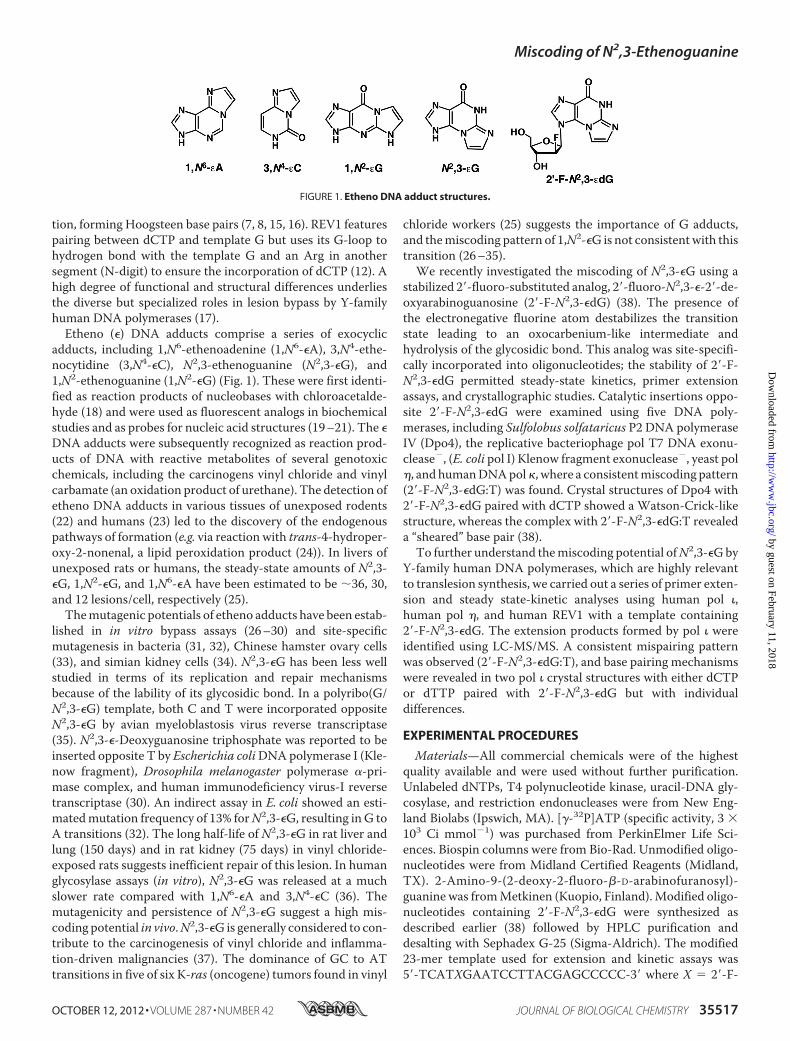
adducts, including 1,N6-ethenoadenine (1,N6-�A), 3,N4-ethe-nocytidine (3,N4-�C), N2,3-ethenoguanine (N2,3-�G), and1,N2-ethenoguanine (1,N2-�G) (Fig. 1). These were first identi-fied as reaction products of nucleobases with chloroacetalde-hyde (18) and were used as fluorescent analogs in biochemicalstudies and as probes for nucleic acid structures (19–21). The �DNA adducts were subsequently recognized as reaction prod-ucts of DNA with reactive metabolites of several genotoxicchemicals, including the carcinogens vinyl chloride and vinylcarbamate (an oxidation product of urethane). The detection ofetheno DNA adducts in various tissues of unexposed rodents(22) and humans (23) led to the discovery of the endogenouspathways of formation (e.g. via reaction with trans-4-hydroper-oxy-2-nonenal, a lipid peroxidation product (24)). In livers ofunexposed rats or humans, the steady-state amounts of N2,3-�G, 1,N2-�G, and 1,N6-�A have been estimated to be �36, 30,and 12 lesions/cell, respectively (25).Themutagenic potentials of etheno adducts have been estab-
lished in in vitro bypass assays (26–30) and site-specificmutagenesis in bacteria (31, 32), Chinese hamster ovary cells(33), and simian kidney cells (34). N2,3-�G has been less wellstudied in terms of its replication and repair mechanismsbecause of the lability of its glycosidic bond. In a polyribo(G/N2,3-�G) template, both C and T were incorporated oppositeN2,3-�G by avian myeloblastosis virus reverse transcriptase(35). N2,3-�-Deoxyguanosine triphosphate was reported to beinserted opposite T by Escherichia coliDNApolymerase I (Kle-now fragment), Drosophila melanogaster polymerase �-pri-mase complex, and human immunodeficiency virus-I reversetranscriptase (30). An indirect assay in E. coli showed an esti-matedmutation frequency of 13% forN2,3-�G, resulting inG toA transitions (32). The long half-life of N2,3-�G in rat liver andlung (150 days) and in rat kidney (75 days) in vinyl chloride-exposed rats suggests inefficient repair of this lesion. In humanglycosylase assays (in vitro), N2,3-�G was released at a muchslower rate compared with 1,N6-�A and 3,N4-�C (36). Themutagenicity and persistence of N2,3-�G suggest a high mis-coding potential in vivo.N2,3-�G is generally considered to con-tribute to the carcinogenesis of vinyl chloride and inflamma-tion-driven malignancies (37). The dominance of GC to ATtransitions in five of six K-ras (oncogene) tumors found in vinyl
chloride workers (25) suggests the importance of G adducts,and themiscoding pattern of 1,N2-�G is not consistentwith thistransition (26–35).We recently investigated the miscoding of N2,3-�G using a
stabilized 2�-fluoro-substituted analog, 2�-fluoro-N2,3-�-2�-de-oxyarabinoguanosine (2�-F-N2,3-�dG) (38). The presence ofthe electronegative fluorine atom destabilizes the transitionstate leading to an oxocarbenium-like intermediate andhydrolysis of the glycosidic bond. This analog was site-specifi-cally incorporated into oligonucleotides; the stability of 2�-F-N2,3-�dG permitted steady-state kinetics, primer extensionassays, and crystallographic studies. Catalytic insertions oppo-site 2�-F-N2,3-�dG were examined using five DNA poly-merases, including Sulfolobus solfataricus P2 DNA polymeraseIV (Dpo4), the replicative bacteriophage pol T7 DNA exonu-clease�, (E. coli pol I) Klenow fragment exonuclease�, yeast pol�, and humanDNApol�, where a consistentmiscoding pattern(2�-F-N2,3-�dG:T) was found. Crystal structures of Dpo4 with2�-F-N2,3-�dG paired with dCTP showed a Watson-Crick-likestructure, whereas the complex with 2�-F-N2,3-�dG:T revealeda “sheared” base pair (38).To further understand themiscoding potential ofN2,3-�Gby
Y-family human DNA polymerases, which are highly relevantto translesion synthesis, we carried out a series of primer exten-sion and steady state-kinetic analyses using human pol �,human pol �, and human REV1 with a template containing2�-F-N2,3-�dG. The extension products formed by pol � wereidentified using LC-MS/MS. A consistent mispairing patternwas observed (2�-F-N2,3-�dG:T), and base pairing mechanismswere revealed in two pol � crystal structures with either dCTPor dTTP paired with 2�-F-N2,3-�dG but with individualdifferences.
EXPERIMENTAL PROCEDURES
Materials—All commercial chemicals were of the highestquality available and were used without further purification.Unlabeled dNTPs, T4 polynucleotide kinase, uracil-DNA gly-cosylase, and restriction endonucleases were from New Eng-land Biolabs (Ipswich, MA). [�-32P]ATP (specific activity, 3 �103 Ci mmol�1) was purchased from PerkinElmer Life Sci-ences. Biospin columns were from Bio-Rad. Unmodified oligo-nucleotides were from Midland Certified Reagents (Midland,TX). 2-Amino-9-(2-deoxy-2-fluoro-�-D-arabinofuranosyl)-guanine was fromMetkinen (Kuopio, Finland).Modified oligo-nucleotides containing 2�-F-N2,3-�dG were synthesized asdescribed earlier (38) followed by HPLC purification anddesalting with Sephadex G-25 (Sigma-Aldrich). The modified23-mer template used for extension and kinetic assays was5�-TCATXGAATCCTTACGAGCCCCC-3� where X � 2�-F-
FIGURE 1. Etheno DNA adduct structures.
Miscoding of N2,3-Ethenoguanine
OCTOBER 12, 2012 • VOLUME 287 • NUMBER 42 JOURNAL OF BIOLOGICAL CHEMISTRY 35517
by guest on February 11, 2018http://w
ww
.jbc.org/D
ownloaded from

N2,3-�dG (MALDI-TOFMS (3-hydroxypicolinic acid)m/z cal-culated for [M � H]�, 6986.5; found, 6985.6) or 2�-fluoro-2�-deoxyarabinoguanosine (2�-F-dG) (MALDI-TOF MS (3-hydroxypicolinic acid) m/z calculated for [M � H]�, 6962.5;found, 6963.5). The 18-mer oligomer used for crystallographicstudies was 5�-TCT(2�-F-N2,3-�dG)GGGTCCTAGGACC-(ddC)-3� (ddC, dideoxy-CMP) (MALDI-TOF MS (3-hy-droxypicolinic acid) m/z calculated for [M � H]�, 5514.9;found, 5515.2). HumanDNApol � catalytic fragments (residues1–420) (16), pol � (39), and pol � (38) were purified followingprotocols described previously.Preparation of Recombinant Catalytic Core of Human REV1—
The gene fragment covering the catalytic core (residues 330–833) (12) of wild-type human REV1 was obtained by PCRamplification from the vector pET-22b(�)/hREV1 (13) as tem-plate using PfuDNApolymerase (Stratagene, La Jolla, CA) witha pair of primers (5�-GGATCCATGTCTACGTTTAGCAAG-GCAG-3� and 5�-GCGGCCGCTTATGTGGAAGGGTTCA-GATTAG-3�). The resulting PCR product of the 1.5-kb hREV1fragment was cloned into the vector pSC-B-Amp/Kan (Strat-agene). Following sequence confirmation, thehREV1 gene frag-ment was cloned into the BamHI and NotI sites of the vectorpBG101 (obtained from the Center for Structural Biology,Vanderbilt University) to generate the cleavable glutathi-one S-transferase (GST)-tagged protein. The GST-taggedhREV1(330–833) was expressed in E. coli BL21 (DE3) cells,which were grown at 37 °C and 220 rpm to anOD600 of 0.6 andthen induced with isopropyl �-D-1-thiogalactopyranoside (0.2mM) for 12 h at 16 °C. The harvested pellets were resuspendedin lysis buffer containing 50 mM Tris-HCl (pH 7.4), 500 mM
NaCl, 10% (v/v) glycerol, 5 mM �-mercaptoethanol, 1 mg ml�1
lysozyme, and protease inhibitor mixture (Roche Applied Sci-ence). Suspensions were sonicated, and the cell lysate was clar-ified by centrifugation at 4 � 104 � g for 60 min at 4 °C. Theresulting supernatants were loaded onto a 1-ml GSTrap 4B col-umn (GE Healthcare), and the column was washed with 20 mlof Buffer A (50mMTris-HCl (pH 7.4) containing 150mMNaCl,10% (v/v) glycerol, and 5 mM �-mercaptoethanol). The GST-tagged REV1(330–833) bound on the columnwas cleavedwithPrescission protease (GE Healthcare) for 14 h at 4 °C. CleavedREV1(330–833) was eluted with Buffer A, and the purity wasanalyzed by SDS-polyacrylamide gel electrophoresis with Coo-massie Brilliant Blue R-250 staining. A typical yield was �760�g from 1 liter of culture.Primer Extension and Steady-state Kinetic Assays—An
18-mer oligomer (5�-GGGGGCTCGTAAGGATTC-3�) was 5�[�-32P]ATP end-labeled and annealed to a 23-mer template (5�-TCATXGAATCCTTACGAGCCCCC-3� where X � 2�-F-N2,3-�dG, 2�-F-dG, or 2�-deoxyguanosine (dG)). Primer exten-sion experiments were performed in 50 mM Tris-HCl buffer(pH 7.5) containing 60 nM primer�template complex, 250 �M
dNTPs, 20 nM polymerase, 5% (v/v) glycerol, 5mMDTT, 50mM
NaCl, 5 mM MgCl2, and 50 �g ml�1 bovine serum albumin(BSA) at 37 °C. Steady-state kinetic experiments were carriedout under the same conditions except using 5–20 nM pol, vary-ing dNTP concentrations, and 2–10-min incubation times.Reactions were quenched with 9 �l of 20 mM EDTA (pH 9.0) in95% (v/v) formamide. Products were separated using 20% (w/v)
acrylamide electrophoresis gels, and results were visualizedusing a phosphorimaging system (Bio-Rad, Molecular Imager�FX) and analyzed by Quantity One software as described (38).LC-MS Analysis of Full-length Extension Products—An
18-mer primer (5�-GGGGGCTCGTAAGGAT(dU)C-3�) wasannealed to the same 23-mer oligomer as described above at a1:1 molar ratio. Reaction conditions were similar to those usedin steady-state kinetic assays except that the final concentra-tions were as follows: 10 �M pol �, 12.5 �M primer�templatecomplex, and 2% (v/v) glycerol in a total volume of 80 �l. Reac-tions were carried out in the presence of four dNTPs (10 mM
each) for 3.5 h at 37 °C. The reactions were terminated by spincolumn separations to extract dNTPs andMg2�, and the result-ing productwas treatedwith 50 units of uracil-DNAglycosylaseand then 0.25 M hot piperidine (40). LC-MS/MS analyses wereperformed using an ACQUITY ultraperformance liquid chro-matography (UPLC) system (Waters Corp.) connected to aFinnigan LTQ mass spectrometer (Thermo Scientific Corp.,San Jose, CA) operating in the electrospray ionization negativeion mode and using an ACQUITY UPLC system BEH octade-cylsilane (C18) column (1.7 �m; 1.0 � 100 mm). UPLC condi-tions were as described (38).Crystallization of Pol ��2�-F-N2,3-�dG�DNA Ternary Com-
plexes—The sequence of the 18-mer oligomer used for co-crys-tallization with pol �, 5�-TCT(2�-F-N2,3-�dG)GGGTCCTAG-GACC(ddC)-3�, was designed based on previous studies (15,16). Crystals were obtained under conditions similar to thosedescribed previously (15, 16). Specifically, 210 �M pol �, 253 �M
annealed DNA, 10 mM MgCl2, and 20 mM dCTP (or dTTP)were mixed and preincubated on ice. Droplets (a 1:1 (v/v) mix-ture of pol�DNA complex mixture and the reservoir solution)were equilibrated against a reservoir solution containing 0.10 M
2-(N-morpholino)ethanesulfonic acid (MES) (sodium salt, pH6.5), 0.3 M (NH4)2SO4, and 15% (w/v) polyethylene glycol 5000for pol �-1 (pol ��2�-F-N2,3-�dG:dCTP) or 17% (w/v) polyethyl-ene glycol 5000 for pol �-2 (pol ��2�-F-N2,3-�dG:dTTP). Crystalswere grown using the hanging drop vapor diffusion method at4 °C and mounted at 4 °C with step soaking in mother liquorsolutions containing 0–25% (w/v) glycerol prior to flash freez-ing in liquid nitrogen.Structure Determination and Refinement—X-ray diffraction
data were collected at the Advanced Photon Source(Argonne National Laboratory, Argonne, IL) on the 21-ID-Fand 21-ID-G (Life Sciences Collaborative Access Team)beam lines. All data sets were recorded from cryoprotectedcrystals using a single wavelength at 100 K. Data wereindexed and scaled with the program HKL2000 (41). Bothcrystal types belonged to space group P6522. X-ray diffrac-tion data collection and processing statistics are listed inTable 3 (see below). Phases were calculated using MOLREPas a part of the CCP4 program suite (42, 43) based on apreviously refined model (Protein Data Bank code 3OSN)(16). Refinements were performed using Refmac 6.0 withrestrained and rigid body refinement (44, 45). Repeatedcycles of manual rebuilding were performed in Coot (46).Structural images were generated in PyMOL (47).
Miscoding of N2,3-Ethenoguanine
35518 JOURNAL OF BIOLOGICAL CHEMISTRY VOLUME 287 • NUMBER 42 • OCTOBER 12, 2012
by guest on February 11, 2018http://w
ww
.jbc.org/D
ownloaded from
RESULTS
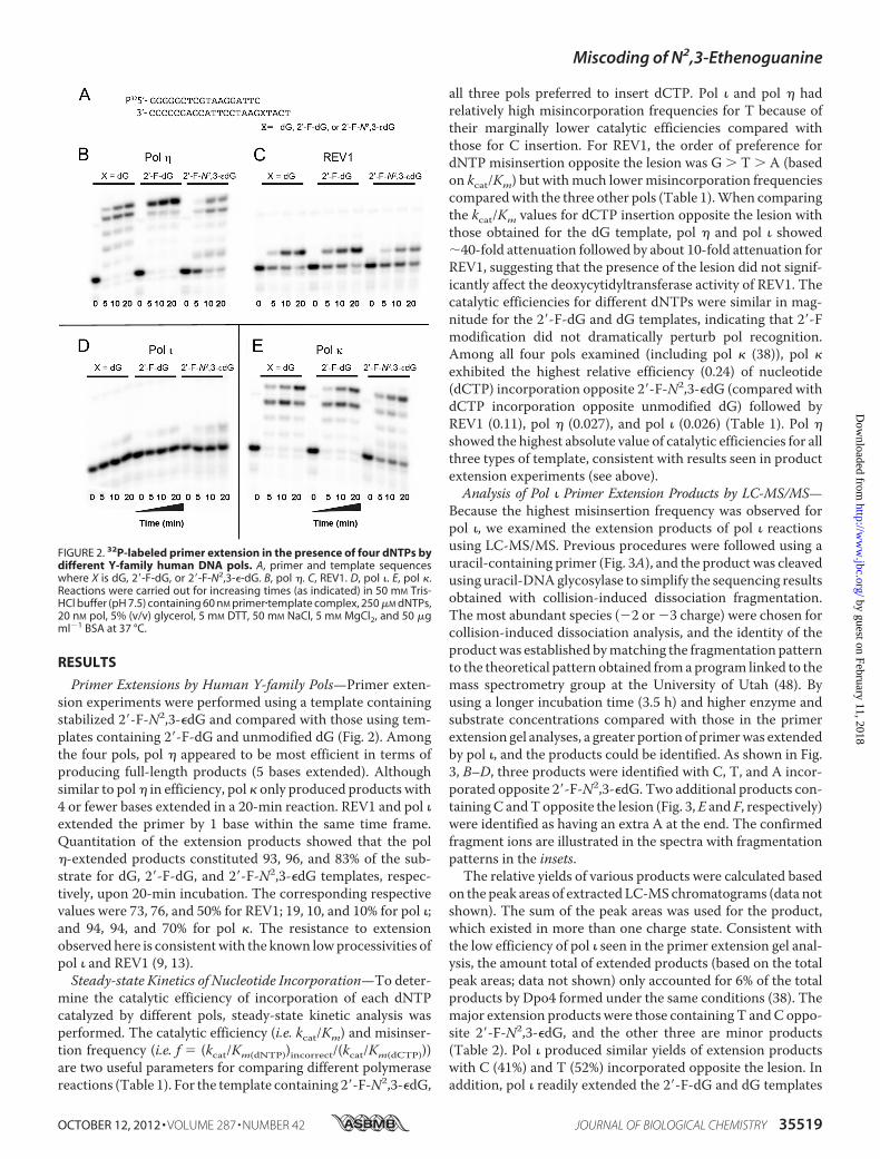
Primer Extensions by Human Y-family Pols—Primer exten-sion experiments were performed using a template containingstabilized 2�-F-N2,3-�dG and compared with those using tem-plates containing 2�-F-dG and unmodified dG (Fig. 2). Amongthe four pols, pol � appeared to be most efficient in terms ofproducing full-length products (5 bases extended). Althoughsimilar to pol� in efficiency, pol � only produced products with4 or fewer bases extended in a 20-min reaction. REV1 and pol �extended the primer by 1 base within the same time frame.Quantitation of the extension products showed that the pol�-extended products constituted 93, 96, and 83% of the sub-strate for dG, 2�-F-dG, and 2�-F-N2,3-�dG templates, respec-tively, upon 20-min incubation. The corresponding respectivevalues were 73, 76, and 50% for REV1; 19, 10, and 10% for pol �;and 94, 94, and 70% for pol �. The resistance to extensionobserved here is consistentwith the known lowprocessivities ofpol � and REV1 (9, 13).Steady-state Kinetics of Nucleotide Incorporation—To deter-
mine the catalytic efficiency of incorporation of each dNTPcatalyzed by different pols, steady-state kinetic analysis wasperformed. The catalytic efficiency (i.e. kcat/Km) and misinser-tion frequency (i.e. f � (kcat/Km(dNTP))incorrect/(kcat/Km(dCTP)))are two useful parameters for comparing different polymerasereactions (Table 1). For the template containing 2�-F-N2,3-�dG,
all three pols preferred to insert dCTP. Pol � and pol � hadrelatively high misincorporation frequencies for T because oftheir marginally lower catalytic efficiencies compared withthose for C insertion. For REV1, the order of preference fordNTP misinsertion opposite the lesion was G � T � A (basedon kcat/Km) but withmuch lowermisincorporation frequenciescomparedwith the three other pols (Table 1).When comparingthe kcat/Km values for dCTP insertion opposite the lesion withthose obtained for the dG template, pol � and pol � showed�40-fold attenuation followed by about 10-fold attenuation forREV1, suggesting that the presence of the lesion did not signif-icantly affect the deoxycytidyltransferase activity of REV1. Thecatalytic efficiencies for different dNTPs were similar in mag-nitude for the 2�-F-dG and dG templates, indicating that 2�-Fmodification did not dramatically perturb pol recognition.Among all four pols examined (including pol � (38)), pol �exhibited the highest relative efficiency (0.24) of nucleotide(dCTP) incorporation opposite 2�-F-N2,3-�dG (compared withdCTP incorporation opposite unmodified dG) followed byREV1 (0.11), pol � (0.027), and pol � (0.026) (Table 1). Pol �showed the highest absolute value of catalytic efficiencies for allthree types of template, consistent with results seen in productextension experiments (see above).Analysis of Pol � Primer Extension Products by LC-MS/MS—
Because the highest misinsertion frequency was observed forpol �, we examined the extension products of pol � reactionsusing LC-MS/MS. Previous procedures were followed using auracil-containing primer (Fig. 3A), and the product was cleavedusing uracil-DNAglycosylase to simplify the sequencing resultsobtained with collision-induced dissociation fragmentation.The most abundant species (�2 or �3 charge) were chosen forcollision-induced dissociation analysis, and the identity of theproductwas established bymatching the fragmentation patternto the theoretical pattern obtained fromaprogram linked to themass spectrometry group at the University of Utah (48). Byusing a longer incubation time (3.5 h) and higher enzyme andsubstrate concentrations compared with those in the primerextension gel analyses, a greater portion of primerwas extendedby pol �, and the products could be identified. As shown in Fig.3, B–D, three products were identified with C, T, and A incor-porated opposite 2�-F-N2,3-�dG. Two additional products con-tainingC andTopposite the lesion (Fig. 3,E and F, respectively)were identified as having an extra A at the end. The confirmedfragment ions are illustrated in the spectra with fragmentationpatterns in the insets.
The relative yields of various products were calculated basedon the peak areas of extracted LC-MS chromatograms (data notshown). The sum of the peak areas was used for the product,which existed in more than one charge state. Consistent withthe low efficiency of pol � seen in the primer extension gel anal-ysis, the amount total of extended products (based on the totalpeak areas; data not shown) only accounted for 6% of the totalproducts by Dpo4 formed under the same conditions (38). Themajor extension products were those containing T andC oppo-site 2�-F-N2,3-�dG, and the other three are minor products(Table 2). Pol � produced similar yields of extension productswith C (41%) and T (52%) incorporated opposite the lesion. Inaddition, pol � readily extended the 2�-F-dG and dG templates
FIGURE 2. 32P-labeled primer extension in the presence of four dNTPs bydifferent Y-family human DNA pols. A, primer and template sequenceswhere X is dG, 2�-F-dG, or 2�-F-N2,3-�-dG. B, pol �. C, REV1. D, pol �. E, pol �.Reactions were carried out for increasing times (as indicated) in 50 mM Tris-HCl buffer (pH 7.5) containing 60 nM primer�template complex, 250 �M dNTPs,20 nM pol, 5% (v/v) glycerol, 5 mM DTT, 50 mM NaCl, 5 mM MgCl2, and 50 �gml�1 BSA at 37 °C.
Miscoding of N2,3-Ethenoguanine
OCTOBER 12, 2012 • VOLUME 287 • NUMBER 42 JOURNAL OF BIOLOGICAL CHEMISTRY 35519
by guest on February 11, 2018http://w
ww
.jbc.org/D
ownloaded from

in an error-free manner. The base insertion pattern obtainedfrom LC-MS/MS analysis agrees with the steady-state kineticanalysis (Table 1) with T insertions being the major misincor-poration events for pol �-catalyzed bypass.Crystal Structures of Pol � with Oligonucleotides Containing
2�-F-N2,3-�dG and dCTP or dTTP—To understand the basepairing mechanisms for dCTP and dTTP observed above, weconducted co-crystallization experiments with pol �, a templatecontaining 2�-F-N2,3-�dG, and dCTP or dTTP. Two types ofcrystals were obtained, i.e. pol �-1 (pol ��2�-F-N2,3-�dG:dCTP;Protein Data Bank code 4FS2) and pol �-2 (pol ��2�-F-N2,3-�dG:dTTP; Protein Data Bank code 4FS1). Structures of these twoternary complexes were determined by molecular replacementusing a previously refined model (Protein Data Bank code3OSN) (16) without the lesion and the incoming nucleotide(Table 3). Clear electron densities around the 2�-F-N2,3-�dGand incoming nucleotide facilitated the unbiased determina-
tion of the base pairing conformations at the active site. Similarto several structures seen previously for template native purines(7, 15) and adducted purines (8, 9, 16), the electron densityaround 2�-F-N2,3-�dG indicated that the lesion was rotatedfrom the anti to the syn conformation (Fig. 4, A and C). Thissubstrate-induced conformational change of template purine isthought to be dictated by the rigid active site of pol � (7).
In both the pol �-1 (Fig. 4A) and pol �-2 (Fig. 4C) structures,the incoming nucleotide served as a donor in hydrogen bondswith the Hoogsteen edge of 2�-F-N2,3-�dG (i.e. the O6 and N7atoms). When 2�-F-N2,3-�dG was paired with dCTP (Fig. 4B),one hydrogen bond was observed between the N4 atom ofdCTP and O6 atom of 2�-F-N2,3-�dG as indicated by a 2.5-Ådistance. The possibility of a second hydrogen bond cannot beruled out based on the distance (3.0 Å) between the N3 atom ofdCTP and the N7 atom of 2�-F-N2,3-�dG provided that the N3atom of dCTP is protonated. This mechanism was proposed in
TABLE 1Steady-state kinetic analysis of polymerase-catalyzed single base insertionThe insertion was opposite X in the template sequence of 3�-CCCCCGAGCATTCCTAAGXTACT-5� where X is 2�-F-N2,3-�dG, 2�-F-dG, or dG.
Polymerase/template dNTP kcat Km(dNTP) kcat/Km(dNTP) f a Relative efficiencyb
min�1 �M min�1 �M�1
pol �2�-F-N2,3-�dG C 0.26 � 0.03 153 � 58 0.0017 0.026
T 0.33 � 0.02 280 � 36 0.0012 0.71A 0.016 � 0.001 956 � 83 0.000017 0.010G 0.007 � 0.001 223 � 71 0.000031 0.018
2�-F-dG C 0.079 � 0.1 14 � 4 0.0056 0.085T 0.049 � 0.005 156 � 46 0.00031 0.055A 0.0067 � 0.0011 88 � 47 0.000076 0.014G 0.0040 � 0.0006 44 � 25 0.000091 0.016
dG C 0.093 � 0.001 1.4 � 0.5 0.066 1T 0.63 � 0.18 1090 � 510 0.00058 0.0088A 0.0052 � 0.0007 111 � 53 0.000047 0.00071G 0.0098 � 0.0009 365 � 69 0.000027 0.00041
Pol �2�-F-N2,3-�dG C 0.88 � 0.04 11 � 2 0.08 0.027
T 1.3 � 0.06 26 � 6 0.05 0.63A 0.40 � 0.03 63 � 21 0.0063 0.079G 0.30 � 0.02 45 � 9 0.0067 0.084
2�-F-dG C 1.7 � 0.06 0.51 � 0.12 3.3 1.1T 0.70 � 0.05 49 � 12 0.014 0.0042A 0.96 � 0.07 15 � 6 0.064 0.019G 0.63 � 0.05 18 � 7 0.035 0.011
dG C 2.3 � 0.14 0.76 � 0.23 3.0 1T 0.51 � 0.03 57 � 11 0.0089 0.0030A 0.88 � 0.03 74 � 9 0.012 0.0040G 0.51 � 0.03 50 � 9 0.010 0.0033
REV12�-F-N2,3-�dG C 0.083 � 0.005 4.8 � 1.4 0.017 0.11
T 0.024 � 0.003 24 � 2 0.0010 0.059A 0.012 � 0.001 32 � 8 0.00038 0.022G 0.060 � 0.018 44 � 13 0.0013 0.080
2�-F-dG C 0.17 � 0.01 1.6 � 0.5 0.11 0.73T 0.37 � 0.02 90 � 13 0.0041 0.037A 0.043 � 0.004 27 � 10 0.0016 0.015G 0.89 � 0.03 10 � 2 0.0089 0.081
dG C 0.12 � 0.01 0.81 � 0.37 0.15 1T 0.024 � 0.001 7.8 � 4.2 0.0031 0.021A 0.043 � 0.004 27 � 11 0.0016 0.011G 0.49 � 0.03 130 � 14 0.0037 0.025
Pol �c
2�-F-N2,3-�dG C 1.6 � 0.1 73 � 13 0.022 0.24T 0.90 � 0.04 111 � 14 0.0082 0.37 0.091A 0.063 � 0.002 55 � 7 0.0011 0.05G 0.22 � 0.01 210 � 22 0.0010 0.045
2�-F-dG C 1.9 � 0.1 2.8 � 0.3 0.68dG C 1.8 � 0.1 20 � 1 0.090 1
a Misinsertion frequency: f � (kcat/Km(dNTP))incorrect/(kcat/Km(dCTP)).b Relative efficiency calculated as the ratio of the kcat/Km for dNTP insertion opposite the adduct to the kcat/Km for dCTP insertion opposite dG.c From Ref. 38.
Miscoding of N2,3-Ethenoguanine
35520 JOURNAL OF BIOLOGICAL CHEMISTRY VOLUME 287 • NUMBER 42 • OCTOBER 12, 2012
by guest on February 11, 2018http://w
ww
.jbc.org/D
ownloaded from

previous studies for dCTP paired with G (15) or adducted G(16).In the case of 2�-F-N2,3-�dG-paired dTTP (Fig. 4D), it is
likely that only one hydrogen bond exists between the O6 atomof 2�-F-N2,3-�Gand theN3 atom of dTTPwith a distance of 2.8Å. Although a distance of 3.2 Å between N7(2�-F-N2,3-�dG)and N3(dTTP) can also be interpreted as a potential hydrogenbonding distance, the asymmetry between the two distances(2.8 versus 3.2 Å) makes it unlikely that a bifurcated hydrogenbonding structure is present. The slightly longer distance (2.8Å) in the case of 2�-F-N2,3-�dG:T hydrogen bond may be anindication of its weaker strength compared with the 2.5-Å dis-tance seen in the 2�-F-N2,3-�dG:C pair, although the resolutionlimit of 2.5 Å does not permit a firm conclusion in this respect.Together with the possibility of two hydrogen bonds in the2�-F-N2,3-�dG:C pair, the base pair modes observed here areconsistent with the slightly favorable insertion of C observed inthe steady-state kinetic analysis.Irrespective of the incoming nucleotide, the pol �-1 and pol
�-2 structures are quite similar with a root mean square devia-
tion value of 0.27 Å for all atom pairs upon superimposition.The superimposition of the incoming nucleotides suggested amovement of 2�-F-N2,3-�dG toward the minor groove for the2�-F-N2,3-�dG:T base pair (Fig. 5A). The pol �-1 structuresuperimposes with the native G:C complex (Protein Data Bankcode 2ALZ; Ref. 15) with a root mean square deviation value of0.27 Å (Fig. 5B), indicating that the presence of the lesion (2�-F-N2,3-�dG) did not significantly affect the conformations ofthe protein and the nucleic acid. The conformation of the 2�-F-N2,3-�dG:C pair also resembles that of an N2-ethylguanine:Cpair crystallizedwith pol � (9) (Fig. 5C).When the conformationof the 2�-F-N2,3-�dG:T mispair is compared with an O6-meth-ylguanine:T base pair (16) (because of the lack of a structurewith the pol � G:T pair in the Protein Data Bank), the confor-mations of both base pairs are similar except that the lesion isslightly moved toward the minor groove in the case of the 2�-F-N2,3-�dG:T pair (Fig. 5D, green). This shift could be due to thebulkier size of 2�-F-N2,3-�dG compared with O6-methylgua-nine. Overall, pol � appears to be able to accommodate the 2�-F-N2,3-�dG pair rather well at the active site without significantprotein and nucleic acid conformational changes. The struc-tures showed that both C and T pair with the lesion in a similarfashion in line with the observation that pol � promoted botherror-free and error-prone bypass in steady-state kinetic andLC-MS/MS analyses.
DISCUSSION
The DNA adduct N2,3-�G is a ubiquitous modification pro-duced from endogenous processes (e.g. lipid peroxidation) orexposure to environmental pollutants (e.g. vinyl chloride orurethane). We recently developed an isostere approach toincorporate the stabilized analog (2�-F-N2,3-�dG) into oligonu-cleotides and investigated the miscoding potential of N2,3-�Gusing several prokaryotic and eukaryotic DNA pols (38). In thepresent work, we extended our previous investigation into the
FIGURE 3. Collision-induced dissociation spectra from LC-MS/MS analysis of the full-length extension assays using pol � and a 23-mer oligomertemplate containing 2�-F-N2,3-�dG in the presence of all four dNTPs. A, template and primer sequences. The confirmed product sequences with thefragmentation patterns shown are 5�-CCATGA-3� (B), 5�-CTATGA-3� (C), 5�-CAATGA-3� (D), 5�-CCATGAA-3� (E), and 5�-CTATGAA-3� (F). The reaction contained12.5 �M DNA complex, 10 mM dNTPs, 10 �M pol �, 5 mM DTT, 50 mM NaCl, 5 mM MgCl2, and 50 �g ml�1 BSA and were incubated at 37 °C for 3.5 h. Underlined Uindicates the cutting site by uracil-DNA glycosylase after DNA polymerase reactions.
TABLE 2Summary of pol � extension products from LC-MS/MS analysisThe results were obtained using template � primer complexes containing 2�-F-N2,3-�dG, 2�-F-dG, or dG. Underlined nucleotides indicate the base incorporated oppo-site the lesion.
Miscoding of N2,3-Ethenoguanine
OCTOBER 12, 2012 • VOLUME 287 • NUMBER 42 JOURNAL OF BIOLOGICAL CHEMISTRY 35521
by guest on February 11, 2018http://w
ww
.jbc.org/D
ownloaded from
other three human Y-family DNA pols and provided the struc-tural basis of the most error-prone bypass enzyme, pol �.
Primer extension gel analysis generated a qualitative com-parison of the capability of bypassing 2�-F-N2,3-�dG by Y-fam-ily pols (Fig. 1). The order of bypassing efficiency (from thepercentage of total product extended) is pol � � pol � �REV1 pol �. Comparedwith pol� and pol �, the higher activityof pol � copying past N2,3-�G observed here is similar to thatseen previously for other etheno adducts, i.e. 1,N2-�G (29),1,N6-�A (26, 49), and 3,N4-�C (50). With regard to DNA poly-merases, the extension pattern is particularly similar to that ofbypass of 1,N2-�G; i.e. pol � readily extended the primer intofull-length products, whereas pol � and pol � showed some sin-gle base incorporation (29).Steady-state kinetic analysis established the preferred base
incorporated opposite the lesion and provided a kinetic ration-ale for primer extension experiments (Table 1). For all fourhuman Y-family DNA pols, the correct base C is marginallypreferred opposite 2�-F-N2,3-�dGwith similar relative efficien-cies in comparison with the insertion of C opposite a regular G(Table 1). Themisinsertion of T is consistent for all four humanY-family DNA pols as well as for several other prokaryotic andeukaryotic DNA polymerases (38). The highest absolute valueof catalytic efficiency (kcat/Km) seen (for pol �) is in line withprimer extension results, which may be partly explained by themore open active site of pol � compared with other poly-
merases (51). The pattern of fidelity for pol � bypassing differ-ent etheno lesions is similar: both error-free and error-pronesyntheses have been observed. Pol � inserted a C oppositeN2,3-�G in a marginally error-free manner with a misinsertionfrequency of 0.63 for T (Table 1). Similarly, pol � copied past1,N6-�A in the order of preference T � C � A � G (49). Theorder was G � A � C for 1,N2-�G (29) and A G � C T for3,N4-�C (50). Pol � has the highest misincorporation frequency(although C is preferred 1.5-fold compared with T), which isconsistent with the view that pol � generally catalyzes error-prone bypass (3). The incorporation patterns seen for pol �bypassing other ethenoDNAadducts are as follows: pol � some-what prefers to incorporate C opposite 1,N6-�A (8) and insertsboth C and T opposite 1,N2-�G with almost the same catalyticefficiencies (29). The fact that REV1 prefers to catalyze dCTPinsertion is not surprising in that REV1 utilizes its G-loop tohydrogen bondwith template G and anArg in another segment(N-digit) to ensure the incorporation of dCTP (12).When com-parisons are made with the catalytic efficiency of dCTP inser-tions opposite native G in the template, the order of relativeefficiency is pol � (0.24) � REV1 (0.11) � pol � (0.027) pol �(0.026) (Table 1), suggesting that 2�-F-N2,3-�dG affects theDNA syntheses of the four Y-family pols to a similar extent.LC-MS/MS analysis of the primer extension products by pol
� provided further insight into the nature of the bases insertedbeyond the lesion in these error-prone reactions. With pol �,
TABLE 3Crystal data collection and refinement statistics for the ternary complexes pol �-1 (pol ��2�-F-N2,3-�dG:dCTP, Protein Data Bank code 4FS2) andpol �-2 (pol ��2�-F-N2,3-�dG:dTTP, Protein Data Bank code 4FS1)
Pol �-1 (2�-F-N2,3-�dG:dCTP) Pol �-2 (2�-F-N2,3-�dG:dTTP)
Data collectionBeamline 21-ID-F 21-ID-GSpace group P6522 P6522Unit cell (a, b, c) (Å) 97.30, 97.30, 202.91 97.47, 97.47, 203.54Unit cell (�, �, �) (°) 90.0, 90.0, 120.0 90.0, 90.0, 120.0Resolution (Å)a 2.05 (2.05–2.09) 2.49 (2.49–2.53)No. of measured reflections 36,517 23,120No. of unique reflections 36,444 23,074Percent possible (%) 99.8 (98.4) 99.8 (100)Redundancy 7.8 (6.3) 6.9 (6.8)Rlinear
b 0.051 (0.581) 0.067 (0.505)Signal to noise (I/I) 32.5 (2.5) 23.0 (3.2)
Coordinate composition (asymmetric unit)No. of protein molecules 1 1No. of amino acid residues 383 383No. of water molecules 179 65No. of Mg2� ions 3 2No. of template nucleotides 8 9No. of primer nucleotides 7 7No. of dCTP 1 0No. of dTTP 0 1
RefinementResolution range (Å) 30.00–2.05 30.00–2.50Reflections 34,557 19,448Rwork (%)c 21.4 21.5Rfree (%)d 26.0 27.0Root mean square deviation bond length (Å) 0.018 0.014Root mean square deviation bond angle (°) 2.05 1.99Mean B-factor 51.3 56.7Wilson B-factor 42.8 49.4Ramachandran summaryIn preferred regions 354 (93.40%) 350 (92.35%)In allowed regions 16 (4.22%) 23 (6.07%)Outliers 9 (2.37%) 5 (1.58%)
a Values for highest resolution bin are given in parentheses.b Rlinear � �I � �I��I where I is the integrated intensity of a given reflection.c Rwork � �Fobserved � Fcalculated�/�Fobserved�.d Rfree was calculated using 5% test size with random selection.
Miscoding of N2,3-Ethenoguanine
35522 JOURNAL OF BIOLOGICAL CHEMISTRY VOLUME 287 • NUMBER 42 • OCTOBER 12, 2012
by guest on February 11, 2018http://w
ww
.jbc.org/D
ownloaded from

approximately half of the products contained T with a highfidelity extension beyond the lesion (Fig. 3 and Table 2). Theobservation of almost equal amounts of products containing Cand T opposite 2�-F-N2,3-�dG (with LC-MS/MS analysis) is inline with results from kinetic analysis (Table 1). The muchlower amount of total extended products (6%) compared withDpo4 (38) agrees with the low bypass efficiency of pol � seen inthe primer extension gel analysis (Fig. 2). These extension prod-ucts are similar to the products generated by Dpo4 (38); how-ever, the pattern of miscoding is considerably different fromthat generated by 1,N2-�G,which yieldsmainly productswithGinserted by human pol � (29) and �1 deletion products byDpo4 (28).
The hydrogen bonding patterns of 2�-F-N2,3-�dG:C and2�-F-N2,3-�dG:T base pairs seen in the crystal structures pro-vided molecular explanations for the error-free and error-prone bypass of pol �. The distance of 2.5 Å is a clear indicationthat a hydrogen bond is established between the O6 atom of2�-F-N2,3-�dG and the N4 atom of dCTP. The possibility of asecond hydrogen bond also exists, i.e. between the N7 atom of2�-F-N2,3-�dG and the N3 atom of dCTP, provided that the N3atom of dCTP is protonated. The tendency for protonation ofthe N3 atom of dCTP has been discussed in several other pol��DNA structures with both native and adducted purines in thetemplates (8, 15, 16). Although theN3 atom of free cytosine hasa pKa �4.5, the local molecular environment could elevate thepKa to 6.2–7.2 at a terminal position or �8.5 at an internalposition in DNA triple helices (52, 53). Nair et al. (8) suggestedthat an elevation of the pKa of dCTP could be due to the base-stacking and long range electrostatic interactions of the activesite residues Asp-126 and Glu-127. The one hydrogen bondobserved in the 2�-F-N2,3-�dG:T pair is an indication that the2�-F-N2,3-�dG:T pair might be less stable compared with the2�-F-N2,3-�dG:C pair. Our crystallization attempts are consis-tentwith this view in that pol �-1 type crystals (with dCTP) grewmore easily and diffracted to higher resolution than the pol �-2crystal (with dTTP). Collectively, the difference in hydrogenbonding may explain a slightly higher catalytic efficiency fordCTP by pol �.
The typical strategy that pol � uses a Hoogsteen base pairingmechanism to accommodate native and adducted purines wasonce again demonstrated in both the pol �-1 and pol �-2 struc-tures albeit with different hydrogen bonding schemes. The sim-ilarity of the two structures consists of their use of the Hoogs-teen edge of 2�-F-N2,3-�dG to hydrogen bond with theincoming nucleotide. The conformation of 2�-F-N2,3-�dG:Calso resembles G:dCTP (15) andN2-ethylG:dCTP (9) at the pol� active site.However, the base pair conformations seen here are quite
different fromwhat has been observed at the active site of Dpo4(38). Specifically, the 2�-F-N2,3-�dG:C pair adopts a Watson-
FIGURE 4. Crystal structures of ternary complexes pol �-1 (pol ��2�-F-N2,3-�dG:dCTP, Protein Data Bank code 4FS2) and pol �-2 (pol ��2�-F-N2,3-�dG:dTTP, Protein Data Bank code 4FS1). A, pol �-1 active site with template2�-F-N2,3-�dG pairing with the incoming dCTP. Green spheres show the twoobserved Mg2� ions. B, 2�-F-N2,3-�dG in syn conformation in pol �-1 with onehydrogen bond formed between the O6 atom of 2�-F-N2,3-�dG and the N4atom of dCTP (2.5 Å); the other potential hydrogen bond formed is indicatedwith a dashed line between the N7 atom of 2�-F-N2,3-�dG and the N3 atom ofdCTP (3.0 Å). C, pol �-2 active site with template 2�-F-N2,3-�dG pairing withincoming dTTP. The green sphere shows the observed single Mg2� ion.D, 2�-F-N2,3-�dG in the syn conformation in pol �-2 with one hydrogen bondformed between the O6 atom of 2�-F-N2,3-�dG and the N3 atom of dTTP (2.8Å). The quality of the data is demonstrated using the Fourier 2Fo � Fc sumelectron density map displayed (blue mesh) at 1.0 in A and C.
FIGURE 5. Comparisons of base pair positions at the active site of pol �complexes based on the superimposition of incoming nucleotides.A, shearing of 2�-F-N2,3-�dG toward the minor groove for the 2�-F-N2,3-�dG:dTTP pair (green) compared with the 2�-F-N2,3-�dG:dCTP pair (red).B, similarity between the 2�-F-N2,3-�dG:dCTP pair (red) and the nativeG:dCTP pair (blue; Protein Data Bank code 2ALZ). C, structural similaritybetween the 2�-F-N2,3-�dG:dCTP pair (red) and the N2-ethylguanine:dCTPpair (gray; Protein Data Bank code 3EPG) crystallized with pol �; D, shearingof 2�-F-N2,3-�dG towards the minor groove for the 2�-F-N2,3-�dG:dTTP pair(green) compared to O6-methylguanine:dTTP pair (16) (orange, ProteinData Bank code 3OSN).
Miscoding of N2,3-Ethenoguanine
OCTOBER 12, 2012 • VOLUME 287 • NUMBER 42 JOURNAL OF BIOLOGICAL CHEMISTRY 35523
by guest on February 11, 2018http://w
ww
.jbc.org/D
ownloaded from

Crick-like conformation, and the 2�-F-N2,3-�dG:T structurecontains a sheared base pair at theDpo4 active site (Fig. 6,C andD, and Ref. 38). That 2�-F-N2,3-�dG was observed to be posi-tioned in the anti conformation by Dpo4 is likely due to therelatively open active site comparedwith pol � (17) (surface viewshown in Fig. 6, A and B). Particularly, the residues adjacent tothe template base are bulkier (Leu-62, Val-64, and Gln-59) inpol � compared with Dpo4 (Ala-42, Ala-44, and Val-32) (54). Inthe Dpo4 structure, hydrophobic interactions are likely to existbetween Val-32 and the imidazole ring of 2�-F-N2,3-�dG (anti).Conversely, residues (Leu-62, Val-64, and Gln-59) may force2�-F-N2,3-�dG to rotate into the syn conformation, whichwould otherwise clash with these residues if the lesion werepositioned in the anti conformation. Despite these conforma-tional differences, similar extents of T misinsertion areobserved in both cases.As mentioned in the Introduction, Singer and co-workers
(30, 32, 35) reported three studies on the miscoding ofN2,3-�Gmore than 20 years ago. These studies were limited by the gen-eral methods available for studying miscoding at the time aswell as the inherent lability of the glycosidic bond of N2,3-�dG.The uncorrected mutation frequency for N2,3-�dG insertedinto anM13 phage systemwas only 0.5%, but in that study (32),an in vitro study with a polyribo(G/N2,3-�G) template and areverse transcriptase (35), and a study on “reverse” incorpora-tion of N2,3-�dG triphosphate (30), the general pattern was
pairing ofN2,3-�Gwith T and C. This pattern, despite any defi-ciencies in the earlier work, is similar to those seen in our ownstudies (Ref. 38 and the present work). The N2,3-�G:T wobblepairing proposed in that early work (35) had no experimentalbasis and has not been observed in our crystal structures withDpo4 (38) or human pol � (Fig. 4).
More recently, theoretical studies (55) have predicted that Gshould be the base most likely to pair withN2,3-�G followed byT � A � C, a prediction that is clearly inconsistent with theresults obtained with all DNA polymerases thus far (Tables 1and 2) (38). The pairing patterns predicted in the theoreticalstudy (55) are also inconsistent with our N2,3-�G:C and N2,3-�G:T structures observed in theDpo4 (38) andhumanpol � (Fig.4) crystals.As mentioned in the Introduction, the goal of the 2�-fluoro
substitutionwas to stabilize the glycosidic bondby destabilizingthe transition state leading to an oxocarbenium-like intermedi-ate in hydrolysis. The substitution was clearly successful in sta-bilizing the residue in oligonucleotides (38). Although miscod-ing by N2,3-�G (specifically, 2�-F-N2,3-�dG) was clearlydemonstrated relative to both dG and 2�-F-dG (Table 1), itshould be noted that the substitution of fluorine for hydrogen atthe C2� sugar position is not without effect; i.e. the substitutioncaused up to a 12-fold change in kcat/Km (primarily in the Kmparameter) among fourY-familyDNApolymerases: an�8-folddecrease of kcat/Km with pol � and a �12-fold increase ofkcat/Km with pol � but no changes with pol � or REV1. There-fore, 2�-fluoro substitution seems to slightly interfere withpol � activity but to facilitate pol � activity, which might berelated to a possible stabilizing effect of 2�-fluorine to exert a(intra- and/or inter-residual) pseudo-hydrogen bondinginteraction with purine H8 as shown previously with 2�-fluo-roarabinonucleic acid (56–58). Such a conformational effect(preferentially to an anti conformation) by 2�-F at dG mightaffect catalysis differently with the various polymerases byinterfering with the (syn-anti) Hoogsteen base pairingadopted by pol � but facilitating the (anti-anti) Watson-Crick base pairing utilized by pol � (albeit not with pol �).Nevertheless, these points regarding the influence of the flu-orine do not affect our conclusions about the miscodingproperties of N2,3-�G reported here.In conclusion, we have utilized a recently developed stabi-
lized analog, 2�-F-N2,3-�dG, to discern the mutation potentialof a ubiquitous but unstable DNA lesion,N2,3-�dG. Kinetic andextension analyses allow qualitative and quantitative assess-ments of themiscoding pattern of this lesion for Y-family DNApolymerases, which are particularly relevant to translesion syn-thesis. Structural insights provided the molecular bases oferror-free and error-prone synthesis by pol �. The consistencyof T misinsertion with all polymerases studied thus far under-scores themiscoding potential ofN2,3-�G.Themiscoding for Tsuggests the relevance of N2,3-�G to vinyl chloride-inducedangiosarcomas in which prevailing GC to AT transition muta-tions were found in the second base of codon 13 of the K-rasgene (59). Our study supports the hypothesis thatN2,3-�Gmaycontribute to the carcinogenesis of vinyl chloride and inflam-mation-driven malignancies (25, 37).
FIGURE 6. A comparison of the active site conformations of pol � (A) and Dpo4(B) (protein shown in surface view) is shown. The conformations of 2�-F-N2,3-�dG paired with incoming nucleotides at the Dpo4 active site are shown in C(dCTP) and D (dTTP) (from Ref. 38).
Miscoding of N2,3-Ethenoguanine
35524 JOURNAL OF BIOLOGICAL CHEMISTRY VOLUME 287 • NUMBER 42 • OCTOBER 12, 2012
by guest on February 11, 2018http://w
ww
.jbc.org/D
ownloaded from

Acknowledgment—The use of the Advanced Photon Source and LifeSciences Collaborative Access Team Sector 21 was supported byUnited States Department of Energy Grant DE-AC02-06CH11357andMichiganEconomicDevelopmentCorporation and theMichiganTechnology Tri-Corridor Grant 085P1000817.
REFERENCES1. Lord, C. J., and Ashworth, A. (2012) The DNA damage response and
cancer therapy. Nature 481, 287–2942. Bebenek, K., and Kunkel, T. A. (2004) Functions of DNA polymerases.
Adv. Protein Chem. 69, 137–1653. Hubscher, U., Maga, G., and Spadari, S. (2002) Eukaryotic DNA poly-
merases. Annu. Rev. Biochem. 71, 133–1634. Lange, S. S., Takata, K., and Wood, R. D. (2011) DNA polymerases and
cancer. Nat. Rev. Cancer 11, 96–1105. Johnson, R. E., Washington, M. T., Prakash, S., and Prakash, L. (2000)
Fidelity of human DNA polymerase �. J. Biol. Chem. 275, 7447–74506. Yang,W. (2011) Surviving the sun: Repair and bypass of DNAUV lesions.
Protein Sci. 20, 1781–17897. Nair, D. T., Johnson, R. E., Prakash, L., Prakash, S., and Aggarwal, A. K.
(2006) An incoming nucleotide imposes an anti to syn conformationalchange on the templating purine in the human DNA polymerase-� activesite. Structure 14, 749–755
8. Nair, D. T., Johnson, R. E., Prakash, L., Prakash, S., and Aggarwal, A. K.(2006) Hoogsteen base pair formation promotes synthesis opposite the1,N6-ethenodeoxyadenosine lesion by human DNA polymerase �. Nat.Struct. Mol. Biol. 13, 619–625
9. Pence, M. G., Blans, P., Zink, C. N., Hollis, T., Fishbein, J. C., and Perrino,F. W. (2009) Lesion bypass of N2-ethylguanine by human DNA polymer-ase �. J. Biol. Chem. 284, 1732–1740
10. Fukuda, H., Takamura-Enya, T., Masuda, Y., Nohmi, T., Seki, C., Kamiya,K., Sugimura, T., Masutani, C., Hanaoka, F., and Nakagama, H. (2009)Translesional DNA synthesis through a C8-guanyl adduct of 2-amino-1-methyl-6-phenylimidazo[4,5-b]pyridine (PhIP) in vitro: REV1 inserts dCopposite the lesion, and DNA polymerase � potentially catalyzes exten-sion reaction from the 3�-dC terminus. J. Biol. Chem. 284, 25585–25592
11. Minko, I. G., Harbut, M. B., Kozekov, I. D., Kozekova, A., Jakobs, P. M.,Olson, S. B., Moses, R. E., Harris, T. M., Rizzo, C. J., and Lloyd, R. S. (2008)Role for DNA polymerase � in the processing of N2-N2-guanine inter-strand cross-links. J. Biol. Chem. 283, 17075–17082
12. Swan, M. K., Johnson, R. E., Prakash, L., Prakash, S., and Aggarwal, A. K.(2009) Structure of the human Rev1-DNA-dNTP ternary complex. J. Mol.Biol. 390, 699–709
13. Choi, J. Y., and Guengerich, F. P. (2008) Kinetic analysis of translesionsynthesis opposite bulky N2- and O6-alkyl guanine DNA adducts by hu-man DNA polymerase REV1. J. Biol. Chem. 283, 23645–23655
14. Yang, W., and Woodgate, R. (2007) What a difference a decade makes:insights into translesion DNA synthesis. Proc. Natl. Acad. Sci. U.S.A. 104,15591–15598
15. Nair, D. T., Johnson, R. E., Prakash, L., Prakash, S., and Aggarwal, A. K.(2005) Human DNA polymerase � incorporates dCTP opposite templateG via a G.C� Hoogsteen base pair. Structure 13, 1569–1577
16. Pence, M. G., Choi, J. Y., Egli, M., and Guengerich, F. P. (2010) Structuralbasis for proficient incorporation of dTTP oppositeO6-methylguanine byhuman DNA polymerase �. J. Biol. Chem. 285, 40666–40672
17. Sale, J. E., Lehmann, A. R., and Woodgate, R. (2012) Y-family DNA poly-merases and their role in tolerance of cellular DNAdamage.Nat. Rev.Mol.Cell Biol. 13, 141–152
18. Kochetko, N. K., Shibaev, V. N., and Kost, A. A. (1971) New reaction ofadenine and cytosine derivatives, potentially useful for nucleic acids mod-ification. Tetrahedron Lett. 12, 1993–1996
19. Secrist, J. A., 3rd, Barrio, J. R., Leonard, N. J., and Weber, G. (1972) Fluo-rescent modification of adenosine-containing coenzymes: biological ac-tivities and spectroscopic properties. Biochemistry 11, 3499–3506
20. Sattsangi, P. D., Leonard, N. J., and Frihart, C. R. (1977) 1,N2-Ethenogua-nine and N2,3-ethenoguanine synthesis and comparison of electronic
spectral properties of these linear and angular triheterocycles related to Ybases. J. Org. Chem. 42, 3292–3296
21. Leonard, N. J. (1993) Etheno-bridged nucleotides in enzyme reactions andprotein binding. Chemtracts Biochem. Mol. Biol. 4, 251–284
22. Fedtke, N., Boucheron, J. A., Walker, V. E., and Swenberg, J. A. (1990)Vinyl chloride-induced DNA adducts. 2. Formation and persistence of7-(2�-oxoethyl)guanine and N2,3-ethenoguanine in rat-tissue DNA. Car-cinogenesis 11, 1287–1292
23. Gonzalez-Reche, L.M., Koch, H.M.,Weiss, T., Muller, J., Drexler, H., andAngerer, J. (2002) Analysis of ethenoguanine adducts in human urineusing high performance liquid chromatography-tandem mass spectrom-etry. Toxicol. Lett. 134, 71–77
24. Lee, S. H., Arora, J. A., Oe, T., and Blair, I. A. (2005) 4-Hydroperoxy-2-nonenal-induced formation of 1,N2-etheno-2�-deoxyguanosine adducts.Chem. Res. Toxicol. 18, 780–786
25. Swenberg, J. A., Lu, K., Moeller, B. C., Gao, L., Upton, P. B., Nakamura, J.,and Starr, T. B. (2011) Endogenous versus exogenous DNA adducts: theirrole in carcinogenesis, epidemiology, and risk assessment. Toxicol. Sci.120, S130-S145
26. Hang, B., Chenna, A., Guliaev, A. B., and Singer, B. (2003) Miscodingproperties of 1,N6-ethanoadenine, a DNA adduct derived from reactionwith the antitumor agent 1,3-bis(2-chloroethyl)-1-nitrosourea. Mutat.Res. 531, 191–203
27. Shibutani, S., Suzuki, N., Matsumoto, Y., and Grollman, A. P. (1996) Mis-coding properties of 3,N4-etheno-2�-deoxycytidine in reactions catalyzedby mammalian DNA polymerases. Biochemistry 35, 14992–14998
28. Zang, H., Goodenough, A. K., Choi, J. Y., Irimia, A., Loukachevitch, L. V.,Kozekov, I. D., Angel, K. C., Rizzo, C. J., Egli, M., and Guengerich, F. P.(2005) DNA adduct bypass polymerization by Sulfolobus solfataricusDNA polymerase Dpo4. Analysis and crystal structures of multiple basepair substitution and frameshift products with the adduct 1,N2-ethe-noguanine. J. Biol. Chem. 280, 29750–29764
29. Choi, J. Y., Zang, H., Angel, K. C., Kozekov, I. D., Goodenough, A. K.,Rizzo, C. J., and Guengerich, F. P. (2006) Translesion synthesis across1,N2-ethenoguanine by humanDNApolymerases.Chem. Res. Toxicol. 19,879–886
30. Singer, B., Kusmierek, J. T., Folkman, W., Chavez, F., and Dosanjh, M. K.(1991) Evidence for the mutagenic potential of the vinyl-chloride inducedadduct, N2,3-etheno-deoxyguanosine, using a site-directed kinetic assay.Carcinogenesis 12, 745–747
31. Basu, A. K.,Wood,M. L., Niedernhofer, L. J., Ramos, L. A., and Essigmann,J. M. (1993) Mutagenic and genotoxic effects of three vinyl chloride-in-duced DNA lesions: 1,N6-ethenoadenine, 3,N4-ethenocytosine, and 4-amino-5-(imidazol-2-yl)imidazole. Biochemistry 32, 12793–12801
32. Cheng, K. C., Preston, B. D., Cahill, D. S., Dosanjh, M. K., Singer, B., andLoeb, L. A. (1991) The vinyl-chlorideDNAderivativeN2,3-ethenoguanineproducesG3A transitions inEscherichia coli.Proc. Natl. Acad. Sci. U.S.A.88, 9974–9978
33. Akasaka, S., and Guengerich, F. P. (1999) Mutagenicity of site-specificallylocated 1,N2-ethenoguanine in Chinese hamster ovary cell chromosomalDNA. Chem. Res. Toxicol. 12, 501–507
34. Moriya,M., Zhang,W., Johnson, F., andGrollman, A. P. (1994)Mutagenicpotency of exocyclic DNA adducts: marked differences between Esche-richia coli and simian kidney cells. Proc. Natl. Acad. Sci. U.S.A. 91,11899–11903
35. Singer, B., Spengler, S. J., Chavez, F., and Kusmierek, J. T. (1987) The vinylchloride-derived nucleoside, N2,3-ethenoguanosine, is a highly efficientmutagen in transcription. Carcinogenesis 8, 745–747
36. Dosanjh,M. K., Chenna, A., Kim, E., Fraenkel-Conrat, H., Samson, L., andSinger, B. (1994) All four known cyclic adducts formed in DNA by thevinyl chloride metabolite chloroacetaldehyde are released by a humanDNA glycosylase. Proc. Natl. Acad. Sci. U.S.A. 91, 1024–1028
37. Nair, U., Bartsch, H., and Nair, J. (2007) Lipid peroxidation-induced DNAdamage in cancer-prone inflammatory diseases: a review of publishedadduct types and levels in humans. Free Radic. Biol. Med. 43, 1109–1120
38. Zhao, L., Christov, P. P., Kozekov, I. D., Pence, M. G., Pallan, P. S., Rizzo,C. J., Egli,M., andGuengerich, F. P. (2012) Replication ofN2,3-ethenogua-nine by DNA polymerases. Angew. Chem. Int. Ed. Engl. 51, 5466–5469
Miscoding of N2,3-Ethenoguanine
OCTOBER 12, 2012 • VOLUME 287 • NUMBER 42 JOURNAL OF BIOLOGICAL CHEMISTRY 35525
by guest on February 11, 2018http://w
ww
.jbc.org/D
ownloaded from

39. Choi, J. Y., and Guengerich, F. P. (2005) Adduct size limits efficient anderror-free bypass across bulky N2-guanine DNA lesions by human DNApolymerase �. J. Mol. Biol. 352, 72–90
40. Sagripanti, J. L., and Kraemer, K. H. (1989) Site-specific oxidative DNAdamage at polyguanosines produced by copper plus hydrogen peroxide.J. Biol. Chem. 264, 1729–1734
41. Otwinowski, Z., andMinor,W. (1997) Processing of x-ray diffraction datacollected in oscillation mode.Methods Enzymol. 276, 307–326
42. Vagin, A., and Teplyakov, A. (1997) MOLREP: an automated program formolecular replacement. J. Appl. Crystallogr. 30, 1022–1025
43. Collaborative Computational Project, Number 4 (1994) The CCP4 suite:programs for protein crystallography. Acta Crystallogr. D. Biol. Crystal-logr. 50, 760–763
44. Winn, M. D., Murshudov, G. N., and Papiz, M. Z. (2003)MacromolecularTLS refinement in REFMAC at moderate resolutions.Methods Enzymol.374, 300–321
45. Winn, M. D., Isupov, M. N., and Murshudov, G. N. (2001) Use of TLSparameters to model anisotropic displacements in macromolecular re-finement. Acta Crystallogr. D. Biol. Crystallogr. 57, 122–133
46. Emsley, P., and Cowtan, K. (2004) Coot: model-building tools for molec-ular graphics. Acta Crystallogr. D. Biol. Crystallogr. 60, 2126–2132
47. DeLano, W. L. (2002) The PyMOL Molecular Graphics System,Schrodinger, LLC, New York
48. Rozenski, J., and McCloskey, J. A. (2002) SOS: a simple interactive pro-gram for ab initio oligonucleotide sequencing by mass spectrometry.J. Am. Soc. Mass Spectrom. 13, 200–203
49. Levine, R. L., Miller, H., Grollman, A., Ohashi, E., Ohmori, H., Masutani,C., Hanaoka, F., and Moriya, M. (2001) Translesion DNA synthesis cata-lyzed by humanpol� and pol� across 1,N6-ethenodeoxyadenosine. J. Biol.Chem. 276, 18717–18721
50. Singer, B., Medina, M., Zhang, Y., Wang, Z., Guliaev, A. B., and Hang, B.(2002) 8-(Hydroxymethyl)-3,N4-etheno-C, a potential carcinogenic gly-cidaldehyde product, miscodes in vitro using mammalian DNA poly-
merases. Biochemistry 41, 1778–178551. Silverstein, T. D., Johnson, R. E., Jain, R., Prakash, L., Prakash, S., and
Aggarwal, A. K. (2010) Structural basis for the suppression of skin cancersby DNA polymerase �. Nature 465, 1039–1043
52. Asensio, J. L., Lane, A. N., Dhesi, J., Bergqvist, S., and Brown, T. (1998) Thecontribution of cytosine protonation to the stability of parallel DNA triplehelices. J. Mol. Biol. 275, 811–822
53. Plum, G. E., and Breslauer, K. J. (1995) Thermodynamics of an intramo-lecular DNA triple-helix: a calorimetric and spectroscopic study of the pHand salt dependence of thermally-induced structural transitions. J. Mol.Biol. 248, 679–695
54. Nair, D. T., Johnson, R. E., Prakash, S., Prakash, L., and Aggarwal, A. K.(2004) Replication by human DNA polymerase-� occurs by Hoogsteenbase-pairing. Nature 430, 377–380
55. Srinivasadesikan, V., Sahu, P. K., and Lee, S. L. (2011) Model calculationsfor the misincorporation of nucleotides opposite five-membered exocy-clicDNAadduct:N2,3-ethenoguanine. J. Phys. Chem. B115, 10537–10546
56. Berger, I., Tereshko, V., Ikeda, H., Marquez, V. E., and Egli, M. (1998)Crystal structures of B-DNA with incorporated 2�-deoxy-2�-fluoro-ara-bino-furanosyl thymines: implications for conformational preorganiza-tion for duplex stability. Nucleic Acids Res. 26, 2473–2480
57. Li, F., Sarkhel, S.,Wilds, C. J.,Wawrzak, Z., Prakash, T. P.,Manoharan,M.,and Egli, M. (2006) 2�-Fluoroarabino- and arabinonucleic acid show dif-ferent conformations, resulting in deviating RNA affinities and processingof the heteroduplexeswithRNAbyRNaseH.Biochemistry 45, 4141–4152
58. Anzahaee, M. Y., Watts, J. K., Alla, N. R., Nicholson, A.W., and Damha,M. J. (2011) Energetically important C–H���F–C pseudohydrogen bondingin water: evidence and application to rational design of oligonucleotideswith high binding affinity. J. Am. Chem. Soc. 133, 728–731
59. Marion, M. J., De Vivo, I., Smith, S., Luo, J. C., and Brandt-Rauf, P. W.(1996) The molecular epidemiology of occupational carcinogenesis in vi-nyl chloride exposed workers. Int. Arch. Occup. Environ. Health 68,394–398
Miscoding of N2,3-Ethenoguanine
35526 JOURNAL OF BIOLOGICAL CHEMISTRY VOLUME 287 • NUMBER 42 • OCTOBER 12, 2012
by guest on February 11, 2018http://w
ww
.jbc.org/D
ownloaded from

Choi, Carmelo J. Rizzo, Martin Egli and F. Peter GuengerichLinlin Zhao, Matthew G. Pence, Plamen P. Christov, Zdzislaw Wawrzak, Jeong-Yun
DNA Polymerases,3-Ethenoguanine by Human Y-family2NBasis of Miscoding of the DNA Adduct
doi: 10.1074/jbc.M112.403253 originally published online August 21, 20122012, 287:35516-35526.J. Biol. Chem.
10.1074/jbc.M112.403253Access the most updated version of this article at doi:
Alerts:
When a correction for this article is posted•
When this article is cited•
to choose from all of JBC's e-mail alertsClick here
http://www.jbc.org/content/287/42/35516.full.html#ref-list-1
This article cites 58 references, 13 of which can be accessed free at
by guest on February 11, 2018http://w
ww
.jbc.org/D
ownloaded from