emergency ultrasound | ucsf emergency medicine ......emergency medicine (saem), the ultrasound...
TRANSCRIPT
EDUCATIONAL ADVANCE
CORD-AEUS: Consensus Document for theEmergency Ultrasound Milestone ProjectResa E. Lewiss, MD, Michelle Pearl, DO, RDMS, Jason T. Nomura, MD, RDMS, Gillian Baty,MD, RimonBengiamin, MD, RDMS, Kael Duprey, MD, Michael Stone, MD, Daniel Theodoro, MD, MSCI, andSaadia Akhtar, MD
AbstractIn 2012, the Accreditation Council for Graduate Medical Education (ACGME) designated ultrasound (US)as one of 23 milestone competencies for emergency medicine (EM) residency graduates. With increasingscrutiny of medical educational programs and their effect on patient safety and health care delivery, it isimperative to ensure that US training and competency assessment is standardized. In 2011, amultiorganizational committee composed of representatives from the Council of Emergency MedicineResidency Directors (CORD), the Academy of Emergency Ultrasound of the Society for AcademicEmergency Medicine (SAEM), the Ultrasound Section of the American College of Emergency Physicians(ACEM), and the Emergency Medicine Residents’ Association was formed to suggest standards forresident emergency ultrasound (EUS) competency assessment and to write a document that addressesthe ACGME milestones. This article contains a historical perspective on resident training in EUS and atable of core skills deemed to be a minimum standard for the graduating EM resident. A surveysummary of focused EUS education in EM residencies is described, as well as a suggestion forstructuring education in residency. Finally, adjuncts to a quantitative measurement of residentcompetency for EUS are offered.
ACADEMIC EMERGENCY MEDICINE 2013; 20:740–745 © 2013 by the Society for Academic EmergencyMedicine
Emergency ultrasound (EUS) in emergency medi-cine (EM) has developed substantially since theAmerican College of Emergency Physicians
(ACEP) first published a position statement in 1990 sup-porting the performance of EUS by appropriatelytrained physicians.1 Soon thereafter, the Society forAcademic Emergency Medicine (SAEM) endorsed thisposition and recommended the development of a train-ing curriculum.2 In 1994, Mateer and colleagues3 pub-lished the model curriculum for physician training inEUS and by 1996 the EM core content curriculumrequired EUS competency for residency graduates. Alandmark resolution by the American Medical Associa-tion in 1999 (Resolution 802 and policy H-230.960) statedthat ultrasound (US) is “within the scope of practice ofappropriately trained physicians” and that each spe-cialty should decide the necessary training requirementsfor sonography competency.4 ACEP further developedthe standard recognition of EUS as “a skill integral tothe practice of EM” in the 2001 Model of the ClinicalPractice of Emergency Medicine (EM Model), whichresulted in the Accreditation Council for Graduate Med-ical Education (ACGME) mandating that all EM resi-dents attain competency in the use of EUS by thecompletion of residency training.5 In 2008, as an updateand revision, ACEP published more comprehensive spe-cialty-specific guidelines as a standard for EUS.6 Subse-quently, SAEM, the Council of Emergency Medicine
From the Department of Emergency Medicine, St. Luke’s Roo-sevelt Hospital Center (REL), New York, NY; the Department ofEmergency Medicine, Cedars-Sinai Medical Center (MP), LosAngeles, CA; the Department of Emergency Medicine, Christi-ana Care Health System (JTN), Newark, DE; the Department ofEmergency Medicine, University of New Mexico (GB), Albu-querque, NM; the Department of Emergency Medicine, Univer-sity of California San Francisco at Fresno (RB), Fresno, CA;Long Island Jewish Medical Center (KD), New Hyde Park, NY;the Department of Emergency Medicine, Brigham & Women’sHospital (MS), Boston, MA; the Department of EmergencyMedicine, Washington University Hospital Center (DT), St.Louis, MO; and the Department of Emergency Medicine, BethIsrael Hospital Center (SA), New York, NY.Received January 1, 2013; revision received March 23, 2013;accepted March 24, 2013.Contributors: John Bailitz, MD, Ashley Bean, MD, Robert Blan-kenship, MD, Erin Broderick, MD, Beth Cadigan, MD, EitanDickman, MD, Katja Goldflam, MD, Robert Hyde, MD, JoshuaJacquet, MD, Annahieta Kalantari, MD, Michelle Mendoza,MD, Joseph Minardi, MD, Christopher Raio, MD, TurandotSaul, MD, Vivek S. Tayal, MD, and Sandra Werner, MD.The authors have no relevant financial information or potentialconflicts of interest to disclose.Supervising Editor: John Burton, MD.Address for correspondence and reprints: Resa E. Lewiss, MD;email: [email protected].
740 PII ISSN 1069-6563583 doi: 10.1111/acem.12164ISSN 1069-6563 © 2013 by the Society for Academic Emergency Medicine
Residency Directors (CORD), and the American Instituteof Ultrasound in Medicine have recognized this docu-ment.7–9
The 2008 document “Resident Training in EmergencyUltrasound: Consensus Recommendations from the2008 Council of Emergency Medicine Residency Direc-tors Conference” introduced a suggested model EUScurriculum to assist program directors by providingminimum education standards for the integration ofEUS into resident education.7 Given the rapid growth ofEUS at all levels of residency training, and the newlyannounced ACGME milestones for resident EUS educa-tion, there has been an emerging need for an updatedframework for EM resident education with suggestedstandardized components.
The specific term “emergency ultrasound” (or EUS) isused throughout this document to maintain clarity andsimplicity and to acknowledge the role EM has playedin the history and development of the field. Associatedterminology such as “point-of-care ultrasound,” “bed-side ultrasound,” “focused ultrasound,” or “limited ultra-sound” will not appear. These terms are often usedinterchangeably as EUS skills apply to other clinicalspecialties and are a part of a larger field of bedside,clinical, provider-performed, point-of-care US.
IMPACT OF EUS TRAINING
Established in 1989, the Agency for HealthcareResearch and Quality (AHRQ) focuses on improving thequality, safety, efficiency, and effectiveness of healthcare for all Americans. The AHRQ Evidence Reportsand Technology Assessments are published to improvethe quality of patient care in the United States. In 2001,the AHRQ Evidence Report 43 highlighted real-time USguidance for central line placement as one of 11 mosthighly rated practices with respect to the degree of evi-dence supporting implementation.10 In 2011, Moore andCopel11 published an article summarizing the currentstate of EUS. This article reviewed the status of US per-formed and interpreted by the clinician at the bedside,gave examples of its use across medical specialties, andended with a call for training and assessment to ensurecompetent use of this technology.
With a decade of graduates having received trainingin EUS, there is now a body of literature demonstratingthe real and potential effect on health care delivery andsafety. Emergency physicians are using EUS andpatients are receiving safer care as a result. In 2010,Dean et al.12 published data regarding the degree towhich EM graduates used their EUS skills after com-pleting residency training. Of those who responded tothe survey, 98% had used EUS within the past3 months.
ACGME MILESTONES AND EUS
In May 2012, the ACGME designated US as patient careskill number 12 (PC12) of 24 total subcompetenciesunder the next accreditation system for all EM residen-cies. In October 2012, a joint statement from theACGME and the American Board of Emergency
Medicine (ABEM) finalized 23 subcompetencies withoutsubstantive changes to PC12.13
RESIDENCY TRAINING
While still very general, the subcompetencies for UStraining provide some structure for residency programsto use to develop and refine their EUS education. Thereis currently appreciable variability in EUS residenttraining in programs across the country. A 2010 surveystudy by Ahern et al.14 reported the survey results ofEUS training in 149 EM residency programs. Of the 65programs that responded, 40% had EM EUS fellow-ships, which suggests that the respondents had a morerobust EUS education program. A structured EUS edu-cation program was in place for 51 of 64 (79%) of theprograms; however, resident “self-directed EUS educa-tion” was reported in 21% of responding programs.With respect to faculty credentialing, 29 of 62 (47%) ofresponding programs reported that more than 50% offaculty were credentialed. Details regarding the educa-tional content were not included in the publication. Nooutcome data were reported except for the number ofUS scans completed by residents.14
In June 2012, we conducted a survey of residencyprogram directors through the CORD listserv (n = 108respondents). We requested that only program direc-tors respond to the survey, so we estimate that this rep-resents a response rate of 68% (108 of 159 programs).The survey questions and responses revealed a signifi-cant discrepancy in the total number of scans requiredof residents, the number of weeks the residents havededicated to an EUS rotation, and the means by whichresident EUS competency is assessed (see Data Supple-ment S1, available as supporting information in theonline version of this paper). Notably, 25% of EM pro-gram directors reported that no minimum number ofscans was required before graduation. Although themajority of programs dedicate 3 to 4 weeks of their res-idency curriculum to EUS, there were nearly 20% thatdevote 1 to 2 weeks only. Of note, the authors do notfeel that 1 to 2 weeks is sufficient to ensure progressionin the EUS subcompetencies.
ORGANIZATION OF AN EUS RESIDENT ROTATION
This section provides a suggested framework for anEUS rotation as a tool and guideline for program direc-tors and EUS educators. Emphasis is placed on asyn-chronous Web-based learning coupled with activehands-on learning. Movement away from the traditionalpassive learning with a majority of the rotation com-posed of didactic lectures in the classroom is encour-aged. These recommendations are based on theexperience of the writing group as well as the consen-sus opinion of EUS resident educators in online discus-sion groups, listservs, and published documents.7
Rotation LengthFor novice sonographers in the postgraduate year(PGY)-1 or PGY-2 years, an EUS beginner track rotationshould ideally be 4 weeks in duration. Currently,
ACADEMIC EMERGENCY MEDICINE • July 2013, Vol. 20, No. 7 • www.aemj.org 741

roughly two-thirds of EM program director respon-dents allocate this amount of time for a rotation.
Beginner TrackEUS educators note that consideration should be madeto create a beginner and an advanced EUS educationtrack. Dividing residents into tracks may allow a bettereducational experience for the resident and a betterability of the educator to assess the appropriate sub-competency progression. A focused introduction toEUS will ensure that residents obtain the necessaryfund of knowledge and essential technical skills for self-directed learning throughout residency. While residentswill use this training period to develop an understand-ing of examination indications and technique, theyshould finish the rotation with the understanding thatthe goal of EUS education is to foster a mature integra-tion of these newly acquired skills into routine EDpatient care (Level 1, 2, and 3 subcompetencies). A lon-gitudinal model that schedules 2 weeks in the first yearand 2 weeks in the second year of training could offerthe benefit of clinical experience in integrating a mean-ingful understanding of EUS. That being said, manyEUS educators recommend that the 4-week rotationoccur in the first year of training.
Advanced TrackAn organized advanced track for residents in the PGY-3or PGY-4 years should focus on fostering the incorpora-tion of EUS into appropriate clinical scenarios duringroutine patient care (Level 4 and Level 5 subcompeten-cies). This track would offer senior residents an oppor-tunity not only to teach the more junior residents, butalso to learn advanced EUS techniques and participatein the quality assessment of US studies performed inthe department. Optional additional rotations, such assenior electives, independent study, and/or researchefforts in EUS should be encouraged and may betailored for interested residents.
Allocation of Rotation Hours and Didactic LearningWith the increasing availability of online education forEUS (e.g., Web-based lectures, podcasts, case-basedlearning, and online tutorials), traditional didactic teach-ing has a lot of competition. Online tools have beendemonstrated to be as effective as traditional didacticteaching when learning US-guided procedures such asvascular access.15 Although the evidence that asynchro-nous EUS learning can be equal to traditional didacticlecturing has not been proven for all applications, thereis great support for the majority of in-person EUS edu-cation occurring at the patient’s bedside. Dynamic USsimulators or static task trainers may be acceptablealternatives for bedside learning. Damewood et al.16
demonstrated that image acquisition and interpretationskills for novice physician sonographers performing thefocused assessment of sonography in trauma examina-tion were similar whether using a multimedia simulatoror a human model. We recommend that the majority ofthe in-person rotation hours be directly supervised byattending physicians or EUS fellows (if present at theinstitution) to foster the early development of properscanning technique. All resident US images should be
reviewed during weekly quality assurance meetings toensure that timely and instructive feedback is providedduring the acquisition of these core skills.
PGY-1 residents are likely first exposed to US in astructured fashion during the EM residency orientationperiod. With increasing frequency, they will have beenexposed to US in medical school; however, the exposurewill remain variable for at least the near future. Theinformation covered during the orientation periodshould serve as a means of standardizing knowledgeand skills among the incoming interns. The EUS rota-tion can then build on the introductory concepts andskills.
Timing of EUS RotationWe suggest that the EUS rotation should be scheduledduring the PGY-1 year to ensure that residents completea beginner track and develop EUS skills early in theirresidency training. This will allow adequate time in resi-dency to focus on the integration of EUS into clinicalcare.
Resident US SkillsThe 2008 CORD consensus recommendations,7 theACEP 2008 practice guidelines,6 and consensus throughdiscussion forums and list-serves form the basis for thefollowing skills table.
Core and Advanced SkillsCore skills represent the expected minimum US applica-tion content learned by EM residents. Advanced skillsare those specific to an EUS fellowship curriculum corecontent (see Data Supplement S2, available as supportinginformation in the online version of this paper). Anexample of how the EUS subcompetency could be inte-grated into the care of patients with different clinicalsyndromes is demonstrated in Data Supplement S3(available as supporting information in the online versionof this paper).
RESIDENT COMPETENCY ASSESSMENT
Meeting the ACGME Milestones for EUS in EMrequires not only resident education but also knowledgeand performance assessment. This section will focus onthe concepts of effective assessment and evaluation forEUS competency. Residents should be prepared forpractice consistent with the EM Model for which EUS isan integral procedural skill.17
Because of the clinical nature of EUS, the progressionof learners and the related assessment is well character-ized by Miller’s framework or pyramid.18 This frame-work describes a process of acquisition of knowledge,understanding of how to apply that knowledge, applica-tion and demonstration of that knowledge, and integra-tion of that knowledge into practice. Each stage shouldbe evaluated separately and competent performance inone area does not guarantee similar performance inanother. It is also clear that assessment of more com-plex tasks, such as the integration of US into clinicaldecision-making and patient care, cannot be assessed ina single format, and requires multiple assessmentmethods.18,19
742 Lewiss et al. • CORD-AEUS ULTRASOUND MILESTONE CONSENSUS
The resident’s fund of knowledge, or simple factrecall, can be assessed through the use of standardizedtesting, which is a well-known, accepted, and validatedformat.18 The use of essay-type questions or clinicalvignettes can evaluate the application of knowledge andfacts.20 Standardized tests with both types of questionformats are widely available for use. Selected questionsfrom each content area can assess fund of knowledge,ideally for more basic level competencies.
Higher-level assessments become more complex andinvolve evaluating the effective transition from knowl-edge recall into performance. It is at this level that com-petency assessments can begin and learners move pastthe purely cognitive elements of fact recall and on to theapplication. At this level the residents demonstrate theirability to perform and interpret EUS as they apply previ-ously acquired knowledge.7 Competency in EUS can beassessed in several formats, including simulation environ-ments, direct observation, and structured examinations.
Simulation has been an effective educational tool forteaching learners the application of knowledge and skilland can be equally effective in assessing the learner.21–24
Another method for assessment of competency is theobjective structured clinical examination (OSCE) for-mat.25 An OSCE consists of several stations where thelearner can be assessed as she or he performs stan-dardized clinical tasks, which could include the use ofUS phantoms and standardized patients.22,25,26 In thistype of assessment, the learner demonstrates her or hisability and skills for the evaluator. However, it is unclearif performance in a limited simulated clinical environ-ment adequately predicts clinical performance.19,27
Resident performance in the clinical setting can beassessed through direct observation by the supervisingfaculty. The benefit of direct observation is that it allowsthe evaluation of the learner’s performance in true clini-cal situations.18,28 It can be difficult to maintain stan-dardization in scoring and evaluation with directobservation as an assessment method.19 Direct observa-tion is resource-intensive and has the limitation of theHawthorne effect if the learner is aware of the assess-ment. Also, due to the resource-intensive nature ofdirect observation, the sampling can be limited.
One method to maintain and improve the standardi-zation of direct observation assessments is the use of astandardized tool or checklist. These standardized directobservation tools (SDOTs) can be developed indepen-dently, based on a review of relevant literature oradapted from those developed and distributed byCORD.29,30 Data Supplement S4 (available as supportinginformation in the online version of this paper) providessample EUS SDOTs.
Maintenance and review of a quality assurance data-base is an effective way to integrate self-reflection into thelearning process.28 This also provides the ability to reviewlearner performance in image capture and interpretationby the use of indirect observation of images, clips, orvideos that are obtained by the learner.23 Ongoing qualityassurance review as an assessment tool removes the simu-lation and Hawthorne effect limitations of the OSCE anddirect observation methods. This review method alsoallows continuous performance improvement and pro-gression through the higher-level subcompetencies.
A quality assurance database also allows residentsand programs to fulfill a minimum requisite number forgraduation, i.e., 150 US examinations. The authors feelthat the number of completed US examinations is notsufficient to imply competency or adequate perfor-mance; other assessment methods beyond a purelynumerical evaluation are necessary.30 Because learningcurves for US can vary by learner and application, anumerical approach can only demonstrate exposure andopportunity to achieve competency.31–33
Achieving adequate scores in knowledge and applica-tion of knowledge does not imply that learners haveachieved the higher-level functions of competency andperformance. And no singular assessment will be ade-quate for all learners at all levels.31–33 Because educationmethods and learning curves for EUS can vary depend-ing upon the environment, so too can assessment meth-ods be customized to match learner environments.31–33
Advanced-level performance is achieved through theuse of assessment, focused feedback, and deliberatepractice to improve.34 Assessment tool types and theirrole in the milestones subcompetency are summarizedin Data Supplement S5 (available as supportinginformation in the online version of this paper). Eachmethod of assessment requires different types ofresources. Consequently, each residency programshould evaluate the equipment, faculty time, and otherrequired resources to determine which combination ofassessment methods will best fit the program, learners,faculty, and curriculum.
Data Supplement S2 represents the solicited consen-sus opinion of the EUS community. Yet, as practiceenvironments and teaching styles differ, controversyregarding designation of core versus advanced skills isexpected. Moreover, the five subcompetencies for EUShave elicited controversy among EUS educators and theEUS community. Since the completion of a requisitenumber of US scans does not ensure a resident’s com-petency, future iterations of the five skill levels may beadjusted in their wording to be less focused on num-bers. A meaningful assessment of a given resident’scompetency in EUS requires evaluation of her or hisability to incorporate US into clinical care and rotationsother than those dedicated to US.
Meaningful EUS competency assessment requiresevaluation of the residents’ ability to incorporate USinto clinical care, and therefore a portion of this assess-ment should be conducted on rotations not otherwisededicated to EUS. The EUS community and the authorgroup suggest a focus on methods of evaluation thatmeasure the ability of the EM resident to integrate EUSinto patient care and clinical decision-making, asopposed to emphasizing the completion of a specificnumber of EUS examinations. Future directions includemedical education and competency assessment researchthat further delineate effective methods to evaluate resi-dent performance in this core clinical skill.
LIMITATIONS
We recognize that the ability to structure a resident USrotation as outlined may not be possible at all residencyprograms. We also recognize that there are programs
ACADEMIC EMERGENCY MEDICINE • July 2013, Vol. 20, No. 7 • www.aemj.org 743
lacking EUS fellowship-trained faculty and/or access toEUS fellows who may serve as resident educators. Wedo, however, believe that all EM residency programsshould identify dedicated EUS faculty to organize, per-form, and assess the progression of EM residentsthrough the subcompetencies for all core skills. Dedi-cated faculty are especially necessary as proving compe-tency across the large variety of EUS core skills createsa burden that may otherwise be difficult to meet.
CONCLUSIONS
The Emergency Medicine Milestones Project, as a jointinitiative of the Resident Review Committee for Emer-gency Medicine under the Accreditation Council forGraduate Medical Education and American Board ofEmergency Medicine, has designated emergency ultra-sound as a core competency skill for emergency medi-cine residents. A multiorganizational committee ofresidency and emergency ultrasound leaders formed tooffer updated recommendations to emergency medicineprogram directors, educators, and residents. A cleardelineation of a minimum standard of emergencyultrasound skills for the graduating emergency medicineresident is offered, and tools to assess competency arediscussed.
References
1. American College of Emergency Physicians. CouncilResolution on Ultrasound. ACEP News 1990;November.
2. Society for Academic Emergency Medicine. Ultra-sound Position Statement. SAEM Newsletter 1991;Summer.
3. Mateer J, Plummer D, Heller M, et al. Model curricu-lum for physician training in emergency ultrasonog-raphy. Ann Emerg Med. 1994;23:95–102.
4. American Medical Association. Policy Sunset Reportfor 2000 AMA Socioeconomic Policies. Privilegingfor Ultrasound Imaging. Available at: http://www.ama-assn.org/resources/doc/cms/a10-cms-rpt-6.pdf.Accessed Apr 30, 2013.
5. Hockberger RS, Binder LS, Graber MA, et al. Themodel of the clinical practice of emergency medi-cine. Ann Emerg Med. 2001;37:745–70.
6. American College of Emergency Physicians. Emer-gency ultrasound guidelines. Ann Emerg Med.2009;53:550–70.
7. Akhtar S, Theodoro D, Gaspari R, et al. Residenttraining in emergency ultrasound: consensus recom-mendations from the 2008 Council of EmergencyMedicine Residency Directors Conference. AcadEmerg Med. 2009;16(Suppl2):S32–6.
8. American College of Emergency Physicians. AIUMOfficially Recognizes ACEP Emergency UltrasoundGuidelines. Available at: http://www.acep.org/News-Media-top-banner/AIUM-Officially-Recognizes-ACEP-Emergency-Ultrasound-Guidelines/. AccessedApr 30, 2013.
9. Moak J. SAEM Endorses the 2008 ACEP UltrasoundGuidelines. Available at: http://new.sinaiem.us/
saem-endorses-the-2008-acep-ultrasound-guidelines/.Accessed Apr 30, 2013.
10. Shojania KG, Duncan BW, McDonald KM, WachterRM, Markowitz AJ. Making health care safer: a crit-ical analysis of patient safety practices. Evid RepTechnol Assess (Summ). 2001;43:1–668.
11. Moore CL, Copel JA. Point-of-care ultrasonography.N Engl J Med. 2011;364:749–57.
12. Dean AJ, Breyer MJ, Ku BS, Mills AM, Pines JM.Emergency ultrasound usage among recent emer-gency medicine residency graduates of a conve-nience sample of 14 residencies. J Emerg Med.2010;38:214–20.
13. American Board of Emergency Medicine. TheEmergency Medicine Milestone Project. Available at:https://www.abem.org/PUBLIC/_Rainbow/Documents/EMMilestonesMeeting4_Final1092012.pdf. AccessedApr 30, 2013.
14. Ahern M, Mallin MP, Weitzel S, Madsen T, Hunt P.Variability in ultrasound education among emer-gency medicine residencies. West J Emerg Med.2010;11:314–8.
15. Chenkin J, Lee S, Huynh T, Bandiera G. Procedurescan be learned on the web: a randomized study ofultrasound-guided vascular access training. AcadEmerg Med. 2008;15:949–54.
16. Damewood S, Jeanmonod D, Cadigan B. Compari-son of a multimedia simulator to a human model forteaching FAST exam image interpretation andimage acquisition. Acad Emerg Med. 2011;18:413–9.
17. Perina DG, Beeson MS, Char DM, et al. The 2007Model of the Clinical Practice of Emergency Medi-cine: the 2009 update 2009 EM Model Review TaskForce. Ann Emerg Med. 2011;57:e1–15.
18. Miller GE. The assessment of clinical skills/compe-tence/performance. Acad Med. 1990;65(9 Suppl):S63–7.
19. Swanson DB, Norman GR, Linn RL. Performance-based assessment: lessons from the health profes-sions. Educ Researcher. 1995;24:5–11.
20. Case SM, Swanson DB. Constructing Written TestQuestions for the Basic and Clinical Sciences. Phila-delphia, PA: National Board of Medical Examiners,2002. Available at: http://www.nbme.org/pdf/item-writing_2003/2003iwgwhole.pdf. Accessed Apr 30,2013.
21. Cook DA, Hatala R, Brydges R, et al. Technology-enhanced simulation for health professions educa-tion: a systematic review and meta-analysis. JAMA.2011;306:978–88.
22. Rodriguez-Paz JM, Kennedy M, Salas E, et al.Beyond “see one, do one, teach one”: toward a dif-ferent training paradigm. Postgrad Med J.2009;85:244–9.
23. Latif RK, Bautista AF, Memon SB, et al. Teachingaseptic technique for central venous access underultrasound guidance: a randomized trial comparingdidactic training alone to didactic plus simulation-based training. Anesth Analg. 2012;114:626–33.
24. Sites BD, Gallagher JD, Cravero J, Lundberg J,Blike G. The learning curve associated with a simu-lated ultrasound-guided interventional task by
744 Lewiss et al. • CORD-AEUS ULTRASOUND MILESTONE CONSENSUS
inexperienced anesthesia residents. Reg AnesthPain Med. 2004;29:544–8.
25. Newble D. Techniques for measuring clinical com-petence: objective structured clinical examinations.Med Educ. 2004;38:199–203.
26. Hofer M, Kamper L, Sadlo M, Sievers K, HeussenN. Evaluation of an OSCE assessment tool forabdominal ultrasound courses. Ultraschall Med.2011;32:184–90.
27. Wallenstein J, Heron S, Santen S, Shayne P, AnderD. A core competency-based objective structuredclinical examination (OSCE) can predict future resi-dent performance. Acad Emerg Med. 2010;17(Sup-pl2):S67–71.
28. Rethans JJ, Norcini JJ, Bar�on-Maldonado M, et al.The relationship between competence and perfor-mance: implications for assessing practice perfor-mance. Med Educ. 2002;36:901–9.
29. Dorfsman ML, Wolfson AB. Direct observation ofresidents in the emergency department: a structurededucational program. Acad Emerg Med.2009;16:343–51.
30. Craig S. Direct observation of clinical practice inemergency medicine education. Acad Emerg Med.2011;18:60–7.
31. Jang TB, Ruggeri W, Kaji AH. Emergency ultra-sound of the gall bladder: comparison of a concen-trated elective experience vs. longitudinal exposureduring residency. J Emerg Med. 2013;44:198–203.
32. De Oliveira Filho GR, Helayel PE, Da Conceic�~ao DB,Garzel IS, Pavei P, Ceccon MS. Learning curves andmathematical models for interventional ultrasoundbasic skills. Anesth Analg. 2008;106:568–73.
33. Gaspari RJ, Dickman E, Blehar D. Learning curve ofbedside ultrasound of the gallbladder. J EmergMed. 2009;37:51–6.
34. Ericsson KA. Deliberate practice and the acquisitionand maintenance of expert performance in medicineand related domains. Acad Med. 2004;10(Suppl):S70–81.
Supporting Information
The following supporting information is available in theonline version of this paper:
Data Supplement S1. 2012 CORD list-serve Emer-gency Ultrasound education survey questions andresponses.
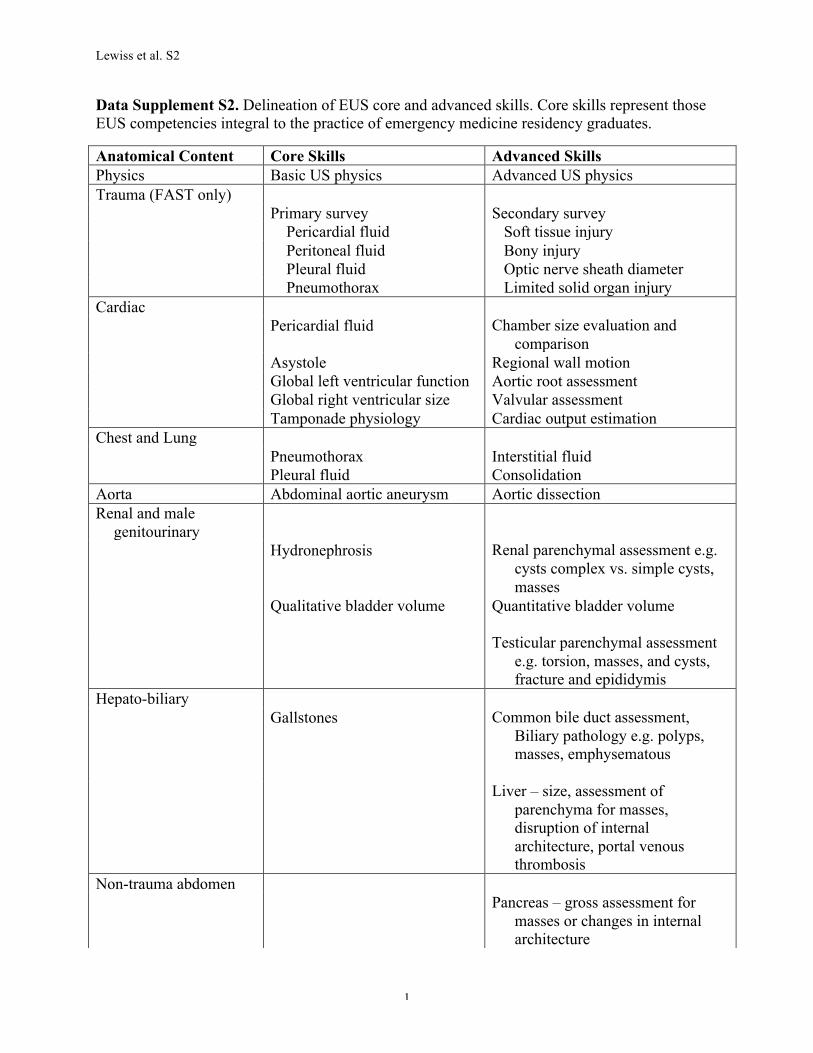
Data Supplement S2. Delineation of EUS core andadvanced skills.
Data Supplement S3. Clinical syndromes and asample of EUS skill competencies that would bedemonstrated by the learner.
Data Supplement S4. EUS SDOTs.Data Supplement S5. Competency assessment
methods.
ACADEMIC EMERGENCY MEDICINE • July 2013, Vol. 20, No. 7 • www.aemj.org 745
Lewiss et al. S2
1
Data Supplement S2. Delineation of EUS core and advanced skills. Core skills represent those EUS competencies integral to the practice of emergency medicine residency graduates.
Anatomical Content Core Skills Advanced Skills Physics Basic US physics Advanced US physics Trauma (FAST only) Primary survey Secondary survey Pericardial fluid Soft tissue injury Peritoneal fluid Bony injury Pleural fluid Optic nerve sheath diameter Pneumothorax Limited solid organ injury Cardiac Pericardial fluid Chamber size evaluation and
comparison Asystole Regional wall motion Global left ventricular function Aortic root assessment Global right ventricular size Valvular assessment Tamponade physiology Cardiac output estimation Chest and Lung Pneumothorax Interstitial fluid Pleural fluid Consolidation Aorta Abdominal aortic aneurysm Aortic dissection Renal and male
genitourinary
Hydronephrosis Renal parenchymal assessment e.g. cysts complex vs. simple cysts, masses
Qualitative bladder volume Quantitative bladder volume
Testicular parenchymal assessment e.g. torsion, masses, and cysts, fracture and epididymis
Hepato-biliary Gallstones Common bile duct assessment,
Biliary pathology e.g. polyps, masses, emphysematous
Liver – size, assessment of
parenchyma for masses, disruption of internal architecture, portal venous thrombosis
Non-trauma abdomen Pancreas – gross assessment for
masses or changes in internal architecture

Lewiss et al. S2
2
Spleen – size, assessment of parenchyma for masses, disruption of internal architecture
Gastrointestinal Appendix Hernia assessment Bowel obstruction or ileus Diverticulitis Pneumoperitoneum Ocular Undifferentiated vitreous
chamber pathology Retinal detachment Vitreous detachment
Optic nerve sheath diameter Foreign body Lens dislocation Orbital emphysema Retro-bulbar hematoma Obstetrics/gynecology
Trans-abdominal Identification of intrauterine
pregnancy with fetal heart rate
1st, 2nd, and 3rd trimester gestational dating and presentation
Identification of free fluid in pelvis
Placental location
Trans-vaginal Identification of intrauterine
pregnancy with fetal heart rate
1st, 2nd, and 3rd trimester gestational dating
Identification of free fluid in the pelvis
Adnexal assessment for cysts or masses e.g. ectopic or tubo-ovarian abscess
Ovarian torsion Uterine masses
Procedures Central venous access Evaluation of tubes – Foley,
gastrostomy-tube, jejunostomy-tube
Peripheral venous access Arterial line placement Thoracentesis Joint aspiration Paracentesis Endo-tracheal tube confirmation Pericardiocentesis Lumbar puncture Abscess drainage Pacer wire placement Foreign body detection Venous/Arterial Assessment

Lewiss et al. S2
3
Deep venous thrombosis evaluation - two region compression lower extremity
Deep venous thrombosis evaluation – upper extremity and neck
Inferior vena cava evaluation Doppler evaluation of arterial and venous structures
Soft Tissue Abscess vs. cellulitis Necrotizing fasciitis Foreign body detection Peri-tonsillar abscess Musculoskeletal Assessment of bones and joints Assessment of tendons and
ligaments Assessment of muscles Nerve blocks Brachial plexus, forearm Intercostal, transversus abdominus Femoral, sciatic, tibial Pediatrics All appropriate imaging listed
above Hip evaluation
Appendicitis Pylorus stenosis Intussusception Lumbar puncture Head and Neck Evaluation of neck masses for
airway compromise Vocal cord assessment

DataSupplementS4.EUSSDOTS
1.Aorta
2.Biliary
3.Cardiac
4.Centralvenouslineaccess.
5.Deepveinthrombosis
6.FAST
7.Lung
8.Renal
9.Trans‐abdominalpelvic
10.Trans‐vaginalpelvic
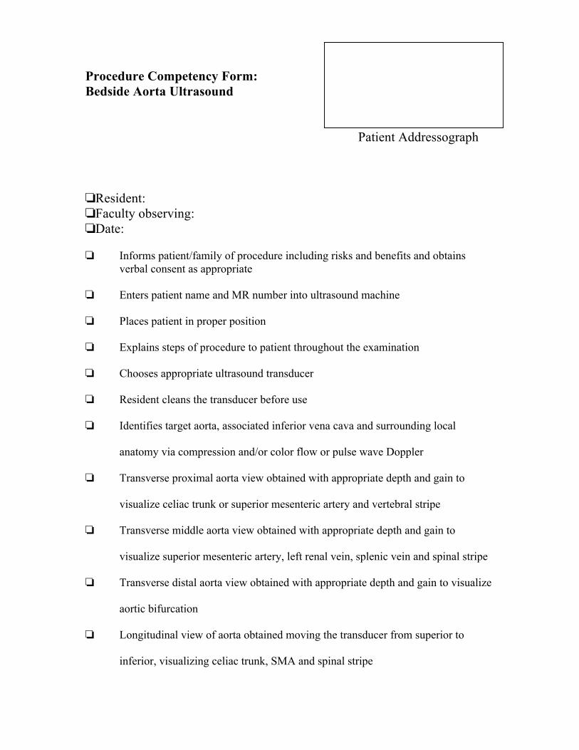
Procedure Competency Form: Bedside Aorta Ultrasound Patient Addressograph
Resident: Faculty observing: Date: Informs patient/family of procedure including risks and benefits and obtains verbal consent as appropriate Enters patient name and MR number into ultrasound machine Places patient in proper position Explains steps of procedure to patient throughout the examination Chooses appropriate ultrasound transducer
Resident cleans the transducer before use
Identifies target aorta, associated inferior vena cava and surrounding local
anatomy via compression and/or color flow or pulse wave Doppler
Transverse proximal aorta view obtained with appropriate depth and gain to
visualize celiac trunk or superior mesenteric artery and vertebral stripe
Transverse middle aorta view obtained with appropriate depth and gain to
visualize superior mesenteric artery, left renal vein, splenic vein and spinal stripe
Transverse distal aorta view obtained with appropriate depth and gain to visualize
aortic bifurcation
Longitudinal view of aorta obtained moving the transducer from superior to
inferior, visualizing celiac trunk, SMA and spinal stripe

Accurately identifies abdominal aortic aneurysm or normal aortic diameter
Performs a caliper measurement of each anatomic area from the outer wall to the
outer wall of the vessel in anterior-posterior and transverse planes
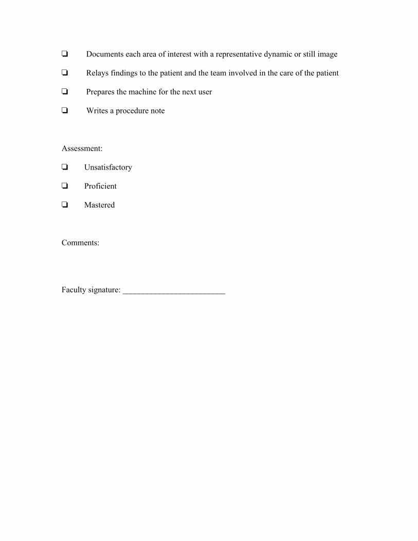
Documents each area of interest with either a representative still image or
video clip
Relays the findings to the patient and the team involved in the care of the patient
Prepares the machine for the next user
Procedure note written
Assessment:
Unsatisfactory
Proficient
Mastered
Comments:
Faculty signature:

Procedure Competency Form: Bedside Biliary Ultrasound Patient Addressograph
Resident: Faculty observing: Date: Informs patient/family of procedure including risks and benefits and obtains verbal consent as appropriate Enters patient name and MR number into ultrasound machine Places patient in proper position Explains steps of procedure to patient throughout the procedure Chooses appropriate ultrasound transducer
Resident cleans the transducer before use
Identifies gallbladder (GB) correctly
Visualizes gallbladder in long and short axis in its entirety, including GB neck
Measures length of GB in long axis and the transverse diameter in short axis
Measures GB wall thickness (anteriorly) and notes upper limit of normal
thickness
Accurately visualizes and measures CBD in association with the portal vein and
notes the upper limit of normal diameter
Accurately identifies the presence or absence of gallstones, pericholecystic fluid
and sonographic Murphy’s sign
Documents each area of interest with either a representative still image or
a video clip and correctly labels images/clips

Relays the findings to the patient and the team involved in the care of the patient
Prepares the machine for the next user
Procedure note written
Assessment:
Unsatisfactory
Proficient
Mastered
Comments:
Faculty signature: _____________________________________

Procedure Competency Form: Bedside Limited Cardiac Ultrasound Patient Addressograph
Resident: Faculty observing: Date:
Informs patient/family of procedure including risks and benefits and obtains verbal consent as appropriate Enters patient name and MR number into ultrasound machine Places patient in proper position Explains steps of procedure to patient throughout the examination Chooses appropriate ultrasound transducer
Resident cleans the transducer before use
Obtains subxiphoid view with appropriate depth to visualize entire
pericardium
Obtains para-sternal long axis view with appropriate depth to visualize
descending thoracic aorta, measures aortic outflow tract appropriately
Obtains para-sternal short axis view with adequate visualization of left and right
ventricles, at approximately the level of the papillary muscles
Obtains apical four chamber view with adequate visualization of all four
chambers
Accurately identifies presence or absence of pericardial fluid
Accurately estimates global cardiac function
Accurately assesses left ventricular versus right ventricular chamber size
Documents each area of interest with a representative dynamic or still image
Relays findings to the patient and the team involved in the care of the patient
Prepares the machine for the next user
Writes a procedure note
Assessment:
Unsatisfactory
Proficient
Mastered
Comments:
Faculty signature: _________________________

Procedure Competency Form: Ultrasound guided Central Venous Access Patient Addressograph
Resident: Faculty observing: Date: Informs patient of procedure and obtains consent consistent with hospital policy
Enters patient name and MR number into ultrasound machine Places patient in proper position Explains steps of procedure to patient throughout the procedure Chooses appropriate ultrasound transducer Resident cleans the transducer before use and performs surveillance of local
anatomy and vessel location
Confirms correct location of probe marker and orientation
Identifies target vein, associated artery and surrounding local anatomy via
compression and/or color flow Doppler
Prepares self and patient using proper sterile technique
Observes sterile technique to place sterile probe cover on ultrasound probe
Adequately anesthetizes target tissue area
Measures depth of target vein to determine needle insertion point
Observes proper needle angle during insertion
Accurately identifies needle tip vs. needle down artifact prior to advancement of
needle

Documents needle insertion during cannulation of vessel with either a
representative still image or a video clip
Uses ultrasound to evaluate causes of difficulty advancing wire when applicable
Properly secures line and orders chest radiograph to confirm placement when
applicable
Prepares the machine for the next user
Procedure note written
Assessment:
Unsatisfactory
Proficient
Mastered
Comments:
Faculty signature: _________________________
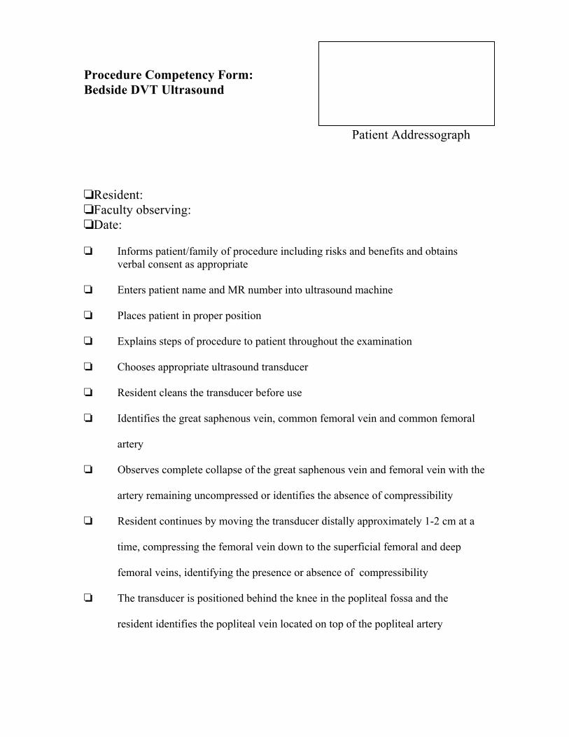
Procedure Competency Form: Bedside DVT Ultrasound Patient Addressograph
Resident: Faculty observing: Date: Informs patient/family of procedure including risks and benefits and obtains verbal consent as appropriate Enters patient name and MR number into ultrasound machine Places patient in proper position Explains steps of procedure to patient throughout the examination Chooses appropriate ultrasound transducer
Resident cleans the transducer before use
Identifies the great saphenous vein, common femoral vein and common femoral
artery
Observes complete collapse of the great saphenous vein and femoral vein with the
artery remaining uncompressed or identifies the absence of compressibility
Resident continues by moving the transducer distally approximately 1-2 cm at a
time, compressing the femoral vein down to the superficial femoral and deep
femoral veins, identifying the presence or absence of compressibility
The transducer is positioned behind the knee in the popliteal fossa and the
resident identifies the popliteal vein located on top of the popliteal artery

The popliteal vein is compressed down to the trifurcation of the popliteal vein
and the resident identifies the presence or absence of compressibility
Documents each area of interest with either a representative still image or
video clip
Relays the findings to the patient and the team involved in the care of the patient
Prepares the machine for the next user
Procedure note written
Assessment:
Unsatisfactory
Proficient
Mastered
Comments:
Faculty signature:

Procedure Competency Form: FAST exam Patient Addressograph
Resident: Faculty observing: Date: Informs patient/family of procedure including risks and benefits and obtains verbal consent as appropriate Enters patient name and MR number into ultrasound machine Chooses appropriate ultrasound transducer
Resident cleans the transducer before use
Subxiphoid view obtained with appropriate depth to visualize entire
pericardium
RUQ view obtained, scanning through Morrison’s pouch, making sure to
visualize the tip of the liver and the inferior pole of the right kidney
LUQ view obtained, scanning through the splenorenal recess, making sure to
visualize the inferior pole of the left kidney
Pelvic view obtained, scanning through the entire bladder in transverse and
sagittal planes
Accurately identifies free fluid or lack of free fluid
Documents each area of interest with either a representative still image or
video clip
Relays the findings to the patient and the team involved in the care of the patient
Procedure note written

Assessment:
Unsatisfactory
Proficient
Mastered
Comments:
Faculty signature:
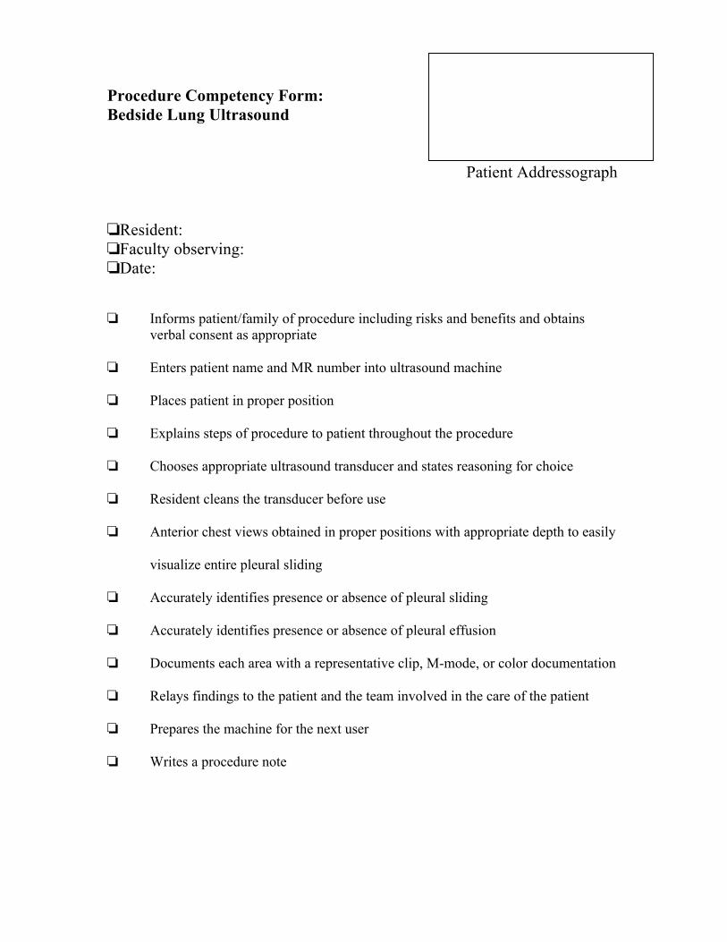
Procedure Competency Form: Bedside Lung Ultrasound Patient Addressograph
Resident: Faculty observing: Date: Informs patient/family of procedure including risks and benefits and obtains verbal consent as appropriate Enters patient name and MR number into ultrasound machine Places patient in proper position Explains steps of procedure to patient throughout the procedure Chooses appropriate ultrasound transducer and states reasoning for choice
Resident cleans the transducer before use
Anterior chest views obtained in proper positions with appropriate depth to easily
visualize entire pleural sliding
Accurately identifies presence or absence of pleural sliding
Accurately identifies presence or absence of pleural effusion
Documents each area with a representative clip, M-mode, or color documentation
Relays findings to the patient and the team involved in the care of the patient
Prepares the machine for the next user
Writes a procedure note
Assessment:
Unsatisfactory
Proficient
Mastered
Comments:
Faculty signature: _________________________

Procedure Competency Form: Bedside Renal Ultrasound Patient Addressograph
Resident: Faculty observing: Date:
Informs patient/family of procedure including risks and benefits and obtains verbal consent as appropriate Enters patient name and MR number into ultrasound machine Places patient in proper position Explains steps of procedure to patient throughout the procedure
Identifies the indication for the examination
Resident chooses the appropriate transducer
Resident cleans the transducer before use
Performs adequate examination of each kidney and the bladder including image
optimization using depth, gain, focus and mode as needed
Recognizes clinical indications for simultaneous aorta ultrasound
Trouble shoots technical limitations (body habitus, bowel gas, tenderness, empty
bladder, inability to position patient)
Correctly identifies normal anatomy and pathology (presence or absence of
hydronephrosis, renal stones)
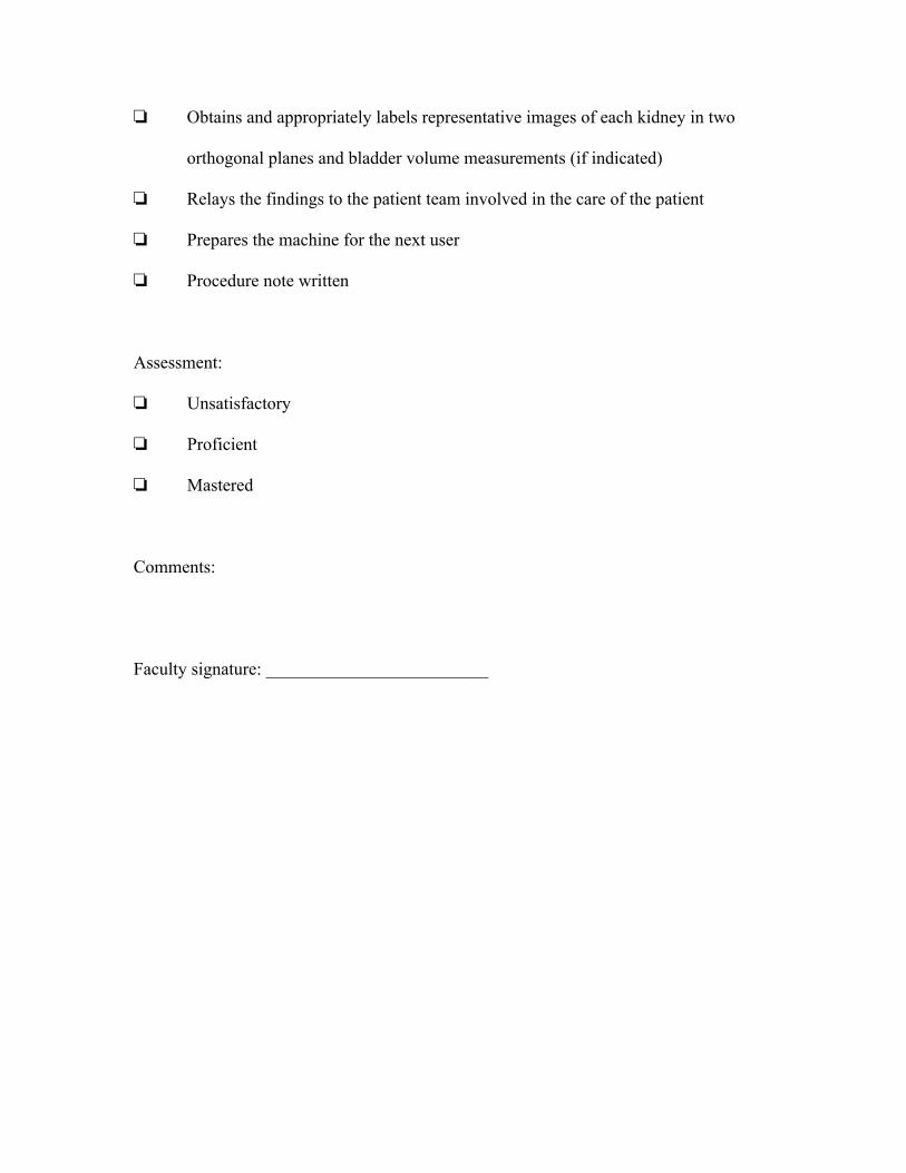
Obtains and appropriately labels representative images of each kidney in two
orthogonal planes and bladder volume measurements (if indicated)
Relays the findings to the patient team involved in the care of the patient
Prepares the machine for the next user
Procedure note written
Assessment:
Unsatisfactory
Proficient
Mastered
Comments:
Faculty signature: _________________________

Procedure Competency Form: Bedside Trans-abdominal Pelvic Ultrasound Patient Addressograph
Resident: Faculty observing: Date: Informs patient/family of procedure including risks and benefits and obtains verbal consent as appropriate Enters patient name and MR number into ultrasound machine Places patient in proper position Explains steps of procedure to patient throughout the examination Chooses appropriate ultrasound transducer
Resident cleans the transducer before use
Obtains long and short axis views of the uterus scanning the entirety from fundus
to cervix
Accurately identifies the presence or absence of free fluid in the cul-de-sac
Scans through both ovaries in two planes (if visible)
Accurately identifies the presence or absence of an intrauterine pregnancy
Measures the endo-myometrial mantle and recognizes that thickness less than 7
mm is concerning for an interstitial pregnancy
Performs a FAST exam if the patient has a positive pregnancy test with absence
of a visualized intrauterine pregnancy

Documents each area of interest with either a representative still image or
a video clip
Relays findings to the patient and the team involved in the care of the patient
Prepares the machine for the next user
Writes a procedure note
Assessment:
Unsatisfactory
Proficient
Mastered
Comments:
Faculty signature: _________________________
Procedure Competency Form: Bedside Trans-vaginal Pelvic Ultrasound Patient Addressograph
Resident: Faculty observing: Date: Informs patient/family of procedure including risks and benefits and obtains verbal consent as appropriate Enters patient name and MR number into ultrasound machine Places patient in proper position Explains steps of procedure to patient throughout the examination Chooses appropriate ultrasound transducer
Resident cleans the transducer before use
Uses appropriate clean probe cover
Applies sterile surgilube gel to a covered clean transducer
Obtains coronal and sagittal views of uterus with appropriate depth to
visualize the posterior cul-de-sac
Scans through entire uterus in two orthogonal planes
Scans lateral of uterus on left and right to identify adnexae
Evaluates adnexae by scanning through in two orthogonal planes
Accurately identifies free fluid or lack of free fluid
Accurately identifies presence or absence of definitive intra-uterine pregnancy
(yolk sac and beyond)
Measures endomyometrial mantle at thinnest point and recognizes that thickness
less than 7 mm is concerning for an interstitial pregnancy
If identified, measures fetal heart rate using M-mode
Documents each area of interest with a representative still image or video clip
Relays findings to the patient and the team involved in the care of the patient
Prepares the machine for the next user
Writes a procedure note
Assessment:
Unsatisfactory
Proficient
Mastered
Comments:
Faculty signature: _________________________