encephalitozoon hellem, isolated from an aids - journal of clinical
TRANSCRIPT

JOURNAL OF CLINICAL MICROBIOLOGY, Nov. 1994, P. 2760-27680095-1137/94/$04.00+0Copyright © 1994, American Society for Microbiology
Polyclonal and Monoclonal Antibody and PCR-AmplifiedSmall-Subunit rRNA Identification of a Microsporidian,
Encephalitozoon hellem, Isolated from an AIDSPatient with Disseminated Infection
GOVINDA S. VISVESVARA,l* GORDON J. LEITCH,2 ALEXANDRE J. DA SILVA,'GIAN PIERO CROPPO,' HERCULES MOURA,' SARA WALLACE,' SUSAN B. SLEMENDA,'DAVID A. SCHWARTZ,3 DELYNN MOSS,' RALPH T. BRYAN,' AND NORMAN J. PIENIAZEK'
Division of Parasitic Diseases, National Center for Infectious Diseases, Centers for Disease Control and Prevention,Morehouse School of Medicine,2 and Department of Pathology, Emory University,3 Atlanta, Georgia
Received 3 May 1994/Returned for modification 27 June 1994/Accepted 11 August 1994
Microsporidia are primitive, spore-forming, mitochondria-lacking, eukaryotic protozoa that are obligateintracellular parasites. They are known to parasitize almost every group of animals including humans.Recently, microsporidia have increasingly been found to infect patients with AIDS. Five genera (Encephalito-zoon, Enterocytozoon, Nosema, Septata, and Pleistophora) of microsporidia are known to infect humans.Enterocytozoon organisms cause gastrointestinal disease in a majority of AIDS patients with microsporidiosis.However, a smaller, but an expanding, number of patients with AIDS are being diagnosed with ocular anddisseminated infection with Encephalitozoon hellem. Although microsporidial spores can be identified in clinicalsamples by a staining technique such as one with Weber's chromotrope stain, identification to the species levelis dependent on cumbersome and time-consuming electron microscopy. We have recently isolated andestablished in continuous culture several strains of E. hellem from urine, bronchoalveolar lavage, and sputumsamples from AIDS patients with disseminated microsporidiosis. We developed polyclonal and monoclonalantibodies and PCR primers to a strain of E. heUem that can be used successfully to identify E. heUem fromother species of microsporidia either in clinical specimens or in cultures established from clinical specimens.Since patients infected with Encephalitozoon spp. are known to respond favorably to albendazole, identificationof the parasite to the species level would be invaluable in the treatment of disseminated microsporidiosis.
Microsporidia are primitive, eukaryotic protozoa that lackmitochondria (2, 6, 18). They are obligate intracellular para-sites and are known to infect a wide variety of cell types.Although they are most often known to parasitize insects andfish, they have been found to infect members of almost everymajor phylum of the animal kingdom (6). During the pastdecade microsporidia have increasingly been recognized toinfect patients with AIDS (2, 3, 5, 7-10, 13, 15, 18, 27). To datemicrosporidia belonging to five genera, Encephalitozoon, En-terocytozoon, Nosema, Pleistophora, and Septata, have beenknown to cause infections in humans (2, 3, 6, 8, 19). A sixthtaxon, Microsporidium, has also been established to includeinsufficiently described microsporidia of undetermined taxo-nomic status (4, 6).We recently reported the in vitro culture of a microsporidian
parasite, Encephalitozoon sp. (CDC:0291:V213), isolated fromthe urine of a patient with AIDS (22) and subsequentlyidentified it as Encephalitozoon hellem (14-16). In this reportwe decribe its growth properties, morphologic characteristics,antigenic profile on the basis of its reactivity with polyclonalantibodies and monoclonal antibodies (MAbs), and sequenceanalysis of the small-subunit rRNA (SSU-rRNA)-coding re-
gion.
* Corresponding author. Mailing address: Division of ParasiticDiseases, M.S.-F/13, Centers for Disease Control and Prevention, 4770Buford Highway NE, Atlanta, GA 30341-3724. Phone: (404) 488-4417.Fax: (404) 488-4108. Electronic mail address: [email protected].
MATERIALS AND METHODS
Sources of parasites. E. hellem CDC:0291:V213 was isolatedfrom the urine of an AIDS patient and was grown on monkeykidney cell (E6) and human lung fibroblast (HLF) monolayers(22). Encephalitozoon cuniculi, isolated from a rabbit andgrown on rabbit kidney cells (17), was obtained from JohnShadduck and Elizabeth Didier and was designated strain JS.E. hellem, isolated from the corneal button of an AIDS patientwith keratoconjunctivitis and grown on MDCK cells anddesignated strain ED (9), and Nosema comeum, isolated fromthe cornea of a human immunodeficiency virus (HIV)-sero-negative patient (19), were also obtained from ElizabethDidier. All of these parasites were subsequently adapted togrow on the E6 cell line.
Parasite growth and harvest. To obtain maximum infectionof the host cells and optimum growth of the parasites, theculture medium was replaced every 3 days. The spent culturemedium from all flasks, which contained extruded spores andunattached host cells infected with developmental stages of theparasite, was centrifuged at 1,500 x g for 20 min at 4°C, andthe supernatant was aspirated. The pellets were put back intothe same culture flasks. This facilitated the infection of a
maximum number of host cells (>70%), as revealed by micro-scopic examination, with the respective parasites. Thereafter,spores that were extruded into the culture supernatants fromall parasites were harvested by centrifugation as describedabove. The spores were suspended in 0.25% sodium dodecylsulfate (SDS), vortexed briefly, and incubated in a water bathfor 20 min at 37°C (9). The spores were next washed threetimes in Hanks' balanced salt solution (HBSS), counted in a
2760
Vol. 32, No. 11
on Decem
ber 23, 2018 by guesthttp://jcm
.asm.org/
Dow
nloaded from

SEROLOGIC AND MOLECULAR ANALYSIS OF E. HELLEM 2761
hemacytometer, suspended in enough HBSS to obtain 109spores per ml, and stored at 4°C until use (22).
Polyclonal antibody. Antibody to E. hellem CDC:0291:V213was produced in female New Zealand White rabbits weighingabout 2 kg. Each of several rabbits were prebled to check forspontaneous antibody to E. cuniculi by indirect immunofluo-rescence (IIF). Two rabbits with no background levels ofantibody to E. cuniculi were selected, and 0.1 ml of sporesuspension containing 108 spores of CDC:0291:V213 was re-peatedly injected into the marginal ear vein. Blood sampleswere taken periodically and were tested by IIF for antibodiesto CDC:0291:V213 (14).MAb. MAb to CDC:0291:V213 was produced in female
BALB/c mice by published methods (11, 23). Each of four8-week-old mice were injected intraperitoneally with 0.1 ml ofwashed parasite suspension containing 106 spores and unat-tached E6 cells containing developing stages on days 0, 7, 14,21, 28, 35, and 42. Spleen cells from two of the mice that hadhigh titers of antibody (>4,096 by IIF) were fused with SP2/0myeloma cells by the addition of polyethylene glycol. Stablehybrids were selected by growth in RPMI 1640 mediumcontaining 10% fetal bovine serum, hypoxanthine, aminop-terin, and thymidine as described earlier (23). Supernatantculture medium was tested for antibody activity against E.hellem CDC:0291:V213 and E. cuniculi JS by the IIF test (22,23). Six of the 24 hybrids showing antibody activity wereselected for further cloning and expansion. The MAbs werealso tested for their isotypes by immunodiffusion by usingmouse immunoglobulin isotype-specific antisera (immuno-globulin M [IgM], IgA, IgGl, IgG2a, IgG2b, and IgG3). One ofthe clones (ED4H1OB11/B12), which showed high levels ofantibody activity, was also injected into BALB/c mice afterpristane priming for ascites production (23).
Scanning and transmission electron microscopy. For scan-ning electron microscopy E6 monolayers were fixed with 2.5%glutaraldehyde in 0.1 M cacodylate buffer (pH 7.4), processedas described previously (22), and examined with a JEOL JSM820 scanning electron microscope. For transmission electronmicroscopy, the glutaraldehyde-fixed samples were postfixed ina solution of 1% OS04, processed, and examined with a JEOL1200 EX transmission electron microscope as described previ-ously (22).
IIF. The IIF test was performed as described earlier (22).Briefly, washed spores of E. cuniculi JS and E. hellem ED andCDC:0291:V213 were suspended in enough 1% buffered For-malin to obtain 107 spores per ml. Antigen slides were pre-pared by depositing 20 ,ul of the spore suspension onto eachwell of several 12-place slides as described previously (22).Rabbit anti-E. cuniculi and rabbit anti-E. hellem or the MAbwere serially diluted with phosphate-buffered saline (PBS; 0.01M; pH 7.6) beginning at 1:2 in U-type microtitration plates(Linbro Scientific Co., Inc., Hamden, Conn.). The seriallydiluted reagents were transferred to the 12-place slides (15 RIof each dilution per well), and the slides were incubated at37°C for 30 min in a moist chamber. The slides were thenwashed three times in PBS (10 min per wash), and then eachwell was covered with 20 ,ul of fluorescein isothiocyanate(FITC)-conjugated goat anti-rabbit IgG (Cappel Laboratories,Westchester, Pa.) at a dilution of 1:2,000 containing Evans blueas the counterstain or FITC-conjugated anti-mouse IgG. Theslides were incubated at 37°C for 30 min and washed threetimes as described above, the specimens were mounted withbuffered glycerin (pH 9.0) and covered with a coverslip, andthe slides were examined with an Olympus BH2 fluorescencemicroscope equipped with epifluorescence illumination.Thin smears of culture-derived N. comeum and Formalin-
fixed stool samples obtained from patients with diarrheaidentified as being caused by Enterocytozoon bieneusi wereprepared, and the IIF test was performed.
IIF was also performed on tissue sections of lungs, kidneys,and prostate obtained from our patient at the time of autopsy.The tissues were fixed in 10% neutral buffered Formalin, and5 to 6-,um-thick sections were cut and stained with hematoxy-lin-eosin and Brown and Hops stains. Additional sections werecut from the blocks that were positive for microsporidia, andthe sections were deparaffinized, hydrated through a gradedseries of ethanol solutions, rinsed in water and PBS, and thencovered with a 1:100 dilution of the rabbit anti-E. hellem or theMAb and processed as described above. The sections werenext covered with a 1:500 dilution of the FITC-conjugated goatanti-rabbit IgG or FITC-conjugated goat anti-mouse Ig andwere incubated at 37°C and processed as described above. Theslides were examined and photographed with an Olympusmicroscope (14-16). Since no fluorescence was observed in thetissue sections when they were reacted with the MAb by thisprotocol, the sections were next pretreated for 30 min with asolution containing 0.2% CaCl2 and 0.2% trypsin, rinsed withwater, and then allowed to react overnight at 4°C with theMAb (12).
SDS-polyacrylamide gel electrophoresis (SDS-PAGE) andimmunoblotting. Proteins were extracted from purified sporesof each parasite by suspending them in sample buffer contain-ing 2.5% SDS and 2.25 M urea and heating the mixture at 65°Cfor 15 min (9, 22). Proteins extracted from approximately 5 x10⁶ spores were loaded onto each lane of a 1.0-mm 2.5 to 27%gradient acrylamide gel (Isolab Inc. Akron, Ohio), and the gelswere subjected to electrophoresis (22). The separated proteinswere transferred onto Immobilon membranes. The mem-branes were reacted with either a 1:500 dilution of a rabbitanti-E. cuniculi serum (courtesy of John Shadduck), a 1:500dilution of rabbit anti-E. hellem CDC:0291:V213 serum, or a1:100 dilution of the MAb ED4H1OB11/B12. After appropri-ate washes, the membranes were next reacted with a 1:5,500dilution of peroxidase-conjugated goat anti-rabbit IgG (CappelLaboratories) or a 1:1,000 dilution of peroxidase-conjugatedanti-mouse IgG. Hydrogen peroxide (3%) and diaminobenzi-dene (0.005%) were used as substrate and chromogen, respec-tively (22).PCR amplification, cloning, and sequencing of SSU-rRNA
coding region. (i) Extraction ofDNA. DNA was extracted fromthe following four different cultures: (i) uninfected (E6) cellculture (control), (ii) E6 culture infected with E. hellem ED,(iii) E6 culture infected with E. hellem CDC:0291:V213, and(iv) E6 culture infected with E. cuniculi JS. All cultures weregrown on 75-cm2 plastic flasks (Corning, Ithaca, N.Y.). Cellsfrom each of the four cultures were scraped separately andwere transferred to 15-ml tubes. After centrifugation at 1,000x g for 20 min at 4°C, the supernatant was aspirated and thesediment was suspended in 0.8 N NaCl (saline) and washedthree times by centrifugation as described above. After thethird wash, the supernatant was aspirated again and the DNAwas extracted from the pellet by using the ONCOR nonorganicDNA extraction kit (Oncor, Gaithersburg, Md.) according tothe manufacturer's instructions. Nucleic acids from each sam-ple were resuspended in 50 ,lI of distilled water and wereamplified by PCR.
(ii) PCR. Primers for amplification of the microsporidialSSU-rRNA-coding region were designed by using the pub-lished sequence of Vairimorpha necatrix SSU-rRNA (24, 25).The forward primer was complementary to bases 1 to 21 of theV necatrix sequence; the reverse primer was complementary tobases 1224 to 1244. In addition, PCR was performed by using
VOL. 32, 1994
on Decem
ber 23, 2018 by guesthttp://jcm
.asm.org/
Dow
nloaded from

2762 VISVESVARA ET AL.
two pairs of primers: the first pair, specific for E. hellem,amplified a 547-bp diagnostic fragment from the E. hellemCDC:0291:V231 SSU-rRNA sequence (N. J. Pieniazek et al.,GenBank accession number L19070). The forward primer(5'-TGAGAAGTAAGATGlTTTAGCA-3') was designed onpositions 358 to 378 and the reverse primer (5'-GTAAAAACACTCTCACACTCA-3') was designed on positions 884 to904 of this sequence. The second pair, specific for E. cuniculi,amplified a 549-bp fragment diagnostic for the E. cuniculiSSU-rRNA (N. J. Pieniazek et al., GenBank accession numberL17072). The forward primer (5'-ATGAGAAGTGATGTGTGTGCG-3') was designed on positions 344 to 364 of thissequence, and the reverse primer (5'-TGCCATGCACTCACAGGCATC-3') was designed on positions 872 to 892 of thissequence. PCR was performed by using a Perkin-Elmer Cetus(Norwalk, Conn.) GenAmp kit according to the manufactur-er's instructions. Denaturation, annealing, and elongation tem-peratures of 94, 55, and 72°C, respectively, were used for atotal of 30 cycles in all reactions. The products of the amplifi-cations were electrophoretically resolved on a 2% agarose gel(Seakem GTG; FMC Bioproducts, Rockland, Maine) andwere visualized for analysis by staining with ethidium bromide.
After amplification with the V necatriJ primers, the PCRproducts were analyzed on an agarose gel, and only when atemplate from an infected cell was used was a band of about1.3 kb seen. PCR products were then purified, cloned, andsequenced. For all PCR products, sequencing was done forthree independent clones for both strands. Sequences weredeposited in the GenBank database under the following acces-sion numbers: L17072 for E. cuniculi and L19070 for E. hellemCDC:0291:V213 and ED.
RESULTS
Growth of parasites. Foci of infection were noticed in themonolayers about a month after the inoculation of the spores.At first, the developmental stages of the parasites (E. hellemED and CDC:0291:V213 and E. cuniculi) began to appear asbluish dots within the E6 cells, which increased in number withtime; finally, the cells became distended with spores (Fig. laand lb), leading to the rupture of cells and the liberation of thespores into the culture medium. All of the three parasitesreadily infected the E6 monolayers and released spores intothe culture medium. The patterns of growth of the threeparasites were similar. In older cultures, especially those of E.hellem CDC:0291:V213, when almost all host cells had beendestroyed and masses of spores were seen attached to thecytoskeletal remains of the host cells, many of the spores wereseen to have extruded their polar tubes (Fig. 2a to d).
Only E. hellem CDC:0291:V213 was evaluated ultrastructur-ally. Developmental or proliferative forms were seen attachedto the parasitophorous vacuole membrane. Many early andlate sporogonial developmental stages suggested that they aredisporous. In the early stages, the sporont membrane thick-ened and the sporont detached from the parasitophorousvacuole membrane. In the maturing sporonts, the polar tubulesin various stages of development were seen (Fig. 3). Theformation of the polaroplast as well as the anchoring disc wasalso clearly discernible.The spores of CDC:0291:V213 were characterized by their
oval shape, birefringence, and gram-positive staining. Whenstained with the chromotrope-based stain of Weber et al. (26),they stained pinkish to red. The spores appeared smoothwalled when examined by phase-contrast microscopy andscanning electron microscopy. They measured 2.25 to 2.8 ,umin length and 1.25 to 1.85 ,um in breadth. When examined by
--W_:......;.Ts.;.s:;: r-i .7.
FIG. 1. Monolayers of HLF (a) and E6 (b) cell cultures infectedwith E. hellem CDC:0291:V213. Note that the cells in both monolayersare distended with spores of E. hellem. Phase-contrast microscopy wasused. Magnification, X600.
transmission electron microscopy the spores were seen to becharacterized by a thin, electron-dense outer wall, the exo-spore, an inner, thick electron-lucent endospore, and a thin cellmembrane that surrounded the spore contents. Other promi-nent features seen in the transmission electron micrographswere cross sections of five to seven coils of the polar tubule oneither side of the spore, the anchoring disc at the anterior endfrom which the polar tubule emanated, the lamellar polaro-plast, also at the anterior end of the spore, and the vacuoleprominently located at the posterior end (Fig. 3).
IIF assay. In the IIF test, both the CDC:0291:V213 and EDstrains of E. hellem reacted with the anti-CDC:0291:V213serum almost to the same extent and gave a titer of >4,096. E.cuniculi, however, reacted less strongly and produced a titer of256. Dramatic differences were, however, noted when E.cuniculi was reacted with the anti-CDC:0291:V213 serum
J. CLIN. MICROBIOL.
on Decem
ber 23, 2018 by guesthttp://jcm
.asm.org/
Dow
nloaded from
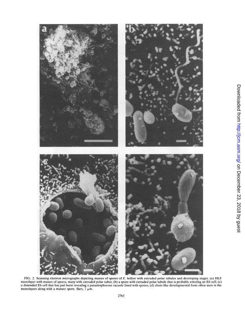
F W JFIG. 2. Scanning electron micrographs depicting masses of spores of E. hellem with extruded polar tubules and developing stages. (a) HLF
monolayer with masses of spores, many with extruded polar tubes; (b) a spore with extruded polar tubule that is probably entering an E6 cell; (c)a distended E6 cell that has just burst revealing a parasitophorous vacuole lined with spores; (d) chain-like developmental form often seen in themonolayers along with a mature spore. Bars, 1 pum.
2763
on Decem
ber 23, 2018 by guesthttp://jcm
.asm.org/
Dow
nloaded from

2764 VISVESVARA ET AL.
C.. v ,1
/
N
A
J. CLIN. MICROBIOL.
...._t :. s- ! ,_y .'. ':
.:
b
A-:|_'.'\_1N br
4
.U ..
.:
..~~~~~~~~~~~~~~~~~~~~~~~~~~~~~~
_Ad0 ..X
ft
- 4
0.
*4#..
Sb9.1
o
.
A.
FIG. 3. Transmission electron micrograph of an E6 cell showing various stages of sporogonial development. The sporont membrane isbeginning to thicken (arrows) and some are binucleated, indicating that they are disporous. Cross sections of the polar tubules are already seenin some of the late stages of the sporonts. Also seen is a cluster of early and late stages of sporoblasts (Sb) showing developing organelles, anchoringdisc (Ad) and cross sections of the polar tubule (arrowheads). Several mature electron-dense spores (S) are also seen. Bar, 1 ,um.
absorbed with E. cuniculi spores. E. cuniculi showed noreactivity with the absorbed serum, indicating that all cross-reacting antigens were absorbed out. Both the CDC:0291:V213and ED strains of E. hellem, however, showed some fluores-cence at the 128 dilution, indicating specific antigens commonto both of these parasites. When either ED or CDC:0291:V213spores were reacted with anti-CDC:0291:V213 serum absorbedwith ED spores or CDC:0291:V213 spores, no fluorescencewas seen.
Dramatic differences were also observed when the spores ofE. hellem and E. cuniculi were reacted with the MAb ED4H1OB11/B12 with IgGl specificity. E. hellem spores reactedwith this MAb and produced a titer of >8,192, whereas E.cuniculi spores failed to react with this MAb at the 1:128dilution but showed a slight reaction at the 1:64 dilution. This
MAb also did not react with the Formalin-fixed stool smearspositive for E. bieneusi as well as culture-derived N. comeum.The parasites in the kidney and the lung tissue sections of
our patient reacted intensely with the anti-CDC:0291:V213serum and produced a bright apple green fluorescence indi-cating that the antiserum can be used successfully to identify E.hellem spores in tissue sections.The MAb, on the other hand, failed to react with the spores
in the Formalin-fixed, paraffin-embedded tissue sections ini-tially. However, bright green fluorescence of the spores wasevident when the sections were treated with trypsin solution for30 min and then reacted with the MAb overnight at 4°C (Fig.4).SDS-PAGE and immunoblotting. Proteins extracted from E.
hellem ED and CDC:0291:V213 after separation and silver
irw-ANML
4011 ,I,!:iI-..f.
on Decem
ber 23, 2018 by guesthttp://jcm
.asm.org/
Dow
nloaded from

SEROLOGIC AND MOLECULAR ANALYSIS OF E. HELLEM 2765
1 2 3
224-
10971-
45-
28-
FIG. 4. Results of IIF test performed on the lung sections of our
patient. (a) Positive fluorescence profiles of E. hellem spores in a
section that was reacted with a 1:100 dilution of MAb ED4H1OB11/B12. Magnification, X400. (b) A negative reaction of spores in a
section similar to that shown in panel a reacted with a 1:100 dilution ofan irrelevant MAb.
staining showed remarkably similar patterns. Although E.hellem shared many proteins with E. cuniculi, differencesbetween the two were clearly apparent, especially in the 14-and 18-kDa and the 28- to 70-kDa regions. When theseproteins were transferred to the Immobilon membranes andthen treated with the rabbit anti-E. hellem serum, characteristicdifferences were seen (Fig. 5). E. hellem proteins (Fig. 5, lanes1 and 3) reacted extensively with the anti-E. hellem serum andproduced many darkly staining bands that were not present inE. cuniculi (Fig. 5, lane 2). These differences were mostobvious, especially in the regions of 14 to 20, 22, 29 to 43, and72 kDa. Both the CDC:0291:V213 and ED proteins alsoreacted with the anti-E. cuniculi serum and produced a number
18-15-
FIG. 5. Immunoblot profiles of E. hellem ED (lane 1) and CDC:0291:V213 (lane 3) and E. cuniculi JS (lane 2) after reaction with rabbitanti-CDC:0291:V213 serum.
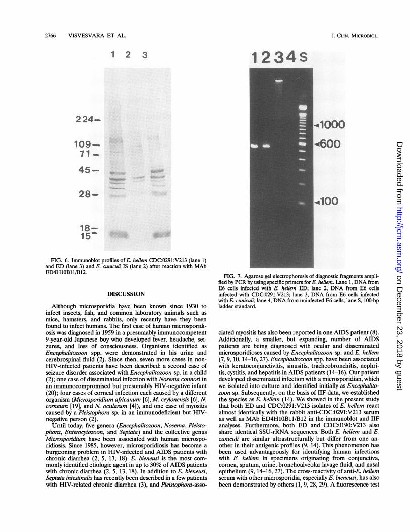
of bands (data not shown), but the reactivities of these wereconsiderably less than those in the homologous reactions. BothCDC:0291:V213 and ED, however, reacted extensively withthe anti-E. hellem serum and produced darkly staining bandsthat were unique to E. hellem. The banding patterns of E.hellem and E. cuniculi were, however, very different when thespore proteins were reacted with MAb ED4 H1OB11/B12 (Fig.6). Both CDC:0291:V213 and ED strains reacted well withMAb ED4H1OB11/B12 and produced darkly staining bands atabout 18, 28.5, 30, 36, and 40 kDa and a doublet at 109 kDa(Fig. 6, lanes 1 and 3). E. cuniculi JS reacted minimally with theMAb and produced relatively strong bands at 40 and 36 kDaand weak bands at 90 and 28 kDa (Fig. 6, lane 2).SSU-rRNA sequence analysis. Species-specific PCR primers
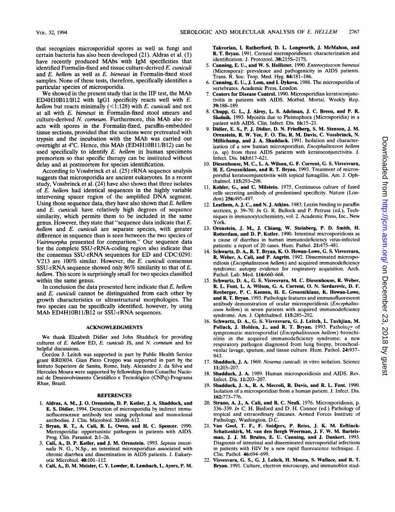
targeting SSU-rRNA-coding sequences selectively amplifiedEncephalitozoon diagnostic fragments with no backgroundfrom mammalian cellular DNA. When DNA isolated fromcultures infected with E. hellem ED and CDC:0291:V213 wasPCR amplified by using E. hellem-specific primers, an expecteddiagnostic band of aproximately 547 bp was detected uponanalysis on an agarose gel (Fig. 7, lane 1 and 2). No such band,however, was seen when DNA from E6 cells infected with E.cuniculi or uninfected cells were used (Fig. 7, lanes 3 and 4). Inaddition, no band was detected when E. hellem template wasamplified with E. cuniculi primers (data not shown). Theconsensus SSU-rRNA sequences for ED and CDC:0291:V213showed 100% similarity. The E. cuniculi consensus SSU-rRNAsequence showed only 86% similarity to that of E. hellem.
VOL. 32, 1994
on Decem
ber 23, 2018 by guesthttp://jcm
.asm.org/
Dow
nloaded from
2766 VISVESVARA ET AL.
1 2 3
224-.
109-_71 -
45-
28-
18-15'
FIG. 6. Immunoblot profiles of E. hellem CDC:0291:V213 (lane 1)and ED (lane 3) and E. cuniculi JS (lane 2) after reaction with MAbED4H1OB11/B12.
DISCUSSION
Although microsporidia have been known since 1930 toinfect insects, fish, and common laboratory animals such asmice, hamsters, and rabbits, only recently have they beenfound to infect humans. The first case of human microsporidi-osis was diagnosed in 1959 in a presumably immunocompetent9-year-old Japanese boy who developed fever, headache, sei-zures, and loss of consciousness. Organisms identified asEncephalitozoon spp. were demonstrated in his urine andcerebrospinal fluid (2). Since then, seven more cases in non-HIV-infected patients have been described: a second case ofseizure disorder associated with Encephalitozoon sp. in a child(2); one case of disseminated infection with Nosema connori inan immunocompromised but presumably HIV-negative infant(20); four cases of corneal infection each caused by a differentorganism (Microsporidium africanum [6],M ceylonensis [6], N.corneum [19], and N. ocularum [4]), and one case of myositiscaused by a Pleistophora sp. in an immunodeficient but HIV-negative person (2).
Until today, five genera (Encephalitozoon, Nosema, Pleisto-phora, Enterocytozoon, and Septata) and the collective genusMicrosporidium have been associated with human microspo-ridiosis. Since 1985, however, microsporidiosis has become aburgeoning problem in HIV-infected and AIDS patients withchronic diarrhea (2, 5, 13, 18). E. bieneusi is the most com-monly identified etiologic agent in up to 30% of AIDS patientswith chronic diarrhea (2, 5, 13, 18). In addition to E. bieneusi,Septata intestinalis has recently been described in a few patientswith HIV-related chronic diarrhea (3), and Pleistophora-asso-
1234s
.41000
4600
.4100
FIG. 7. Agarose gel electrophoresis of diagnostic fragments ampli-fied by PCR by using specific primers for E. hellem. Lane 1, DNA fromE6 cells infected with E. hellem ED; lane 2, DNA from E6 cellsinfected with CDC:0291:V213; lane 3, DNA from E6 cells infectedwith E. cuniculi; lane 4, DNA from uninfected E6 cells; lane S, 100-bpladder standard.
ciated myositis has also been reported in one AIDS patient (8).Additionally, a smaller, but expanding, number of AIDSpatients are being diagnosed with ocular and disseminatedmicrosporidioses caused by Encephalitozoon sp. and E. hellem(7, 9, 10, 14-16, 27). Encephalitozoon spp. have been associatedwith keratoconjunctivitis, sinusitis, tracheobronchitis, nephri-tis, cystitis, and hepatitis in AIDS patients (14-16). Our patientdeveloped disseminated infection with a microsporidian, whichwe isolated into culture and identified initially as Encephalito-zoon sp. Subsequently, on the basis of IIF data, we establishedthe species as E. hellem (14). We showed in the present studythat both ED and CDC:0291:V213 isolates of E. hellem reactalmost identically with the rabbit anti-CDC:0291:V213 serumas well as MAb ED4H1OB11/B12 in the immunoblot and IIFanalyses. Furthermore, both ED and CDC:0190:V213 alsoshare identical SSU-rRNA sequences. Both E. hellem and E.cuniculi are similar ultrastructurally but differ from one an-other in their antigenic profiles (9, 14). This phenomenon hasbeen used advantageously for identifying human infectionswith E. hellem in specimens originating from conjunctiva,cornea, sputum, urine, bronchoalveolar lavage fluid, and nasalepithelium (9, 14-16, 27). The cross-reactivity of anti-E. hellemserum with other microsporidia, especially E. bieneusi, has alsobeen demonstrated by others (1, 9, 28, 29). A fluorescence test
J. CLIN. MICROBIOL.
on Decem
ber 23, 2018 by guesthttp://jcm
.asm.org/
Dow
nloaded from
SEROLOGIC AND MOLECULAR ANALYSIS OF E. HELLEM 2767
that recognizes microsporidial spores as well as fungi andcertain bacteria has also been developed (21). Aldras et al. (1)have recently produced MAbs with IgM specificities thatidentified Formalin-fixed and tissue culture-derived E. cuniculiand E. hellem as well as E. bieneusi in Formalin-fixed stoolsamples. None of these tests, therefore, specifically identifies a
particular species of microsporidia.We showed in the present study that in the IIF test, the MAb
ED4H1OB11/B12 with IgGl specificity reacts well with E.hellem but reacts minimally (<1:128) with E. cuniculi and notat all with E. bieneusi in Formalin-fixed stool smears andculture-derived N. comeum. Furthermore, this MAb also re-acts with spores in the Formalin-fixed, paraffin-embeddedtissue sections, provided that the sections were pretreated withtrypsin and the incubation with the MAb was carried outovernight at 4°C. Hence, this MAb (ED4H1OB11/B12) can beused specifically to identify E. hellem in human specimenspremortem so that specific therapy can be instituted withoutdelay and at postmortem for species identification.According to Vossbrinck et al. (25) rRNA sequence analysis
suggests that microsporidia are ancient eukaryotes. In a recentstudy, Vossbrinck et al. (24) have also shown that three isolatesof E. hellem had identical sequences in the highly variableintervening spacer region of the amplified DNA segment.Using those sequence data, they have also shown that E. hellemand E. cuniculi have relatively high degrees of sequencesimilarity, which permits them to be included in the same
genus. However, they state that "sequence data indicate that E.hellem and E. cuniculi are separate species, with greaterdifference in sequence than is seen between the two species ofVairimorpha presented for comparison." Our sequence datafor the complete SSU-rRNA-coding region also indicate thatthe consensus SSU-rRNA sequences for ED and CDC:0291:V213 are 100% similar. However, the E. cuniculi consensus
SSU-rRNA sequence showed only 86% similarity to that of E.hellem. This score is surprisingly small for two species classifiedwithin the same genus.
In conclusion the data presented here indicate that E. hellemand E. cuniculi cannot be distinguished from each other bygrowth characteristics or ultrastructural morphologies. Thetwo species can be specifically identified, however, by usingMAb ED4H1OB11/B12 or SSU-rRNA sequences.
ACKNOWLEDGMENTS
We thank Elizabeth Didier and John Shadduck for providingcultures of E. hellem ED, E. cuniculi JS, and N. comeum and forhelpful discussions.Gordon J. Leitch was supported in part by Public Health Service
grant RR03034. Gian Piero Croppo was supported in part by theIstituto Superiore de Sanita, Rome, Italy. Alexandre J. da Silva andHercules Moura were supported by fellowships from Conselho Nacio-nal de Desenvolvimento Cientiifico e Tecnol6gico (CNPq)-ProgramaRhae, Brazil.
REFERENCES
1. Aldras, A. M., J. 0. Orenstein, D. P. Kotler, J. A. Shadduck, andE. S. Didier. 1994. Detection of microsporidia by indirect immu-nofluorescence antibody test using polyclonal and monoclonalantibodies. J. Clin. Microbiol. 32:608-612.
2. Bryan, R T., A. Cali, R L. Owen, and H. C. Spencer. 1990.Microsporidia: opportunistic pathogens in patients with AIDS.Prog. Clin. Parasitol. 2:1-26.
3. Cali, A., D. P. Kotler, and J. M. Orenstein. 1993. Septata intesti-nalis N. G., N.Sp., an intestinal microsporidian associated withchronic diarrhea and dissemination in AIDS patients. J. Eukary-otic Microbiol. 40:101-112.
4. Cali, A., D. M. Meisler, C. Y. Lowder, R Lembach, L. Ayers, P. M.
Takvorian, I. Rutherford, D. L. Longworth, J. McMahon, andR T. Bryan. 1991. Corneal microsporidioses: characterization andidentification. J. Protozool. 38:215S-217S.
5. Canning, E. U., and W. S. Hollister. 1990. Enterocytozoon bieneusi(Microspora): prevalence and pathogenicity in AIDS patients.Trans. R. Soc. Trop. Med. Hyg. 84:181-186.
6. Canning, E. U., J. Lom, and I. Dykova. 1986. The microsporidia ofvertebrates. Academic Press, London.
7. Centers for Disease Control. 1990. Microsporidian keratoconjunc-tivitis in patients with AIDS. Morbid. Mortal. Weekly Rep.39:188-189.
8. Chupp, G. L., J. Alroy, L. S. Adelman, J. C. Breen, and P. RSkolnik. 1993. Myositis due to Pleistophora (Microsporidia) in apatient with AIDS. Clin. Infect. Dis. 16:15-21.
9. Didier, E. S., P. J. Didier, D. N. Friedberg, S. M. Stenson, J. M.Orenstein, R W. Yee, F. 0. Tio, R M. Davis, C. Vossbrinck, N.Millichamp, and J. A. Shadduck. 1991. Isolation and character-ization of a new human microsporidian, Encephalitozoon hellem(n. sp) from three AIDS patients with keratoconjunctivitis. J.Infect. Dis. 163:617-621.
10. Diesenhouse, M. C., L. A. Wilson, G. F. Corrent, G. S. Visvesvara,H. E. Grossniklaus, and R T. Bryan. 1993. Treatment of micros-poridial keratoconjunctivitis with topical fumagillin. Am. J. Oph-thalmol. 115:293-298.
11. Kohler, G., and C. Milstein. 1975. Continuous culture of fusedcells secreting antibody of predestined specificity. Nature (Lon-don) 256:495-497.
12. Leathem, A. J. C., and N. J. Atkins. 1983. Lectin binding to paraffinsections, p. 39-70. In G. R. Bullock and P. Petrusz (ed.), Tech-niques in immunocytochemistry, vol. 2. Academic Press, Inc., NewYork.
13. Orenstein, J. M., J. Chiang, W. Steinberg, P. D. Smith, H.Rotterdam, and D. P. Kotler. 1990. Intestinal microsporidiosis asa cause of diarrhea in human immunodeficiency virus-infectedpatients: a report of 20 cases. Hum. Pathol. 21:475-481.
14. Schwartz, D. A., R T. Bryan, K. 0. Hewan-Lowe, G. S. Visvesvara,R. Weber, A. Cali, and P. Angritt. 1992. Disseminated microspo-ridiosis (Encephalitozoon hellem) and acquired immunodeficiencysyndrome: autopsy evidence for respiratory acquisition. Arch.Pathol. Lab. Med. 116:660-668.
15. Schwartz, D. A., G. S. Visvesvara, M. C. Diesenhouse, R. Weber,R. L. Font, L. A. Wilson, G. A. Corrent, 0. N. Serdarevic, D. F.Rosberger, P. C. Keenen, H. E. Grossniklaus, K. Hewan-Lowe,and R T. Bryan. 1993. Pathologic features and immunofluorescentantibody demonstration of ocular microsporidiosis (Encephalito-zoon hellem) in seven patients with acquired immunodeficiencysyndrome. Am. J. Ophthalmol. 115:285-292.
16. Schwartz, D. A., G. S. Visvesvara, G. J. Leitch, L. Tashjian, M.Pollack, J. Holden, J., and R. T. Bryan. 1993. Pathologv ofsymptomatic microsporidial (Encephalitozoon hellem) bronchi-olitis in the acquired immunodeficiency syndrome: a newrespiratory pathogen diagnosed from lung biopsy, bronchoal-veolar lavage, sputum, and tissue culture. Hum. Pathol. 24:937-943.
17. Shadduck, J. A. 1969. Nosema cuniculi: in vitro isolation. Science11:203-207.
18. Shadduck, J. A. 1989. Human microsporidiosis and AIDS. Rev.Infect. Dis. 11:203-207.
19. Shadduck, J. A., R. A. Meccoli, R. Davis, and R. L. Font. 1990.Isolation of a microsporidian from a human patient. J. Infect. Dis.162:773-776.
20. Strano, A. J., A. Cali, and R C. Neafi. 1976. Microsporidiosis, p.336-339. In C. H. Binford and D. H. Connor (ed.) Pathology oftropical and extraordinary diseases. Armed Forces Institute ofPathology, Washington, D.C.
21. Van Gool, T. F., F. Snidjers, P. Reiss, J. K. M. Eeftinck-Schattenkirk, M. van den Bergh Weerman, J. F. W. M. Bartels-man, J. J. M. Bruins, E. U. Canning, and J. Dankert. 1993.Diagnosis of intestinal and disseminated microsporidial infectionsin patients with HIV by a new rapid fluorescence technique. J.Clin. Pathol. 46:694-699.
22. Visvesvara, G. S., G. J. Leitch, H. Moura, S. Wallace, and R. T.Bryan. 1991. Culture, electron microscopy, and immunoblot stud-
VOL. 32, 1994
on Decem
ber 23, 2018 by guesthttp://jcm
.asm.org/
Dow
nloaded from

2768 VISVESVARA ET AL.
ies on a microsporidian parasite isolated from the urine of apatient with AIDS. J. Protozool. 38:105S-111S.
23. Visvesvara, G. S., M. J. Peralta, F. H. Brandt, M. Wilson, C.Aloisio, and E. Franko. 1987. Production of monoclonal antibod-ies to Naegleria fowleri, agent of primary amebic meningoenceph-alitis. J. Clin. Microbiol. 25:1629-1634.
24. Vossbrinck, C. R., M. D. Baker, E. S. Didier, B. A. Debrunner-Vossbrinck, and J. A. Shadduck. 1993. Ribosomal DNA sequencesof Encephalitozoon hellem and Encephalitozoon cuniculi: speciesidentification and phylogenetic construction. J. Eukaryotic Micro-biol. 40:354-362.
25. Vossbrinck, C. R., J. V. Maddox, S. Friedan, B. A. Debrunner-Vossbrinck, and C. R Woese. 1987. Ribosomal RNA sequencesuggests microsporidia are extremely ancient eukaryotes. Nature(London) 326:411-414.
26. Weber, R, R. T. Bryan, R. L. Owen, C. M. Wilcox, L. Gorelkin, andG. S. Visvesvara. 1992. Improved light-microscopical detection of
J. CLIN. MICROBIOL.
microsporidia spores in stool and duodenal aspirates. N. Engl. J.Med. 326:161-166.
27. Weber, R, H. Kuster, G. S. Visvesvara, R T. Bryan, D. A.Schwartz, and R Luthy. 1993. Disseminated microsporidiosis dueto Encephalitozoon hellem: pulmonary colonization, microhematu-ria, and mild conjunctivitis in a patient with AIDS. Clin. Infect.Dis. 17:415-419.
28. Weiss, L. M., A. Cali, E. Levee, D. Laplace, H. Tanowitz, D. Simon,and M. Wittner. 1992. Diagnosis of Encephalitozoon cuniculiinfection by Western blot and the use of cross-reactive antigens forthe possible detection of microsporidiosis in humans. Am. J. Trop.Med. Hyg. 47:456-462.
29. Zeirdt, C. H., V. J. Gill, and W. S. Zeirdt. 1993. Detection ofmicrosporidian spores in clinical samples by indirect fluorescent-antibody assay using whole-cell antisera to Encephalitozoon cunic-uli and Encephalitozoon hellem. J. Clin. Microbiol. 31:3071-3074.
on Decem
ber 23, 2018 by guesthttp://jcm
.asm.org/
Dow
nloaded from