encoding molecular motions in voxel maps - laas...
TRANSCRIPT

Encoding Molecular Motions in Voxel Maps
Juan Cortes, Sophie Barbe, Monique Erard and Thierry Simeon
Abstract— Understanding life at the atomic level requiresthe development of new methodologies, able to overcomethe limitations of available experimental and computationaltechniques for the analysis of processes involving molecularmotions. With this goal in mind, we develop new methods,combining robotic path planning algorithms and molecularmodeling techniques, for computing large-amplitude motions.This paper builds on these new methods, and introduces voxelmaps as a computational tool to encode and to representsuch motions. Voxel maps can be used to represent relativemotions of two molecules, as well as conformational changesin macromolecules. We investigate several applications andshow results that illustrate the interest of such representation.In particular, voxel maps are used to display channels intoproteins, to analyze protein-ligand specificity, and to representsprotein loop and domain motions.
I. INTRODUCTION
Nowadays, the dynamic nature of biological macro-molecules, opposed to the static picture provided by X-raycrystallography, is generally accepted. Furthermore, it hasbeen shown that flexibility plays key roles in molecularinteractions such as protein-ligand [1], [2] and protein-protein docking [3], [4]. Unfortunately, and despite greatadvances achieved in the last years [5], [6], an atomic-resolution structural description of slow-timescale (large-amplitude) molecular motions is out of reach for currentlyavailable experimental methods [7]. Computational methodsare therefore necessary to complement experimentation.
Molecular dynamics (MD) [8], [9] is the most widely usedcomputational method to simulate molecular motions. MDis an appropriate method to analyze motions taking placein a short timescale (up to some nanoseconds). However,it is too computationally expensive for routine simulationsof large-amplitude motions of macromolecules. In somecases, MD simulations can be accelerated by the introductionof artificial forces [10]. Nevertheless, devising such forcesmay require prior knowledge of the particular problem, andthey can excessively bias the resulting trajectories. Simula-tion methods based on Monte Carlo (MC) algorithms [8],[9] have been developed to overcome the limitations ofMD. Such methods present however a major drawback forcomputing large-amplitude motions since the conformationalexploration tends to get trapped into the many local minimaof the complex molecular energy landscape. Alternatives to
J. Cortes and T. Simeon are with the LAAS-CNRS, F-31077 Toulouse,France {jcortes,nic}@laas.fr
S. Barbe is with the LISBP, F-31077 Toulouse, [email protected]
M. Erard is with the IPBS, F-31077 Toulouse, [email protected]
All the authors are with the Universite de Toulouse; UPS, INSA, INP,ISAE; F-31077 Toulouse, France
MD and MC simulations have been proposed using verydifferent methods such as iterative NMA calculations [11],[12], or structurally constrained conformational explorationusing models from rigidity theory [13].
Our method builds on path planning algorithms [14], [15],originally developed in the field of robotics. Such algorithmsare efficient tools for exploring constrained high-dimensionalspaces. In the recent years, path-planning-based methodshave been successfully applied for investigating differentproblems in structural biology such as: protein-ligand accessand docking [16], [17], [18], protein and RNA folding[19], [20], [21], protein loop motions [22], domain motions[23], and motions of pairs of α-helices in transmembraneproteins [24].
This paper recalls our approach for computing molecularmotions (Section II-B) and introduces voxel maps as a newand general computational tool to encode them (Section II-C). Section III illustrates the potential interest of the methodon several structural biology problems. The presented resultsshow how voxel maps can effectively represent relativemotions of two molecules, as well as conformational changesin proteins. In Section III-A, voxel maps are applied toidentify channels in proteins, by exploring and encodingpossible motions of a single atom between the active site andthe surface. The second application (Section III-B) addressesprotein-ligand interactions. Voxel maps permit to reflectdifferences between the access/exit pathways of differentligands to the active site of a protein. Finally, Section III-Cdeals with the representation of conformational changes dueto loop and domain motions.
II. METHODOLOGY
A. Overview
The method presented in this paper builds on the two-stageapproach proposed in [18] for computing large-amplitudemolecular motions. The first and main stage consists in ageometric processing of the strongest molecular constraints(no atom overlaps, no bond breaking). Fast geometric compu-tation [25] combined with efficient path planning algorithms[26] permits our method to generate large-amplitude motionsof flexible molecules with very low computational cost. Inthe second stage, results of the geometric exploration can berefined and analyzed using classic molecular modeling tools(e.g. energy evaluation/minimization).
The voxel-map representation described below can be seenas an intermediate layer between the two stages. It permitsto arrange the information obtained from the explorationof a high-dimensional space (the molecular conformationalspace) into a simple three-dimensional data structure. The
choice of the three dimensions and the size of voxels dependson the application (see Section III). In addition to theinformation structuring, voxel maps permit a visual analysisof the results of the conformational exploration.
B. Exploring geometrically feasible motions
The conformational search method applied in this work(described in more detail in [18]) is based on a mechanisticmodeling of molecules [27]. Groups of bonded atoms formthe bodies of the mechanism, which are linked by articula-tions corresponding to bond torsions. These torsions are themolecular degrees of freedom. The atoms are representedby rigid spheres with (a percentage of) van der Waalsradii. These spheres cannot overlap. Additional distance andorientation constraints can be imposed between elements ofthis mechanistic model in order to simulate interactions suchas hydrogen bonds.
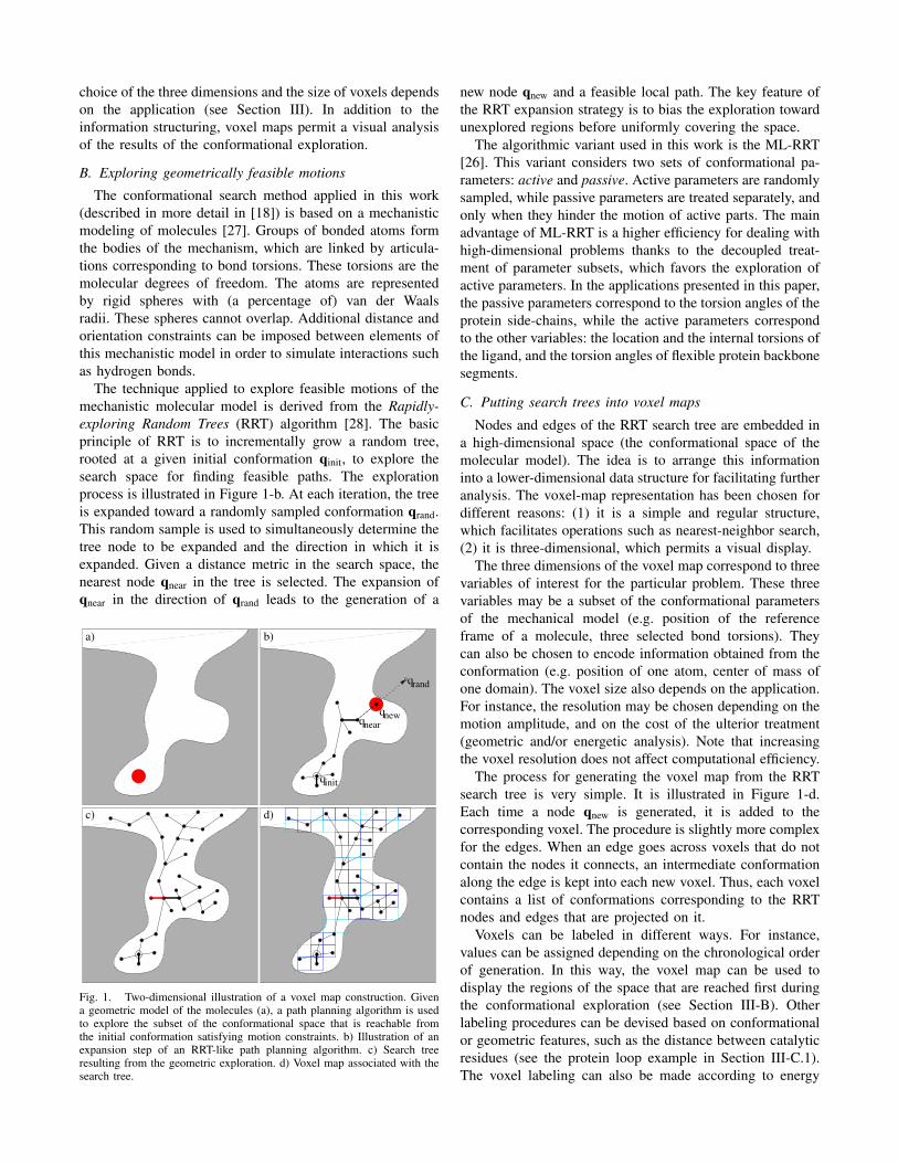
The technique applied to explore feasible motions of themechanistic molecular model is derived from the Rapidly-exploring Random Trees (RRT) algorithm [28]. The basicprinciple of RRT is to incrementally grow a random tree,rooted at a given initial conformation qinit, to explore thesearch space for finding feasible paths. The explorationprocess is illustrated in Figure 1-b. At each iteration, the treeis expanded toward a randomly sampled conformation qrand.This random sample is used to simultaneously determine thetree node to be expanded and the direction in which it isexpanded. Given a distance metric in the search space, thenearest node qnear in the tree is selected. The expansion ofqnear in the direction of qrand leads to the generation of a
a) b)
d)c)
q
q
q
qinit
nearnew
rand
Fig. 1. Two-dimensional illustration of a voxel map construction. Givena geometric model of the molecules (a), a path planning algorithm is usedto explore the subset of the conformational space that is reachable fromthe initial conformation satisfying motion constraints. b) Illustration of anexpansion step of an RRT-like path planning algorithm. c) Search treeresulting from the geometric exploration. d) Voxel map associated with thesearch tree.
new node qnew and a feasible local path. The key feature ofthe RRT expansion strategy is to bias the exploration towardunexplored regions before uniformly covering the space.
The algorithmic variant used in this work is the ML-RRT[26]. This variant considers two sets of conformational pa-rameters: active and passive. Active parameters are randomlysampled, while passive parameters are treated separately, andonly when they hinder the motion of active parts. The mainadvantage of ML-RRT is a higher efficiency for dealing withhigh-dimensional problems thanks to the decoupled treat-ment of parameter subsets, which favors the exploration ofactive parameters. In the applications presented in this paper,the passive parameters correspond to the torsion angles of theprotein side-chains, while the active parameters correspondto the other variables: the location and the internal torsions ofthe ligand, and the torsion angles of flexible protein backbonesegments.
C. Putting search trees into voxel maps
Nodes and edges of the RRT search tree are embedded ina high-dimensional space (the conformational space of themolecular model). The idea is to arrange this informationinto a lower-dimensional data structure for facilitating furtheranalysis. The voxel-map representation has been chosen fordifferent reasons: (1) it is a simple and regular structure,which facilitates operations such as nearest-neighbor search,(2) it is three-dimensional, which permits a visual display.
The three dimensions of the voxel map correspond to threevariables of interest for the particular problem. These threevariables may be a subset of the conformational parametersof the mechanical model (e.g. position of the referenceframe of a molecule, three selected bond torsions). Theycan also be chosen to encode information obtained from theconformation (e.g. position of one atom, center of mass ofone domain). The voxel size also depends on the application.For instance, the resolution may be chosen depending on themotion amplitude, and on the cost of the ulterior treatment(geometric and/or energetic analysis). Note that increasingthe voxel resolution does not affect computational efficiency.
The process for generating the voxel map from the RRTsearch tree is very simple. It is illustrated in Figure 1-d.Each time a node qnew is generated, it is added to thecorresponding voxel. The procedure is slightly more complexfor the edges. When an edge goes across voxels that do notcontain the nodes it connects, an intermediate conformationalong the edge is kept into each new voxel. Thus, each voxelcontains a list of conformations corresponding to the RRTnodes and edges that are projected on it.
Voxels can be labeled in different ways. For instance,values can be assigned depending on the chronological orderof generation. In this way, the voxel map can be used todisplay the regions of the space that are reached first duringthe conformational exploration (see Section III-B). Otherlabeling procedures can be devised based on conformationalor geometric features, such as the distance between catalyticresidues (see the protein loop example in Section III-C.1).The voxel labeling can also be made according to energy

evaluation. An energetic analysis of the conformations as-sociated with voxels may provide very useful informationabout the conformational energy landscape (see the exampleof protein domain motions in Section III-C.2).
III. APPLICATIONS
This section presents several examples illustrating voxelmap computations for different types of problems involvingmolecular motions. The goal is to show the interest of theproposed approach. Therefore, the section does not providein-depth explanations about the considered systems and thebiological importance of the results.
The method has been implemented within our softwareprototype BioMove3D. PyMOL [29] has been used forviewing molecular models and voxel maps. The computingtimes given below correspond to tests run on a single AMDOpteron 148 processor at 2.6 GHz.
A. Findings channels into proteins
The most straightforward application of the method isthe search and representation of channels into proteins.The channels are searched using the ML-RRT algorithm toexplore feasible motions of a ball of arbitrary radius insidethe protein model. This algorithm permits to directly treat allside-chain flexibility with a low computational cost. In thistype of application, the three variables used for the voxel-map representation are the position parameters of the movingball. The voxel resolution can be chosen in relation to theball size.
The chosen benchmark for this application is cytochromeP450. The in/out channels of this enzyme have been recentlycharacterized in [30]. The authors of the cited study appliedthe technique CAVER [31] for computing channels betweenthe buried active site and the surface in different crystalstructures of P450. CAVER is based on the construction ofa vertex-weighted graph from a discrete three-dimensionalgrid model of the protein. The weights are computed fromthe distance to the protein atoms, the lowest weights corre-sponding to nodes with the highest clearance. A variant ofDijkstra’s algorithm is applied to search the shortest low-cost paths. CAVER considers static structures, and performssystematic exploration of the protein interior. Molecularflexibility can only be indirectly treated by applying thetechnique to a set of structures (e.g. samples of a moleculardynamics simulation). The results described below showseveral advantages of the voxel map method over CAVER.
The structure represented in Figure 2 corresponds tobacterial P450-BM3 (PDBid 1JPZ). The voxel map in thefigure represents the channels found by our method for amoving ball of radius 1.2 A. Three of these channels, W,2b and 2f, were also found by CAVER. However, channel2d was not reported for this structure. Nevertheless, thischannel was observed in a small number of other P450-BM3 structures [30]. A further analysis of channel 2d showsthat the exit of the moving ball requires slight motions ofsome side-chains, in particular those of residues Leu20 and
2f
2d
2b
W
Fig. 2. Voxel-map representation of channels connecting the active siteand the surface in bacterial P450-BM3. Channel identifiers follow thenomenclature in related work [30].
Leu29. This result shows the interest of considering side-chain motions when computing channels in proteins.
Also note that very similar channels were obtained fromseveral runs of the voxel map computation with the movingball initially located at different positions in the enzymeactive site. Such a reliability shows the low sensibility to thestarting point, which is another advantage over CAVER1.
Finally note that, even considering side-chain flexibility,the computing time required to construct the voxel maprepresentation of the channels remains low (only 5 minutes).
B. Analyzing ligand access/exit pathways
The proposed approach can be applied to carry outan analysis of access/exit pathways of ligands (or sub-strates/products) to the active site of a protein. Such pathwayscan play important roles in the protein activity and specificity,particularly for proteins presenting a deep binding pocket.For instance, they may be one factor in enzyme enantiose-lectivity [32].
The example illustrated in Figure 3 corresponds to theinteraction between Candida antarctica lipase B (CALB)and (R,S)-enantiomers of 4-methyloctanoic acid [33]. Ourmethod has been applied to explore possible motions of theR and S substrates from its catalytic position. The modelconsiders the flexibility of the substrate and all protein side-chains. The voxel maps computed in this case represent thefeasible positions reached by the substrate center of massduring the conformational exploration. Voxel resolution is0.1 A. This representation shows significant differences be-tween both enantiomers. First, the bottom of the voxel map isnarrower for the (S)-enantiomer than for the (R)-enantiomer,which shows that possible motions of the (S)-enantiomer aremore constrained in this region. Voxels have been colored
1Results reported in [30] indicate that CAVER presents a significantsensibility to the staring point.

R
R S
S
c) d)
b)a)
Num. RRT iterations 20,0000 10,000
Fig. 3. a)-b) Models of the (R,S)-enantiomers of 4-methyloctanoic acidin the active site of CALB [33]. c)-d) Voxel maps representing locationsof the center of mass of the (R,S)-enantiomers reachable from the catalyticposition. Voxels size is 0.1 A, and colors indicate the chronological orderof generation.
depending on the chronological order of generation. Dark-blue voxels correspond to the positions that are reachedfirst. For the (R)-enantiomer (Figure 3-c), dark-blue voxelsreach the middle-part of the map. However, such voxelsare concentrated in the bottom part of the map for the (S)-enantiomer (Figure 3-d). This means that, only consideringgeometric constraints, the access/exit of the (R)-enantiomercan be faster. These results tend to indicate that the topologyof CALB active site is better suited for facilitating thereaction with (R)-4-methyloctanoic acid, which correlateswith experimental results on the enzyme enantioselectivityshowing a significant preference for this enantiomer [33].
Concerning computational performance, the constructionof the voxel map (including the ML-RRT exploration pro-cess) took less than 7 minutes for each enantiomer. Notethat, aiming to get a very good coverage of the space intothe active site cavity, the ML-RRT search tree expansionprocess yielded voxel maps with more than 25,000 and15,000 voxels respectively for the (R)- and (S) enantiomers.This result shows that even such an exhaustive explorationis computationally fast.
22.016.511.0Distance Gln357−Val407 (A)o
Gln357
Val407
Fig. 4. (Right) Structure of PTPase (PDB ID: 1YPT), with the WDP loopin black color. (Left) Detail of the WDP loop and voxel map displayingthe explored locations of the Cα atom of Glu357. Voxel colors indicatethe distance between Cα atoms of Glu357 and Val407. The most open andclosed loop conformations are represented in red and blue respectively.
C. Representing loop/domain motions
Our path-planning-based approach can be applied to com-pute large-amplitude internal molecular motions such as loopand domain motions [22], [18], [23]. Integrating the voxel-map representation in this approach provides a new tool foranalyzing such deformations.
1) Loop motions: The example illustrated in Figure 4concerns the “WDP loop” in Yersinia protein tyrosine phos-phatase (PTPase). The movement of the WDP loop playsa central role in the PTPase-mediated catalytic process [34],[35]. An open conformation of this loop permits the substrateaccess to the protein active site. Then, the WDP loop isrequired to adopt a closed conformation that brings thecatalytic residue Asp356 to a specific location for protein-substrate interaction. Starting from the open conformation ofPTPase [34] (PDB ID: 1YPT), the ML-RRT algorithm wasapplied to explore the mobility of the WDP loop (residues352-361). A voxel map obtained from this exploration isrepresented in the right part of Figure 4. It displays thepositions reached by the Cα atom of the middle loop residueGlu357. Voxels size is 0.5 A, and colors have been assigneddepending on the distance between the referred atom and theCα of Val407, which is located on the bottom of the bindingpocket. These atoms were also chosen in [35] to measure theWDP loop gating during molecular dynamics simulations.The distance in the initial crystal structure is 17 A. Theminimum and maximum distances obtained through theconformational exploration are 11 A and 22 A respectively.The WDP loop reaches conformations very similar to theone in the closed structure [34] (PDB ID: 1YTN), with CαRMSD below 1 A. The voxel map shows that the loop canadopt more open conformations, as also suggested by molec-ular dynamic simulations [35]. Interestingly, the voxel mappresents a marked pipe-like shape. Such a shape indicatesthat the WDP loop in PTPase is “mechanically” designed toperform opening-closure motions, while lateral-motions arenot likely. This mechanical predisposition may explain therapid opening-closure WDP loop motions reported in [35].Finally note that constructing the voxel map required about

5000 iterations of the ML-RRT algorithm, with a computingtime of 5 minutes.
2) Domain motions: Similarly, the proposed approachcan be applied for analyzing protein domain motions. Theexample presented here concerns the N-Oct-3 POU do-main. This molecule, represented in Figure 5, comprisestwo distinct, highly conserved sub-domains, termed POUsand POUh, connected by a flexible linker [36]. It hasbeen shown that, due to its remarkable plasticity, the N-Oct-3 POU domain can adopt different configurations andcorresponding homodimerization patterns, with the POUsand POUh sub-domains acting as sensors for the distinctunderlying structures which characterize the respective DNAtargets [37]. The conformational transitions of the N-Oct-3POU domain are currently being investigated by combiningexperimental techniques (SAXS) and the computational ap-proach presented in this paper. Figure 6 shows preliminaryresults of this work. The voxel map represents possiblelocations of POUh with respect to POUs (relative positionsof the centers of mass) considering the flexibility of the longlinker between them (18 residues are considered to be fullyflexible and 13 have limited flexibility). All the protein side-chains are also potentially flexible (i.e. they move if theyhinder backbone motions). Maximum and minimum valuesof the radius of gyration have been integrated as constraintsfor the conformational exploration. This structural informa-tion has been derived from SAXS experiments coupled tomolecular mechanics [38]. The voxels size is 2 A. Thewideness of the explored region reflects the high flexibilityof the linker. Voxels have been labeled using an energeticscoring. The conformations associated with each voxel havebeen clustered into significantly different sets. Then, oneconformation of each cluster has been energy minimized2
imposing constraints on the backbone atom positions in orderto remain within the voxel. The voxel color is assignedaccording to the resulting lowest-energy conformation. Thegoal of this work is to compute a complete conformationalmap of the protein enabling the identification of low-energyconformations attainable from the initial structure, and possi-ble transition pathways between them. Comparison with freeand pre-bound models of N-Oct-3 POU constructed fromexperimental data and molecular modeling tools will allowus to validate this novel approach.
IV. CONCLUSIONS
We have presented a general framework for computingand representing molecular motions. The basic principle ofthe approach is to apply path-planning-based algorithms toexplore feasible motions of mechanistic molecular models.The efficiency of such conformational exploration permits toattain large-amplitude motions with low computational cost.The voxel-map representation facilitates ulterior treatmentof the explored conformations and permits direct visualinterpretation of results. Besides, voxel maps could be usedto bias or to focus the exploration to specific regions of
2AMBER ff03 force field [39] has been used for the energetic analysis.
POUh
linker
POUs
Fig. 5. Structure of N-Oct-3 POU domain.
−2870Total energy (kcal/mol) −1560
Fig. 6. Two views of the voxel map representing geometrically feasiblemotions of POUh with respect to POUs. The voxels display the relativeposition of the centers of mass of both domains. Voxels size is 2 A, andcolors have been assigned depending on the energies of the associatedconformations. The black voxel indicates the initial conformation.
the conformational space, and could permit to device moreaccurate metrics, considering motion feasibility, for improvedpath-planning algorithms.
Voxel maps can represent relative motions of twomolecules. Such a representation displays the geometric suit-ability of a protein presenting a narrow, deep binding site forinteracting with different ligands. When applied to exploremolecular deformations, voxel maps can provide a globalrepresentation of the conformational space of protein loopsand protein domains undergoing large-amplitude motions.
First results highlight the potential of the proposed ap-proach. Voxel maps can be seen as a general tool that,combined with other computational and experimental meth-ods, will help to investigate the importance of flexibility andmotion in molecular interactions.
ACKNOWLEDGMENTS
The authors thank Maurice Franssen, David Guieysseand Magali Remaud for providing models and informationabout CALB. This work has been partially supported by theRegion Midi-Pyrenees under projets ITAV ALMA and CTPAMOBIO, and by the French National Agency for Research(ANR) under project NanoBioMod.

REFERENCES
[1] H. Carlson, “Protein flexibility is an important component of structure-based drug discovery,” Curr. Pharm. Des., vol. 8, pp. 1571–1578,2002.
[2] C. Cavasotto, A. Orry, and R. Abagyan, “The challenge of consideringreceptor flexibility in ligand docking and virtual screening,” Curr.Comput. Aided. Drug. Des., vol. 1, pp. 423–440, 2005.
[3] J. Janin, “Assessing predictions of protein-protein interaction: theCAPRI experiment,” Protein Sci., vol. 14, pp. 278–283, 2005.
[4] L. Ehrlich, M. Nilges, and R. Wade, “The impact of protein flexibilityon protein-protein docking,” Proteins, vol. 58, pp. 126–133, 2005.
[5] G. Katona, P. Carpentier, V. N. , P. Amara, V. Adam, J. Ohana,N. Tsanov, and D. Bourgeois, “Raman-assisted crystallography revealsend-on peroxide intermediates in a nonheme iron enzyme,” Science,vol. 316, pp. 449–453, 2007.
[6] P. Schanda, V. Forge, and B. Brutscher, “Protein folding and un-folding studied at atomic resolution by fast two-dimensional nmrspectroscopy,” PNAS, vol. 104, pp. 11 257–11 262, 2007.
[7] K. Henzler-Wildman and D. Kern, “Dynamic personalities of pro-teins,” Nature, vol. 450, pp. 964–972, 2007.
[8] A. Leach, Molecular Modeling: Principles and Applications. Cam-bridge: Longman, 1996.
[9] T. Schlick, Molecular Modeling and Simulation - An InterdisciplinaryGuide. New York: Springer, 2002.
[10] S. Izrailev, S. Stepaniants, B. Isralewitz, D. Kosztin, H. Lu, F. Molnar,W. Wriggers, and K. Schulten, “Steered molecular dynamics,” inComputational Molecular Dynamics: Challenges, Methods, Ideas.Vol. 4 of Lecture Notes in Computational Science and Engineering,P. Deuflhard, J. Hermans, B. Leimkuhler, A. Mark, S. Reich, andR. Skeel, Eds. Berlin: Springer-Verlag, 1998, pp. 39–65.
[11] L. Mouawad and D. Perahia, “Motions in hemoglobin studied bynormal mode analysis and energy minimization: evidence for theexistence of tertiary T-like, quaternary R-like intermediate structures,”J. Mol. Biol., vol. 258, pp. 393–410, 1996.
[12] J. Jeong, E. Lattman, and G. Chirikjian, “A method for findingcandidate conformations for molecular replacement using relativerotation between domains of a known structure,” Acta. Cryst., vol.D62, pp. 398–409, 2006.
[13] S. Wells, S. Menor, B. Hespenheide, and M. Thorpe, “Constrainedgeometric simulation of diffusive motion in proteins,” Phys. Biol.,vol. 2, pp. 127–136, 2005.
[14] H. Choset, K. Lynch, S. Hutchinson, G. Kantor, W. Burgard,L. Kavraki, and S. Thrun, Principles of Robot Motion: Theory,Algorithms, and Implementations. Cambridge: MIT Press, 2005.
[15] S. M. LaValle, Planning Algorithms. New York: Cambridge Univer-sity Press, 2006.
[16] A. Singh, J.-C. Latombe, and D. Brutlag, “A motion planning approachto flexible ligand binding,” Proc. Conf. Intell. Syst. Mol. Biol. (ISMB),pp. 252–261, 1999.
[17] M. Apaydin, D. Brutlag, C. Guestrin, D. Hsu, and J.-C. Latombe,“Stochastic conformational roadmaps for computing ensemble prop-erties of molecular motion,” in Algorithmic Foundations of RoboticsV, J.-D. Boissonnat, J. Burdick, K. Goldberg, and S. Hutchinson, Eds.Berlin: Springer-Verlag, 2004, pp. 131–147.
[18] J. Cortes, T. Simeon, V. Ruiz, D. Guieysse, M. Remaud, and V. Tran,“A path planning approach for computing large-amplitude motions offlexible molecules,” Bioinformatics, vol. 21, pp. i116–i125, 2005.
[19] N. M. Amato, K. A. Dill, and G. Song, “Using motion planning tomap protein folding landscapes and analyze folding kinetics of knownnative structures,” J. Comput. Biol., vol. 10, pp. 149–168, 2003.
[20] M. Apaydin, D. Brutlag, C. Guestrin, D. Hsu, J.-C. Latombe, andC. Varma, “Stochastic roadmap simulation: an efficient representationand algorithm for analyzing molecular motion,” J. Comput. Biol.,vol. 10, pp. 257–281, 2003.
[21] X. Tang, B. Kirkpatrick, S. Thomas, G. Song, and N. Amato, “Usingmotion planning to study RNA folding kinetics,” J. Comput. Biol.,vol. 12, pp. 862–881, 2005.
[22] J. Cortes, T. Simeon, M. Remaud-Simeon, and V. Tran, “Geometricalgorithms for the conformational analysis of long protein loops,” J.Comput. Chem., vol. 25(7), pp. 956–967, 2004.
[23] S. Kirillova, J. Cortes, A. Stefaniu, and T. Simeon, “An NMA-guidedpath planning approach for computing large-amplitude conformationalchanges in proteins,” Proteins, vol. 70, pp. 131–143, 2008.
[24] A. Enosh, S. Fleishman, N. Ben-Tal, and D. Halperin, “Predictionand simulation of motion in pairs of transmembrane α-helices,”Bioinformatics, vol. 23, pp. e212–e218, 2007.
[25] V. Ruiz de Angulo, J. Cortes, and T. Simeon, “BioCD: An efficientalgorithm for self-collision and distance computation between highlyarticulated molecular models,” in Robotics: Science and Systems, S. T.snd G. Sukhatme, S. Schaal, and O. Brock, Eds. Cambridge: MITPress, 2005, pp. 6–11.
[26] J. Cortes, L. Jaillet, and T. Simeon, “Disassembly path planning forcomplex articulated objects,” IEEE Transactions on Robotics, vol. 24,pp. 475–481, 2008.
[27] M. Zhang and L. Kavraki, “A new method for fast and accuratederivation of molecular conformations,” J. Chem. Inf. Comput. Sci.,vol. 42(1), pp. 64–70, 2002.
[28] S. M. LaValle and J. J. Kuffner, “Rapidly-exploring random trees:Progress and prospects,” in Algorithmic and Computational Robotics:New Directions (WAFR2000), B. Donald, K. Lynch, and D. Rus, Eds.Boston: A.K. Peters, 2001, pp. 293–308.
[29] W. DeLano, “The PyMOL molecular graphics system,” 2002,http://www.pymol.org.
[30] V. Cojocaru, P. Winn, and R. Wade, “The ins and outs of cytochromeP450s,” Biochim. Biophys. Acta, vol. 1770, pp. 390–401, 2007.
[31] M. Petrek, M. Otyepka, P. Banas, P. Kosinova, J. Koca, andJ. Damborsky, “CAVER: A new tool to explore routes from proteinclefts, pockets and cavities,” BMC Bioinfo., vol. 7, pp. 316–324, 2006.
[32] D. Guieysse, J. Cortes, S. Puech-Guenot, S. Barbe, V. Lafaquiere,P. Monsan, T. Simeon, I. Andre, and M. Remaud-Simeon, “A structure-controlled lipase enantioselectivity investigated by a path planningapproach,” ChemBioChem, vol. 9, pp. 1308–1317, 2008.
[33] M. Franssen, M. Kamp, L. Alessandrini, M. Huibers, and J. Vervoort,“The interaction between candida antarctica lipase and branched chainfatty acids: a kinetic and molecular modelling study,” Proc. Int. Symp.Biocatalysis and Biotransformations (Biotrans), 2005.
[34] J. Stuckey, H. Schubert, E. Fauman, Z.-Y. Zhang, J. E. Dixon, andM. Saper, “Crystal structure of Yersinia protein tyrosine phosphataseat 2.5 A and the complex with tungstate,” Nature, vol. 370, pp. 571–575, 1994.
[35] X. Hu and C. Stebbins, “Dynamics of the WPD loop of the Yersiniaprotein tyrosine phosphatase,” Biophys. J., vol. 91, pp. 948–956, 2006.
[36] D. Latchman, “POU family transcription factors in the nervous sys-tem,” J. Cell. Physiol., vol. 179, pp. 126–133, 1999.
[37] R. Alazard, M. Blaud, S. Elbaz, C. Vossen, G. Icre, G. Joseph,L. Nieto, and M. Erard, “Identification of the ’NORE’ (N-Oct-3responsive element), a novel structural motif and composite element,”Nucleic Acids Res., vol. 33, pp. 1513–1523, 2005.
[38] R. Alazard, L. Mourey, C. Ebel, P. Konarev, M. Petoukhov, D. Sver-gun, and M. Erard, “Fine-tuning of intrinsic N-Oct-3 POU domainallostery by regulatory DNA targets,” Nucleic Acids Res., vol. 35, pp.4420–4432, 2007.
[39] Y. Duan, C. Wu, S. Chowdhury, M. Lee, G. Xiong, W. Zhang, R. Yang,P. Cieplak, R. Luo, T. Lee, J. Caldwell, J. Wang, and P. Kollman,“A point-charge force field for molecular mechanics simulations ofproteins,” J. Comput. Chem., vol. 24, pp. 1999–2012, 2003.