enteropathogenic escherichia coli and vaccinia virus do not
TRANSCRIPT

Enteropathogenic Escherichia coli and Vaccinia Virus Do Not Requirethe Family of WASP-Interacting Proteins for Pathogen-Induced ActinAssembly
John J. Garber,a,b,e Fuminao Takeshima,a,b Inés M. Antón,c,f* Michiko K. Oyoshi,c,f Anna Lyubimova,a,b Archana Kapoor,a,b
Tomoyuki Shibata,a,b Feng Chen,h Frederick W. Alt,d,g,h Raif S. Geha,c,f John M. Leong,i and Scott B. Snappera,b,e,j
Gastrointestinal Unit and Center for the Study of Inflammatory Bowel Disease, Massachusetts General Hospital,a and Departments of Medicine,b Pediatrics,c andGenetics,d Harvard Medical School, Boston, Massachusetts, USA; Division of Gastroenterology/Nutrition and Center for Inflammatory Bowel Disease Treatment andResearche and Division of Immunology,f Howard Hughes Medical Institute,g and Immune Diseases Institute, Children’s Hospital,h Boston, Massachusetts, USA;Department of Molecular Biology and Microbiology, Tufts University School of Medicine, Boston, Massachusetts, USAi; and Division of Gastroenterology and Hepatology,Brigham and Women’s Hospital, Boston, Massachusetts, USAj
The human pathogens enteropathogenic Escherichia coli (EPEC) and vaccinia virus trigger actin assembly in host cells by acti-vating the host adaptor Nck and the actin nucleation promoter neural Wiskott-Aldrich syndrome protein (N-WASP). EPECtranslocates effector molecules into host cells via type III secretion, and the interaction between the translocated intimin recep-tor (Tir) and the bacterial membrane protein intimin stimulates Nck and N-WASP recruitment, leading to the formation of actinpedestals beneath adherent bacteria. Vaccinia virus also recruits Nck and N-WASP to generate actin tails that promote cell-to-cell spread of the virus. In addition to Nck and N-WASP, WASP-interacting protein (WIP) localizes to vaccinia virus tails, andinhibition of actin tail formation upon ectopic expression of WIP mutants led to the suggestion that WIP is required for thisprocess. Similar studies of WIP mutants, however, did not affect the ability of EPEC to form actin pedestals, arguing against anessential role for WIP in EPEC-induced actin assembly. In this study, we demonstrate that Nck and N-WASP are normally re-cruited by vaccinia virus and EPEC in the absence of WIP, and neither WIP nor the WIP family members CR16 and WIRE/WICH are essential for pathogen induced actin assembly. In addition, although Nck binds EPEC Tir directly, N-WASP is re-quired for its localization during pedestal formation. Overall, these data highlight similar pathogenic strategies shared by EPECand vaccinia virus by demonstrating a requirement for both Nck and N-WASP, but not WIP or WIP family members in patho-gen-induced actin assembly.
Manipulation of the host actin cytoskeleton is central to thepathogenesis of a variety of intra- and extracellular patho-
gens. Enteropathogenic Escherichia coli (EPEC), and vaccinia vi-rus are important human pathogens that stimulate actin assemblyto promote colonization and/or spread. Both pathogens mediateactin assembly by recruiting the host adaptor protein Nck, neuralWiskott-Aldrich syndrome protein (N-WASP), and the WASP-interacting protein (WIP) (55). When activated by host (e.g., Nckand Cdc42) or pathogen-encoded factors (see below), WASP fam-ily proteins, including WASP, N-WASP, and three WAVE/Scarmolecules (49), efficiently stimulate the Arp2/3 complex to initi-ate actin assembly (8, 14, 22, 41, 42). This activity is central todiverse cellular processes, including formation of filopodia andlamellipodia (55), endocytosis (7, 26, 37), cell movement (24, 44,51), and surface receptor signaling (6, 28).
During colonization of the intestinal epithelium, EPEC strainsinduce hallmark attaching-and-effacing (AE) lesions character-ized by localized rearrangement of the actin cytoskeleton, the for-mation of actin-rich pedestals beneath adherent bacteria, and thedestruction of adjacent microvilli (10, 12, 20, 21). To generateactin pedestals, EPEC utilizes a type III secretion system to deliverthe translocated intimin receptor (Tir) to the apical plasma mem-brane of the host cell, where its extracellular loop binds the bacte-rial outer membrane protein intimin and stabilizes bacterial at-tachment (25). The C-terminal domain of Tir, localized in thehost cell cytoplasm, initiates host signaling cascades that exploitthe actin polymerizing ability of N-WASP to form actin-rich ped-
estals beneath adherent bacteria (21, 27, 29). EPEC Tir contains acritical C-terminal tyrosine residue (Y474) that is phosphorylatedby host kinases upon translocation into the target cell (9). Thephosphorylation of Y474 in EPEC Tir provides a binding site forthe host adaptor protein Nck (9, 12, 21), and both Tir phosphor-ylation and Nck binding are essential for N-WASP recruitmentand actin pedestal formation (9, 16, 17, 21, 29, 30). The relatedpathogen enterohemorrhagic E. coli (EHEC) also recruitsN-WASP, but in an Nck-independent fashion (52, 53).
Vaccinia virus utilizes a similar signaling cascade to induceactin tails and enhance intercellular movement (46, 55). The vac-cinia virus-encoded A36R coat protein also contains a C-terminaltyrosine (Y112), which, when phosphorylated by host kinases
Received 5 November 2011 Returned for modification 2 December 2011Accepted 23 July 2012
Published ahead of print 10 September 2012
Editor: B. A. McCormick
Address correspondence to Scott B. Snapper,[email protected].
* Present address: Inés M. Antón, Centro Nacional de Biotecnología, Madrid, Spain.
J.J.G. and F.T. contributed equally to this article.
Supplemental material for this article may be found at http://iai.asm.org/.
Copyright © 2012, American Society for Microbiology. All Rights Reserved.
doi:10.1128/IAI.06148-11
December 2012 Volume 80 Number 12 Infection and Immunity p. 4071–4077 iai.asm.org 4071
Dow
nloa
ded
from
http
s://j
ourn
als.
asm
.org
/jour
nal/i
ai o
n 19
Oct
ober
202
1 by
58.
97.1
94.1
99.

(38), provides an Nck binding site required for the recruitment ofNck and N-WASP (18). Immunolocalization studies have re-vealed the presence of Grb2, Nck, WIP, and N-WASP at the viralcoat protein, and more recent fluorescence recovery after photo-bleaching (FRAP) experiments revealed that all four proteins un-dergo dynamic and continuous turnover during actin tail forma-tion by vaccinia virus (54). It has been suggested that WIPmediates the recruitment of N-WASP to vaccinia virus, based onthe observation that overexpression of the WASP-binding do-main (WBD) of WIP acts as a dominant-negative inhibitor ofvaccinia virus-induced actin tail formation and preventedN-WASP recruitment to the viral particle (36). Notably, FRAPstudies also revealed that the baseline rates of exchange of Nck andWIP were nearly identical and significantly more rapid than therate of N-WASP exchange, which is consistent with a model inwhich Nck and WIP may be recruited to A36R as a complex andtogether subsequently recruit N-WASP (54).
In contrast to vaccinia virus, Lommel et al. demonstrated thatoverexpression of the WIP WBD had no effect on the ability ofEPEC to form pedestals on N-WASP-sufficient cells; the recruit-ment of N-WASP to EPEC Tir was also unaffected by expressionof N-WASP lacking the WH1 domain, which is unable to interactwith WIP (32). Based on the presence of functionally analogousphosphotyrosine motifs—Y112 and Y474 of vaccinia virus A36Rand EPEC Tir, respectively—the apparent requirement for WIP inrecruiting N-WASP to vaccinia virus-bound but not EPEC-boundNck was unexpected.
The generation of mice and cell lines lacking N-WASP (47) andits interacting partners Nck (50), Cdc42 (13), and WIP (2) haveenabled us to better characterize the intracellular host signalingcascades targeted by pathogens that manipulate the host actinpolymerization machinery. We sought to characterize here morefully the molecular signaling events required for pathogen-in-duced actin assembly by EPEC and vaccinia virus. Utilizing tar-geted cell lines and immunolocalization studies, we demonstratethat vaccinia virus and EPEC both utilize and require Nck andN-WASP, but not WIP or WIP family members for actin assem-bly, and that Nck and N-WASP appear to be recruited by thesepathogens in a mutually dependent manner.
MATERIALS AND METHODSGene targeting and cell preparations. The generation of N-WASP�/�
and Cdc42�/� fibroblast-like cells (FLC) from embryonic stem cells hasbeen described (13, 47). WIP�/� FLC were isolated from lung tissue iso-lated from WIP knockout (KO) mice (2). Briefly, lung pieces from WT orWIP KO mice were washed with phosphate-buffered saline (PBS), mincedand cultured in Iscove medium supplemented with 10% fetal bovine se-rum and penicillin-streptomycin (50 U/ml) for several days. After theremoval of unattached debris, the adherent cells were treated with trypsinand maintained in culture.
Bacterial and viral strains. EPEC JPN15/pMAR7 strain and EHECTUV 93-0 were cultured in lysogeny broth for at least 8 h to late log phasethe day prior to infection and then diluted 1:10 and grown overnight at37°C without agitation. For EPEC, the media were supplemented withampicillin (100 �g/ml). Shigella flexneri 2a strain 2457T was culturedovernight prior to infection. Vaccinia virus was either provided by Ram-nik Xavier (Massachusetts General Hospital, Boston, MA) or produced inone of our laboratories (R.S.G., Children’s Hospital, Boston, MA).
Retroviruses and vectors. Retrovirus expressing rat N-WASP (19)was generated by cloning the coding sequence into a replication-defectiveretrovirus chicken matrix metalloproteinase plasmid (pCMMP) (31). N-WASP/GFP bicistronic viruses were constructed by cloning green fluores-
cent protein (GFP; containing an internal ribosome entry site) down-stream of N-WASP, and recombinant virus was produced by transienttransfection of the 293-GPG packaging cell line as previously described(39). Wild-type (WT) WIP was cloned in phase with GFP at the C termi-nus of WIP in the vector pcDNA3 as previously described (15). pELGFP-WIP was provided by Michael Way (36), and pEBB myc-tagged Nck SH3-1,2,3 mutant was provided by Bruce J. Mayer (50).
Infection assays. Infection assays were described previously (13, 47).Briefly, FLC were inoculated and grown overnight on coverslips in Dul-becco modified Eagle medium supplemented with 10% fetal calf serumand overlaid with the bacterial or viral suspension. In bacterial experi-ments, to promote adhesion to the cells, the samples were centrifuged for10 min at 2,000 � g at 23°C. The cells were incubated for 1 h at 37°C at amultiplicity of infection (MOI) of 200 for EPEC and S. flexneri and at anMOI of 400 for EHEC, followed by incubation in medium supplementedwith gentamicin (50 �g/ml) for 3 h. The efficiency of Shigella and EPEC/EHEC infections were more than 10 and 90%, respectively. For vacciniavirus invasion, cells were infected at an MOI of 20 for 8 h without genta-micin, which resulted in an infection efficiency of 50 to 90%. After infec-tion, coverslips were washed three to five times with PBS, fixed for 10 minwith 3.7% fresh paraformaldehyde, and stained.
CR16 Western blotting. Protein extracts from lung fibroblasts, as wellas WT testes tissue (positive control), were separated by sodium dodecylsulfate-gel electrophoresis and transferred onto nitrocellulose membraneas previously described (8). Immunoblotting was performed with rabbitpolyclonal anti-CR16 (1:500) (generated in the R.S.G. laboratory), followedby horseradish peroxidase-conjugated goat anti-rabbit secondary antibody(1:3,000; Cell Signaling, Danvers, MA), and visualized using enhanced chemi-luminescence (GE Healthcare Life Sciences, Piscataway, NJ).
RNAi. For WIRE knockdown, an ON-TARGETplus SMARTpoolagainst mouse WIRE (Thermo Fisher Scientific/Dharmacon, Lafayette,CO) was transfected into WIP�/� and WT control fibroblasts using Lipo-fectamine 2000 according to the manufacturer’s protocol. Control shortinterfering RNAs (siRNAs) for SMARTpool RNA interference (RNAi)included the ON-TARGETplus nontargeting pool (Thermo Fisher Scien-tific/Dharmacon). WIRE protein levels were determined 48 and 96 h aftersiRNA transfection by Western blotting and detection with rabbit poly-clonal anti-WIRE antibody (Sigma, St. Louis, MO). Greater than 85%knockdown was confirmed by comparison of the optical density of WIREbands relative to tubulin loading controls in ImageJ software.
Transfections. For WIP and DN-Nck expression, DNA was trans-fected into 105 FLC 16 h prior to infection using Lipofectamine Plus (In-vitrogen, Carlsbad, CA) or FuGENE6 (Roche Applied Science, Indianap-olis, IN). The efficiency of transfection for each of these constructs rangedbetween 20 and 40%, with an overall transfection and infection efficiencyof 2 to 15%.
Immunofluorescence microscopy. The fixed cells were permeabil-ized with 0.5% Triton X-100 in PBS for 20 min and washed five times with1% bovine serum albumin in PBS. Nck, N-WASP, and WIP were localizedby incubating with anti-Nck rabbit polyclonal antibody (Upstate Biotech-nology, Waltham, MA) at a 1:200 dilution, anti-N-WASP rabbit poly-clonal antibody (provided by Rajat Rohatgi), or anti-WIP rabbit poly-clonal antibody (provided by Raif Geha), respectively, for 1 h at roomtemperature. Subsequently, the cells were incubated with fluorescein iso-thiocyanate (FITC)-conjugated anti-rabbit IgG antibody (Sigma) at a1:200 dilution for 1 h. Myc-tagged DN-Nck was detected by incubationwith anti-myc mouse monoclonal antibody (Oncogene, Cambridge, MA)at a 1:200 dilution for 1 h at room temperature. The cells were then incu-bated with FITC-conjugated anti-mouse IgG or TRITC (tetramethyl rho-damine isothiocyanate)-conjugated anti-mouse IgG antibody (Sigma, St.Louis, MO) at 1:200 dilution, followed by 1 �g of rhodamine-labeledphalloidin (Molecular Probes, Eugene, OR)/ml for 20 min at room tem-perature for F-actin staining. To identify bacterially infected cells, perme-abilized cells were incubated with DAPI (4=,6=-diamidino-2-phenylin-dole; Molecular Probes, Eugene, OR) at 300 nM for 3 min at room
Garber et al.
4072 iai.asm.org Infection and Immunity
Dow
nloa
ded
from
http
s://j
ourn
als.
asm
.org
/jour
nal/i
ai o
n 19
Oct
ober
202
1 by
58.
97.1
94.1
99.
temperature. Standard epifluorescence microscopy was performed usingan AX70 upright microscope (Olympus, Tokyo, Japan). To determine theefficiency of pedestal formation for EPEC, the actin cytoskeleton of 100bacterially infected cells were examined in five independent experimentsby immunofluorescence. Cells containing at least 10 bacteria by DAPIstaining were considered infected. To determine the relative tail lengths invaccinia virus-infected or Shigella-infected WT and WIP�/� FLC, thelengths of 50 phalloidin-stained tails were measured using Photoshop 5.0software (Adobe, San Jose, CA) by random sampling in three independentexperiments.
RESULTSCdc42 is not required for EPEC-induced actin pedestal forma-tion. The Rho family GTPase Cdc42 interacts with the GTPase-binding domain (GBD) of N-WASP, and it was reported thatWASP proteins lacking the GBD fail to localize to EPEC-inducedactin pedestals, raising the possibility that Cdc42 plays a role inactin pedestal formation (27). However, inactivation of Cdc42with Clostridium difficile toxin B or overexpression of N-WASPGBD did not prevent EPEC pedestal formation (5, 8), and anN-WASP mutant that is insensitive to Cdc42 activation can none-theless mediate actin assembly by EPEC (32). To determinewhether Cdc42 is required for pedestal formation, WT andCdc42-deficient cells (13) were infected with EPEC. EHEC, whichtranslocates the effector EspFU/TccP that directly activatesN-WASP (14, 43), was included as a control. EPEC, like EHEC,efficiently formed pedestals in the absence of Cdc42 (Fig. 1), de-finitively ruling out an essential role for Cdc42 in EPEC pedestalformation.
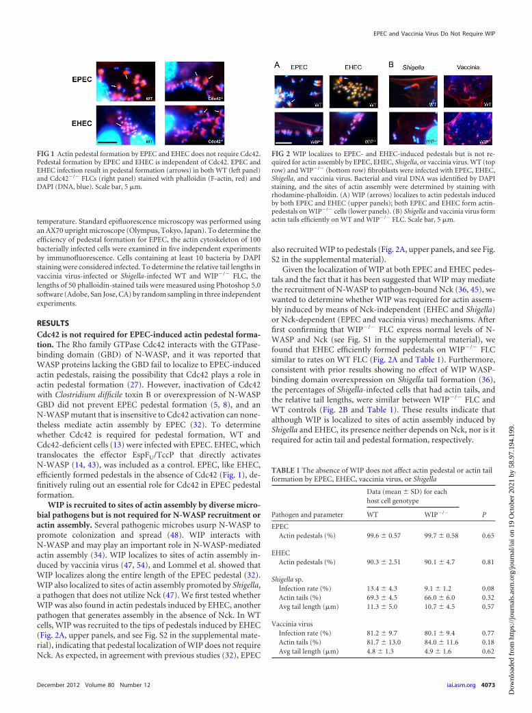
WIP is recruited to sites of actin assembly by diverse micro-bial pathogens but is not required for N-WASP recruitment oractin assembly. Several pathogenic microbes usurp N-WASP topromote colonization and spread (48). WIP interacts withN-WASP and may play an important role in N-WASP-mediatedactin assembly (34). WIP localizes to sites of actin assembly in-duced by vaccinia virus (47, 54), and Lommel et al. showed thatWIP localizes along the entire length of the EPEC pedestal (32).WIP also localized to sites of actin assembly promoted by Shigella,a pathogen that does not utilize Nck (47). We first tested whetherWIP was also found in actin pedestals induced by EHEC, anotherpathogen that generates assembly in the absence of Nck. In WTcells, WIP was recruited to the tips of pedestals induced by EHEC(Fig. 2A, upper panels, and see Fig. S2 in the supplemental mate-rial), indicating that pedestal localization of WIP does not requireNck. As expected, in agreement with previous studies (32), EPEC
also recruited WIP to pedestals (Fig. 2A, upper panels, and see Fig.S2 in the supplemental material).
Given the localization of WIP at both EPEC and EHEC pedes-tals and the fact that it has been suggested that WIP may mediatethe recruitment of N-WASP to pathogen-bound Nck (36, 45), wewanted to determine whether WIP was required for actin assem-bly induced by means of Nck-independent (EHEC and Shigella)or Nck-dependent (EPEC and vaccinia virus) mechanisms. Afterfirst confirming that WIP�/� FLC express normal levels of N-WASP and Nck (see Fig. S1 in the supplemental material), wefound that EHEC efficiently formed pedestals on WIP�/� FLCsimilar to rates on WT FLC (Fig. 2A and Table 1). Furthermore,consistent with prior results showing no effect of WIP WASP-binding domain overexpression on Shigella tail formation (36),the percentages of Shigella-infected cells that had actin tails, andthe relative tail lengths, were similar between WIP�/� FLC andWT controls (Fig. 2B and Table 1). These results indicate thatalthough WIP is localized to sites of actin assembly induced byShigella and EHEC, its presence neither depends on Nck, nor is itrequired for actin tail and pedestal formation, respectively.
FIG 1 Actin pedestal formation by EPEC and EHEC does not require Cdc42.Pedestal formation by EPEC and EHEC is independent of Cdc42. EPEC andEHEC infection result in pedestal formation (arrows) in both WT (left panel)and Cdc42�/� FLCs (right panel) stained with phalloidin (F-actin, red) andDAPI (DNA, blue). Scale bar, 5 �m.
FIG 2 WIP localizes to EPEC- and EHEC-induced pedestals but is not re-quired for actin assembly by EPEC, EHEC, Shigella, or vaccinia virus. WT (toprow) and WIP�/� (bottom row) fibroblasts were infected with EPEC, EHEC,Shigella, and vaccinia virus. Bacterial and viral DNA was identified by DAPIstaining, and the sites of actin assembly were determined by staining withrhodamine-phalloidin. (A) WIP (arrows) localizes to actin pedestals inducedby both EPEC and EHEC (upper panels); both EPEC and EHEC form actin-pedestals on WIP�/� cells (lower panels). (B) Shigella and vaccinia virus formactin tails efficiently on WT and WIP�/� FLC. Scale bar, 5 �m.
TABLE 1 The absence of WIP does not affect actin pedestal or actin tailformation by EPEC, EHEC, vaccinia virus, or Shigella
Pathogen and parameter
Data (mean � SD) for eachhost cell genotype
PWT WIP�/�
EPECActin pedestals (%) 99.6 � 0.57 99.7 � 0.58 0.65
EHECActin pedestals (%) 90.3 � 2.51 90.1 � 4.7 0.81
Shigella sp.Infection rate (%) 13.4 � 4.3 9.1 � 1.2 0.08Actin tails (%) 69.3 � 4.5 66.0 � 6.0 0.32Avg tail length (�m) 11.3 � 5.0 10.7 � 4.5 0.57
Vaccinia virusInfection rate (%) 81.2 � 9.7 80.1 � 9.4 0.77Actin tails (%) 81.7 � 13.0 84.0 � 11.6 0.18Avg tail length (�m) 4.8 � 1.3 4.9 � 1.6 0.62
EPEC and Vaccinia Virus Do Not Require WIP
December 2012 Volume 80 Number 12 iai.asm.org 4073
Dow
nloa
ded
from
http
s://j
ourn
als.
asm
.org
/jour
nal/i
ai o
n 19
Oct
ober
202
1 by
58.
97.1
94.1
99.

In contrast to Shigella and EHEC, vaccinia virus and EPECutilize Nck to recruit host N-WASP, and previous reports sug-gested that WIP, by mediating the interaction between Nck andN-WASP, plays an important role in actin-tail formation by vac-cinia virus (36) but, somewhat unexpectedly, not in actin pedestalformation by EPEC (32). To clarify this apparent dichotomy in therequirement for WIP in actin assembly, we assessed actin pedestalor tail formation by EPEC and vaccinia virus in WIP-deficientcells. Consistent with previous WIP mutant overexpression stud-ies (32), EPEC efficiently formed pedestals on WIP�/� FLC simi-lar to rates on WT FLC (Fig. 2A and Table 1). Moreover, we foundthat the percentage of vaccinia virus-infected cells that had actintails was similar between WIP�/� FLC and WT controls (Fig. 2Band Table). There was also no difference in the relative tail lengthin vaccinia virus-infected WT or WIP�/� cells (Fig. 2B and Table1). Together, these results demonstrate that WIP is not essentialfor EPEC-induced pedestal formation or for the actin-based mo-tility of vaccinia virus.
Given that WIP was not required for EPEC- and vaccinia virus-based actin assembly, we hypothesized that WIP would not berequired for either Nck or N-WASP recruitment. We first showedthat N-WASP, but not Nck, was also recruited to EHEC actinpedestals on WIP-deficient cells, an observation consistent withthe ability of EHEC to form pedestals in an Nck-independentmanner (Fig. 3, bottom row). Importantly, N-WASP and Nckwere also normally recruited by vaccinia virus and EPEC inWIP�/� cells (Fig. 3, top and middle rows).
WIP is recruited to EHEC, but not EPEC or vaccinia virus,upon ectopic expression of a dominant-negative Nck derivative.Since WIP can bind to both Nck and N-WASP, WIP recruitmentmay be dependent on a direct interaction with either Nck and/orN-WASP. In order to test the role of Nck in WIP recruitment toboth EPEC and vaccinia virus, we utilized a dominant-negativeinhibitor of endogenous Nck (DN-Nck) that contains mutationsin each of the SH3 domains (50). We first confirmed that theconstruct was effective by demonstrating that DN-Nck specificallyblocked EPEC-induced but not EHEC-induced pedestal forma-
tion (21) (see Fig. S3 in the supplemental material) and blockedthe recruitment of N-WASP to EPEC but not EHEC (Fig. 4, leftpanels). WIP recruitment to EHEC pedestals did not appear todepend on Nck because ectopic expression of the DN-Nck had noeffect on pedestal localization by WIP (Fig. 4, bottom right). Incontrast, DN-Nck expression blocked WIP recruitment to EPEC(Fig. 4, upper right panel). Similarly, and consistent with previ-ously published work (13, 27), DN-Nck expression blocked bothN-WASP and WIP recruitment to vaccinia virus (data notshown).
Localization of both WIP and Nck to EPEC and vaccinia vi-rus is N-WASP dependent. Given that the second SH3 domainwithin Nck interacts with WIP (4), the observation that WIP lo-calization to sites of attachment by vaccinia virus or EPEC wasblocked by the expression of an Nck derivative with defective SH3domains is consistent with the simple model that Nck normallydirectly recruits WIP to sites of actin assembly induced by thesepathogens. Alternatively, given that WIP and N-WASP are capa-ble of direct interaction (15, 23, 34), it is also possible that theexpression of a dominant-negative Nck derivative inhibited WIPrecruitment by blocking the localization of N-WASP. Indeed, theability of EHEC to recruit WIP to sites of pedestal formation (Fig.4), in spite of the Nck-independent nature of EHEC pedestal for-mation, is consistent with N-WASP-dependent WIP recruitment.In addition, we previously demonstrated that WIP is recruited tosites of both vaccinia virus and Shigella actin tail formation in anN-WASP-dependent manner (47).
To determine whether N-WASP is required for WIP or Ncklocalization to sites of EPEC or EHEC pedestal formation, we in-fected N-WASP-deficient cells with these bacteria. As a control, inparallel we also infected these cells with vaccinia virus. All threepathogens were incapable of promoting actin assembly in thesecells, an observation consistent with previous findings (21, 47). Inaddition, we confirmed that WIP and Nck recruitment to vacciniavirus actin tails required N-WASP (Fig. 5, lower row) (47). Finally,neither Nck nor WIP was localized at sites of adherent EHEC orEPEC in the absence of N-WASP (Fig. 5, top and middle rows).Since EHEC does not require Nck for either N-WASP recruitment
FIG 3 WIP is not essential for the recruitment of Nck or N-WASP duringinfection with vaccinia virus, EPEC or EHEC. F-actin (phalloidin staining,red) and DNA (DAPI, blue) were visualized in WT and WIP�/� FLC infectedwith vaccinia virus (top row), EPEC (middle row), or EHEC (lower row).Actin tails and pedestals are formed efficiently in WIP�/� FLC, and N-WASP(green) (arrows, left column) or Nck (green) (arrows, right column) are re-cruited to EPEC and vaccinia virus in the absence of WIP. *, Lack of Ncklocalization to EHEC. Scale bar, 5 �m.
FIG 4 Neither N-WASP nor WIP is recruited to EPEC in the absence of Nck.FLC transfected with a dominant-negative inhibitor of Nck (green) were in-fected with either EPEC (upper row) or EHEC (lower row) and were analyzedby DAPI staining of DNA and immunostained for N-WASP (left panel, red) orWIP (right panel, red). N-WASP localizes to EHEC (closed arrowhead), butnot EPEC in DN-Nck transfected cells (open arrowhead). WIP localizes toEHEC (closed arrowhead), but not EPEC (open arrowhead) in DN-Nck trans-fected cells. Green anti-myc staining is omitted from the insets to improvephalloidin and DAPI visualization. Scale bar, 10 �m; inset scale bar, 2.5 �m.
Garber et al.
4074 iai.asm.org Infection and Immunity
Dow
nloa
ded
from
http
s://j
ourn
als.
asm
.org
/jour
nal/i
ai o
n 19
Oct
ober
202
1 by
58.
97.1
94.1
99.

or pedestal formation, these data are consistent with the sugges-tion that WIP is recruited to EHEC-induced actin pedestals indi-rectly through its interaction with N-WASP. Importantly, to-gether with the above finding that ectopic expression of a DN-Nckderivative blocks N-WASP recruitment to sites of EPEC attach-ment, these data also suggest that, as is the case for vaccinia virus,N-WASP and Nck are dependent on each other, but not WIP, forlocalization to sites of EPEC-mediated actin assembly.
WIP family members CR16 and WICH/WIRE do not con-tribute to actin rearrangements induced by EPEC or vacciniavirus in the absence of WIP. In addition to WIP, the WIP familyof proteins includes two additional WIP homologues that are ca-pable of binding N-WASP and may be able to functionally substi-tute for WIP: CR16 and WIRE/WICH (WIP related/WIP CR16homologous) (3, 35). In order to determine whether the apparentdispensability of WIP for actin pedestal and tail formation was aresult of functional redundancy of CR16 or WIRE for WIP, wefirst confirmed by Western blotting that only WIRE, and notCR16, was expressed in fibroblasts (see Fig. 4A in the supplemen-tal material). We next generated WIRE knockdown and WIP�/�/WIRE knockdown lines by siRNA-mediated depletion of WIRE inWT and WIP�/� cells. We confirmed by Western blot and densi-tometry that there was �85% knockdown of WIRE (see Fig. S4Bin the supplemental material). We found no significant differ-ences, compared to WT cells or WIP�/� cells, in the rates of in-fection, the frequency of actin tails or actin pedestals, or the mor-phology of these actin structures during infection with EPEC orvaccinia virus in WIRE-depleted cell lines (Fig. 6; see also Table S1in the supplemental material). These data confirm that neitherWIP, nor CR16, nor WIRE/WICH is required for EPEC- and vac-cinia virus-induced actin rearrangement.
DISCUSSION
In order to characterize the role of N-WASP and its interactingpartners (WIP, Cdc42, and Nck) in regulating the actin cytoskel-
eton, we generated cell lines with germ line deletion or dominant-negative inhibition of these proteins (1, 2, 13, 47). These cell lineshave enabled us to characterize more fully the host molecularsignaling events required for pathogen-induced actin-assembly byEPEC, EHEC, and vaccinia virus and demonstrate conclusivelythat Cdc42 is not required for EPEC- or EHEC-induced pedestalformation.
We have demonstrated that WIP is not required for actin as-sembly induced by either EPEC or vaccinia virus. Although bothN-WASP and WIP are recruited to sites of actin assembly by EPEC(Fig. 2A and 4) and vaccinia virus (Fig. 3), only N-WASP, and notWIP, was required for actin pedestal formation and actin-basedmotility (Fig. 3) (32, 47). For vaccinia virus, Nck has been impli-cated as the link between virally expressed A36R and WIP, whichin turn binds N-WASP (36, 45). Like vaccinia virus, EPEC alsoutilizes Nck as a link between a bacterially expressed protein (Tir)and downstream host signaling molecules (9, 21). We have dem-onstrated that EPEC and vaccinia virus do not require WIP forNck localization or for the recruitment of N-WASP in initiatingactin polymerization. A direct interaction between Nck and N-WASP, which has been confirmed in vitro (40, 42), is consistentwith our findings. However, reports have suggested that the regionof N-WASP previously shown to bind Nck, the proline-rich re-gion domain, is not required for either the localization ofN-WASP to pedestals or for the formation of pedestals by EPEC(27, 33). Given the more recent demonstration that EPEC gener-ates actin pedestals by (at least) three pathways, only one of whichis Nck-dependent (11), the lack of discernible phenotypes associ-ated with N-WASP deletion derivatives might be due to redun-dancy. For example, an Nck-independent pathway involving theN-WASP WH1 domain might facilitate N-WASP localization andpedestal formation in the absence of the Nck-binding N-WASPproline-rich domain.
Our results suggest that the recruitment of Nck, WIP, and N-WASP to EPEC or vaccinia virus does not proceed in a simple linearfashion. Despite the fact that Nck can bind to phosphorylated Tir (9,21), we found that Nck does not localize to EPEC or vaccinia virus inthe absence of N-WASP (Fig. 5). These findings contrast with previ-
FIG 5 N-WASP is required for the recruitment of both Nck and WIP. N-WASP�/� FLC were infected with either EPEC (upper row) or EHEC (middlerow) or vaccinia virus (lower row) and were analyzed by DAPI staining of DNA(blue), phalloidin staining for actin (red), and immunostaining with Nck (leftpanels, green) or transfection with WIP-GFP (right panels, green). NeitherNck nor WIP localized to EPEC (arrows), EHEC (arrows) or vaccinia virus(arrows) in the absence of N-WASP. Scale bar, 5 �m.
FIG 6 The WIP family member WIRE/WICH does not substitute for WIP inWIP�/� cells during infection with EPEC or vaccinia virus. EPEC (upper row)and vaccinia virus (lower row) were equally able to induce actin pedestals andactin tails, respectively, in WIP WT (left panels) or KO cells (right panels)depleted of the WIRE/WICH by siRNA. Red is actin staining with Alexa Fluor594-conjugated phalloidin; insets show DAPI staining of nuclei (omitted fromthe figure for clarity). Scale bar, 5 �m.
EPEC and Vaccinia Virus Do Not Require WIP
December 2012 Volume 80 Number 12 iai.asm.org 4075
Dow
nloa
ded
from
http
s://j
ourn
als.
asm
.org
/jour
nal/i
ai o
n 19
Oct
ober
202
1 by
58.
97.1
94.1
99.

ous reports in which GFP-tagged Nck was observed beneath extracel-lular vaccinia virus particles in N-WASP�/� cells (54). That we didnot see endogenous Nck staining suggests that N-WASP may be re-quired to stabilize the nascent interaction between Tir and Nck ormay facilitate the translocation of Nck to the cytoplasmic tail of Tir,but that GFP-Nck, ectopically expressed at relatively high levels,might still localize to vaccinia virus in the absence of N-WASP. Wepropose that EPEC Tir and vaccinia virus A36R require a complexthat contains N-WASP and Nck for recruitment to the pathogen (Fig.7). A role for N-WASP upstream of Nck is also consistent with FRAPstudies demonstrating that the rate of N-WASP exchange is the majordeterminant of both Nck and WIP turnover at the vaccinia virus coatprotein (54). It has been proposed that the observed differences in therates of Nck and N-WASP exchange may be due to additional stabi-lizing interactions between the WASP-homology 2 (WH2) domainof N-WASP and free barbed ends of growing actin filaments (54), andthis model is compatible with our data demonstrating that the pres-ence of both Nck and N-WASP at the viral particle is required forWIP recruitment to vaccinia virus. Although WIP can be found aspart of this Nck/N-WASP complex at the site of actin assembly, itspresence is not required for actin assembly induced by EPEC or vac-cinia virus.
We also determined that it is unlikely that other WIP familymembers—CR16 and WIRE/WICH—serve a functionally redun-dant role in stabilizing the Nck/N-WASP complex. CR16 is notexpressed in the fibroblasts used in these experiments, and deple-tion of WIRE/WICH did not affect the rates of infection or theability of EPEC or vaccinia virus to induce actin rearrangements inWIP�/� cells. Our findings are also consistent with prior studiesinvolving the overexpression of either the WBD of WIP orWIP�WBD that indicated that an N-WASP/WIP interaction wasdispensable for EPEC pedestal formation (32). The observationthat overexpression of the WBD of WIP blocked vaccinia virusactin tail formation, suggesting an essential role for WIP in thisprocess (36), may have resulted from reduced actin polymeriza-tion activity through nonspecific sequestration of N-WASP.Given that we show here that N-WASP is required for Nck recruit-ment by vaccinia virus, the prior observation that overexpressionof the WBD of WIP also blocked the recruitment of Nck to vac-cinia virus (36) may also have been a consequence of its effect onN-WASP.
Overall, our studies highlight similar pathogenic strategiesshared by EPEC and vaccinia virus in the Nck-dependent recruit-ment of an N-WASP/WIP complex to homologous phosphoty-rosine motifs in EPEC Tir and vaccinia virus A36R. We demon-
strate a dual requirement for both Nck and N-WASP, but not WIPor WIP family members, in actin assembly induced by EPEC andvaccinia virus.
ACKNOWLEDGMENTS
This study was supported by National Institutes of Health grantsR01AI46454 (to J.M.L.), P01HL059561 (to R.S.G. and S.B.S.),R01AI052354 (to S.B.S.), and F32DK088442 (to J.J.G.), as well as a CareerDevelopment Award from the Crohn’s and Colitis Foundation of Amer-ica (to J.J.G.) and the Harvard Digestive Disease Center (5P30DK034854to S.B.S. and J.J.G.).
REFERENCES1. Anton I, et al. 2003. WIP participates in actin reorganization and ruffle
formation induced by PDGF. J. Cell Sci. 116:2443–2451.2. Anton IM, et al. 2002. WIP deficiency reveals a differential role for WIP
and the actin cytoskeleton in T and B cell activation. Immunity 16:193–204.
3. Anton IM, Jones GE. 2006. WIP: a multifunctional protein involved inactin cytoskeleton regulation. Eur. J. Cell Biol. 85(3– 4):295–304.
4. Anton IM, Lu W, Mayer BJ, Ramesh N, Geha RS. 1998. The Wiskott-Aldrich syndrome protein-interacting protein (WIP) binds to the adaptorprotein Nck. J. Biol. Chem. 273:20992–20995.
5. Ben-Ami G, et al. 1998. Agents that inhibit Rho, Rac, and Cdc42 do notblock formation of actin pedestals in HeLa cells infected with entero-pathogenic Escherichia coli. Infect. Immun. 66:1755–1758.
6. Benesch S, et al. 2005. N-WASP deficiency impairs EGF internalizationand actin assembly at clathrin-coated pits. J. Cell Sci. 118(Pt 14):3103–3115.
7. Bu W, Chou AM, Lim KB, Sudhaharan T, Ahmed S. 2009. The Toca-1-N-WASP complex links filopodial formation to endocytosis. J. Biol.Chem. 284:11622–11636.
8. Campellone KG, et al. 2008. Repetitive N-WASP-binding elements of theenterohemorrhagic Escherichia coli effector EspF(U) synergistically acti-vate actin assembly. PLoS Pathog. 4 :e1000191. doi:10.1371/journal.ppat.1000191.
9. Campellone KG, Giese A, Tipper DJ, Leong JM. 2002. A tyrosine-phosphorylated 12-amino-acid sequence of enteropathogenic Escherichiacoli Tir binds the host adaptor protein Nck and is required for Nck local-ization to actin pedestals. Mol. Microbiol. 43:1227–1241.
10. Campellone KG, Leong JM. 2003. Tails of two Tirs: actin pedestal for-mation by enteropathogenic Escherichia coli and enterohemorrhagic E.coli O157:H7. Curr. Opin. Microbiol. 6:82–90.
11. Campellone KG, Leong JM. 2005. Nck-independent actin assembly ismediated by two phosphorylated tyrosines within enteropathogenic Esch-erichia coli Tir. Mol. Microbiol. 56:416 – 432.
12. Campellone KG, et al. 2004. Clustering of Nck by a 12-residue Tir phos-phopeptide is sufficient to trigger localized actin assembly. J. Cell Biol.164:407– 416.
13. Chen F, et al. 2000. Cdc42 is required for PIP(2)-induced actin poly-merization and early development but not for cell viability. Curr. Biol.10:758 –765.
14. Cheng HC, Skehan BM, Campellone KG, Leong JM, Rosen MK. 2008.Structural mechanism of WASP activation by the enterohaemorrhagicEscherichia coli effector EspF(U). Nature 454:1009 –1013.
15. de la Fuente MA, et al. 2007. WIP is a chaperone for Wiskott-Aldrichsyndrome protein (WASP). Proc. Natl. Acad. Sci. U. S. A. 104:926 –931.
16. DeVinney R, Puente JL, Gauthier A, Goosney D, Finlay BB. 2001.Enterohaemorrhagic and enteropathogenic Escherichia coli use a differentTir-based mechanism for pedestal formation. Mol. Microbiol. 41:1445–1458.
17. Finlay BB, Rosenshine I, Donnenberg MS, Kaper JB. 1992. Cytoskeletalcomposition of attaching and effacing lesions associated with entero-pathogenic Escherichia coli adherence to HeLa cells. Infect. Immun. 60:2541–2543.
18. Frischknecht F, et al. 1999. Actin-based motility of vaccinia virus mimicsreceptor tyrosine kinase signalling. Nature 401:926 –929.
19. Fukuoka M, Miki H, Takenawa T. 1997. Identification of N-WASPhomologs in human and rat brain. Gene 196:43– 48.
20. Garmendia J, et al. 2004. TccP is an enterohaemorrhagic Escherichia coli
FIG 7 Model for strategies of Nck/N-WASP recruitment and activationshared by EPEC and vaccinia virus and contrasted with EHEC and Shigella.
Garber et al.
4076 iai.asm.org Infection and Immunity
Dow
nloa
ded
from
http
s://j
ourn
als.
asm
.org
/jour
nal/i
ai o
n 19
Oct
ober
202
1 by
58.
97.1
94.1
99.

O157:H7 type III effector protein that couples Tir to the actin-cytoskeleton. Cell Microbiol. 6:1167–1183.
21. Gruenheid S, et al. 2001. Enteropathogenic Escherichia coli Tir binds Nckto initiate actin pedestal formation in host cells. Nat. Cell Biol. 3:856 – 859.
22. Higgs HN, Pollard TD. 2000. Activation by Cdc42 and PIP(2) of Wiskott-Aldrich syndrome protein (WASp) stimulates actin nucleation by Arp2/3complex. J. Cell Biol. 150:1311–1320.
23. Ho HY, et al. 2004. Toca-1 mediates Cdc42-dependent actin nucleationby activating the N-WASP-WIP complex. Cell 118:203–216.
24. Isaac BM, et al. 2010. N-WASP has the ability to compensate for the lossof WASP in macrophage podosome formation and chemotaxis. Exp. CellRes. 316:3406 –3416.
25. Jerse AE, Yu J, Tall BD, Kaper JB. 1990. A genetic locus of enteropatho-genic Escherichia coli necessary for the production of attaching and effac-ing lesions on tissue culture cells. Proc. Natl. Acad. Sci. U. S. A. 87:7839 –7843.
26. Kaksonen M, Sun Y, Drubin DG. 2003. A pathway for association ofreceptors, adaptors, and actin during endocytic internalization. Cell 115:475– 487.
27. Kalman D, et al. 1999. Enteropathogenic Escherichia coli acts throughWASP and Arp2/3 complex to form actin pedestals. Nat. Cell Biol. 1:389 –391.
28. Kempiak SJ, Yip SC, Backer JM, Segall JE. 2003. Local signaling by theEGF receptor. J. Cell Biol. 162:781–787.
29. Kenny B. 1999. Phosphorylation of tyrosine 474 of the enteropathogenicEscherichia coli (EPEC) Tir receptor molecule is essential for actin nucle-ating activity and is preceded by additional host modifications. Mol. Mi-crobiol. 31:1229 –1241.
30. Kenny B, et al. 1997. Enteropathogenic E. coli (EPEC) transfers its recep-tor for intimate adherence into mammalian cells. Cell 91:511–520.
31. Klein C, Bueler H, Mulligan RC. 2000. Comparative analysis of geneti-cally modified dendritic cells and tumor cells as therapeutic cancer vac-cines. J. Exp. Med. 191:1699 –1708.
32. Lommel S, Benesch S, Rohde M, Wehland J, Rottner K. 2004. Entero-haemorrhagic and enteropathogenic Escherichia coli use different mecha-nisms for actin pedestal formation that converge on N-WASP. Cell Mi-crobiol. 6:243–254.
33. Lommel S, et al. 2001. Actin pedestal formation by enteropathogenicEscherichia coli and intracellular motility of Shigella flexneri are abolishedin N-WASP-defective cells. EMBO Rep. 2:850 – 857.
34. Martinez-Quiles N, et al. 2001. WIP regulates N-WASP-mediated actinpolymerization and filopodium formation. Nat. Cell Biol. 3:484 – 491.
35. Meng L, Rajmohan R, Yu S, Thanabalu T. 2007. Actin binding andproline-rich motifs of CR16 play redundant role in growth of vrp1� cells.Biochem. Biophys. Res. Commun. 357:289 –294.
36. Moreau V, et al. 2000. A complex of N-WASP and WIP integrates sig-nalling cascades that lead to actin polymerization. Nat. Cell Biol. 2:441–448.
37. Naqvi SN, Zahn R, Mitchell DA, Stevenson BJ, Munn AL. 1998. TheWASp homologue Las17p functions with the WIP homologue End5p/verprolin and is essential for endocytosis in yeast. Curr. Biol. 8:959 –962.
38. Newsome TP, Weisswange I, Frischknecht F, Way M. 2006. Abl collab-orates with Src family kinases to stimulate actin-based motility of vacciniavirus. Cell Microbiol. 8:233–241.
39. Ory DS, Neugeboren BA, Mulligan RC. 1996. A stable human-derivedpackaging cell line for production of high titer retrovirus/vesicular stoma-titis virus G pseudotypes. Proc. Natl. Acad. Sci. U. S. A. 93:11400 –11406.
40. Rivero-Lezcano OM, Marcilla A, Sameshima JH, Robbins KC. 1995.Wiskott-Aldrich syndrome protein physically associates with Nckthrough Src homology 3 domains. Mol. Cell. Biol. 15:5725–5731.
41. Rohatgi R, et al. 1999. The interaction between N-WASP and the Arp2/3complex links Cdc42-dependent signals to actin assembly. Cell 97:221–231.
42. Rohatgi R, Nollau P, Ho HY, Kirschner MW, Mayer BJ. 2001. Nck andphosphatidylinositol 4,5-bisphosphate synergistically activate actin po-lymerization through the N-WASP-Arp2/3 pathway. J. Biol. Chem. 276:26448 –26452.
43. Sallee NA, et al. 2008. The pathogen protein EspF(U) hijacks actinpolymerization using mimicry and multivalency. Nature 454:1005–1008.
44. Sanchez AM, et al. 2010. Estrogen receptor-alpha promotes breast cancercell motility and invasion via focal adhesion kinase and N-WASP. Mol.Endocrinol. 24:2114 –2125.
45. Scaplehorn N, et al. 2002. Grb2 and Nck act cooperatively to promoteactin-based motility of vaccinia virus. Curr. Biol. 12:740 –745.
46. Smith GL, Murphy BJ, Law M. 2003. Vaccinia virus motility. Annu. Rev.Microbiol. 57:323–342.
47. Snapper SB, et al. 2001. N-WASP deficiency reveals distinct pathways forcell surface projections and microbial actin-based motility. Nat. Cell Biol.3:897–904.
48. Stevens JM, Galyov EE, Stevens MP. 2006. Actin-dependent movementof bacterial pathogens. Nat. Rev. Microbiol. 4:91–101.
49. Takenawa T, Suetsugu S. 2007. The WASP-WAVE protein network:connecting the membrane to the cytoskeleton. Nat. Rev. Mol. Cell. Biol.8:37– 48.
50. Tanaka M, Gupta R, Mayer BJ. 1995. Differential inhibition of signalingpathways by dominant-negative SH2/SH3 adapter proteins. Mol. Cell.Biol. 15:6829 – 6837.
51. Tsuboi S. 2006. A complex of Wiskott-Aldrich syndrome protein withmammalian verprolins plays an important role in monocyte chemotaxis.J. Immunol. 176:6576 – 6585.
52. Vingadassalom D, et al. 2009. Insulin receptor tyrosine kinase substratelinks the Escherichia coli O157:H7 actin assembly effectors Tir and Es-pF(U) during pedestal formation. Proc. Natl. Acad. Sci. U. S. A. 106:6754 –6759.
53. Weiss SM, et al. 2009. IRSp53 links the enterohemorrhagic Escherichiacoli effectors Tir and EspFU for actin pedestal formation. Cell Host Mi-crobe 5:244 –258.
54. Weisswange I, Newsome TP, Schleich S, Way M. 2009. The rate ofN-WASP exchange limits the extent of ARP2/3-complex-dependent ac-tin-based motility. Nature 458:87–91.
55. Welch MD, Mullins RD. 2002. Cellular control of actin nucleation.Annu. Rev. Cell Dev. Biol. 18:247–288.
EPEC and Vaccinia Virus Do Not Require WIP
December 2012 Volume 80 Number 12 iai.asm.org 4077
Dow
nloa
ded
from
http
s://j
ourn
als.
asm
.org
/jour
nal/i
ai o
n 19
Oct
ober
202
1 by
58.
97.1
94.1
99.