modelling of infection by enteropathogenic escherichia...
TRANSCRIPT

INFECTION AND IMMUNITY, Apr. 2009, p. 1304–1314 Vol. 77, No. 40019-9567/09/$08.00�0 doi:10.1128/IAI.01351-08Copyright © 2009, American Society for Microbiology. All Rights Reserved.
Modelling of Infection by Enteropathogenic Escherichia coli Strainsin Lineages 2 and 4 Ex Vivo and In Vivo by Using
Citrobacter rodentium Expressing TccP�
Francis Girard,† Valerie F. Crepin,† and Gad Frankel*Centre for Molecular Microbiology and Infection, Division of Cell and Molecular Biology, Imperial College London,
SW7 2AZ London, United Kingdom
Received 5 November 2008/Returned for modification 31 December 2008/Accepted 28 January 2009
Enteropathogenic Escherichia coli (EPEC) strains colonize the human gut mucosa via attaching-and-effacing(A/E) lesion formation, while in vitro they employ diverse strategies to trigger actin polymerization. Strainsbelonging to the EPEC-1 lineage trigger strong actin polymerization via tyrosine phosphorylation of the typeIII secretion system (T3SS) effector Tir, recruitment of Nck, and activation of N-WASP. Strains belonging toEPEC-2 and EPEC-4 can trigger strong actin polymerization by dual mechanisms, since while employing theTir-Nck pathway they can additionally activate N-WASP via the T3SS effectors TccP2 and TccP, respectively.It is currently not known if the ability to trigger actin polymerization by twin mechanisms increases in vivovirulence or fitness. Since mice are resistant to EPEC infection, in vivo studies are frequently done using themurine model pathogen Citrobacter rodentium, which shares with EPEC-1 strains the ability to induce A/Elesions and trigger strong actin polymerization via the Tir:Nck pathway. In order to model infections withEPEC-2 and EPEC-4, we constructed C. rodentium strains expressing TccP. Using a mouse intestinal in vitroorgan culture model and oral gavage into C57BL/6 mice, we have shown that TccP can cooperate with Tir ofC. rodentium. The recombinant strains induced typical A/E lesions ex vivo and in vivo. Expression of TccP didnot alter C. rodentium colonization dynamics or pathology. In competition with the wild-type strain, expressionof TccP in C. rodentium did not confer a competitive advantage.
Enteropathogenic Escherichia coli (EPEC) and enterohem-orrhagic Escherichia coli (EHEC) are diarrheal pathogenswhich colonize the gut epithelium via attaching-and-effacing(A/E) lesion formation (reviewed in references 9 and 10). A/Elesions are characterized by effacement of the brush bordermicrovilli and intimate bacterial attachment to the mammaliancell plasma membrane (24). The genes required for A/E lesionformation are carried on the locus of enterocyte effacement(26), which encodes transcriptional regulators (21), the adhe-sin intimin (20), a type III secretion system (T3SS) (19), chap-erones, translocators, and several effector proteins (reviewedin references 10 and 12).
While infecting cultured cells in vitro, EPEC and EHECtrigger strong actin polymerization at the site of bacterial at-tachment. The principal T3SS effector protein needed for A/Elesion formation on mucosal surfaces and actin polymerizationin vitro is Tir (23, 33). Once translocated, Tir is integrated intothe plasma membrane of the mammalian cell in a hairpin looptopology (18). The extracellular loop, presented above theplasma membrane, serves as an intimin receptor (reviewed inreference 11). In EPEC, actin polymerization in vitro is initi-ated once clustering of Tir by intimin (34) leads to phosphor-ylation of a Tir tyrosine (Y) residue at position 474 in theprototype EPEC strain E2348/69 (22), which is present in the
context of a consensus binding site (YPDEP/D/V) for themammalian adaptor protein Nck (5, 17). Binding of Nck tophosphorylated Tir leads to recruitment and activation of theneuronal Wiskott-Aldrich syndrome protein (N-WASP) andactin polymerization via the actin-related protein 2/3 complex(reviewed in reference 7). Tir from E2348/69 can also triggerweak actin polymerization in the absence of Nck recruitment(6). This Nck-independent pathway is dependent on a univer-sally conserved NPY Tir motif (2).
Studying the interaction of EHEC O157:H7 with culturedcells revealed that activation of the actin polymerization cas-cade occurs by a mechanism that is distinct from that ofE2348/69 (reviewed in reference 9) since in contrast to EPECTir, EHEC O157:H7 Tir lacks a Y474 equivalent and hencecannot assemble the Tir-Nck signaling complex. Instead,EHEC O157:H7 employs the T3SS effector TccP (also knownas EspFU) (3, 13), which mimics Nck in terms of linking Tir tothe N-WASP actin polymerization machinery. Recruitmentof TccP to Tir is dependent on the conserved NPY Tir motif(1, 4).
Recently, while screening for the presence of tccP in clinicalEPEC isolates, we unexpectedly found that strains belongingto EPEC-2 (represented by EPEC O111:NM strain B171) andEPEC-4 (represented by EPEC O119:H6 strain ICC199) (25,36) encode TccP and TccP2, respectively, which are function-ally interchangeable (36). In contrast, strains belonging to theEPEC-1 lineage (represented by EPEC O127:H6 strain E2348/69) are tccP/tccP2 gene negative (25, 36). As such, strainsbelonging to EPEC-1, EPEC-2, and EPEC-4 are able to triggerstrong actin polymerization via the Tir-Nck pathway, whilestrains belonging to EPEC-2 and EPEC-4 can additionally
* Corresponding author. Mailing address: CMMI, Flowers Building,Imperial College London, London SW7 2AZ, United Kingdom.Phone: 44 020 2594 5253. Fax: 44 020 5794 3069. E-mail: [email protected].
† These two authors contributed equally to this work.� Published ahead of print on 2 February 2009.
1304
on June 2, 2018 by guesthttp://iai.asm
.org/D
ownloaded from
assemble the Tir-TccP/2 actin polymerization complex (35). Itis currently not known if the ability to simultaneously triggerstrong actin polymerization by the twin Tir-Nck and Tir-TccPpathways changes the course of the disease or providesEPEC-2 and EPEC-4 strains with an in vivo advantage.
A difficulty in studying EPEC infection in vivo is attributedto the fact that after oral gavage into mice, the bacteria behavesimilarly to commensal E. coli, showing continuous populationdecline to clearance (27). For this reason, the mouse pathogenCitrobacter rodentium, which colonizes the mouse colon viaA/E lesion formation, has become a popular surrogate modelfor in vivo studies (reviewed in reference 28). C. rodentium is adiarrheal mouse pathogen that while causing colonic hyperpla-sia shares many virulence factors with EPEC, including thelocus of enterocyte effacement and Tir, which can triggerstrong actin polymerization in vitro via the Tir:Nck pathway. C.rodentium is missing tccP/tccP2 and as such serves as an idealmodel for EPEC-1. The aim of the study was to construct a C.rodentium strain that expresses TccP, which would model in-fection with EPEC-2 and EPEC-4 in vivo. In particular, wewanted to determine if expression of TccP changes the mor-phology of the A/E lesions, the infection characteristics, or thein vivo fitness.
MATERIALS AND METHODS
Strains and plasmids. The strains and plasmids used in this study are listed inTable 1. For mouse intestinal in vitro organ culture (mIVOC) experiments,strains were grown for 8 h in Luria-Bertani (LB) broth and then transferred tosterile Dulbecco’s modified Eagle medium (DMEM) containing 4,500 mg liter�1
of glucose and grown overnight (static) at 37°C in 5% CO2. For in vivo experi-ments, strains were grown overnight in LB and the pellet was concentrated 10times in sterile phosphate-buffered saline (PBS) prior to oral gavage. Whenappropriate, nalidixic acid, chloramphenicol, and kanamycin were used at con-centrations of 50 �g ml�1, 34 �g ml�1, and 50 �g ml�1, respectively.
Construction of pACYC-tccP expression vector. Full-length tccP was amplifiedusing EHEC O157:H7 EDL933 genomic DNA as a template and the primer pairEcoRV-rbsTccP-Fw and BamHI-TccP-Rv (Table 1). The PCR product wasdigested and ligated into the EcoRV/BamHI sites of pACYC184 to produce theplasmid pICC439, which constitutively expresses tccP from the tetracycline pro-moter (Table 1). The construction was confirmed by DNA sequencing.
Construction of a xylE-tccP C. rodentium strain. For expression of tccP fromthe chromosomal xylE locus, the 5� and 3� ends of the C. rodentium xylE genewere amplified using the primer pairs AseI-N-XylE-Fw and AseI-N-XylE-Rv andXmnI-C-XylE-Fw and XmnI-C-XylE-Rv, respectively (Table 1). The PCR prod-ucts were digested with AseI and XmnI, respectively, and sequentially clonedinto pACYC184 to produce the plasmid pICC440 (Table 1). Full-length tccPfrom EHEC O157:H7 EDL933 was then cloned into the EcoRV/BamHI sites ofpICC440 to produce the plasmid pICC441 (Table 1). A fragment of pICC441consisting of 5� xylE, the tetracycline promoter, tccP, the chloramphenicol resis-tance cassette of pACYC184, and 3� xylE was amplified by PCR using the primerpair AseI-N-XylE-Fw and XmnI-C-XylE-Rv. The PCR product was electropo-rated into C. rodentium strain ICC169 containing pKD46, which encodes � Redrecombinase (8). Transformants were selected on chloramphenicol plates, andthe insertion of tccP in the xylE locus was confirmed by PCR.
Preparation of polyclonal N-WASP antiserum. The EVHI (Ena/VASP andHomer/Vesl domains) domain of N-WASP was PCR amplified from a transfec-tion vector harboring full-length N-WASP (kindly provided by David Holden,Imperial College London) using primers NdeI-N-WASP-Fw and EcoRI-nwasp-Rv (Table 1). The resulting PCR product was NdeI-EcoRI digested andligated into pET28a, generating the plasmid pICC431, for expression as anN-terminal His-tagged protein. His-EVHI was then purified from induced BL21-star culture using a nickel column as previously described (30). Rabbit polyclonalN-WASP antiserum was generated commercially at CovaLab United Kingdom.
Cell culture procedures. Swiss 3T3 and embryonic Nck1� Nck2� and Nck1�
Nck2� fibroblast cell lines were grown in DMEM supplemented with 10% fetalcalf serum and 2 mM glutamine at 37°C in 5% CO2. Cells were seeded onto glasscoverslips in 24-well plates at a density of 5 � 104 cells per well 24 h beforeinfection. C. rodentium strains used for in vitro assays were grown for 8 h in LBbroth and then transferred into fresh, sterile DMEM containing 1,000 mg liter�1
of glucose and subjected to static incubation at 37°C in 5% CO2 overnight priorto infection. Each coverslip was infected with 100 �l of the appropriate overnightculture, centrifuged at 1,000 rpm for 5 min at room temperature, and thenincubated at 37°C in 5% CO2 for 6 h. The cell culture medium was renewed
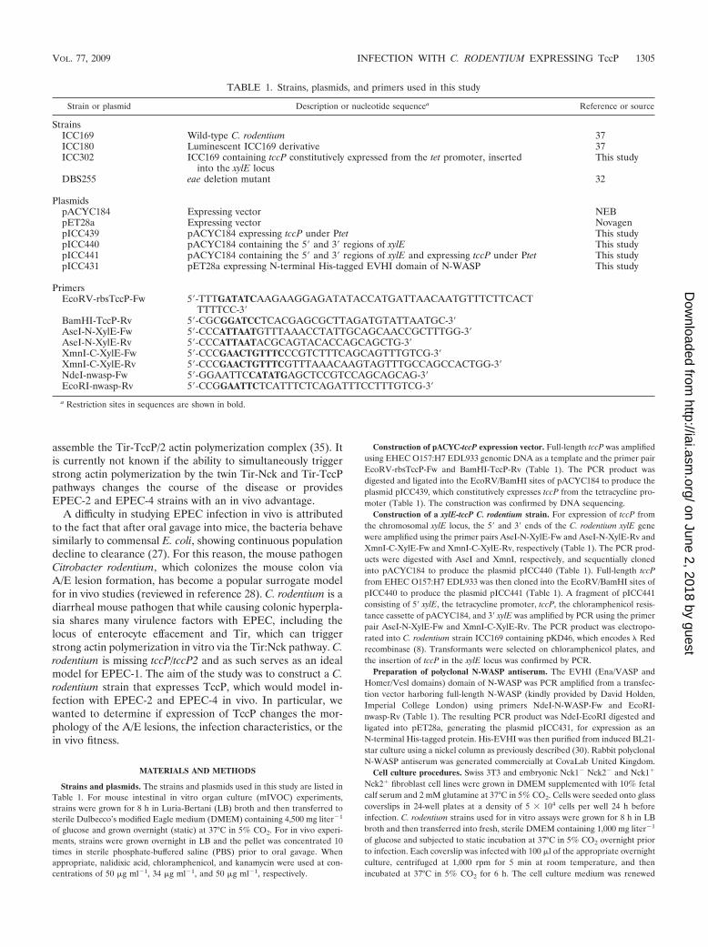
TABLE 1. Strains, plasmids, and primers used in this study
Strain or plasmid Description or nucleotide sequencea Reference or source
StrainsICC169 Wild-type C. rodentium 37ICC180 Luminescent ICC169 derivative 37ICC302 ICC169 containing tccP constitutively expressed from the tet promoter, inserted
into the xylE locusThis study
DBS255 eae deletion mutant 32
PlasmidspACYC184 Expressing vector NEBpET28a Expressing vector NovagenpICC439 pACYC184 expressing tccP under Ptet This studypICC440 pACYC184 containing the 5� and 3� regions of xylE This studypICC441 pACYC184 containing the 5� and 3� regions of xylE and expressing tccP under Ptet This studypICC431 pET28a expressing N-terminal His-tagged EVHI domain of N-WASP This study
PrimersEcoRV-rbsTccP-Fw 5�-TTTGATATCAAGAAGGAGATATACCATGATTAACAATGTTTCTTCACT
TTTTCC-3�BamHI-TccP-Rv 5�-CGCGGATCCTCACGAGCGCTTAGATGTATTAATGC-3�AseI-N-XylE-Fw 5�-CCCATTAATGTTTAAACCTATTGCAGCAACCGCTTTGG-3�AseI-N-XylE-Rv 5�-CCCATTAATACGCAGTACACCAGCAGCTG-3�XmnI-C-XylE-Fw 5�-CCCGAACTGTTTCCCGTCTTTCAGCAGTTTGTCG-3�XmnI-C-XylE-Rv 5�-CCCGAACTGTTTCGTTTAAACAAGTAGTTTGCCAGCCACTGG-3�NdeI-nwasp-Fw 5�-GGAATTCCATATGAGCTCCGTCCAGCAGCAG-3�EcoRI-nwasp-Rv 5�-CCGGAATTCTCATTTCTCAGATTTCCTTTGTCG-3�
a Restriction sites in sequences are shown in bold.
VOL. 77, 2009 INFECTION WITH C. RODENTIUM EXPRESSING TccP 1305
on June 2, 2018 by guesthttp://iai.asm
.org/D
ownloaded from
halfway through the infection period. Immunostaining of the coverslips was doneas described previously (35).
Mouse in vitro organ cultures. A mIVOC model was developed using condi-tions similar to those used for bovine intestinal organ culture (14). Briefly, sixmice were used in four independent experiments. Fresh segments from theterminal ileum and colon were inoculated with 50 �l of the appropriate overnightbacterial culture, corresponding to approximately 107 CFU, and incubated at37°C in a 5%-CO2 atmosphere on a seesaw rocker (18 cycles min�1) for 8 h.Uninfected explants were cultured in each experiment as controls.
Explants were gently rinsed with PBS and fixed in 10% buffered formalin formicroscopic examination. Formalin-fixed tissues were processed, paraffin embed-ded, sectioned at 5 �m, and stained with hematoxylin and eosin (H&E) accord-ing to standard techniques. Sections were then examined by light microscopy,and the mucosal epithelium located between adjacent crypts (designated theintercrypt mucosal epithelium [ICME]) was examined for the presence of inti-mately adherent bacteria, as described previously (16). The mean percentage ofICME demonstrating intimately adherent bacteria per section was calculated.Additional explants were fixed in 2.5% glutaraldehyde for electron microscopyanalysis.
Oral inoculation of mice. Pathogen-free female C57BL/6 mice (18 to 20 g)were purchased from Charles River, Inc. All animals were housed in individuallyHEPA-filtered cages with sterile bedding and free access to sterilized food andwater. All animal experiments were performed in accordance with the AnimalsScientific Procedures (Act 1986) and were approved by the local Ethical ReviewCommittee. For in vivo experiments, independent single-infection experimentswere performed twice using four to eight mice per group; wild-type C. rodentium-infected and uninfected mice were included alongside every experiment as con-trols.
Mice were inoculated by oral gavage with 200 ml overnight LB-grown C.rodentium suspension in PBS (�5 � 109 CFU). The number of viable bacteriaused as an inoculum was determined by retrospective plating onto LB agarcontaining antibiotics. Stool samples were recovered aseptically at various timepoints after inoculation, and the number of viable bacteria per gram of stool wasdetermined after homogenization in PBS and plating on LB agar containingantibiotics (38).
In mixed-infection experiments, the two overnight LB-grown bacterial cultureswere combined in a ratio of 1:1 (approximately 2 � 109 CFU for each strain) in200 �l PBS and used to inoculate female C57BL/6 mice by oral gavage. Dilutionsof the inoculum were plated on respective antibiotic-containing plates to deter-mine the ratio of the two bacterial strains (test strain/reference strain) in theinoculum. Stool samples were collected at regular intervals, and the competitiveindex (CI) was calculated by dividing the ratio of test strain CFU and referencestrain CFU from the stools by the ratio of test strain CFU to reference strainCFU in the inoculum (29). The CI was analyzed using five animals per group andwas determined at days 7, 9, 11, and 15 postinoculum.
Histopathology and indirect immunofluorescence. Tissues were harvested at 8and 17 days postinoculation. Segments of the terminal colon, collected postmor-tem, were rinsed of their content, fixed in 10% buffered formalin, processed,paraffin embedded, sectioned at 5 �m, and stained with H&E according tostandard techniques. Sections were examined by light microscopy for the pres-ence of intimately adhering bacteria on intestinal cells. Crypt length, as a mea-sure of crypt hyperplasia, was evaluated from at least six well-oriented crypts.Additional segments were fixed in 2.5% glutaraldehyde or embedded in optimal-cutting-temperature medium (Raymond A Lamb Limited, United Kingdom),snap-frozen in liquid nitrogen, and kept at �80°C for further electron micros-copy analysis and cryosectioning, respectively.
Indirect immunofluorescence (IFA) using formalin-fixed and paraffin-embed-ded sections was used for detection of O152-positive bacteria (corresponding tothe C. rodentium serogroup), as described previously (14, 15). Cryosections (twoserial sections per strain per mouse per experiment), fixed in 3% paraformalde-hyde in PBS, were used for detection of C. rodentium, TccP, Nck and N-WASP.Sections were examined with an Axio Imager M1 microscope (Carl Zeiss Mi-croImaging GmbH); images were acquired using an AxioCam MRm mono-chrome camera and computer processed using the AxioVision (Carl Zeiss Mi-croImaging GmbH) and Photoshop 5.0 and Illustrator 8.0 (Adobe SystemsIncorporated) software programs.
Antibodies and reagents. Sections were immunostained using the followingantibodies: rabbit anti-O152 (kindly provided by Lothar Beutin, The NationalReference Laboratory for Escherichia coli, Federal Institute for Risk Assess-ment, Berlin, Germany), chicken anti-intimin (kindly provided by John M. Fair-brother, Escherichia coli Laboratory, Faculte de Medecine Veterinaire, Univer-site de Montreal, Montreal, Canada), rabbit anti-TccP (31), rabbit anti-N-WASP(this study), and rabbit anti-Nck (Millipore Upstate, Lake Placid, NY). Cy2-
conjugated donkey antichicken and tetramethyl rhodamine isocyanate-conju-gated donkey antirabbit (Jackson ImmunoResearch Europe Ltd., Soham, Cam-bridgeshire, United Kingdom) were used as secondary antibodies. Phalloidin-Alexa Fluor 633 (Invitrogen, United Kingdom) was used to stain F-actin, whileDNA was counterstained with Hoechst 33342.
Electron microscopy. Ex vivo- and in vivo-derived tissue samples were pro-cessed for electron microscopy, as described previously (14). Samples for scan-ning electron microscopy (SEM) were examined blindly at an accelerating volt-age of 25 kV using a Jeol JSM-5300 scanning electron microscope. Samples fortransmission electron microscopy (TEM) were observed using a Phillips 201transmission electron microscope at an accelerating voltage of 60 kV.
Statistical analysis. Results are presented as a scatter plot with the median(ICME with adherent bacteria and CI), as a line plot with the mean and itsstandard deviation (colonization), or as a vertical bar chart with the mean and itsstandard deviation (crypt length). Fisher’s exact test (number of explants withadherent bacteria) or a Mann-Whitney test (median percentage of ICME withadherent bacteria, colonization, crypt length, and CI) was performed using com-mercially available GraphPad InStat v3.06 software (GraphPad Software Inc.,San Diego, CA). A P value of �0.05 was considered significant.
RESULTS
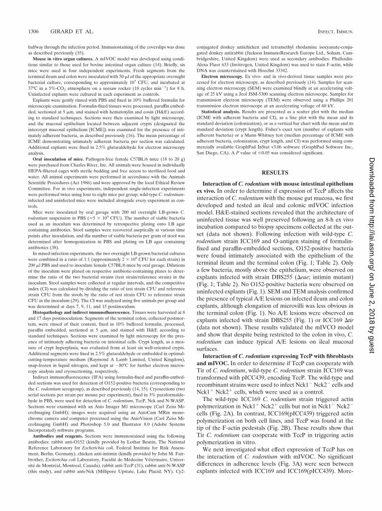
Interaction of C. rodentium with mouse intestinal epitheliumex vivo. In order to determine if expression of TccP affects theinteraction of C. rodentium with the mouse gut mucosa, we firstdeveloped and tested an ileal and colonic mIVOC infectionmodel. H&E-stained sections revealed that the architecture ofuninfected tissue was well preserved following an 8-h ex vivoincubation compared to biopsy specimens collected at the out-set (data not shown). Following infection with wild-type C.rodentium strain ICC169 and O-antigen staining of formalin-fixed and paraffin-embedded sections, O152-positive bacteriawere found intimately associated with the epithelium of theterminal ileum and the terminal colon (Fig. 1; Table 2). Onlya few bacteria, mostly above the epithelium, were observed onexplants infected with strain DBS255 (�eae; intimin mutant)(Fig. 1; Table 2). No O152-positive bacteria were observed onuninfected explants (Fig. 1). SEM and TEM analysis confirmedthe presence of typical A/E lesions on infected ileum and colonexplants, although elongation of microvilli was less obvious inthe terminal colon (Fig. 1). No A/E lesions were observed onexplants infected with strain DBS255 (Fig. 1) or ICC169 �tir(data not shown). These results validated the mIVCO modeland show that despite being restricted to the colon in vivo, C.rodentium can induce typical A/E lesions on ileal mucosalsurfaces.
Interaction of C. rodentium expressing TccP with fibroblastsand mIVOC. In order to determine if TccP can cooperate withTir of C. rodentium, wild-type C. rodentium strain ICC169 wastransformed with pICC439, encoding TccP. The wild-type andrecombinant strains were used to infect Nck1� Nck2� cells andNck1� Nck2� cells, which were used as a control.
The wild-type ICC169 C. rodentium strain triggered actinpolymerization in Nck1� Nck2� cells but not in Nck1� Nck2�
cells (Fig. 2A). In contrast, ICC169(pICC439) triggered actinpolymerization on both cell lines, and TccP was found at thetip of the F-actin pedestals (Fig. 2B). These results show thatTir C. rodentium can cooperate with TccP in triggering actinpolymerization in vitro.
We next investigated what effect expression of TccP has onthe interaction of C. rodentium with mIVOC. No significantdifferences in adherence levels (Fig. 3A) were seen betweenexplants infected with ICC169 and ICC169(pICC439). More-
1306 GIRARD ET AL. INFECT. IMMUN.
on June 2, 2018 by guesthttp://iai.asm
.org/D
ownloaded from
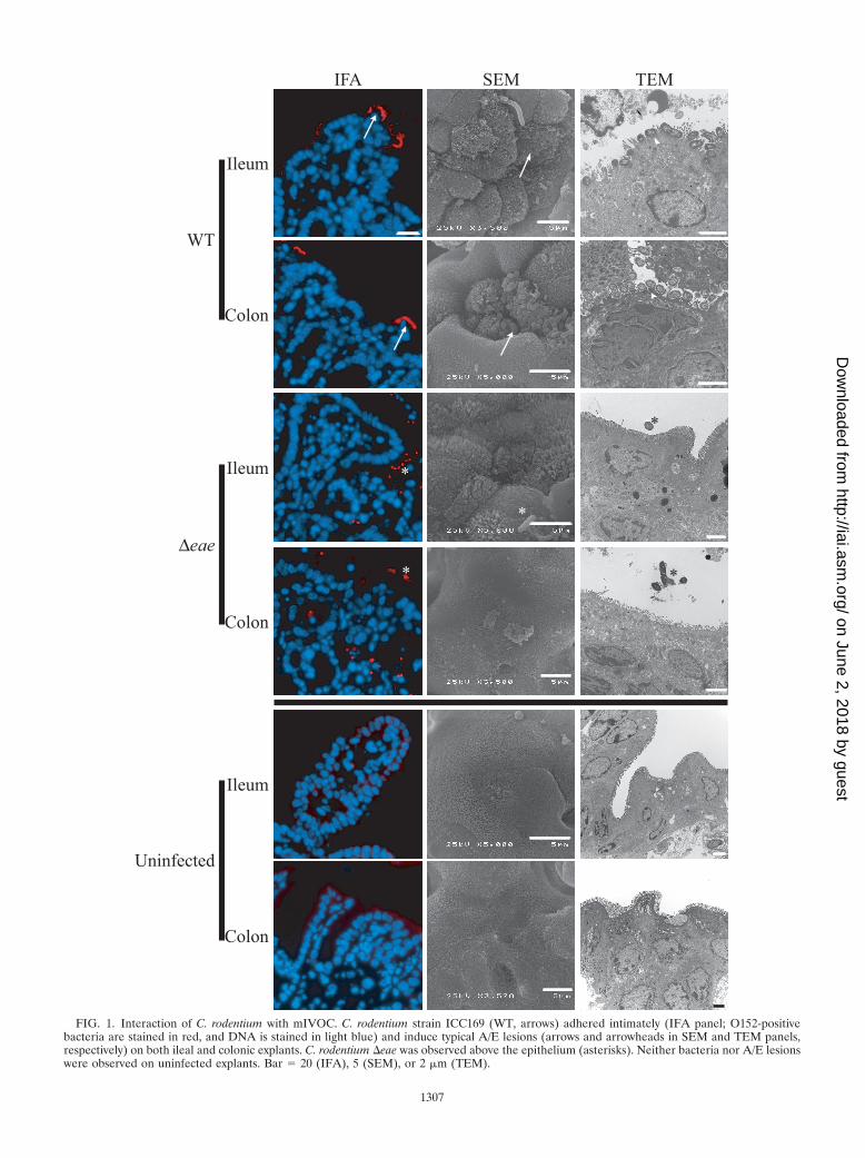
FIG. 1. Interaction of C. rodentium with mIVOC. C. rodentium strain ICC169 (WT, arrows) adhered intimately (IFA panel; O152-positivebacteria are stained in red, and DNA is stained in light blue) and induce typical A/E lesions (arrows and arrowheads in SEM and TEM panels,respectively) on both ileal and colonic explants. C. rodentium �eae was observed above the epithelium (asterisks). Neither bacteria nor A/E lesionswere observed on uninfected explants. Bar 20 (IFA), 5 (SEM), or 2 �m (TEM).
1307
on June 2, 2018 by guesthttp://iai.asm
.org/D
ownloaded from
over, ICC169(pICC439) adhered intimately to, and inducedtypical A/E lesions on, ileal and colonic mIVOC (Fig. 3B).
Role of TccP during mouse infection. Finally, we aimed todetermine if expression of TccP changes the course of C. ro-dentium infection in vivo. However, preliminary studiesshowed that pICC439 is unstable in vivo since it was lost fromICC169 as early as 3 days post-oral gavage into C57BL mice(data not shown).
In order to overcome the problem of plasmid instability invivo, we constructed C. rodentium strain ICC302, which ex-presses tccP constitutively from the tet promoter at the chro-mosomal xylE locus; we selected this site since we have previ-ously shown that disruption of xylE by insertion of theluxCDABE operon and a kanamycin resistance cassette (C.rodentium strain ICC180) did not affect the overall growth andvirulence capacity of ICC169 (37).
In vitro characterization showed that expression of TccP inICC302 enabled the strain to trigger strong actin polymeriza-
tion in both Nck1� Nck2� and Nck1� Nck2� cells (Fig. 2C).When tested ex vivo on mIVOC, ICC302 adhered intimately toileal and colonic mIVOC (Fig. 3C) and induced typical A/Elesions (Fig. 3D).
Since constitutive expression of tccP did not affect the abilityof ICC302 to infect mammalian cells in vitro and mucosalsurfaces ex vivo, the recombinant strain was tested in vivo.C57BL/6 mice were challenged by oral gavage with ICC302expressing TccP; wild-type C. rodentium strain ICC169 wasused as a reference. In comparison to results for wild-type C.rodentium, expression of TccP did not affect the colonizationdynamics, as reflected by the number of CFU per gram ofstools recovered over time (Fig. 4A).
Histopathological examination revealed that the constitutiveexpression of tccP did not influence colonic hyperplasia (asreflected by the crypt length) (Fig. 4B and C) compared toresults for wild-type C. rodentium. Colonic sections fromICC169 and ICC302 challenge showed similar levels of inti-mately adherent O152-positive bacteria at the apical surface ofthe mucosal epithelium (Fig. 4C).
Recruitment of TccP, Nck, and N-WASP and A/E lesionformation in vivo. Examination of infected cryosections re-vealed that C. rodentium strain ICC302 was able to recruitTccP, Nck, and N-WASP underneath intimately adherent bac-teria while Nck and N-WASP, in the absence of TccP, weredetected under adherent ICC169 (Fig. 5). This suggests that invivo C. rodentium can recruit both TccP and Nck, although inthe absence of double staining it is not possible to assess if
FIG. 2. Interaction of C. rodentium with Nck1� Nck2� (Nck�/� panel) or Nck1� Nck2� (Nck�/� panel) embryonic fibroblasts. Wild-type C.rodentium strain ICC169 (A) adhered to both cell lines (arrowheads) but was able to trigger actin polymerization only in the Nck�/� cells(arrowheads). Expression of tccP from pICC439 in C. rodentium ICC169 (B) or from the xylE locus in ICC302 (C) enabled the strains to triggerstrong actin polymerization in both Nck�/� and Nck�/� cells (arrows). TccP (asterisks) was detected in both strains at the tip of the actin-richpedestals. DNA was stained blue, TccP was stained red, and actin was stained green. Bar 2 �m.
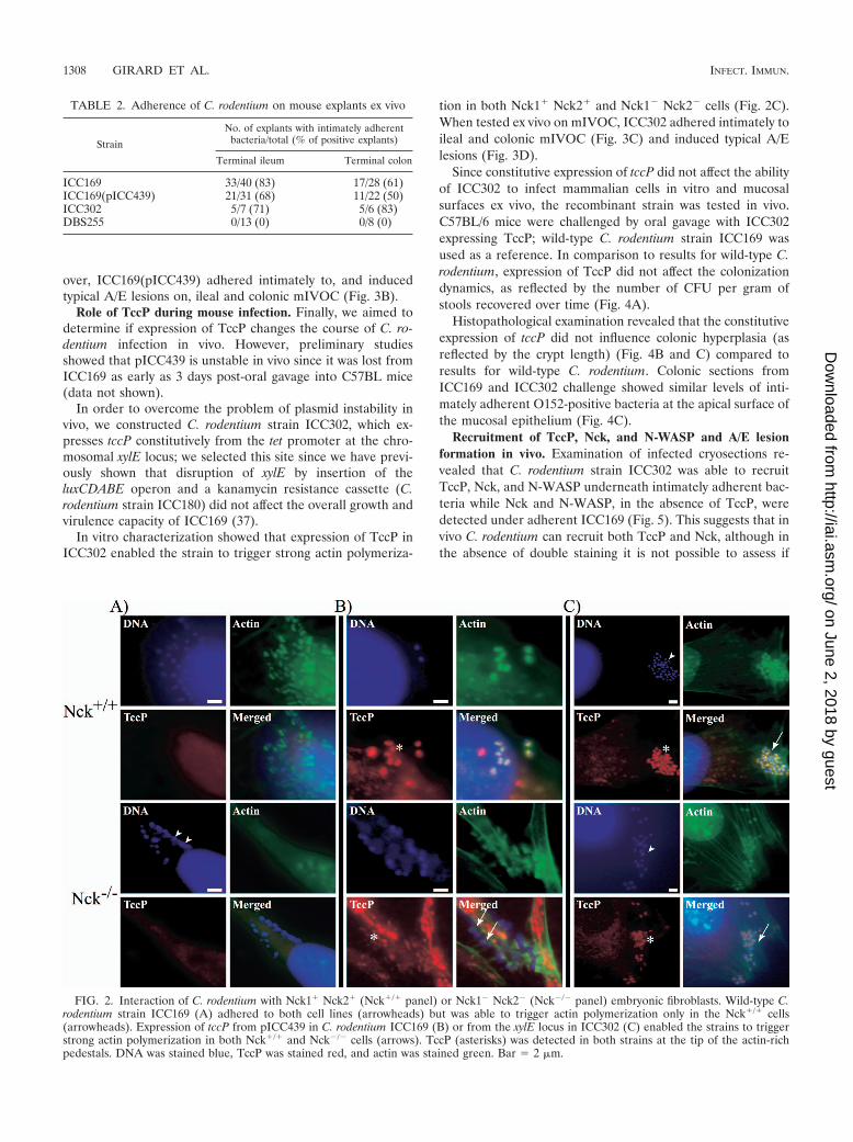
TABLE 2. Adherence of C. rodentium on mouse explants ex vivo
Strain
No. of explants with intimately adherentbacteria/total (% of positive explants)
Terminal ileum Terminal colon
ICC169 33/40 (83) 17/28 (61)ICC169(pICC439) 21/31 (68) 11/22 (50)ICC302 5/7 (71) 5/6 (83)DBS255 0/13 (0) 0/8 (0)
1308 GIRARD ET AL. INFECT. IMMUN.
on June 2, 2018 by guesthttp://iai.asm
.org/D
ownloaded from

TccP and Nck are colocalized. SEM (Fig. 6A) and TEM (Fig.6B) examination revealed that C. rodentium strains ICC169and ICC302 exhibited indistinguishable colonization patternsand A/E lesions.
Expression of TccP does not change in vivo fitness ofICC302. Finally, we determined if expression of TccP providesthe bacterium with a competitive advantage. To this end wedetermined the competitive index of ICC302 during mixed
FIG. 3. Interaction of C. rodentium expressing TccP with mIVOC. (A) In comparison to results for wild-type (WT columns) C. rodentium,expression of tccP in ICC169 in trans (ptccP columns) did not significantly influence the median percentage of intercrypt mucosal epithelium withadherent bacteria (ICME�) on either ileal or colonic explants ex vivo. (B) Intimately adherent, O152-positive C. rodentium ICC169(pICC439)(DNA was stained in light blue, and bacteria were stained red; arrows) were observed on both ileal and colonic explants; typical A/E lesions wereconfirmed by SEM (arrows) and TEM (arrowheads). (C) Adhesion of C. rodentium ICC302 constitutively expressing TccP from the xylE locus tothe ileal and colonic mIVOC (DNA was stained in light blue, and bacteria were stained red; arrows). (D) C. rodentium ICC302 triggers typical A/Elesions on ileal and colonic mIVOC. Bar 20 (IFA), 5 (SEM), or 1 �m (TEM) (bar in inset 500 nm).
VOL. 77, 2009 INFECTION WITH C. RODENTIUM EXPRESSING TccP 1309
on June 2, 2018 by guesthttp://iai.asm
.org/D
ownloaded from

FIG. 4. Colonization dynamic, hyperplasia, and CI of C. rodentium ICC302. ICC302 (xylE-tccP) exhibits colonization dynamics (A) and crypthyperplasia (B and C) similar to those of wild-type C. rodentium, which was significantly different from results for uninfected mice; ���, P 0.001.Both strains intimately adhered to the colonic mucosa (C) (O152-positive bacteria are stained in red, and DNA is stained in light blue). Noadherent bacteria were observed on colonic sections derived from uninfected mice (Uninfected). Bar 100 (bar in inset 20) (C). (D) The invivo fitness of ICC302 was investigated in mixed-infection experiments with the ICC169 (WT) strain. The CI was determined on days 7, 9, 11, and15 postinoculation for each of the five mice per group, and individual CI values (open circles) are represented as log10 CI values. The median CIof each group is indicated on the graph by a horizontal line, with the corresponding numerical values shown beneath the line. No statisticaldifferences were observed between the CI of ICC302 and the CI of ICC180, used as a control, indicating that none of the strains had a competitiveadvantage compared to ICC169.
1310 GIRARD ET AL. INFECT. IMMUN.
on June 2, 2018 by guesthttp://iai.asm
.org/D
ownloaded from
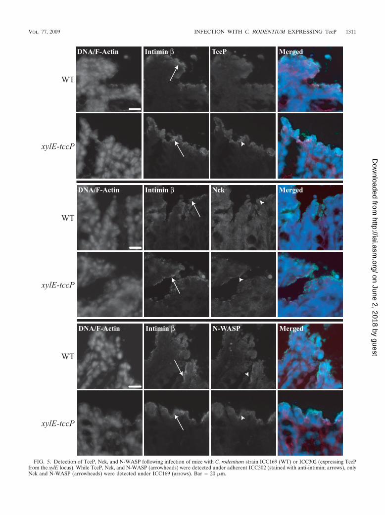
FIG. 5. Detection of TccP, Nck, and N-WASP following infection of mice with C. rodentium strain ICC169 (WT) or ICC302 (expressing TccPfrom the xylE locus). While TccP, Nck, and N-WASP (arrowheads) were detected under adherent ICC302 (stained with anti-intimin; arrows), onlyNck and N-WASP (arrowheads) were detected under ICC169 (arrows). Bar 20 �m.
VOL. 77, 2009 INFECTION WITH C. RODENTIUM EXPRESSING TccP 1311
on June 2, 2018 by guesthttp://iai.asm
.org/D
ownloaded from

infection with ICC169. In order to neutralize any potentialeffects of a chromosomal insertion into the xylE locus on invivo fitness, the CI of ICC302 was directly compared to the CIof ICC180, which harbors a chromosomal insertion in the samelocus (37). This showed that expression of TccP did not affectthe in vivo fitness of ICC302 in comparison to that of ICC180at any time point after mixed infection (Fig. 4D). This resultsuggests that EPEC-1 (modeled by ICC169) and EPEC-2 andEPEC-4 (modeled by ICC302) are as infectious and that theability to trigger strong actin polymerization by twin mecha-nisms in vitro does not increase the virulence potential orfitness in vivo.
DISCUSSION
The ability to polymerize actin is considered central toEPEC infection. While EPEC-1 strains can only assemble theTir-Nck actin signaling complex, EPEC-2 and EPEC-4 strainsare equipped with the twin Tir-Nck and Tir-TccP mechanisms(reviewed in reference 9). However, it was not known if theability to assemble more than one actin polymerization com-plex increases virulence or fitness.
In this study we compared the infectious properties of wild-type C. rodentium (ICC169), which can trigger strong actinpolymerization in vitro exclusively by the Tir-Nck pathway,with a C. rodentium strain that can additionally express TccP(ICC302). First, we demonstrated that TccP can be translo-cated from C. rodentium and cooperate with its Tir proteinduring infection of Nck-deficient cells. In order to determine if
expression of TccP affects the ability of ICC169 to interact withmucosal surfaces, we developed an mIVOC infection model.As far as we know, this is the first time such a model has beendescribed; we believe the mIVOC model provides an impor-tant tool for studying enteric bacterial infection; moreover, itwould contribute to the reduction, replacement, and refine-ment (three Rs) of animal experimentation. Interestingly, al-though following inoculation by the oral route C. rodentium isfound attached only to the cecum and colon, ex vivo C. roden-tium can efficiently induce A/E lesions on the ileum. Impor-tantly, in comparison with infection of mIVOC with ICC169,expressing TccP from a plasmid did not enhance the ability ofC. rodentium to attach and induce A/E lesions on either theileal or the colonic mucosa.
We next aimed to determine if expression of TccP influencescolonization dynamics, pathology, recruitment of signalingmolecules to the site of bacterial attachment, and competitive-ness in vivo. Since the tccP-encoding plasmid was unstable invivo, we constructed a recombinant C. rodentium strain thatconstitutively expresses tccP from the tet promoter. We choseto express TccP from the xylE locus, since we have shownbefore that it can tolerate gene insertions without virulencebeing affected (37). Comparison of infection profiles ofICC169 and ICC302 has shown that both strains exhibited thesame colonization dynamics; infection with both strains peakedat 7 days postchallenge and started to clear 15 days later.Expression of TccP did not exacerbate the overall disease/pathology since similar levels of colonic hyperplasia were seen
FIG. 6. Typical A/E lesions were observed by SEM (arrows) and TEM (arrowheads) following infection with C. rodentium ICC169 and ICC302.Neither adherent bacteria nor A/E lesions were observed on colonic sections derived from uninfected mice (Uninfected). Bar 5 �m (A) or 2�m (bar in insets 500 nm) (B).
1312 GIRARD ET AL. INFECT. IMMUN.
on June 2, 2018 by guesthttp://iai.asm
.org/D
ownloaded from

following infection with ICC302 and the control ICC169 C.rodentium strains.
Immunofluorescence analysis of frozen sections derivedfrom infected colons has shown that TccP is accumulated un-der adherent ICC302; no signal was seen under the wild-typeICC169 control. This shows that TccP is translocated from C.rodentium in vivo and recruited to Tir. Importantly, TccP doesnot bind Tir directly but via an adaptor protein encoded by thehost (3, 13). Our results imply that the adaptor linking Tir andTccP is also expressed in the murine gut. Importantly, quali-tative data suggest that TccP competitively excludes Nck, sincefewer Nck-positive bacterial foci were seen under adherentICC302 (data not shown). This suggests that although in vitroNck and TccP are seen together under adherent bacteria (35),in vivo either Nck or TccP appears to be dominant. It is notcurrently known if the difference between the in vivo and invitro data is due to Tir density or to special constraints at thesite of bacterial attachment. Nonetheless, both Nck and TccPare capable of efficient recruitment of N-WASP. Finally, weinvestigated if the ability to activate N-WASP by both Nck andTccP confers a competitive advantage in vivo. Comparison ofthe competitive indexes of ICC302 and ICC180 in competitionwith ICC169 has shown that expression of TccP does not in-crease in vivo competitiveness.
Infections with C. rodentium strains ICC169 and ICC302model those of EPEC-1 and of EPEC-2 and EPEC-4, respec-tively. Our data show that although EPEC-2 and EPEC-4 canassemble two actin-signaling complexes (compared to one inEPEC-1), this did not have a measurable impact during infec-tion in vivo. These finds are consistent with epidemiologicaland clinical data, which did not reveal any significant differ-ences between EPEC-1 and EPEC-2. However, the fact thatthe ability to polymerize actin by the Tir-Nck and Tir-TccPactin polymerization pathways is conserved in EPEC-2 andEPEC-4 suggests that they are under selective pressure. Atpresent it is not known if the selection is at the level of thehuman host, the animal reservoir, or the environment.
ACKNOWLEDGMENTS
We thank Anthony J. FitzGerald for technical help and Alan Phillips(UCL) for making his SEM available for this project. We thank LotharBeutin from the Nationales Referenzlabor fur Escherichia coli(NRL-E. coli) in Berlin for the O152 antiserum.
Francis Girard was supported by a postdoctoral fellowship from theNatural Sciences and Engineering Research Council (NSERC) ofCanada and by a Canada-United Kingdom Millennium ResearchAward, awarded by the NSERC and the Royal Society of London,United Kingdom. This work was supported by the BBSRC and theWellcome Trust.
REFERENCES
1. Allen-Vercoe, E., B. Waddell, M. C. Toh, and R. DeVinney. 2006. Amino acidresidues within enterohemorrhagic Escherichia coli O157:H7 Tir involved inphosphorylation, alpha-actinin recruitment, and Nck-independent pedestalformation. Infect. Immun. 74:6196–6205.
2. Brady, M. J., K. G. Campellone, M. Ghildiyal, and J. M. Leong. 2007.Enterohaemorrhagic and enteropathogenic Escherichia coli Tir proteinstrigger a common Nck-independent actin assembly pathway. Cell. Microbiol.9:2242–2253.
3. Campellone, K., D. Robbins, and J. Leong. 2004. EspF(U) is a translocatedEHEC effector that interacts with Tir and N-WASP and promotes Nck-independent actin assembly. Dev. Cell 7:217–228.
4. Campellone, K. G., M. J. Brady, J. G. Alamares, D. C. Rowe, B. M. Skehan,D. J. Tipper, and J. M. Leong. 2006. Enterohaemorrhagic Escherichia coliTir requires a C-terminal 12-residue peptide to initiate EspFu-mediated
actin assembly and harbours N-terminal sequences that influence pedestallength. Cell. Microbiol. 8:1488–1503.
5. Campellone, K. G., A. Giese, D. J. Tipper, and J. M. Leong. 2002. A tyrosine-phosphorylated 12-amino-acid sequence of enteropathogenic Escherichiacoli Tir binds the host adaptor protein Nck and is required for Nck local-ization to actin pedestals. Mol. Microbiol. 43:1227–1241.
6. Campellone, K. G., and J. M. Leong. 2005. Nck-independent actin assemblyis mediated by two phosphorylated tyrosines within enteropathogenic Esch-erichia coli Tir. Mol. Microbiol. 56:416–432.
7. Caron, E., V. F. Crepin, N. Simpson, S. Knutton, J. Garmendia, and G.Frankel. 2006. Subversion of actin dynamics by EPEC and EHEC. Curr.Opin. Microbiol. 9:40–45.
8. Datsenko, K. A., and B. L. Wanner. 2000. One-step inactivation of chromo-somal genes in Escherichia coli K-12 using PCR products. Proc. Natl. Acad.Sci. USA 97:6640–6645.
9. Frankel, G., and A. D. Phillips. 2008. Attaching effacing Escherichia coli andparadigms of Tir-triggered actin polymerization: getting off the pedestal.Cell. Microbiol. 10:549–556.
10. Frankel, G., A. D. Phillips, I. Rosenshine, G. Dougan, J. B. Kaper, and S.Knutton. 1998. Enteropathogenic and enterohemorrhagic Escherichia coli:more subversive elements. Mol. Microbiol. 30:911–921.
11. Frankel, G., A. D. Phillips, L. R. Trabulsi, S. Knutton, G. Dougan, and S.Matthews. 2001. Intimin and the host cell—is it bound to end in Tir(s)?Trends Microbiol. 9:214–218.
12. Garmendia, J., G. Frankel, and V. F. Crepin. 2005. Enteropathogenic andenterohemorrhagic Escherichia coli infections: translocation, translocation,translocation. Infect. Immun. 73:2573–2585.
13. Garmendia, J., A. D. Phillips, M. F. Carlier, Y. Chong, S. Schuller, O.Marches, S. Dahan, E. Oswald, R. K. Shaw, S. Knutton, and G. Frankel.2004. TccP is an enterohaemorrhagic Escherichia coli O157:H7 type IIIeffector protein that couples Tir to the actin-cytoskeleton. Cell. Microbiol.6:1167–1183.
14. Girard, F., F. Dziva, P. van Diemen, A. D. Phillips, M. P. Stevens, and G.Frankel. 2007. Adherence of enterohemorrhagic Escherichia coli O157, O26,and O111 strains to bovine intestinal explants ex vivo. Appl. Environ. Mi-crobiol. 73:3084–3090.
15. Girard, F., G. Frankel, A. D. Phillips, W. Cooley, U. Weyer, A. H. Dugdale,M. J. Woodward, and R. M. La Ragione. 2008. Interaction of enterohemor-rhagic Escherichia coli O157:H7 with mouse intestinal mucosa. FEMS Mi-crobiol. Lett. 283:196–202.
16. Girard, F., I. P. Oswald, I. Taranu, P. Helie, G. D. Appleyard, J. Harel, andJ. M. Fairbrother. 2005. Host immune status influences the development ofattaching and effacing lesions in weaned pigs. Infect. Immun. 73:5514–5523.
17. Gruenheid, S., R. DeVinney, F. Bladt, D. Goosney, S. Gelkop, G. D. Gish, T.Pawson, and B. B. Finlay. 2001. Enteropathogenic E. coli Tir binds Nck toinitiate actin pedestal formation in host cells. Nat. Cell Biol. 3:856–859.
18. Hartland, E. L., M. Batchelor, R. M. Delahay, C. Hale, S. Matthews, G.Dougan, S. Knutton, I. Connerton, and G. Frankel. 1999. Binding of intiminfrom enteropathogenic Escherichia coli to Tir and to host cells. Mol. Micro-biol. 32:151–158.
19. Jarvis, K. G., and J. B. Kaper. 1996. Secretion of extracellular proteins byenterohemorrhagic Escherichia coli via a putative type III secretion system.Infect. Immun. 64:4826–4829.
20. Jerse, A. E., J. Yu, B. D. Tall, and J. B. Kaper. 1990. A genetic locus ofenteropathogenic Escherichia coli necessary for the production of attachingand effacing lesions on tissue culture cells. Proc. Natl. Acad. Sci. USA87:7839–7843.
21. Kaper, J. B., J. P. Nataro, and H. L. Mobley. 2004. Pathogenic Escherichiacoli. Nat. Rev. Microbiol. 2:123–140.
22. Kenny, B. 1999. Phosphorylation of tyrosine 474 of the enteropathogenicEscherichia coli (EPEC) Tir receptor molecule is essential for actin nucle-ating activity and is preceded by additional host modifications. Mol. Micro-biol. 31:1229–1241.
23. Kenny, B., R. DeVinney, M. Stein, D. J. Reinscheid, E. A. Frey, and B. B.Finlay. 1997. Enteropathogenic E. coli (EPEC) transfers its receptor forintimate adherence into mammalian cells. Cell 91:511–520.
24. Knutton, S., D. R. Lloyd, and A. S. McNeish. 1987. Adhesion of entero-pathogenic Escherichia coli to human intestinal enterocytes and culturedhuman intestinal mucosa. Infect. Immun. 55:69–77.
25. Lacher, D. W., H. Steinsland, T. E. Blank, M. S. Donnenberg, and T. S.Whittam. 2007. Molecular evolution of typical enteropathogenic Escherichiacoli: clonal analysis by multilocus sequence typing and virulence gene allelicprofiling. J. Bacteriol. 189:342–350.
26. McDaniel, T. K., K. G. Jarvis, M. S. Donnenberg, and J. B. Kaper. 1995. Agenetic locus of enterocyte effacement conserved among diverse enterobac-terial pathogens. Proc. Natl. Acad. Sci. USA 92:1664–1668.
27. Mundy, R., F. Girard, A. J. FitzGerald, and G. Frankel. 2006. Comparisonof colonization dynamics and pathology of mice infected with enteropatho-genic Escherichia coli, enterohaemorrhagic E. coli and Citrobacter roden-tium. FEMS Microbiol. Lett. 265:126–132.
28. Mundy, R., T. T. MacDonald, G. Dougan, G. Frankel, and S. Wiles. 2005.Citrobacter rodentium of mice and man. Cell. Microbiol. 7:1697–1706.
VOL. 77, 2009 INFECTION WITH C. RODENTIUM EXPRESSING TccP 1313
on June 2, 2018 by guesthttp://iai.asm
.org/D
ownloaded from

29. Mundy, R., D. Pickard, R. K. Wilson, C. P. Simmons, G. Dougan, and G.Frankel. 2003. Identification of a novel type IV pilus gene cluster requiredfor gastrointestinal colonization of Citrobacter rodentium. Mol. Microbiol.48:795–809.
30. Neves, B. C., R. K. Shaw, G. Frankel, and S. Knutton. 2003. Polymorphismswithin EspA filaments of enteropathogenic and enterohemorrhagic Esche-richia coli. Infect. Immun. 71:2262–2265.
31. Ogura, Y., T. Ooka, A. Whale, J. Garmendia, L. Beutin, S. Tennant, G.Krause, S. Morabito, I. Chinen, T. Tobe, H. Abe, R. Tozzoli, A. Caprioli, M.Rivas, R. Robins-Browne, T. Hayashi, and G. Frankel. 2007. TccP2 ofO157:H7 and non-O157 enterohemorrhagic Escherichia coli (EHEC): chal-lenging the dogma of EHEC-induced actin polymerization. Infect. Immun.75:604–612.
32. Schauer, D. B., and S. Falkow. 1993. The eae gene of Citrobacter freundiibiotype 4280 is necessary for colonization in transmissible murine colonichyperplasia. Infect. Immun. 61:4654–4661.
33. Schuller, S., Y. Chong, J. Lewin, B. Kenny, G. Frankel, and A. D. Phillips.2007. Tir phosphorylation and Nck/N-WASP recruitment by enteropatho-genic and enterohaemorrhagic Escherichia coli during ex vivo colonization ofhuman intestinal mucosa is different to cell culture models. Cell. Microbiol.9:1352–1364.
34. Touze, T., R. D. Hayward, J. Eswaran, J. M. Leong, and V. Koronakis. 2004.Self-association of EPEC intimin mediated by the beta-barrel-containinganchor domain: a role in clustering of the Tir receptor. Mol. Microbiol.51:73–87.
35. Whale, A. D., J. Garmendia, T. A. Gomes, and G. Frankel. 2006. A novelcategory of enteropathogenic Escherichia coli simultaneously utilizes theNck and TccP pathways to induce actin remodelling. Cell. Microbiol. 8:999–1008.
36. Whale, A. D., R. T. Hernandes, T. Ooka, L. Beutin, S. Schuller, J. Garmen-dia, L. Crowther, M. A. Vieira, Y. Ogura, G. Krause, A. D. Phillips, T. A.Gomes, T. Hayashi, and G. Frankel. 2007. TccP2-mediated subversion ofactin dynamics by EPEC 2—a distinct evolutionary lineage of enteropatho-genic Escherichia coli. Microbiology 153:1743–1755.
37. Wiles, S., S. Clare, J. Harker, A. Huett, D. Young, G. Dougan, and G.Frankel. 2004. Organ specificity, colonization and clearance dynamics in vivofollowing oral challenges with the murine pathogen Citrobacter rodentium.Cell. Microbiol. 6:963–972.
38. Wiles, S., G. Dougan, and G. Frankel. 2005. Emergence of a ‘hyperinfec-tious’ bacterial state after passage of Citrobacter rodentium through the hostgastrointestinal tract. Cell. Microbiol. 7:1163–1172.
Editor: A. J. Baumler
1314 GIRARD ET AL. INFECT. IMMUN.
on June 2, 2018 by guesthttp://iai.asm
.org/D
ownloaded from