epidemiology of some diseases caused by gram +ve bacteria
TRANSCRIPT
Epidemiology of Diseases
Subtitle By,A. Ajay Kumar Reddy,
B. Pharmacy-III/IV,Reg No: 13421R0003,
Sri Padmavathi School of Pharmacy.
Diseases Caused by Gram ‘+’ Bacteria
Diphtheria, Tuberculosis, Leprosy, Tetanus &Food Poisoning.
Introduction
Study of aetiology,
Signs & symptoms
Diagnosis,
Mode of Transmission,
Treatment,
Immunization methods,
Prevention & Control Methods.
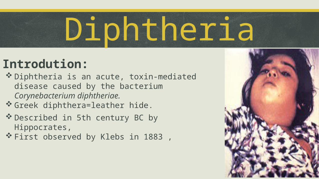
DiphtheriaIntrodution: Diphtheria is an acute, toxin-mediated disease caused by the
bacterium Corynebacterium diphtheriae. Greek diphthera=leather hide.
Described in 5th century BC by Hippocrates, First observed by Klebs in 1883 ,
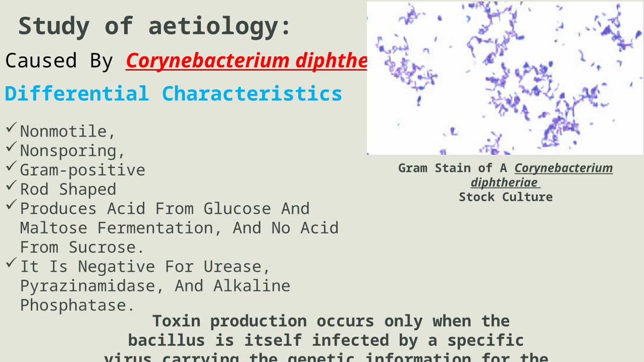
Caused By Corynebacterium diphtheriae
Study of aetiology:
Differential Characteristics
Nonmotile, Nonsporing,Gram-positive Rod Shaped Produces Acid From Glucose And Maltose
Fermentation, And No Acid From Sucrose. It Is Negative For Urease, Pyrazinamidase, And
Alkaline Phosphatase.
Gram Stain of A Corynebacterium diphtheriae Stock Culture
Toxin production occurs only when the bacillus is itself infected by a specific virus carrying the genetic information for the toxin
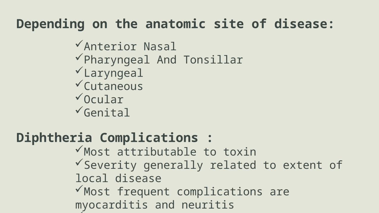
Depending on the anatomic site of disease:Anterior Nasal Pharyngeal And Tonsillar Laryngeal Cutaneous Ocular Genital
Diphtheria Complications :Most attributable to toxin Severity generally related to extent of local disease Most frequent complications are myocarditis and neuritis Death occurs in 5%-10%

Signs and symptoms:
Symptoms of diphtheria include: Fever Of 38 °C (100.4 °F) Or Above, Chills, Fatigue, Bluish Skin Coloration (Cyanosis), Sore Throat, Hoarseness, Cough, Headache, Difficulty in Swallowing, Difficulty in Breathing, Foul-smelling Bloodstained Nasal Discharge & Lymphadenopathy.[
Symptoms Can Also Include Cardiac Arrhythmias, Myocarditis, And Cranial And Peripheral Nerve Palsies.
Pseudomembrane Covering The Tonsils
A diphtheria skin lesion on the leg
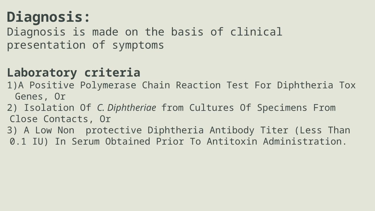
Diagnosis:Diagnosis is made on the basis of clinical presentation of symptoms
Laboratory criteria1)A Positive Polymerase Chain Reaction Test For Diphtheria Tox Genes, Or 2) Isolation Of C. Diphtheriae from Cultures Of Specimens From Close Contacts, Or3) A Low Non protective Diphtheria Antibody Titer (Less Than 0.1 IU) In Serum Obtained Prior To Antitoxin Administration.

Mode of TransmissionSpreads by
Droplet infection (major route) Infected cutaneous lessions Nasopharyngeal secretions
Portal of Entry Mostly by Respiratory tract Breeches in skin, eye, genitalia, & middle ear
Incubation Time 2-7 days

Treatment Tracheotomy Diptheria antitoxin Metronidazole Antibiotics
Erythromycin Procaine penicillin G (IM14 days) Rifampin or clindamycin
Immunization MethodsDiptheria Vaccines are used in combination with Tetanus, Pertusis4 Types of Diptheria vaccines:
i. DTaP- For infants & childrenii. DT- For infants & childreniii. Tdap- For adolescents and adultsiv. Td- For adolescents and adults

EpidemiologyOccurs Worldwide
Particularly in Tropical countries Rare in Industrialised countries(USA)
Occurs mostly in non vaccinated childrenCaused several deaths in USA before vaccination
Diphtheria cases reported to the World Health Organization
About 1 out of 10 people who gets diptheria will die
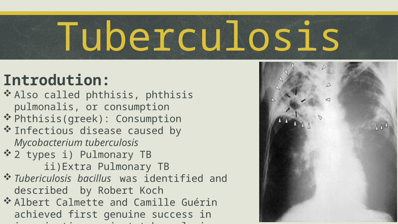
TuberculosisIntrodution: Also called phthisis, phthisis pulmonalis, or consumption Phthisis(greek): Consumption Infectious disease caused by Mycobacterium tuberculosis 2 types i) Pulmonary TB
ii)Extra Pulmonary TB Tubericulosis bacillus was identified and described by
Robert Koch Albert Calmette and Camille Guérin achieved first genuine
success in immunization against tuberculosis
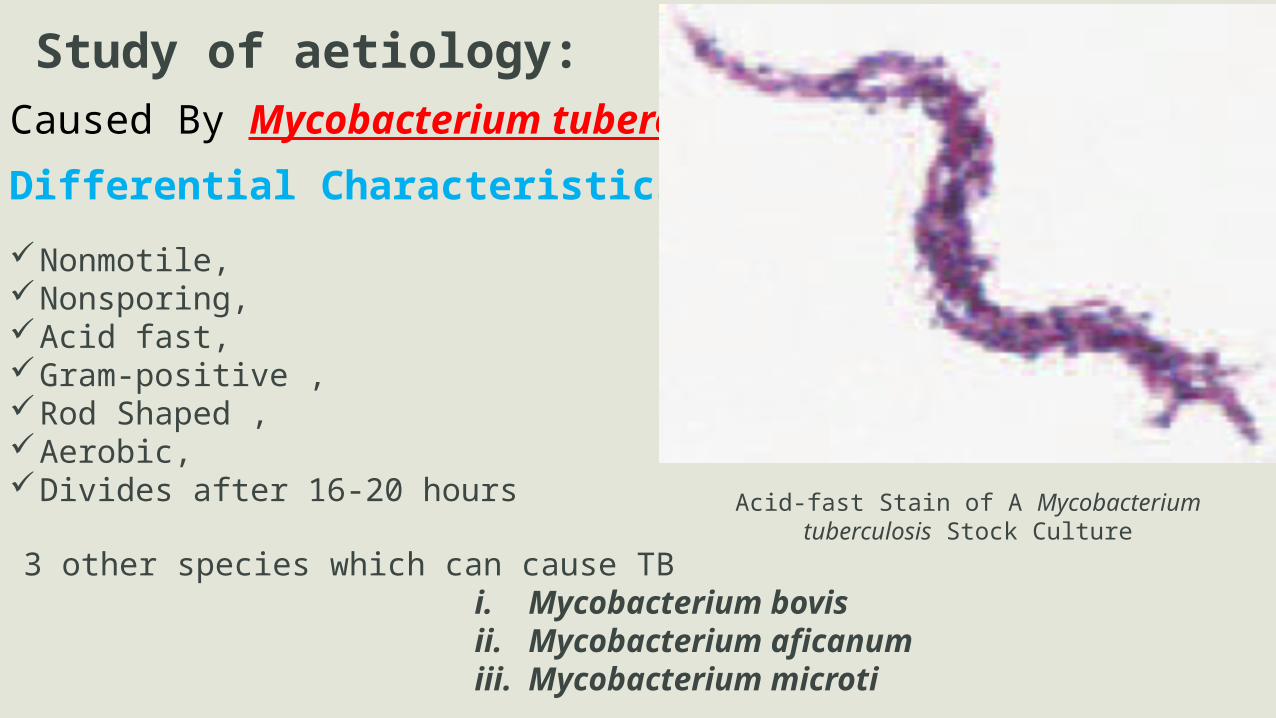
Caused By Mycobacterium tuberculosis
Study of aetiology:
Differential Characteristics
Nonmotile, Nonsporing,Acid fast,Gram-positive ,Rod Shaped ,Aerobic,Divides after 16-20 hours Acid-fast Stain of A Mycobacterium tuberculosis Stock Culture
3 other species which can cause TBi. Mycobacterium bovisii. Mycobacterium aficanumiii. Mycobacterium microti

Signs and symptoms:Pulmonary TB Extra Pulmonary TB
Tuberculosis infection becomes active, Commonly involves the lungs (90% of cases).,
Symptoms include i)Chest pain ii)Prolonged cough producing sputum.
About 25% of people do not have any symptoms Occasionally,
People may cough up blood in small amounts,
the infection may erode into the pulmonary artery or a Rasmussen's aneurysm,
resulting in massive bleeding(rarely)
Notable extrapulmonary infection sites include :i. Pleura (in tuberculous pleurisy), ii. The central nervous system (in tuberculous
meningitis), iii. The lymphatic system (in scrofula of the neck), iv. The genitourinary system (in urogenital
tuberculosis),v. The bones and joints (in Pott disease of the spine). When it spreads to the bones, it is also known as
"osseous tuberculosis". Bursting of a tubercular abscess through skin results
in tuberculous ulcer. Potentially more serious, widespread form of TB is
called "disseminated" TB, commonly known as miliary tuberculosis.
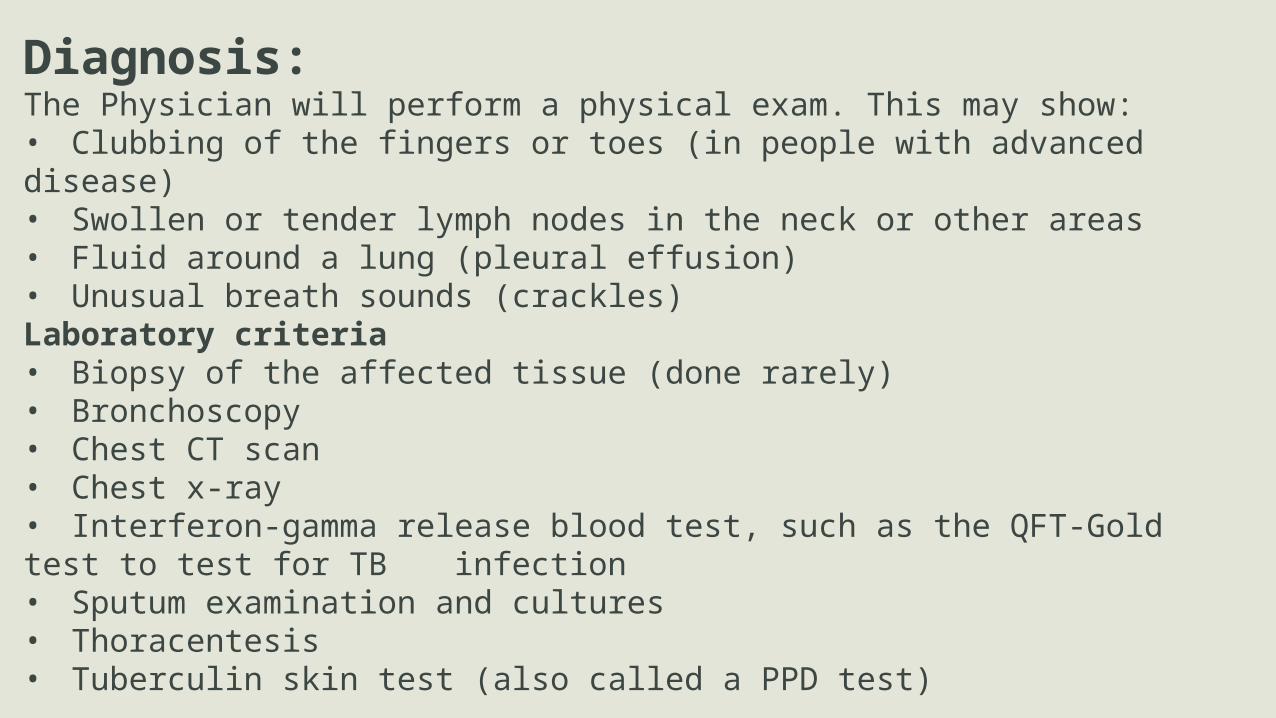
Diagnosis:The Physician will perform a physical exam. This may show:• Clubbing of the fingers or toes (in people with advanced disease)• Swollen or tender lymph nodes in the neck or other areas• Fluid around a lung (pleural effusion)• Unusual breath sounds (crackles)Laboratory criteria• Biopsy of the affected tissue (done rarely)• Bronchoscopy• Chest CT scan• Chest x-ray• Interferon-gamma release blood test, such as the QFT-Gold test to test for TB infection• Sputum examination and cultures• Thoracentesis• Tuberculin skin test (also called a PPD test)

Mode of TransmissionPeople with active pulmonary TB
• Cough,• Sneeze, • Speak, • Sing, or • Spit.
Prolonged, frequent, or close contact with people with TB (22% infection rate)Probability of transmission from one to another depends upon
the number of infectious droplets expelled by the carrier, the effectiveness of ventilation, the duration of exposure, the virulence of the M. tuberculosis strain, the level of immunity in the uninfected person, and others.
} Expel infectious aerosol droplets 0.5 to 5.0 µm in diameter

TreatmentAntibiotics are mostly used to kill the bacteria
Latent TB- Single antibioticActive TB- Combiantion of several antibiotics In multiple drug-resistant TB-Bedaquiline Immunization MethodsOnly available vaccine is Bacillus Calmette-Guérin (BCG).In children it decreases the risk of getting infection by 20% and
the risk of infection turning into disease by nearly 60%.Most widely used vaccine worldwide, >90% of all children being vaccinated. BCG is also administered to only those people at high risk.A number of new vaccines are currently in development.

Epidemiology Roughly 1/3 of the world's population has been
infected with TB Tuberculosis is more common in developing
countries Asian and African countries-about 80%test
positive US population –Only 5–10% test positive India had the largest incidence-estimated 2 million
new cases The rates of TB varies with age. In Africa, it primarily affects adolescents and
young adults. TB is mainly a disease of older people and the
immunocompromised
Prevalence of TB was highest in sub-Saharan Africa, and was also relatively
high in Asia.
Tuberculosis is the 2nd most common cause of death from infectious disease
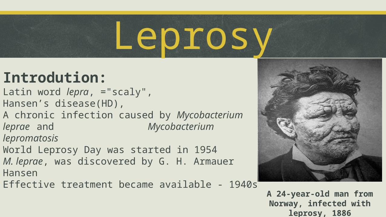
LeprosyIntrodution:Latin word lepra, ="scaly",Hansen’s disease(HD),A chronic infection caused by Mycobacterium leprae and
Mycobacterium lepromatosisWorld Leprosy Day was started in 1954M. leprae, was discovered by G. H. Armauer HansenEffective treatment became available - 1940s
A 24-year-old man from Norway, infected with leprosy, 1886

Caused By Mycobacterium lepraeStudy of aetiology:
Differential Characteristics
Nonmotile, Anaerobic, Acid-fast, Nonsporing,Rod Shaped, M.leprae is uncultivable in vitro, Intracellular parasite
Acid-fast Stain of A Mycobacterium lepraeStock Culture
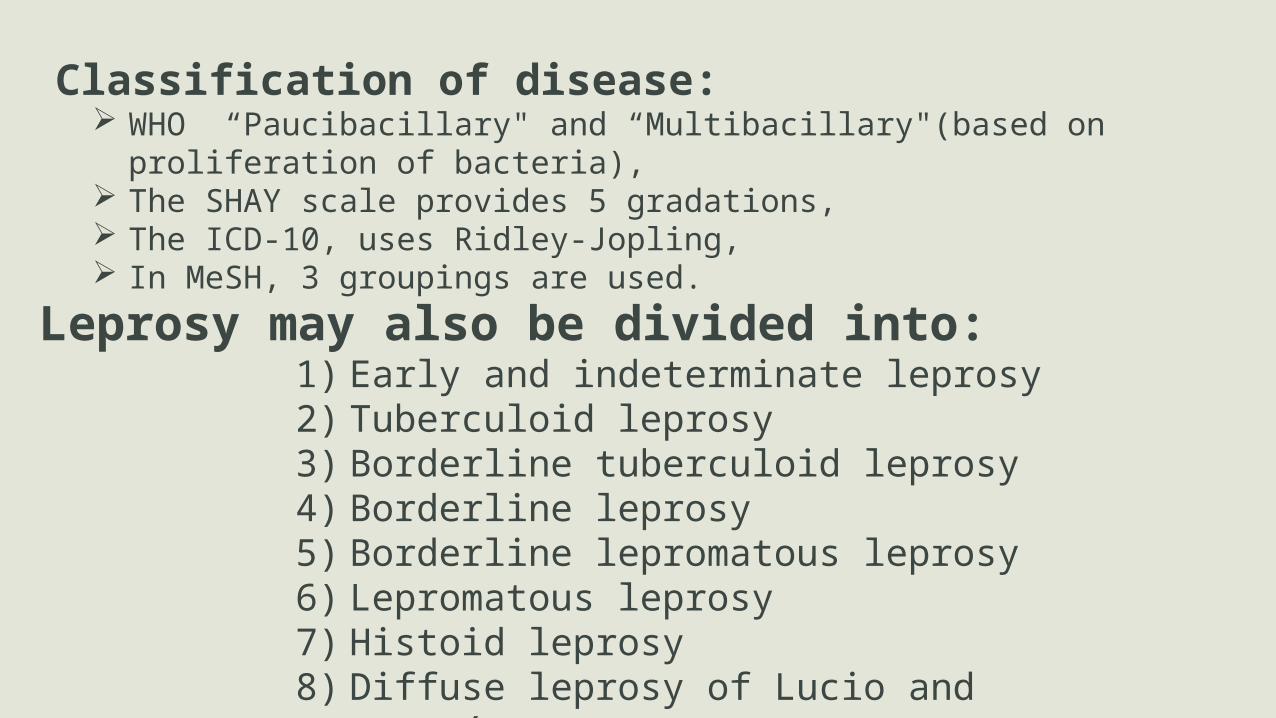
Classification of disease: WHO “Paucibacillary" and “Multibacillary"(based on proliferation of
bacteria), The SHAY scale provides 5 gradations, The ICD-10, uses Ridley-Jopling, In MeSH, 3 groupings are used.
Leprosy may also be divided into:1) Early and indeterminate leprosy2) Tuberculoid leprosy3) Borderline tuberculoid leprosy4) Borderline leprosy5) Borderline lepromatous leprosy6) Lepromatous leprosy7) Histoid leprosy8) Diffuse leprosy of Lucio and Latapí

Signs and symptoms:
Symptoms of diphtheria include: Skin lesions (light or dark patches)-Primary external Leprosy progress can cause permanent damage to the
skin, nerves, limbs, and eyes.
Secondary infections Body parts can become numb or diseased tissue loss causing fingers and toes to become
shortened and deformed, as cartilage is absorbed into the body
Hands deformed by leprosy

Diagnosis:According to WHO, diagnosis in areas where people are frequently infected is based on one of these main signs:
• Skin lesion consistent with leprosy and with definite sensory loss• Positive skin smears
Skin lesions can be • single or multiple, • usually hypopigmented, • reddish or copper-colored. • macules (flat), papules (raised), or nodular.
Diagnosis is confirmed by acid-fast bacilli in a biopsy of the skin or by detecting the DNA using polymerase chain reaction.
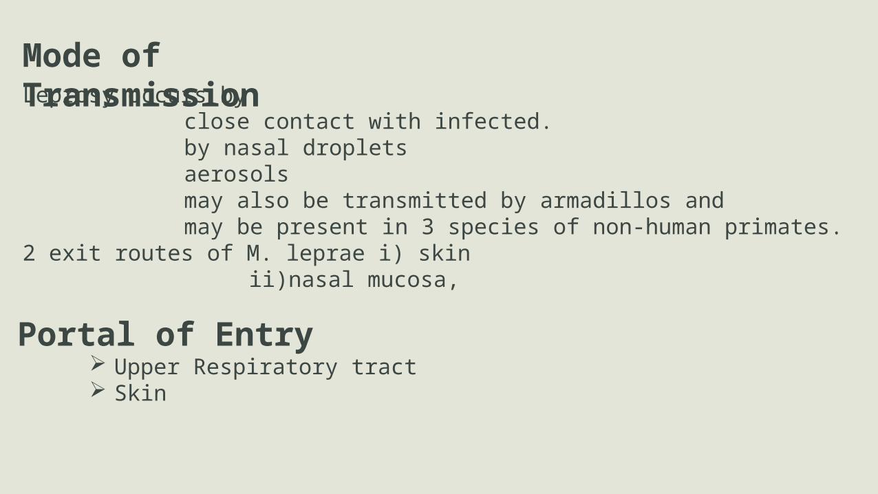
Mode of TransmissionLeprosy occurs by
close contact with infected.by nasal dropletsaerosolsmay also be transmitted by armadillos and may be present in 3 species of non-human primates.
2 exit routes of M. leprae i) skinii)nasal mucosa,
Portal of Entry Upper Respiratory tract Skin

TreatmentA number of leprostatic agents are available for treatment. Paucibacillary (PB or tuberculoid) cases-daily dapsone and monthly rifampicin for 6 months Multibacillary (MB or lepromatous) cases,- daily dapsone and clofazimine along with monthly rifampicin for 12 months is recommended.
Immunization MethodsBCG Vaccine is used arround the world
Diaminodiphenyl sulfone(DDS)1950
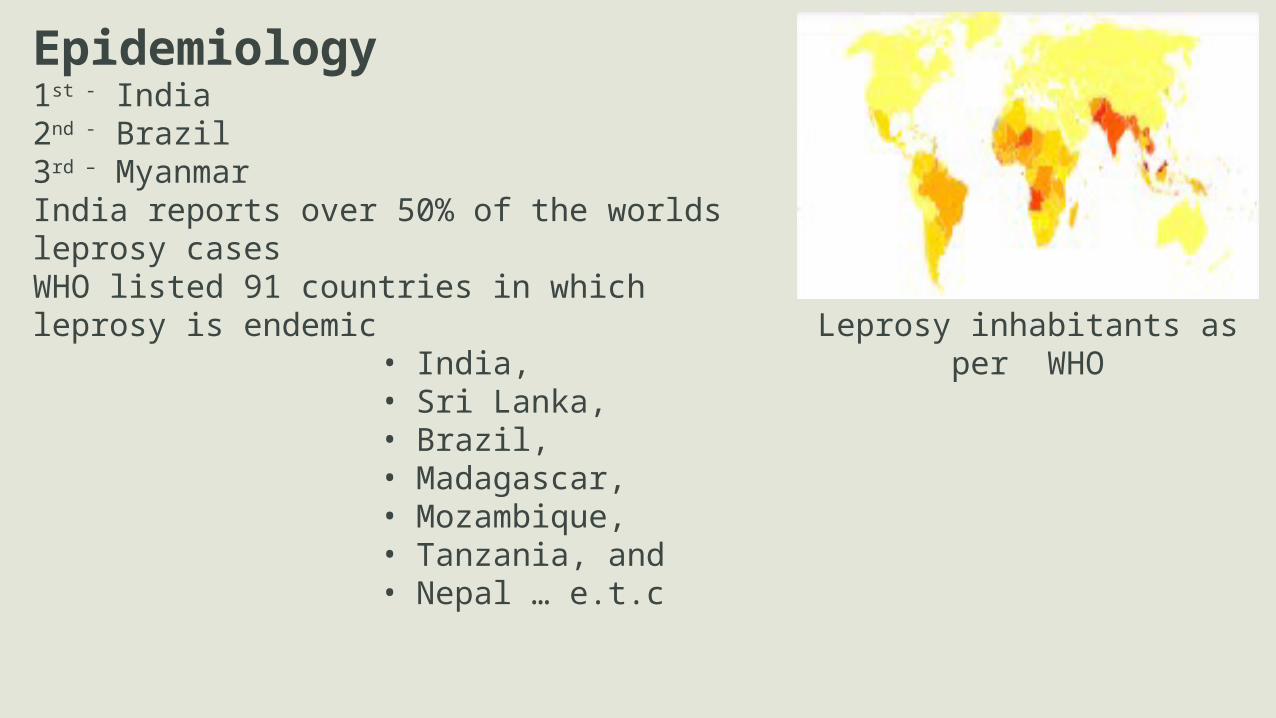
Epidemiology1st - India 2nd - Brazil 3rd – MyanmarIndia reports over 50% of the worlds leprosy casesWHO listed 91 countries in which leprosy is endemic
• India,• Sri Lanka, • Brazil, • Madagascar, • Mozambique,• Tanzania, and• Nepal … e.t.c
Leprosy inhabitants as per WHO

TetanusIntrodution: Tetanus also known as lockjaw, An acute, often fatal, disease caused by exotoxin
produced by the bacteria An infection characterized by muscle spasms Etiology discovered in 1884 by Carle and Rattone Passive immunization used for treatment and prophylaxis
during World War I Tetanus toxoid first widely used during World War II

Caused By Clostridium tetani
Study of aetiology:
Differential Characteristics
Motile, Endospore forming,Gram-positive Rod Shaped AnaerobicSurvive autoclaving at 249.8°F (121°C) by 10–15 mntsSpores found in soil, animal feces Produces 2 exotoxins
i. tetanolysin and ii. tetanospasmin
Tetanospasmin estimated human lethal dose = 2.5 ng/kg
Gram Stain of A Clostridium tetani Stock Culture
Types In Disease:Generalized tetanusNeonatal tetanusLocal tetanusCephalic tetanus
Tetanus Complications :LaryngospasmFracturesHypertension and/orAbnormal heart rhythmNosocomial infectionsPulmonary embolismAspiration pneumoniaDeath
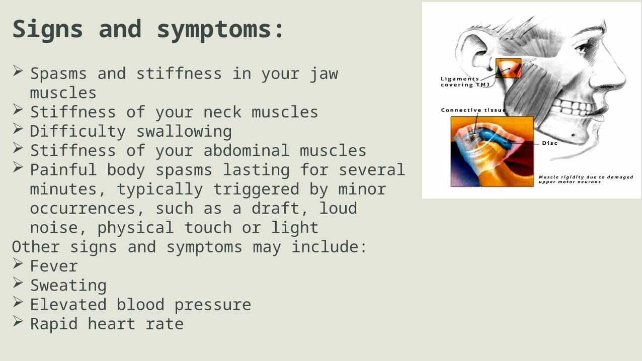
Signs and symptoms:
Spasms and stiffness in your jaw muscles Stiffness of your neck muscles Difficulty swallowing Stiffness of your abdominal muscles Painful body spasms lasting for several minutes, typically
triggered by minor occurrences, such as a draft, loud noise, physical touch or light
Other signs and symptoms may include: Fever Sweating Elevated blood pressure Rapid heart rate

Diagnosis: There are no blood tests for diagnosing tetanus. Diagnosis is made on the basis of clinical presentation of symptoms Laboratory identification of C. tetani can be demonstrated only by production
of tetanospasmin in mice. Having recently experienced head trauma may indicate cephalic tetanus if no
other diagnosis has been made. The "spatula test" is a clinical test for tetanus that involves touching the
posterior pharyngeal wall with a soft-tipped instrument and observing the effect. + test result is the involuntary contraction of the jaw (biting down on
"spatula") - test result would normally be a gag reflex attempting to expel foreign
object.
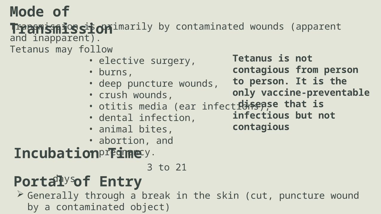
Mode of TransmissionTransmission is primarily by contaminated wounds (apparent and inapparent). Tetanus may follow
• elective surgery, • burns, • deep puncture wounds, • crush wounds, • otitis media (ear infections), • dental infection,• animal bites, • abortion, and • pregnancy.
Portal of Entry Generally through a break in the skin (cut, puncture wound by a contaminated object)
Incubation Time 3 to 21 days
Tetanus is not contagious from person to person. It is the only vaccine-preventable disease that is infectious but not contagious
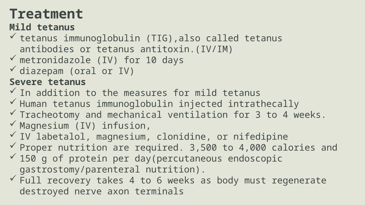
TreatmentMild tetanus tetanus immunoglobulin (TIG),also called tetanus antibodies or tetanus
antitoxin.(IV/IM) metronidazole (IV) for 10 days diazepam (oral or IV)Severe tetanus In addition to the measures for mild tetanus Human tetanus immunoglobulin injected intrathecally Tracheotomy and mechanical ventilation for 3 to 4 weeks. Magnesium (IV) infusion, IV labetalol, magnesium, clonidine, or nifedipine Proper nutrition are required. 3,500 to 4,000 calories and 150 g of protein per day(percutaneous endoscopic gastrostomy/parenteral nutrition). Full recovery takes 4 to 6 weeks as body must regenerate destroyed nerve axon
terminals
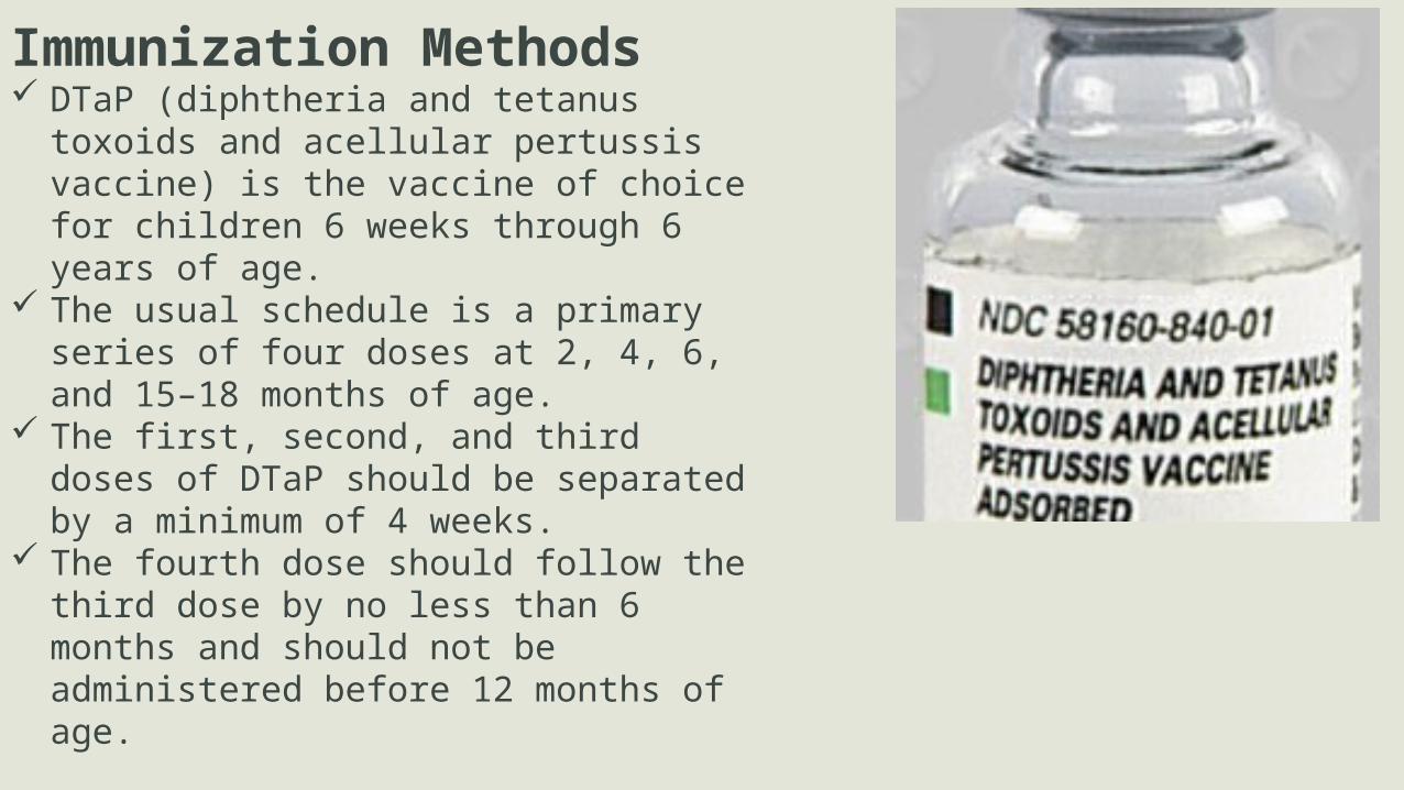
Immunization Methods DTaP (diphtheria and tetanus toxoids and
acellular pertussis vaccine) is the vaccine of choice for children 6 weeks through 6 years of age.
The usual schedule is a primary series of four doses at 2, 4, 6, and 15–18 months of age.
The first, second, and third doses of DTaP should be separated by a minimum of 4 weeks.
The fourth dose should follow the third dose by no less than 6 months and should not be administered before 12 months of age.
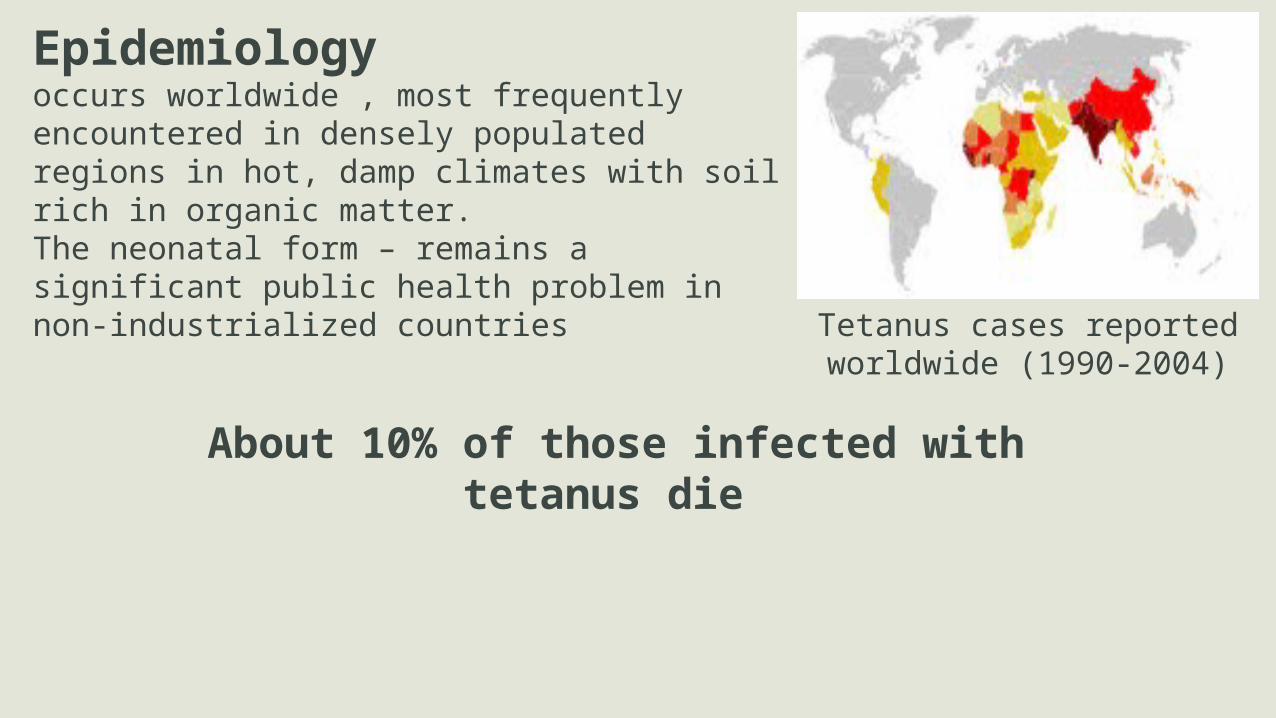
Epidemiologyoccurs worldwide , most frequently encountered in densely populated regions in hot, damp climates with soil rich in organic matter. The neonatal form – remains a significant public health problem in non-industrialized countries
Tetanus cases reported worldwide (1990-2004)
About 10% of those infected with tetanus die
Food PoisoningIntrodution:Food Poisoning =foodborne disease, foodborne illness Illness resulting from the consumption of
• contaminated food• pathogenic bacteria, • viruses, • parasites • chemical or natural toxins(poisonous
mushrooms)Cause severe illness and even death.
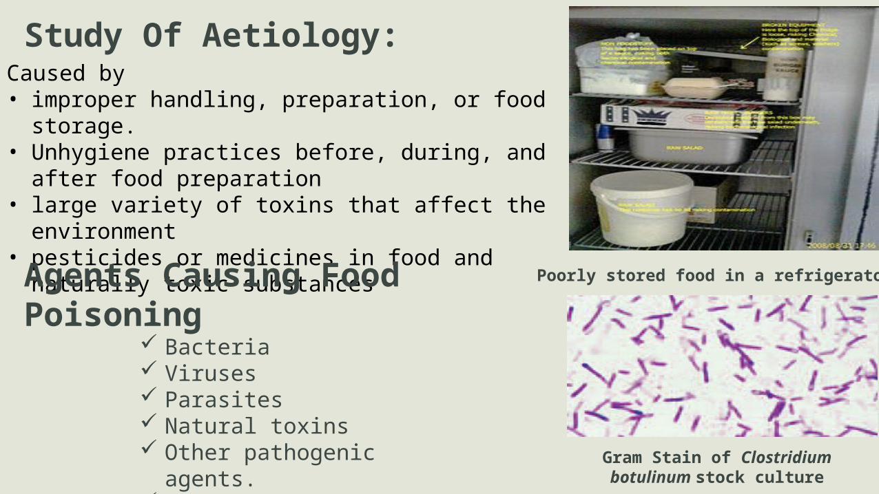
Study Of Aetiology:
Poorly stored food in a refrigerator
Caused by• improper handling, preparation, or food storage. • Unhygiene practices before, during, and after food
preparation• large variety of toxins that affect the environment• pesticides or medicines in food and naturally toxic
substances
Agents Causing Food Poisoning Bacteria Viruses Parasites Natural toxins Other pathogenic agents. "Ptomaine poisoning"
Gram Stain of Clostridium botulinum stock culture

Signs and symptoms:
The most common symptoms of food poisoning are: • Nausea • Vomiting • Diarrhoea • Stomach pains
Other symptoms include:• Abdominal cramps • Loss of appetite • A high temperature (fever) • Muscle pains • Chills
Diagnosis: Diagnosis for food poisoning is made from the symptoms. A laboratory diagnosis for food poisoning is usually only needed if:
•Your symptoms are severe •Your symptoms persist despite treatment •You are showing signs of dehydration and/or •There has been an outbreak of similar cases linked to a possible source of contamination.
Further testing is only needed if the symptoms indicate that infection has spread from digestive system to other parts of the body.
You may be asked to have investigations including blood tests (for infection) and examination of stool sample.
Incubation Time • Varies from type to type• May range from hours to days
Treatment• No specific treatment for food poisoning• Depends on sympotoms• Antibiotics are mostly used in case of bacteria
Protection and prevention Prevent food poisoning by:
Cleaning Cooking Chilling Cross-contamination…e.t.c.
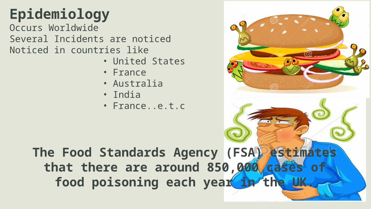
EpidemiologyOccurs WorldwideSeveral Incidents are noticedNoticed in countries like
• United States• France• Australia• India• France..e.t.c
The Food Standards Agency (FSA) estimates that there are around 850,000 cases of food poisoning each year in the UK.

ReferencesText Book Of Microbiology by Anantha NarayanSource
www.google.comhttps://en.wikipedia.org/wiki/Tetanushttps://en.wikipedia.org/wiki/Tuberculosishttps://en.wikipedia.org/wiki/Foodpoisoninghttps://en.wikipedia.org/wiki/Diptheriahttps://en.wikipedia.org/wiki/Leprosywww.vaccines.netwww.cdc.gov

Thank You