essentials of lung tumor cytology - home ... of lung tumor cytology is the result of my experience...
TRANSCRIPT

ESSENTIALS OF LUNG TUMOR CYTOLOGY
Gia-Khanh Nguyen 2008

2
ESSENTIALS OF LUNG TUMOR CYTOLOGY Gia-Khanh Nguyen, M.D. Professor Emeritus Department of Laboratory Medicine and Pathology Faculty of Medicine and Dentistry University of Alberta Edmonton, Alberta, Canada Revised first edition, 2008. All rights reserved. Legally deposited at the Library and Archives Canada. ISNB: 0-9780929-0-2

TABLES OF CONTENTS Table of contents 3 Preface 4 Dedication 5 Related material and Key to abbreviations 6 Chapter 1: Cytologic investigations of lung tumors 7 Chapter 2: Usual lung cancers 17 Chapter 3: Neuroendocrine carcinomas 38 Chapter 4: Other primary tumors and tumorlike lesions 48 Chapter 5: Metastatic cancers 64 Chapter 6: Pleural tumors 76

4
PREFACE Cytology plays a very important role in the diagnosis of lung cancers. This monograph on Essentials of Lung Tumor Cytology is the result of my experience gained in over 20 years of active involvement in the cytodiagnosis of lung tumors at the University of Alberta Hospital, Edmonton, Alberta, Canada. It is written mainly for practicing pathologists in community hospitals, residents in pathology and cytotechnologists who are interested in making safe and accurate cytodiagnoses of important tumors of the lung and pleura. The text is concise and illustrations are abundant, and several histologic images are included for cytohistologic correlation. In the first edition of the monograph (2007), cytodiagnostic criteria of lung tumors were emphasized. In this revised edition, immunocytochemical features of lung tumor cells that are important for tumor typing and differential diagnosis are stressed. A number of important references are listed at the end of each chapter for further consultation. For improvement of the future editions of this monograph, constructive comments and suggestions from the readers will be highly appreciated. Gia-Khanh Nguyen, M.D. Surrey, BC, Canada [email protected] Winter 2008

5
To my family with love.

6
RELATED MATERIAL BY THE SAME AUTHOR Essentials of Needle Aspiration Biopsy Cytology. Igaku-Shoin, New York,1991 Essentials of Exfoliative Cytology. Igaku-Shoin, New York, 1992 Essentials of Cytology: An Atlas. Igaku-Shoin, New York, 1993 Critical Issues in Cytopathology. Igaku-Shoin, New York, 1996 Essentials of Abdominal Fine Needle Aspiration Cytology. UBC Pathology, 2008
KEY TO ABBREVIATIONS FNA: Fine needle aspiration or Fine needle aspirate TBFNA: Transbronchial/mucosal FNA TTFNA: Transthoracic FNA Pap: Papanicolaou stain HE: hematoxylin and eosin stain ABC: Avidin-biotin complex technique

7
Chapter 1
CYTOLOGIC INVESTIGATIONS OF LUNG TUMORS Investigation of lung diseases using cytologic materials has a long history that can be traced back to the 19th century. It began with the identification of exfoliated bronchial epithelial cells in sputa by Donne in 1845 and it was followed by the description of lung cancer cells by Walshe in 1846 and by Hampeln in 1887. Pulmonary cytology had no remarkable developments in the early years of the 20th century until the 1950s when a large number of papers reporting on the ability to detect and type lung cancers were published. In the 1960s the technique of TTFNA of lung cancer under chest fluoroscopic guidance was developed and the early years of 1980s marked the development of TBFNA via a flexible fiberoptic bronchoscope that allowed cytologic diagnoses of submucosal lesions and enlarged peribronchial lymph nodes. THE RESPIRATORY TRACT The respiratory tract is divided into upper and lower parts. The upper respiratory tract is composed of the nose and larynx, and the lower respiratory tract consists of the trachea and lung. The tracheobronchial tree contains cartilage and submucosal mucus-secreting glands and is lined by a pseudostratified, ciliated columnar epithelium that contains, in addition, goblet cells, Clara cells and Kulchitsky cells (neuroendocrine cells). (Figure 1.1).
Figure 1.1. Histology of normal tracheobronchial wall containing submucosal mucus-secreting glands. (HE, x100).

8
The bronchi ultimately branch into bronchioles that do not have cartilage and submucosal glands. The terminal bronchioles are purely conducting ducts that divide into respiratory bronchioles which merge into alveolar ducts and alveoli. (Figure 1.2).
Figure 1.2. Normal ciliated pseudotratified columnar bronchial epithelium. (HE, x 250). The alveoli are lined by type I and II epithelial cells. (Figure 1.3). Type I cells account for 40% of the alveolar cells, covers 95% of the alveolar surface and facilitate gas exchange. Type II cells produce surfactant and can reconstitute the alveolar surface after injury. The lung and the inner aspect of the thoracic cavities are covered by a layer of mesothelial cells.
Figure 1.3. Normal lung parenchyma showing alveolar spaces. (HE, x 100). DIFFERENT TYPES OF RESPIRATORY CELL SAMPLES The lower respiratory tract is the target of respiratory cytology that can be studied by one or a variable combination of the following 7 types of cell sample: sputum, bronchial suction, bronchial wash, bronchial brush, bronchoalveolar lavage, TBFNA and TTFNA.

9
Tumors of the pleura can be investigated by cytologic examination of associated serous effusions or TTFNA that will be discussed in Chapter 6. 1. Sputum Sputum cell samples are obtained by early morning deep cough after mouth washing. These are excellent specimens for screening of cancers arising from the tracheobronchial tree. Usually 3 samples collected on 3 consecutive days are required. The commonly used fixatives are 70% ethanol and Saccomanno solution (50% ethanol and 2% polyethylene glycol or carbowax). If the patient is unable to expectorate properly, the sputum expectoration can be induced by inhaling nebulized water or saline. For a sputum specimen collected in 70% ethanol, the classic “pick and smear” technique is used. Two to 4 smears are prepared, immediately fixed in 95% ethanol and stained by the Papanicolaou technique. The rest of the specimen is fixed in formalin and embedded in paraffin for cell block sections. Sputum collected in Saccomanno solution is homogenized in a blender and concentrated by centrifugation. It can also be processed using a thin layer method. The sputum processing must be performed under a biologic safety hood to minimize the risk of infection by inhalation. A sputum cell sample must contain alveolar macrophages and other cells derived from the lung. (Figure 1.4).
Figure 1.4. Adequate sputum cell sample showing alveolar macrophages. (Pap, x 500). Sputum cytology is more efficient in detecting cancers involving large proximal bronchi. Its sensitivity rate is low with one specimen (27-41%) and when 3 samples are used it increases to 57-89%. If 5 samples are used a sensitivity rate as high as 96.1% may be reached. 2. Bronchial Washing and Brushing Bronchial secretions may be aspirated from the trachea via a tracheal tube or a tracheotomy stoma. Bronchial washing is performed during bronchoscopy by instilling vials of 5 to 10 mL of warm normal saline into a bronchus. The fluid is then aspirated and

10
usually 4 cytospin smears are prepared and stained by the Papanicolaou method. A bronchial wash from a normal individual should show a few bronchial columnar cells admixed with polymorphonuclear leukocytes and macrophages. (Figure 1.5). It is often contaminated with squamous cells exfoliated from the upper respiratory tract. Bronchial washing is contraindicated in patients with respiratory failure or uncontrolled coughing.
Figure 1.5. Bronchial washing showing normal bronchial epithelial cells, alveolar macrophages and metaplastic squamous cells. (Pap, x 500). Bronchial brushing is performed during bronchoscopy. A cytobrush is used to scrape the surface of a bronchial lesion. The entrapped cells are transferred to a frosted slide by circular movements. Usually 2 smears are prepared and stained by the Papanicolaou technique. It can be done 2 to 3 times to secure an adequate number of diagnostic cells. Cytologic material obtained by bronchial brushing contains abundant bronchial epithelial cells and a small number of neutrophils as well as a few squamous cells exfoliated from the upper airways. (Figures 1.6 and 1.7). Bronchial brushing is contraindicated in patients with respiratory failure and uncontrolled coughing.
Figure 1.6. Bronchial brushing showing 2 bronchial epithelial fragments with ciliated columnar cells with terminal plates and a benign metaplastic squamous cell. (Pap, x 500).

11
Figure 1.7. Bronchial brushing showing a few columnar bronchial epithelial cells and goblet cells with intracytoplasmic mucous vacuoles. (Pap, x 500). The sensitivity rate of a bronchial washing in the diagnosis of lung cancer varies from 61 to 76%, and that of a bronchial brushing ranges from 70 to 77%. 3. Bronchoalveolar lavage (BAL) A bronchoscope is wedged into position as far as it can advance. The distal airways are flushed with several vials of warm normal saline totaling 300 mL. The flushed samples are then aspirated. The first sample contains mainly bronchial secretion and is discarded. Other samples are pooled together and usually 4 cytospin smears are prepared and stained by the Papanicolaou and/or Diff-Quik technique. BAL reflects the cellular changes within alveolar spaces. A satisfactory BAL cell sample should contain abundant alveolar macrophages and a few lymphocytes and polymorphonuclear leukocytes. (Figure 1.8). The number of epithelial cell (bronchial columnar and squamous cells) should be less than 5% of all cells present in the sample. Differential cell counts are obtained by evaluating 200 cells. In normal, nonsmoking individuals polymorphonuclear leukocytes account for about 1% of all cells present. Neutrophils, up to 4%, can be found in the BAL from a cigarette smoker without any lung disease. BAL is contraindicated in patients with respiratory failure and uncontrolled coughing. BAL has a sensitivity rate of 37.5% in detecting lung cancer.

12
Figure 1.8. BAL sample from a city resident showing numerous alveolar macrophages. A few of them contain dust and carbon particles. (Pap, x 500). 4. Transbronchial/transmucosal fine needle aspiration By TBFNA cell samples from a submucosal mass lesion or a paratracheal or parabronchial lesion or enlarged lymph node can be obtained by a 22-gauge needle via the suction tube of a flexible bronchoscope. The sample is commonly contaminated with bronchial secretions containing exfoliated bronchial epithelial cells and submucosal glandular cells may rarely be seen. (Figure 1.9). For TBFNA, the sensitivity rate of the procedure alone is about 52%. When TBFNA is combined with bronchial washing and brushing and bite biopsy its sensitivity rate increases to 72%. The specificity rate of the biopsy technique is 70 - 74% and its positive and negative predictive values are 100% and 53 - 70%, respectively.
Figure 1.9. Acini of a normal bronchial submucosal gland in a TBFNA. (Pap, x 500).

13
An adequate TBFNA cell sample from a lymph node should show abundant lymphocytes. (Figure 1.10). TBFNA is almost free of complications. However, transient hemoptysis is common and pneumothorax is exceedingly rare. It is contraindicated in patients with uncontrolled coughing, respiratory failure and bleeding disorders.
Figure 1.10. Adequate TBFNA of an enlarged peribronchial lymph node showing abundant lymphoid cells. (Pap, x 500). 5. Transthoracic fine needle aspiration TTFNA is used for investigation of patients with a lung mass lesion, usually peripheral, showing no diagnostic cells in sputum, bronchial washing and brushing, BAL and TBFNA. It is contraindicated in patients with chronic obstructive lung disease, uncontrolled coughing, bleeding disorders, severe pulmonary hypertension, arteriovenous malformation and suspected hydatid cyst. The most common complication of TTFNA is pneumothorax which is minor and detectable by chest roentgenogram in 21-34% of patients. However, only 10% of pneumothoraces require a chest tube drainage. Transient hemoptysis occurs in 5-10% of cases. Other complications include hemothorax, air embolism, tumor seeding along the needle tract and rare sudden death. An adequate TTFNA cell sample from a normal lung tissue should show alveolar macrophages, bronchial epithelial cells and sheets of mesothelium. (Figure 1.11). For TTFNA of lung cancers, the sensitivity and specificity rates are 89% and 96%, respectively. Its positive and negative predictive values are 98% and 70%, respectively; and a false-positive and false-negative rates are 0.85% and 6%, respectively.

14
Figure 1.11. TTFNA from a normal lung showing a large fragment of mesothelium with folding and several alveolar macrophages. (Pap, x 500). ANCILLARY TECHNIQUES In recent years, with the availability of numerous commercially available antibodies cytologic typing of lung tumors, in particular metastatic cancers, has become more feasible. An accurate cytodiagnosis of a metastatic tumor to the lung and an identification of a primary lung cancer arising in a patient with a malignant tumor in remission are very important for patient management. Cytochemical and immunocytochemical studies can be done with satisfactory results on previously stained smears without prior destaining. However, they are best performed on formalin-fixed minute tumor tissue fragments in cell blocks prepared from materials procured by bronchial brushing or FNA. Any grossly identified minute tissue fragments in an FNA should be removed and fixed in formalin for histologic, cytochemical and immunohistochemical studies. They may also be fixed in 2% glutaraldehyde for ultrastructural evaluation. It should be born in mind that ethanol is not a suitable fixative for electron microscopy as it destroys cellular ultrastructures. ACCURACY OF TUMOR TYPING The sensitivity and specificity rates and predictive values of different types of respiratory specimen in the diagnosis of lung cancer vary with the tumor location and the type and number of specimens. In general a combination of different types of cell sample offers higher sensitivity and specificity rates and predictive value for a positive result than a single sample. For tumor typing, the cytohistologic correlation rates of sputum and bronchoscopy cytologic materials, as reported by Johnston and Bossen, were 85% for squamous cell carcinoma, 79% for adenocarcinoma, 30% for large cell carcinoma and 93% for small cell carcinoma of the bronchial tree. Those investigators have also reported that the cytohistologic correlation rates of TTFNA were 80%, 96%, 42% and

15
95% for squamous cell carcinoma, adenocarcinoma, large cell carcinoma and small cell carcinoma of the lung, respectively. BIBLIOGRAPHY Bedrossian CWM, Rybka DL. Bronchial brushing during fiberoptic bronchoscopy for cytodiagnosis of lung cancer: comparison with sputum and bronchial washings. Acta cytol. 1976;20: 446. Caglayan B, et al. Transbronchial needle aspiration in the diagnosis of endobronchial malignant lesions: a 3-year experience. Chest. 2005;128: 704. Erozan YS, Frost JK. Cytopathologic diagnosis of cancer in pulmonary material: a critical histopathologic correlation. Acta Cytol. 1970;14: 560. French CA. Respiratory tract. In Cytology. Diagnostic principles and clinical correlates. 2nd ed, 2003. Cibas ES, Ducatman BS, eds. Philadelphia, Saunders. p 61 Garg S, et al. Comparative analysis of various cytotechnical techniques in diagnosis of lung diseases. Diagn Cytopathol. 2007;35:26. Johnston WW. Cytodiagnosis of lung cancer. Principles and problems. Path Res Pract. 1986;181:1. Johnston WW, Bossen EH. Ten years of respiratory cytopathology at Duke University Medical Center. I. The cytopathologic diagnosis of lung cancer during the years 1970-1974, noting the the significance of specimen number and type. Acta Cytol.1981;25: 103. Johnson WW, Bossen EH. Ten years of respiratory cytopathology at Duke University Medical Center. II. A comparison between cytopathology and histopathology in typing of lung cancer during the years 1970-1974. Acta Cytol. 1981;25:499. Koss LG, et al. pulmonary cytology-a brief survey of diagnostic results from July 1st, 1952 until December 31st, 1960. Acta Cytol. 8:104. Layfield LJ, et al. Guidelines of the Papanicolaou Society of Cytopathology for the examination of cytologic specimens obtained from the respiratory tract. Diagn Cytopathol.1999;21:61. Ng ABP, Horak GC. Factors significant in the diagnostic accuracy of lung cytology in bronchial washings and sputum samples. I Bronchial washings. Acta Cytol. 1983;27: 391.

16
Ng ABP, Horak GC. Factors significant in the diagnostic accuracy of lung cytology results in bronchial washing and sputum samples. I. Sputum samples. Acta cytol. 27: 397. Nguyen GK, et al. Transmucosal needle aspiration biopsy via the fiberoptic bronchoscope. Value and limitations in the cytodiagnosis of tumors and tumor like-lesions of the lung. Pathol Annu. 1992; 27 (1):105. Pilotti S, et al. Sputum cytology for the diagnosis of carcinoma of the lung. Acta Cytol. 1982;26: 649. Pilotti S, et al. Cytologic diagnosis of pulmonary carcinoma on bronchial brushing material. Acta Cytol. 1982;26: 655. Powers CN. Complications of fine needle aspiration biopsy: the reality behind myths. Cytopathology. Chicago, Am Soc Cytol. 1996, p. 69. Raab SS, et al. Metastatic tumors in the lung: a practical approach to diagnosis. In Practical Pulmonary Pathology, Leslie KO and Wick MR, eds, Philadelphia, Churchill Livingtone, 2005, p 603. Tanaka T, et al. Cytologic and histologic correlation in primary lung cancer: a study of 154 cases with respectable tumors. Acta Cytol. 1985;29:49. Truong et al. Diagnosis and typing of lung carcinomas by cytopathologic methods: a review of 108 cases. Acta Cytol. 1985;29:379. Weisbrod GL.Transthoracic percutaneous lung biopsy. Radiol Clin N Am. 1990; 28:647.

17
Chapter 2
USUAL LUNG CANCERS Bronchogenic carcinoma is the commonest cause of cancer death worldwide and it is caused by cigarette smoking in the vast majority of cases. The association between smoking and lung cancer is not solely based on epidemiological studies. Lung cancers in smokers frequently contain a typical, though not specific, molecular characteristic in the form of G:C > T:A mutations in the TP53 gene that are probably caused by benzo[a]pyrene, one of the many carcinogens in tobacco smoke. About 215,000 new cases of lung cancer are diagnosed in 2008 in the United States. Lung cancer usually occurs between 60 and 70 years of age and has a male predominance, but the number of affected women is increasing. A number of genetic alterations have been implemented in the development of lung cancer, including K-ras oncogen mutations, Myc oncogen overexpression, p53 mutations, Rb mutations and Bcl-2 protooncogene expressions. Over 90% of usual bronchogenic carcinomas may be classified into four major histologic types: squamous cell carcinoma, adenocarcinoma, large cell carcinoma and small cell carcinoma. Since most bronchogenic carcinomas show a mixed histologic pattern and the difference in therapeutic options for small cell carcinoma and other bronchogenic carcinomas, a correct identification of a small cell or nonsmall cell carcinoma of the lung is practical and desirable for patient management. The clinical manifestations of bronchogenic cancers have some common features: cough, dyspnea, hemoptysis, chest pain, obstructive pneumonia and pleural effusion. A Pancoast syndrome may be present when an apex lung cancer invades the eighth cervical and first and second thoracic nerves. A Horner syndrome is observed if the apex lung cancer (Pancoast tumor) invades cervical sympathetic nerves. When a lung cancer involves the mediastinum a superior vena cava syndrome may develop. In general, about 30% of all bronchogenic carcinomas are surgically resectable when diagnosed. The prognosis of lung cancer is poor and its 5-year survival rate is about 10% in most countries. Bronchogenic carcinomas consist of many histologic types that are classified as follows by the 2004 World Health Organization. Of these neoplasms the 4 major ones (squamous cell carcinoma, adenocarcinoma, small cell carcinoma, large cell carcinoma) account for about 95% of all lung tumors. The cytologic features of some more commonly encountered tumors of the aforementioned 4 usual bronchogenic cancers are described in this chapter. However, those of the other neoplasms are discussed elsewhere in this monograph.

18
CLASSIFICATION OF MALIGNANT EPITHELIAL LUNG TUMORS Squamous cell carcinoma Papillary subtype Clear cell subtype Small cell subtype Basaloid subtype Adenocarcinoma Acinar subtype Papillary subtype Solid subtype Bronchioloalveolar subtype Mixed subtypes subtype Large-cell carcinoma Large cell neuroendocrine carcinoma Basaloid carcinoma Lymphoepithelioma-like carcinoma Clear cell carcinoma Large cell carcinoma with rhabdoid features Small-cell carcinoma Combined small-cell carcinoma Adenosquamous carcinoma Sarcomatoid carcinomas Spindle cell carcinoma Giant-cell carcinoma Pleomorphic carcinoma Carcinosarcoma Pulmonary blastoma Carcinoid tumor Typical carcinoid Atypical carcinoid Salivary gland tumors Mucoepidermoid carcinoma Adenoid cystic carcinoma Epithelial-myoepithelial carcinoma

19
SQUAMOUS CELL CARCINOMA This tumor accounts for about 30% of all primary lung cancer. It commonly arises from a major or segmental bronchus and invades the surrounding lung parenchyma. Central cavitation may occur. Bronchogenic squamous cell carcinoma may be well- or poorly differentiated. (Figures 2.1 and 2.2). A well-differentiated neoplasm shows keratin pearls and intercellular bridges. A poorly differentiated tumor may mimic a poorly differentiated adenocarcinoma or large cell carcinoma histologically. The cytologic manifestations of a well-differentiated squamous cell carcinoma in the sputum and in materials obtained by bronchial washing, bronchial brushing and FNA are basically similar and consist of malignant keratinizing squamous cells present predominantly singly. The individual tumor cell shows well-defined cytoplasmic contours, orangeophilic, eosinophilic or basophilic, densely granular cytoplasm and hyperchromatic, “ink-dark” pleomorphic nuclei. Tumor cells forming epithelial pearls and intercellular bridges may be seen. A poorly differentiated tumor shows cohesive clusters on non-keratinizing malignant epithelial cells with ill-defined, opaque cytoplasm and hyperchromatic nuclei with prominent nucleoli. (Figures 2.3 to 2.9).
Figure 2.1. Histology of a bronchogenic well-differentiated squamous cell carcinoma. (HE,x 250)

20
Figure 2.2. Histology of a bronchogenic poorly differentiated squamous cell carcinoma. (HE, x 250). Histologic subtypes of bronchogenic squamous cell carcinoma such as clear cell or small cell variants may yield cells mimicking those of a large cell carcinoma, adenocarcinoma of the lung with extensive clear cell change and metastatic clear cell carcinoma from the kidney and ovary or cells derived from a small cell lung cancer. In these situations immunocytochemical studies of the obtained neoplastic cells may yield important information for a more accurate tumor typing. Most lung squamous cell carcinomas express high molecular weigh keratin, CK5/6 and carcinoembryonic antigen (CEA). Many react to low molecular weigh keratin antibody and very few express thyroid transcription factor-1 (TTF-1) and CK7. Bronchogenic large cell carcinoma and adenocarcinoma usually express CEA, CK7 and TTF-1. Cells derived from a small cell lung cancer are positive for TTF-1 and neuroendocrine markers (chromogranin, synaptophysin and NCAM/CD56). Cells from a renal cell carcinoma stain weakly positively with CK7 and react strongly positively with vimentin and renal cell carcinoma antibodies. Cells from an ovarian carcinoma are positive for CA125, vimentin, estrogen receptor and negative for CEA.
Figure 2.3. Necrotic and viable keratinized malignant squamous cells in sputum of a patient with well-differentiated bronchogenic squamous cell carcinoma. (Pap, x 500).

21
Figure 2.4. Sputum cell block section from the same case illustrated above showing single and loosely clustered keratinized malignant squamous cells. (HE,x 250).
Figure 2.5. A syncytial cluster of malignant epithelial cells in the sputum of a patient with a poorly differentiated bronchogenic squamous cell carcinoma. (Pap, x 500).
Figure 2.6. Sputum cell block section from the same case (Figure 2.5) showing fragments of nonkeratinized malignant squamous epithelium. (HE, x 250).

22
Figure 2.7. Bronchial brushing from a bronchogenic well-differentiated squamous cell carcinoma showing isolated keratinized malignant squamous cells. (Pap, x 500).
Figure 2.8. TBFNA from a bronchogenic well-differentiated squamous cell carcinoma showing dyshesive keratinized tumor cells. (Pap, x 500).
23
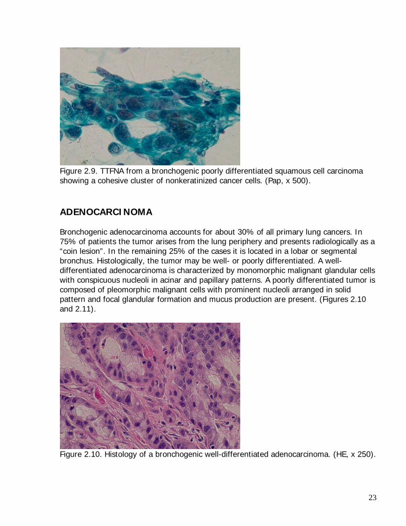
Figure 2.9. TTFNA from a bronchogenic poorly differentiated squamous cell carcinoma showing a cohesive cluster of nonkeratinized cancer cells. (Pap, x 500). ADENOCARCINOMA Bronchogenic adenocarcinoma accounts for about 30% of all primary lung cancers. In 75% of patients the tumor arises from the lung periphery and presents radiologically as a “coin lesion”. In the remaining 25% of the cases it is located in a lobar or segmental bronchus. Histologically, the tumor may be well- or poorly differentiated. A well-differentiated adenocarcinoma is characterized by monomorphic malignant glandular cells with conspicuous nucleoli in acinar and papillary patterns. A poorly differentiated tumor is composed of pleomorphic malignant cells with prominent nucleoli arranged in solid pattern and focal glandular formation and mucus production are present. (Figures 2.10 and 2.11).
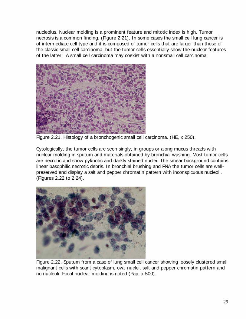
Figure 2.10. Histology of a bronchogenic well-differentiated adenocarcinoma. (HE, x 250).

24
Figure 2.11. Histology of a bronchogenic poorly differentiated adenocarcinoma. (HE, x 250). The cytologic manifestations of bronchogenic adenocarcinomas in sputum and in materials obtained by bronchial washing and brushing and FNA are similar. The malignant glandular cells are present predominantly in small groups with acinar arrangement or in large clusters. Cells from a well-differentiated tumor show fairly uniform nuclei with smooth nuclear contours and conspicuous nucleoli. Cells from a poorly differentiated adenocarcinoma are more pleomorphic and show single or multiple macronucleoli. Intracellular mucus may be demonstrated with mucicarmine or periodic acid-Schiff (PAS) stain with prior diastase digestion. (Figures 2.12-2.15).
Figure 2.12. A bronchogenic well-differentiated adenocarcinoma showing in sputum clustered monomorphic tumor cells with vacuolated cytoplasm and conspicuous nucleoli (Pap, x 500).

25
Figure 2.13. A bronchogenic poorly differentiated adenocarcinoma showing in sputum clustered pleomorphic malignant glandular cells with prominent nucleoli. (Pap, x 500). Cells from a bronchogenic adenocarcinoma contain intracytoplasmic mucin and stain positively with PAS and with PAS with prior diastase digestion. From the immunocytochemical point of view, these cells are CEA, CK7, villin and TTF-1 positive and CK20 negative.
Figure 2.14. Sputum cell block showing a cluster of malignant glandular cells with vacuolated cytoplasm. (HE, x 250).

26
Figure 2.15. TTFNA from a bronchogenic adenocarcinoma showing a cohesive cluster of malignant glandular cells with prominent nucleoli. (Pap, x 500). Bronchioloalveolar carcinoma is a rare subtype of lung adenocarcinoma and it has not been definitely linked to cigarette smoking. It accounts for 1-5% of all primary lung cancers and can be unifocal or multifocal. The tumor is characterized by cuboidal or low columnar tumor cells with conspicuous nucleoli growing along preexisting alveolar walls. It can be mucinous or nonmucinous and intranuclear cytoplasmic inclusions may be present. (Figures 2.16 and 2.17). In sputum, small cuboidal tumor cells with oval nuclei are seen predominantly in tridimensional clusters. In materials obtained by bronchial brushing or FNA the tumor cells are commonly seen in large monolayered sheets with nuclear crowding and overlapping. Intranuclear cytoplasmic inclusions may be noted. (Figures 2.18-2.20). Cells from a mucinous bronchioloalveolar carcinoma are CK7 and CK20 positive and TTF-1 negative. Tumor cells from a nonmucinous tumor may express surfactant proteins (SP-A, pro-SP-B, pro-SP-C).
Figure 2.16. Histology of a nonmucinous bronchioloalveolar carcinoma. (HE, x 250).

27
Figure 2.17. Histology of a mucinous bronchioloalveolar carcinoma. (HE, x 250).
Figure 2.18. Sputum from a patient with nonmucinous bronchioloalveolar carcinoma showing a cohesive cluster of tumor cells with nuclear crowding and molding. (Pap, x 500).
Figure 2.19. TTFNA from a mucinous bronchioloalveolar carcinoma showing a cohesive sheet of mucus-secreting tumor cells with nuclear crowding. (Pap, x 400).

28
A
B Figure 2.20. A. TTFNA from a bronchioloalveolar carcinoma showing tumor cells predominantly in irregular, large, cohesive sheets. (Pap, x 100). B. At higher magnification focal glandular spaces, crowded tumor cells with slightly pleomorphic nuclei and conspicuous nucleoli are observed, as well as intranuclear cytoplasmic inclusions. (Pap, x 500). SMALL CELL CARCINOMA Small cell carcinoma or “oat cell carcinoma” accounts for about 20% of all primary lung cancers. The tumor is related to cigarette smoking and may be associated with a paraneoplastic syndrome (diabetes insipidus or Cushing syndrome). It arises most commonly from major bronchi and forms a perihilar mass and has a rapid growth with early hilar lymph node and distant metastases. About 70% of patients with small cell carcinoma present at an advanced stage when it is detected. Rarely, a small cell carcinoma presents as a “coin lesion”. Histologically, the tumor has a solid growth pattern with extensive necrosis. The tumor cells are small, two to three times the size of a mature lymphocyte and show scant cytoplasm, oval nuclei with finely granular chromatin pattern and inconspicuous
29
nucleolus. Nuclear molding is a prominent feature and mitotic index is high. Tumor necrosis is a common finding. (Figure 2.21). In some cases the small cell lung cancer is of intermediate cell type and it is composed of tumor cells that are larger than those of the classic small cell carcinoma, but the tumor cells essentially show the nuclear features of the latter. A small cell carcinoma may coexist with a nonsmall cell carcinoma.
Figure 2.21. Histology of a bronchogenic small cell carcinoma. (HE, x 250). Cytologically, the tumor cells are seen singly, in groups or along mucus threads with nuclear molding in sputum and materials obtained by bronchial washing. Most tumor cells are necrotic and show pyknotic and darkly stained nuclei. The smear background contains linear basophilic necrotic debris. In bronchial brushing and FNA the tumor cells are well-preserved and display a salt and pepper chromatin pattern with inconspicuous nucleoli. (Figures 2.22 to 2.24).
Figure 2.22. Sputum from a case of lung small cell cancer showing loosely clustered small malignant cells with scant cytoplasm, oval nuclei, salt and pepper chromatin pattern and no nucleoli. Focal nuclear molding is noted (Pap, x 500).

30
Figure 2.23. Small cell carcinoma showing in bronchial brushing tumor cells with salt and pepper chromatin pattern and linear, basophilic nuclear debris. (Pap, x 500).
Figure 2.24. Small cell carcinoma, intermediate cell type showing larger tumor cells and linear basophilic nuclear debris. (Pap, x 500). About 90% bronchogenic small cell carcinomas are chromogranin, synaptophysin, CD56 and TTF-1 positive. LARGE CELL CARCINOMA Large cell carcinoma constitutes about 10% of all bronchogenic carcinomas. Most of these tumors arise from segmental or lobar bronchi. The histologic diagnosis of large cell carcinoma is a diagnosis of exclusion: the tumor does not show any patterns characteristic for a squamous cell carcinoma, adenocarcinoma or small cell carcinoma. Histologically, it is composed of large malignant cells with abundant, granular cytoplasm

31
and macronucleoli. By electron microscopy large cell carcinoma almost always shows focal squamous or glandular differentiation. In cytologic material of all types (sputum, bronchial washing and brushing, FNA) the tumor cells are seen singly and in loose or cohesive aggregates. These are large malignant cells with variably abundant cytoplasm, large nuclei with single or multiple eosinophilic macronucleoli. (Figures 2.25 and 2.26). Cells from a bronchogenic large cell carcinoma are usually CEA, CK7 and TTF-1 positive, and CK20 negative.
Figure 2.25. Single and clustered large tumor cells with single or multiple macronucleoli in bronchial washing of a bronchogenic large cell carcinoma. (Pap, x 500).
Figure 2.26. A cohesive cluster of large tumor cells from a bronchogenic large cell carcinoma in a TBFNA. Single and multiple macronucleoli are present. (Pap, x 500). Giant cell carcinoma is a rare variant of large cell carcinoma (1%) with very poor prognosis. Histologically, it is characterized by giant, bizarre malignant cells with single or

32
multiple nuclei. The tumor yields in sputum and in materials obtained by bronchial washing and brushing or FNA single and loosely clustered giant, bizarre malignant cells with variably abundant cytoplasm, single, multiple and lobulated nuclei with macronucleoli. (Figures 2.27 and 2.28).
Figure 2.27. Histology of a bronchogenic giant cell carcinoma showing bizarre multinucleated giant malignant cells. (HE, x 250).
Figure 2.28. A multinucleated large malignant cell in bronchial brushing of a giant cell carcinoma of the lung. (Pap, x 500). DIAGNOSTIC PITFALLS Cytologic diagnosis of lung cancers is compounded with diagnostic pitfalls. Reactive, hyperplastic or regenerative bronchial epithelial cells, reactive alveolar lining cells, atypical metaplastic squamous cells may be mistaken for malignant cells. Hyperplastic bronchial epithelial cells in patients with chronic obstructive pulmonary disease may form tridimensional clusters with smooth contours or Creola bodies. Patients with viral

33
pneumonitis may exfoliate reactive bronchial epithelial cells in tridimensional clusters with prominent nucleoli, mimicking cells derived from a bronchogenic adenocarcinoma. These cells usually disappear within 2 weeks after the recovery of the lung infection. Patients receiving hyperbaric oxygen therapy for respiratory failure may exfoliate highly atypical reactive alveolar cells mimicking malignant glandular cells. Radiation and chemotherapy may also induced cellular changes, readily mistaken for cancer cells. (Figures 2.29 to 2.37). Those above-mentioned cells lack unequivocal cytologic features of malignant cells such as a high nuclear:cytoplasmic ratio, irregular nuclear contours and hyperchromatic coarsely granular chromatin clumping. Vegetable cells of food origin may sometimes be mistaken for malignant squamous cells by an inexperienced observer. A thick cell wall of a vegetable cell is the clue for a correct cytodiagnosis. Hyperplastic reserve cells and lymphocytes may be mistaken for cells derived from a small cell carcinoma by an inexperienced observer.
Figure 2.29. Reactive bronchial epithelial cells seen in bronchial brushing of a patient with viral pneumonitis. (Pap, x 500).
Figure 2.30. Reactive/regenerative bronchial epithelial cells in bronchial brushing of a patient with viral pneumonitis. (Pap, x 500).

34
Figure 2.31. Hyperplastic bronchial epithelial cells forming a Creola body. (Pap, x 500)
Figure 2.32. A Creola body seen in the sputum of a patient with chronic bronchitis. (Pap, x 250).
Figure 2.33. A cluster of hyperplastic reserve cells showing small cuboidal cells with scant cytoplasm and focal nuclear molding. (Pap, x 500).

35
Figure 2.34. A cluster of hyperplastic alveolar cells in bronchial washing of a patient recovering from a diffuse alveolar cell damage. (Pap, x 500).
Figure 2.35. Atypical metaplastic squamous cells in bronchial brushing. (Pap, x 500).
Figure 2.36. Highly atypical or suspicious epithelial cells in sputum of a patient receiving radiation therapy for mediastinal germ cell tumor. (Pap, x 500).

36
Figure 2.37. Highly atypical epithelial cells of probable alveolar origin in sputum of a patient receiving chemotherapy for acute myelogenous leukemia (Pap, x 500). BIBLIOGRAPHY Colby TV, et al. Tumors of the Lower Respiratory Tract. In Atlas of Tumor Pathology, 3rd series, 1995. Washington DC, Armed Forces Institutes of Pathology. Erozan YS. Cytopathology in pulmonary biopsy procedures. In Biopsy Techniques in Pulmonary Disorders. Wang KP, ed. New York, Raven Press, 1989, p 139. Geisinger KR, et al. Localized lung diseases. In Modern Cytopathology. Philadelphia, Churchill Livingston, 2004, p 399. Jemal A, et al. Cancer statistics, 2008. CA Cancer J Clin. 2008; 58:71. Johnston WW. Cytodiagnosis of lung cancer. Principles and problems. Pathol Res Pract. 1986; 181:1. Koss LG, Melamed MR. Tumors of the lung: conventional cytology and aspiration biopsy. In Koss’ Diagnostic Cytology and Its Histopathologic Bases, Koss LG and Melamed MR, eds. 5th ed, 2006. Philadelphia, Lippincott Williams & Wilkin, p 643. Nguyen GK, et al. Transmucosal needle aspiration biopsy via the fiberoptic bronchoscope. Value and limitations in the cytodiagnosis of tumors and tumor-like lesions of the lung. Pathol Annu. 1992; 27(1):105. Nguyen GK, Kline TS. Essentials of Cytology. An Atlas. New York, Igaku-Shoin, 1993, p 43.

37
Nguyen GK, et al. Cytodiagnosis of bronchogenic carcinoma and neuroendocrine tumor of the lung by transthoracic fine needle aspiration. Diagn Cytopathol.2000;23:431. Shimosato Y, Noguchi M. Pulmonary neoplasms. In Sternberg’s Diagnostic Surgical Pathology, 4th ed, 2004. Mills SE, et al (eds). Philadelphia, Lippincott Williams & Wilkins. p 1173 Singh HK, Silverman JF. Lung, chest wall and pleura. In Fine Needle Aspiration Cytology. Orell SR et al, eds. 4th ed, 2005. Philadelphia, Churchill Livingston, p 227. Tao LC. Lung, Pleura and Mediastinum. In Guides to Clinical Aspiration Biopsy, Kline TS, ed. New York, Igaku-Shoin. 1988. Travis WD, et al. Pathology and Genetics of Tumours of the Lung, Pleura, Thymus and Heart. In WHO Classification of Tumours, Lyon, IARCPress, 2004.

38
Chapter 3
NEUROENDOCRINE CARCINOMAS Pulmonary neuroendocrine neoplasms are one of the most complicated and confusing topics in human pathology. The histogenesis of these neoplasms has been controversial, and their classification has undergone several revisions. Pulmonary neuroendocrine tumors are generally believed to arise from the epithelial neuroendocrine cells. These neoplasms share some common features with other neuroendocrine tumors arising from other anatomic sites, such as neuroendocrine growth patterns (organoid, ribbon/trabecular...), positive reactions to neuroendocrine markers or antibodies (neuron-specific enolase, chromogranin, synaptophysin, and specific peptide hormone, such as calcitonin, serotonin, glucagon antibodies ….), and presence of intracytoplasmic membrane-bound and dense-core neurosecretory granules at ultrastructural levels. Several lung tumors such as small cell carcinoma, well-differentiated adenocarcinoma of fetal type and pulmonary blastoma and a small percentage of nonsmall cell bronchogenic carcinomas show neuroendocrine differentiation by immunohistochemical and ultrastructural studies. TYPICAL CARCINOID TUMOR Typical carcinoid tumors (TCT) of the lung account for 1-2% of all primary lung cancers, occur in all age groups (20-70 years), with a mean of 55 years, and affect men and women equally. About 80% of TCTs are centrally located and 10-20% are found in the periphery of the lung. Most patients with pulmonary TCTs are asymptomatic. However, patients with tumors arising in proximal bronchi may present with dyspnea, hemoptysis and obstructive pneumonia. 2-7% of the patients develop a carcinoid syndrome that is due to an increased production of serotonin, and the majority of these patients have liver metastasis. Some patients present with Cushing syndrome which is secondary to ACTH production by the tumor. At initial diagnosis, metastasis to hilar lymph nodes is present in about 20% of cases. TCTs usually pursue an indolent course, and the 5-year-disease-free survival rate is about 100%. TCT is usually covered with an intact bronchial or squamous metaplastic epithelium and it is composed of uniform small round or cuboidal cells arranged in neuroendocrine growth patterns. The tumor cell nuclei are oval and show a granular chromatin pattern, conspicuous nucleoli, and a scant or moderate amount of pale, clear or eosinophilic cytoplasm. Peripheral TCTs are well-circumscribed, non-encapsulated and generally unrelated to the bronchial tree. These uncommon peripheral tumors account for about 5% of all pulmonary carcinoid tumors and are usually composed of uniformly spindle cells

39
with oblong nuclei showing granular chromatin pattern and inconspicuous nucleoli. Areas showing a TCT may be present elsewhere within the tumor. Fewer than 2 mitoses per 2 square mm and no foci of necrosis are present in TCTs. (Figures 3.1 and 3.2).
Figure 3.1 Histology of a typical carcinoid tumor. (HE, x 250).
Figure 3.2. Histology of a typical carcinoid tumor (HE, x 250). TCT cells may be detected in sputum and bronchial wash if the overlying bronchial mucosa is destroyed by ulceration or tumor invasion. Bronchial brush, TTFNA or TBFNA are effective means to diagnose carcinoid tumors. The cytologic manifestations of a TCT in cell samples obtained by bronchial brush and FNA have characteristic features that are diagnostic of the tumor. The tumor cells are seen singly, in loose aggregates or syncytial clusters. They are polygonal in shape and show either a well-defined, moderately abundant, granular cytoplasm or an ill-defined, scant, pale cytoplasm. The nuclei are oval in shape and show a granular chromatin pattern and conspicuous nucleoli, and nuclear molding are rarely observed. Tumor cells wrapping around capillary blood vessels may be present. The tumor cell cytoplasm stains positively with neuron-specific enolase (NSE) and chromogranin antibodies. (Figures 3.3 to 3.7). It is important to note that the tumor

40
cell nuclei of central TCTs show some similarities with those of benign bronchial glandular epithelial cells. Therefore, cautions should be exercised when interpreting naked nuclei in cell samples taken by bronchial brush or FNA. A TCT may show oncocytic change and yield cells with abundant, granular and eosinophilic cytoplasm mimicking those of a granular cell tumor. (Figure 3.8). Occasionally, a TCT is composed of cells with large intracytoplasmic vacuoles and it yields in TBFNA cells mimicking those of a signet-ring cell adenocarcinoma. Immunocytochemical staining of the tumor cells with NSE and chromogranin antibodies will be helpful for confirmation of the neuroendocrine differentiation of the tumor.
Figure 3.3. Typical carcinoid tumor showing in sputum monomorphic tumor cells with round nuclei and scant cytoplasm. (Pap, x 500).
Figure 3.4. Typical carcinoid tumor showing in bronchial brushing dyshesive monomorphic tumor cells with plasmacytoid configuration. (Pap, x 500).

41
Figure 3.5. Typical carcinoid tumor showing in TBFNA single and clustered monomorphic tumor cells. (Pap, x 500).
A
B Figure 3.6. TBFNA of a typical carcinoid tumor showing tumor cells wrapping around a capillary blood vessel. (Pap, A x 100, B x 500).

42
Figure 3.7. Single and clustered tumor cells aspirated from a typical carcinoid tumor showing immunopositive cytoplasmic reaction with chromogranin antibody. (ABC, x 500).
Figure 3.8. Carcinoid tumor oncocytic change showing clustered tumor cells with abundant, eosinophilic cytoplasm and minimally atypical nuclei. (Pap, x 500). Peripheral TCTs with spindle cells yield in needle aspirate randomly arranged, uniform, spindle tumor cells with oval or spindle nuclei displaying a granular chromatin pattern and inconspicuous nucleoli. (Figures 3.9 and 3.10). Cells from a central TCT should be differentiated from hyperplastic reserve cells, lymphoid cells, cells from a small-cell adenocarcinoma or small-cell carcinoma. Cells from a spindle-cell tumor may be mistaken for those of a metastatic melanoma, spindle-cell squamous cell carcinoma, metastatic thyroid medullary carcinoma, spindle-cell thymoma and soft tissue tumors. Immunocytochemical staining with NSE and chromogranin and/or calcitonin and CEA antibodies is helpful in difficult cases.

43
Figure 3.9. Peripheral typical carcinoid tumor with spindle tumor cells. (HE, x 250).
Figure 3.10. TTFNA of a peripheral carcinoid tumor showing dyshesive spindle tumor cells with elongated nuclei and scant cytoplasm in no specific pattern. (Pap, x 500). ATYPICAL CARCINOID TUMOR Atypical carcinoid tumors (ACT) are rare neoplasms and account for less than 25% of all pulmonary carcinoid tumors. At initial diagnosis 70% of patients with ACT already have hilar lymph node metastasis, and distant metastasis is present in about 20% of the cases. The treatment of choice for an ACT is surgical resection. Post-operative adjuvant chemotherapy with or without radiotherapy has limited affects, and the 5-year survival rate is about 70%. ACTs are composed of more pleomorphic and larger tumor cells arranged in neuroendocrine patterns. Mitoses are abundant and tumor necrosis is common. As TCT, an ACT may be covered by an intact bronchial mucosa, and therefore, it may not show any tumor cells in sputum. In materials obtained by bronchial brushing or FNA the tumor

44
cells are seen singly and in loose or tight aggregates. Nuclear pleomorphism with granular chromatin pattern and conspicuous nucleoli are prominent features. 2-10 mitoses per 2 square mm and/or foci of necrosis are present. An ACT may have an endobronchial component that is composed of a TCT. In this case cell samples procured by bronchial brushing may show only cells with features of a TCT. (Figures 3.11 and 3.12).
Figure 3.11. Histology of an atypical carcinoid tumor showing more pleomorphic neoplastic cells. (HE, x 250).
Figure 3.12. Atypical carcinoid tumor showing in TBFNA more pleomorphic tumor cells. Small and conspicuous nucleoli are present in some tumor cells. (Pap, x 500). In some cases, cells derived from a small-cell cancer may simulate those of a TCT. Staining of the tumor cells with a proliferative cell marker such as Ki-67 or MIB-1 may provide useful information for tumor grading. Over 50% of tumor cells from a lung small cell carcinoma show an immuno-positive nuclear reaction while fewer than 25% of the tumor cell nuclei derived from a TCT or ACT stain positively with this antibody.

45
(Figure 3.13). For TTF-1 varying results have been reported. According to some studies cells of a TCT and ACT are usually TTF-1 negative. In other studies about 30% of lung TCTs and ACTs are TTF-1 positive. Several lung carcinoid tumors are positive for CD99.
Figure 3-13. Cell block section from TBFNA of an atypical carcinoid tumor showing positive nuclear staining with Ki-67 antibody (ABC, x 200). LARGE CELL NEUROENDOCRINE CARCINOMA Large cell neuroendocrine carcinoma (LCNC) is rare and highly aggressive tumor occurring in adults with a median age of 64 years (range, 35 to 75 years). Most patients are heavy smokers and ectopic hormone production is not observed. The neoplasm may be centrally or peripherally located and averages 3 cm in greatest dimensions (range 1.3 to 10 cm). This tumor does not appear to be a specific entity and behaves similarly to a bronchogenic large cell carcinoma. LCNC consists of large pleomorphic malignant cells arranged in neuroendocrine pattern with focal rosette formation. Mitotic figures are abundant and geographic necrosis is common.
Figure 3.13. Histology of a lung large cell neuroendocrine carcinoma. (HE, x 250).

46
In cell samples obtained by bronchial brushing or FNA the tumor cells are seen singly and in loose aggregates. They are large, pleomorphic and display well-defined, granular cytoplasm and oval nuclei with granular chromatin pattern and prominent nucleoli, mimicking those of a large cell carcinoma. Tumor cells arranged in tridimensional clusters may be seen. Necrotic debris, naked nuclei, nuclear streaking and tumor cells arranged in linear pattern and in rosettes have been reported (Figures 3.13 and 3.14). Staining with NSE and chromogranin antibodies will be helpful for confirming the neuroendocrine differentiation of the tumor cells examined. About 50% of large cell neuroendocrine carcinomas are TTF-1 positive.
Figure 3.14. TTFNA of a large cell neuroendocrine carcinoma showing large, pleomorphic malignant epithelial cells with abundant cytoplasm, oval nuclei and prominent nucleoli. Some tumor cells show a plasmacytoid configuration. (Diff-Quik, x 400). Acknowledgement: Professor N. Shapiro, Editor-in-Chief of Russian Journal of Clinical Cytology, Moscow, Russia, has kindly granted his permission for reusing of some parts of the text and microphotographs from the author’s paper in this chapter (Nguyen GK. Cytology of neuroendocrine cancer of the lung. Russian Journal of Clinical Cytology. 2004; 8 (3-4):19-23). BIBLIOGRAPHY Anderson C, et al. Fine needle aspiration cytology of pulmonary carcinoid tumors. Acta Cytol 190;34:505. Brambilla E. Classification of broncho-pulmonary cancers (WHO 1999). Rev Mal Respir. 2002; 19:409. Colby TV, et al. Tumors of the lower respiratory tract. In Atlas of tumor pathology, 3rd series, Washington DC, Armed Forces Institute of Pathology, 1995, p 235, 287.

47
Ionescu DN, et al. Nonsmall cell lung carcinoma with neuroendocrine differentiation-an entity of no clinical or prognostic significance. Am J Surg Pathol. 2007; 31:26. Kakinuma H, et al. Diagnostic findings of bronchial brush cytology for pulmonary large cell neuroendocrine carcinomas. Comparison with poorly differentiated adenocarcinomas, squamous cell carcinomas, and small cell carcinomas. Cancer (Cancer Cytopathol) 2003; 99:247. Lin O, et al. Immunohistochemical staining of cytologic smears with MIB-1 helps distinguish low-grade from high-grade neuroendocrine neoplasms. Am J Clin Pathol 2003; 120:209-216. Mitchell MI, Parker FP. Capillaries: a cytologic feature of pulmonary carcinoid tumors. Acta Cytol 1991; 35: 183. Nguyen GK, et al. Transmucosal needle aspiration biopsy via the fiberoptic bronchoscope. Value and limitations in the cytodiagnosis of tumors and tumor-like lesions of the lung. Pathol Annu 1992; 27 (1):105. Nguyen GK. Cytopathology of pulmonary carcinoid tumors in sputum and bronchial brushing. Acta Cytol 1995; 39:1152. Nguyen GK, et al. Cytodiagnosis of bronchogenic carcinoma and neuroendocrine tumor of the lung by transthoracic fine-needle aspiration. Diagn Cytopathol 2000; 23:431. Ogino S, et al. Cytopathology of oncocytic carcinoid tumor of the lung mimicking granular cell tumor. A case report. Acta Cytol 2000; 44:247-250. Szyfelbein WM, Ross SS. Carcinoids, atypical carcinoids and small cell carcinomas of the lung. Diagn Cytopathol 1988; 4:1. Travis WD, et al. Pathology and Genetics of Tumours of the Lung, Pleura, Thymus and Heart. In WHO Classification of Tumours, Lyon, IARCPress, 2004. Wiatrowska BA, et al. Large cell neuroendocrine carcinoma of the lung: proposed criteria for cytologic diagnosis. Diagn Cytopathol 2001;24:58.

48
Chapter 4
OTHER PRIMARY TUMORS AND TUMORLIKE LESIONS MALIGNANT TUMORS BRONCHIAL GLAND CARCINOMA Bronchial gland carcinomas are rare neoplasms occuring in adult patients. These neoplasms may manifest with hemoptysis or bronchial obstruction with distal lung infection. They account for about 1% of all primary lung cancers and consist of two lesions: Adenoid cystic carcinoma and mucoepidermoid carcinoma. Adenoid Cystic Carcinoma is the most common salivary gland-like tumor of the lower respiratory tract. It accounts for about 0.2% of all primary lung cancers. The neoplasm usually arises from the trachea, main stem bronchi or lobar bronchi. The patient’s age ranges from 18 to 79 years. It is a less aggressive neoplasm and has a distinct histologic pattern of growth consisting of cribiforms and glandular arrays or tubules surrounding central spaces filled with epithelial mucin and solid foci. (Figure 4.1). The tumor yields in bronchial brushing and TBFNA single and clustered small, round cells with scant cytoplasm and round basophilic bodies. Tumor cells wrapping around basophilic bodies are a diagnostic feature of the lesion. (Figures 4.2 and 4.3).
Figure 4.1. Histology of a bronchial adenoid cystic carcinoma. (HE, x 250).

49
A
B Figures 4.2. TBFNA of a bronchial adenoid cystic carcinoma showing in A single and clustered small cuboidal neoplastic cells and in B an eosinophilic amorphous round body wrapped by small tumor cells. (HE, x 400).
Figure 4.3. TTFNA from a pulmonary adenoid cystic carcinoma reveals small tumor cells wrapping around ball-like, basophilic, amorphous bodies and a basophilic round body at the left lower corner of the figure. (Pap, x 250).

50
Mucoepidermoid Carcinoma is a rare tumor comprising 0.1 to 0.2% of all primary lung carcinomas. The patients range in age from 4 to 78 years but about 50% are younger than 30 years. The tumor most commonly arises from the main or lobar bronchus and can measures up to 6 cm in greatest dimension. Histologically, it consists of a variable population of mucus-secreting cells, squamous cells and intermediate cells that display no particular differentiating characteristics. Bronchial mucoepidermoid carcinomas can be classified as low- and high-grade tumors, depending on the degree of cellular atypia. About 75% to 80% of mucoepidermoid carcinomas arising from the lung are of low histologic grade. A low-grade tumor yields in FNA single and clustered benign-appearing squamous cells admixed with benign-appearing mucus-secreting cells. A variable number of intermediate cells may be present. A high-grade tumor yields loosely clustered malignant squamoid cells containing intracytoplasmic mucus. (Figures 4.4 to 4.7).
Figure 4.4. Histology of a bronchial low-grade mucoepidermoid carcinoma showing polygonal squamoid cells admixed with glandular cells with clear cytoplasm. (HE, x 250).
Figure 4.5. FNA of a low-grade mucoepidermoid carcinoma showing loosely clustered squamoid tumor cells with some cells showing vacuolated “clear” cytoplasm. A few small cuboidal tumor cells in the lower part of the figure are of intermediate type. (Pap, x 500).

51
Figure 4.6. Histology of a high-grade mucoepidermoid carcinoma of the bronchus showing nests of invasive malignant squamous cells with pleomorphic, hyperchromatic nuclei. Some tumor cells have a “clear” cytoplasm. (HE, x 250).
Figure 4.7. TBFNA of a bronchial high-grade mucoepidermoid carcinoma showing clustered malignant squamoid cells with intracytoplasmic mucus that stains positively with PASD. (PASD, x 400). WELL DIFFERENTIATED FETAL ADENOCARCINOMA This rare cancer is related to cigarette smoking and commonly occurs in 5th or 6th decade of life. It usually pursues a less aggressive clinical course. Histologically, the tumor is composed of low-grade malignant glandular cells arranged in acinar pattern. Focal tumor cell morulae containing intracytoplasmic neurosecretory granules, as demonstrated by electron microscopy and by immunohistochemical staining with neuron-specific enolase and chromogranin antibodies, are present. (Figure 4.8). Only a few tumors of this type with cytologic evaluation have been encountered in the literature. In one case the TTFNA

52
revealed large monolayered and folded sheets of low-grade malignant columnar epithelial cells with clear or granular cytoplasm and uniformly oval, small nuclei with inconspicuous nucleoli. Focal glandular arrangement may be visualized within a tumor cell sheet. (Figure 4.9)
Figure 4.8. Histology of a pulmonary well-differentiated adenocarcinoma, fetal type (WDAFT) showing an intraglandular morule of tumor cells. (HE, x 250).
Figure 4.9. TTFNA of a lung WDAFT showing a large sheet of tumor cells with honeycomb pattern. A round glandular space is noted. (Pap, x 250). PULMONARY BLASTOMA This rare lung cancer is seen in adult patients and it is related to cigarette smoking. It is composed of fetal-type glandular elements admixed with spindle-shaped cells and cartilage and bone may be present. A few examples of this neoplasm with cytologic evaluation by TTFNA have been reported and both types of above-mentioned cells were observed.

53
LUNG SARCOMA Primary lung sarcomas are exceedingly rare neoplasms and account for less than 1% of all primary lung cancers. Almost all histologic types of soft-tissue sarcomas have been reported in the lung. Practically, a diagnosis of primary lung sarcoma can only be made if the patient has no history of a treated soft-tissue sarcoma, and no sarcoma is detected by extensive clinical and diagnostic imaging studies. In one large surgical series there were one sarcoma for every 500 bronchogenic carcinomas. Of the primary lung sarcomas, leiomyosarcoma is the most common one, and about 100 cases of this neoplasm have been reported in the literature. The tumor occurs mainly in adults and rarely in children. It may arise from a bronchus or from intraparenchymal blood vessels. The cytologic manifestations of a primary lung leiomyosarcoma in bronchial brushing and in FNA are similar and consist of scattered loosely aggregated, elongated slightly pleomorphic, hyperchromatic, naked tumor cell nuclei with blunt ends. Bundle of smooth muscle cells may be present in materials obtained by bronchial brushing (Figures 4.10 and 4.11). Cells derived from a fibrosarcoma or neurogenic sarcoma are morphologically similar to those of a leiomyosarcoma. A positive cytoplasmic reaction with smooth muscle and desmin antibodies will confirm the diagnosis of a leiomyosarcoma.
Figure 4.10. TTFNA of a well-differentiated leiomyosarcoma of the lung showing single and loosely clustered tumor cells with elongated nuclei with blunt ends (Pap, x 400).

54
Figure 4.11. Bronchial brushing from a low-grade leiomyosarcoma arising from a lobar bronchus reveals bundles of malignant smooth muscle cells with enlarged, elongated, oval or polygonal nuclei (Pap, x 400). Embryonal or alveolar rhadomyosarcoma of the lung yields malignant small round cells with scant cytoplasm. (Figure 4.12). A positive reaction of the tumor cell cytoplasm with MyoD1 or myogenin antibodies will be helpful for a more accurate tumor typing.
Figure 4.12. Primary lung embryonal rhabdomyosarcoma showing in TTFNA small round malignant cells with hyperchromatic nuclei and scant cytoplasm (Pap, x 400). Pulmonary artery angiosarcoma is a rare neoplasm of adults. In one reported case the tumor showed in bronchial washing single and clustered small malignant cells with scant cytoplasm and hyperchromatic oval nuclei. Focal luminal formation was noted in some tumor cell clusters. A positive reaction of the tumor cell cytoplasm with factor VIII related antigen antibody will confirm the diagnosis of this lung cancer.

55
BENIGN LUNG TUMORS AND TUMORLIKE LESIONS HAMARTOMA This benign tumor most commonly occurs in 6th decade of life. It is usually asymptomatic and often discovered incidentally by chest roentgenograms. It is usually located in the peripheral zone of the lung. If located in a bronchus it may cause bronchial obstruction with distal bronchial infection. The lesion is well-circumscribed, lobulated and usually measures 2 cm in greatest dimension. It is formed by elements that are normally present in the lung such as cartilage, fibromyxoid connective tissue, fat, smooth muscles and respiratory epithelium. It shows in TTFNA an admixture of the above-mentioned cytologic elements. (Figures 4.13 and 4.14).
Figure 4.13. Histology of a lung hamartoma. (HE, x 100).
A

56
B Figures 4.14.TTFNA of a lung hamartoma showing in A myxoid material, chondrocytes and clusters of benign bronchial glandular cells, and in B a large fragment of benign cartilaginous tissue. (Pap, x 400). GRANULAR CELL TUMOR This is a rare benign neoplasm arising from the Schwann cell. In over 90% of cases, the tumor has an endobronchial component, and in less than 10% of patients it presents as a parenchymal lesion and appears on chest roentgenograms as a “coin lesion”. It yields in bronchial brushing or submucosal needle aspirate sheets of benign tumor cells with eosinophilic, granular and PAS-positive cytoplasm and small, oval nuclei with conspicuous nucleoli. (Figures 4.15 and 4.16).
Figure 4.15. Histology of a bronchial granular cell tumor showing neoplastic cells with granular and slightly PAS-positive cytoplasm. (PAS, x 200).

57
Figure 4.16. A thick fragment of tumor tissue in a submucosal FNA showing benign neoplastic cells with oval nuclei and ill-defined, granular cytoplasm. (Pap, x 400). CLEAR CELL “SUGAR” TUMOR This is a rare benign lung tumor of unknown histogenesis. It consists of spindle-shaped cells with “glycogen-rich”, clear cytoplasm. In one reported case the tumor yielded in TTFNA large clusters of spindle cells with clear cytoplasm and oval or elongated bland nuclei. Intracytoplasmic glycogen can be demonstrated by staining of the tumor cells with PAS reagent. (Figures 4.17 and 4.18).
Figure 4.17. Histology of a benign clear cell “sugar” lung tumor showing spindle tumor cells with clear cytoplasm and round or elongated nuclei. (HE, x 250).

58
A
B Figures 4.18. TTFNA from a benign clear cell tumor of the lung showing in A a large cluster of spindle tumor cells with oval or elongated, bland nuclei, and in B, an aggregate of benign epithelial-like cells with ill-defined cytoplasm, round or oval nuclei and thin, semitransparent, “clear” cytoplasm. (HE, A x 250, B x 400). SQUAMOUS CELL AND GLANDULAR CELL PAPILLOMAS These are very rare benign endobronchial lesions that may cause hemoptysis or bronchial obstruction with distal bronchial and pulmonary infection. Biopsy of the lesion may cause severe hemorrhage. The squamous cell papilloma may be solitary, multiple, exophytic or endophytic. Solitary squamous cell papilloma is seen mainly in men in their fifth decade of life and is more commonly exophytic. It may be associated with human papilloma virus (HPV) subtypes 6 and 11, suggesting a possible pathogenetic role for the virus. HPV subtypes 16, 18 and 31/33/35 in squamous cell papillomas associated with carcinomas and in squamous cell carcinomas have been reported, suggesting that HPV infection might be related to tumoral progression. Depending on the histologic type, benign squamous cells and glandular cells are seen in materials obtained by bronchial washing or brushing. The squamous cell tumor associated with HPV infection may show histologic features of a papillary condyloma and yields in bronchial cytologic materials dyskaryotic squamous cells with perinuclear halos. (Figures 4.19 and 4.20). The glandular cell

59
papilloma exfoliates benign bronchial glandular cells and cannot be identified cytologically.
A
B Figure 4.19. Histology of a solitary bronchial squamous cell papilloma associated with HPV 6 infection showing its squamous epithelial lining with mild dysplasia and dyskaryotic koilocytes. (HE, A x 4, B x 250).
Figure 4.20. Dyskaryotic squamous cells with one showing koilocytic change in bronchial washing of a patient with bronchial squamous cell papilloma. (Pap, x 500).

60
PULMONARY AMYLOIDOSIS This lesion most commonly occurs in patients over 60 years of age. It usually diffusely involves the submucosa of the tracheobronchial tree but it may appear as a parenchymal mass lesion. The bronchial lesion may mimic a submucosal tumor and it yields in bronchial brushing or TBFNA irregular masses of amorphous, granular, waxy material that stains slightly eosinophilic or basophilic with the Papanicolaou stain and orangeophilic with Congo red. (Figures 4.21 and 4.22).
Figure 4.21. Histology of a bronchial amyloid deposit covered with a benign metaplastic squamous epithelium. (HE, x 250).
Figure 4.22. Bronchial brushing showing irregular, ill-defined masses of amorphous, waxy, granular and orangeophilic amyloid material. (Pap, x 500). WEGENER GRANULOMATOSIS This systemic necrotizing vasculitis of unknown etiology is characterized by granulomatous lesions in the nose, nasal sinuses, lung and kidney. Over 90% of patients

61
have ANCA in their blood. Of those, 75% are C-ANCA. Persistent bilateral pneumonitis and chronic sinusitis are prominent clinical manifestations, and hematuria and proteinuria are indicative of renal involvement. In the lung the granulomata may measure up to 5 cm in greatest dimension and may mimic a neoplasm radiologically. Untreated disease is fatal, and immunosuppressive treatment with cyclophosphamide usually results in marked improvement. A TTFNA or bronchial brushing of the lung lesion reveals granular debris of necrotic collagen admixed with chronic inflammatory cells. Multinucleated giant cells and epithelioid cells may be seen. INFLAMMATORY PSEUDOTUMOR This lesion is also known as inflammatory fibroblastic tumor of the lung is a rare lesion that usually develops after a nonspecific pulmonary inflammation. It occurs in men or women, usually before the age of 40. Most of these lesions are contained within the lung and appear as a circumscribed, nodular mass consisting of an admixture of fibroblastic cells, myoepithelial cells and chronic inflammatory cells such as lymphocytes, plasma cells and macrophages. The above-mentioned cellular elements may be seen in TTFNA. (Figures 4.23). The majority of these lesions are benign, but about 5% of them are aggressive and invade adjacent structures such as esophagus, mediastinum, diaphragm and chest walls.
A

62
B Figures 4.23. TTFNA of a pseudo-inflammatory tumor of the lung reveals irregular bundles of fibroblastic cells admixed with scattered chronic inflammatory cells. (Pap, A x 100, B x 400). BIBLIOGRAPHY Awasthi A, et al. Pitfalls in the diagnosis of Wegener’s granulomatosis on fine needle aspiration cytology. Cytopathology. 2007;18:8. Colby TV, et al. Tumors of the Lower Respiratory Tract. In Atlas of Tumor Pathology, 3rd series,1995. Geisinger KR, et al. Localized lung diseases. In Modern Cytopathology, Geisinger KR, et al, eds. Philadelphia, Churchill Livingstone.2004, p.399. Gray JA, Nguyen GK. Primary pulmonary rhabdomyosarcoma diagnosed by fine needle aspiration cytology. Diagn Cytopathol.2003;29:181. Husain M, Nguyen GK. Cytopathology of granular cell tumor of the lung. Diagn Cytopathol.2000;23: 294. Machicao CN, et al. Transthoracic needle aspiration biopsy of inflammatory pseudotumors of the lung,. Diagn Cytopathol.1989;5:400. Naryshskin S, Young NA. Respiratory cytology: a review of non-neoplastic mimics of malignancy. Diagn Cytopathol.1993;9:89. Micheal CW, Flint A. The cytologic features of Wegener’s granulomatosis. Am J Clin Pathol. 1998;110:10.

63
Nguyen GK. Exfoliative cytology of angiosarcoma of the pulmonary artery. Acta Cytol. 1985;29:627. Nguyen GK. Cytology of bronchial gland carcinoma. Acta Cytol.1988;32:235. Nguyen GK. Aspiration biopsy cytology of benign clear-cell “sugar” tumor of the lung. Acta Cytol.1989;33:511. Nguyen GK. Fine needle aspiration cytology of well-differentiated fetal adenocarcinoma (endodermal tumor) of the lung. Acta Cytol. 2001;45:475. Odashiro AN, et al. Primary lung leiomyosarcoma detected by bronchoscopy cytology. Diagn Cytopathol.2005;33:220. Shimosato Y, Noguchi M. Pulmonary neoplasms. In Sternberg’s Diagnostic Surgical Pathology. 4th ed, 2004. Mills SE, et al, eds. Philadelphia, Lippincott Williams & Wilkins, 1173. Tao LC. Lung, Pleura and Mediastinum. In Guides to Clinical Aspiration Biopsy, Kline TS, ed. New York, Igaku-Shoin, 1988. Travis WD, et al. Pathology and Genetics of Tumours of the Lung, Pleura, Thymus and Heart. In WHO Classification of Tumours. Lyon, IARCPress,2004.

64
Chapter 5 METASTATIC CANCERS The lung is one of the most common sites of metastasis from extrathoracic cancers. From 20 to 60% of individuals with extrathoracic solid cancers show, at autopsy, lung metastases; and lung is the only site of metastasis in 15 to 25% of these cases. Carcinomas arising from the breast, prostate, testicles and kidney, cutaneous melanoma, Ewing sarcoma, osteogenic sarcoma and rhabdomyosarcoma frequently metastasize to the lung. MACROSCOPIC PATTERNS OF METASTATIC CANCERS Metastatic cancers in the lung display some distinctive patterns of metastasis such as multiple tumor nodules, lymphangitic, endobronchial, endovascular, solitary and pleural. An awareness of these macroscopic patterns of metastasis is helpful for a more accurate cytodiagnosis of secondary lung cancers. A summary of patterns of metastasis in the lung, as described by Colby et al. is summarized below. Multiple and bilateral masses of metastatic tumor of different sizes is the most common pattern of lung metastasis. Concomitant lymphangitic, endobronchial and endovascular tumor deposits may also be present. This pattern of metastasis is most commonly seen in patients with sarcoma, renal cell carcinoma, cutaneous melanoma and colorectal carcinoma. Lymphangitic pattern accounts for 6-8% of all lung metastases. It is characterized by a diffuse, linear and nodular thickening of bronchovascular bundles, interlobular septae and subpleural spaces. About 80% of the metastatic tumors are adenocarcinomas arising from the lung, breast, gastrointestinal tract and pancreas. This pattern of metastasis can be diagnosed by CT scan or chest roentgenograms. Endobronchial metastasis is found at autopsy in 18-51% of patients with extrathoracic cancers. In most cases the bronchus is invaded by metastatic cancer deposits in adjacent lung parenchyma or lymph nodes. Metastasis involving only a bronchus is uncommon and is found in less than 5% of patients with solid cancers arising from the head and neck, breast, colon, kidney and soft tissue. Endobronchial metastases may mimic a bronchogenic cancer at bronchoscopy.

65
Metastatic tumor embolization is not uncommon. It is found at autopsy in 2-26% of patients with solid cancer, and it can be diagnosed during life by cytologic examination of blood obtained by wedge pulmonary artery catheterization. Renal cell, hepatocellular and gastric carcinomas, choriocarcinoma and chondrosarcoma more commonly show this pattern of metastasis. Solitary metastasis is not uncommon. About 1-5% of lung metastases are solitary and 3-9% of all solitary lung nodules are metastatic deposits. Cutaneous melanoma, renal cell carcinoma, and colonic carcinoma, breast carcinoma, urinary bladder carcinoma, soft tissue sarcomas and non-seminomatous testicular cancers most frequently cause of solitary lung metastasis. Pleural metastasis is seen in the setting of lymphatic or vascular spread with contiguous extension from parenchymal tumor deposits. It is commonly associated with malignant pleural effusions. Macroscopically, the pleura shows multiple and scattered tumor nodules of different sizes ranging from miliary to dominant masses. A diffuse infiltrating pattern, as seen in pleural mesothelioma, may be observed. CYTOLOGY OF METASTATIC CANCERS Endobronchial metastatic cancers may exfoliate their cells in sputum and different bronchoscopy cytologic specimens. Lung parenchymal deposits are best diagnosed with TTFNA. BAL may show cancer cells with alveolar spread. The cytologic manifestations of metastatic cancers to the lung are somewhat similar in different types of pulmonary cell samples and display similar features with those of their primary cancers arising from different anatomic locations. Clinical history and a comparison of the metastatic cancer cells with the histologic sections or cytologic samples of the primary cancers, if available, are of diagnostic help in the majority of cases. Diagnosis of solitary metastasis is important for patient care, as a second or a third primary cancer may develop in patients who had a surgically removed primary cancer many years prior. As in the liver, diagnosis of metastatic adenocarcinoma to the lung is challenging. An awareness of the incidences of metastasis of cancers arising from different organs or anatomic sites can be of diagnostic help. Immunocytochemical studies of the cell samples or aspirated minute tumor tissue fragments with selected antibodies may be required for a more accurate tumor typing in some cases. On rare occasions, electron microscopic studies of aspirated tumor tissue fragments are needed for differential diagnoses. Since there are numerous histologic types of solid tumors, therefore, it would be more convenient to discuss the cytologic manifestations of metastases from primary cancers arising in different anatomic locations or sites.

66
Breast Mammary carcinomas frequently metastasize to the lung. The tumors yield malignant glandular cells with nonspecific features. Tumor cells arranged in linear pattern may be observed. (Figure 5.1). These cells usually express estrogen and progesterone receptors and CK7 and stain negatively with CK20 and TTF-1 antibodies.
A
B Figure 5.1. Metastatic mammary duct carcinoma showing in: A. TTFNA clustered malignant glandular cells with focal nuclear crowding. Tumor cells in linear arrangement are noted elsewhere. (Diff-Quik, x 400). B. Tumor cells in a cell block section showing positive nuclear staining with ER antibody. (ABC, x 200). Thyroid About 15% of thyroid carcinomas metastasize to the lung. Metastatic cancers are most frequently derived from an anaplastic carcinoma then from a poorly differentiated or well-differentiated carcinoma. Metastatic papillary carcinoma shows papillary tissue fragments, sheets or groups of tumor cells with nuclear crowding, intranuclear cytoplasmic inclusions

67
and nuclear grooves. (Figure 5.2). A follicular carcinoma yields cells in clusters with focal acinar arrangement. A Hürthle cell carcinoma shows tumor cells with granular cytoplasm singly and in monolayered sheets. A medullary carcinoma may show single and clustered polygonal and/or spindle tumor cells with elongated nuclei. Intranuclear cytoplasmic inclusions may be noted and cytoplasmic azurophil granules may be observed in tumor cells stained with MGG or Diff-Quik technique (Figure 5.3). Cells derived from an anaplastic carcinoma are either large pleomorphic or spindle. Tumor cells from a papillary, follicular, Hürthle cell and insular carcinoma express thyroglobulin and TTF-1 while those of a medullary carcinoma stain positively with calcitonin and carcinoembryonic antigen antibodies. Cells derived from an anaplastic carcinoma may not show any positive staining reactions with thyroglobulin or TTF-1 antibodies.
Figure 5.2. Metastatic papillary carcinoma of the thyroid, follicular variant showing clustered tumor cells displaying nuclear crowding. Intranuclear cytoplasmic inclusions are observed in some tumor cells. (Diff-Quik, x 500).
Figure 5.3. Metastatic thyroid medullary carcinoma to the lung showing tumor cells with plasmacytoid configuration. (Diff-Quik, x 500).

68
Gastrointestinal tract, pancreas and biliary tree Metastatic tumors from a poorly differentiated adenocarcinomas arising from the stomach, small and large bowels, pancreas or biliary tree yields malignant cells with no specific features. Staining of the tumor cells with CDX2, CK7, CK20, MUC-2 and MUC-5 antibodies may be useful for determining the site of the primary tumor. Cells from a biliary or pancreatic tumor are usually monoclonal CEA (+), CDX2 (-), CK7 (+), CK20 (-) and MUC-5 (+) while those of colorectal origin are usually CDX2 (+), CK7 (-), CK20 (+) and MUC-2 (+). Cells with signet-ring configurations are most commonly derived from a signet-ring cell carcinoma of the stomach. Cells from a well- or moderately differentiated colonic adenocarcinoma are seen in sheets with elongated nuclei in picket fence pattern. A large amount of necrotic debris is commonly noted in a FNA from a metastatic colonic adenocarcinoma. (Figures 5.4 and 5.5).
Figure 5.4. Irregular sheets of a metastatic colonic adenocarcinoma to the lung showing a large amount of necrotic debris and irregular sheets of tumor cells with cells at periphery arranged in palisade. (Pap, x 250).

69
Figure 5.5. An endobronchial metastatic colonic adenocarcinoma yields in bronchial brushing tumor cells with nuclei at periphery arranged in palisade. (Pap, x 500). Liver Hepatocellular carcinomas commonly spread to the lung. Single and clustered polygonal cells with granular or vacuolated cytoplasm are seen, and intracytoplasmic globular inclusions may be observed. (Figures 5.6 and 5.7). Cells derived from a hepatocellular carcinoma do not express CK7 or CK20. A positive staining of the tumor cell cytoplasm with alpha-fetoprotein or HepPar 1 antibody will confirm the diagnosis of a metastatic hepatocellular carcinoma.
Figure 5.6. Metastatic hepatocellular carcinoma showing in BAL a tumor cell with intra cytoplasmic globular inclusion. (Pap, x 500).
Figure 5.7. Histology of a moderately differentiated hepatocellular carcinoma metastatic to the lung showing tumor cells in Figure 5.6. Note the presence of numerous intracytoplasmic globular inclusions. (HE, x 250).

70
Salivary glands Of the salivary gland carcinomas, adenoid cystic carcinoma most commonly metastasizes to the lung. It is characterized in FNA small hyperchromatic cells in acinar arrangement. Globular bodies of amorphous, basophilic material may be present in smear background and globular bodies wrapped with tumor cells are commonly seen. Urinary tract and adrenal Renal cell carcinomas (RCC) commonly metastasize to the lung. A conventional RCC yields cells with clear or granular cytoplasm singly, in clusters and in monolayered sheets. (Figures 5.8). A metastatic papillary RCC yields in FNA monolayered sheets of monomorphic tumor cells with clear or granular cytoplasm, and papillary tumor tissue fragments with fibrovascular core may be present. RCC cells stain positively with RCC and negatively with CK7 and CK20 antibodies.
A
B Figures 5.8. Metastatic grade 3/3 renal cell carcinoma to the lung showing in TTFNA: A. Single tumor cells with granular, thick cytoplasm and pleomorphic, single or multiple nuclei. B. Sheets of tumor cells with clear or granular cytoplasm and prominent nucleoli. (A. HE, x 200; B. Pap, x 500).

71
Cells from a conventional RCC and an adrenal cortical carcinoma are morphologically similar and express both cytokeratin and vimentin. Adrenal cortical carcinoma cells express, in addition, melan A or A103. By electron microscopy, adrenal cortical tumor cells show abundant intracytoplasmic smooth endoplasmic reticula (ultrastructural features of steroid secreting cells) while those of a RCC contains intracytoplasmic fat droplets, glycogen granules and a normal number of smooth endoplasmic reticula. A metastatic chromophobe RCC yields cells similar to those of a conventional RCC. Perinuclear clear spaces and positive cytoplasmic staining with colloidal iron are other characteristic cellular features of the tumor. Abundant intracytoplasmic microvesicles are seen by electron microscopic study of aspirated minute tumor tissue fragments. A metastatic high-grade transitional cell carcinoma of the renal pelvis or urinary bladder is characterized by pleomorphic malignant epithelial cells singly and in clusters. Tumor cells with cytoplasmic extension or “tail” (cercariform cells) are commonly seen and constitute a fairly reliable feature for this type of neoplasm. (Figures 5.9). Urothelial cancer cells may express uroplakin III (URO III), CK7 and CK20.
A
B Figures 5.9. A. TTFNA of a metastatic grade 3/3 transitional cell carcinoma of the urinary bladder to the lung showing pleomorphic malignant cells. A few tumor cells with cytoplasmic tails or “cercariform cells” are noted in Figure 5.9 B. (Pap, A x 500, B x 200).

72
Prostate A metastatic prostatic adenocarcinoma shows clusters and sheets of small glanfular cells with clear cytoplasm and round nuclei with prominent nucleoli. The tumor cell cytoplasm characteristically stains positively with prostatic specific antigen antibody. (Figures 5.10).
A
B Figures 5.10. Figure A showing cohesive small malignant cells in bronchial brushing of an endobronchial metastatic prostatic adenocarcinoma. The tumor cells in Figure B stain positively with prostatic specific antigen antibody. (A: Pap, x 500; B: ABC, x 500). Uterus Cervical squamous cell carcinomas frequently spread to the lung while endocervical adenocarcinomas rarely do so. Endometrial adenocarcinoma also rarely metastasizes to the lung. Its tumor cells express CA 125. A metastatic low-grade endometrial stromal sarcoma shows in TTFNA abundant single and clustered small round cells with scant

73
cytoplasm. A metastatic myometrial leiomyosarcoma to the lung shows malignant spindle cells with elongated unclei with blunt ends. Ovary Ovarian carcinoma metastatic to the lung frequently involves the pleura with associated malignant effusion. Parenchymal pread is uncommon and occurs late in the disease. Cells derived from an ovarian adenocarcinoma usually express CA125, vimentin and estrogen receptor and they are CEA negative. Skin Cutaneous melanoma frequently spreads to the lung and commonly yields single pleomorphic malignant cells. Intranuclear cytoplasmic inclusions are commonly seen and intracytoplasmic melanin pigment granules may be noted. The tumor cell cytoplasm characteristically expresses S100 protein, HMB-45, MART1 and melan A. (Figures 5.11).
A
B Figures 5.11. A. TTFNA of a metastatic cutaneous melanoma to the lung showing pleomorphic dyshesive malignant cells that stain positively with HMB-45 antibody (A: Pap, x 500; B: ABC, x 500).

74
Soft tissue and bone Soft tissue sarcomas commonly spread to the lung. Bone sarcomas rarely metastasize to the lung with the exception of Ewing sarcoma. Metastatic deposits of soft tissue sarcomas in the lung are usually parenchymal and are most commonly diagnosed by TTFNA. In clinical practice, the presence of malignant nonepithelial cells in a pulmonary cell sample from a patient with a known soft tissue or bone sarcoma is often diagnostic of metastatic sarcoma. Testicular and extragonadal germ cell tumors Testicular seminomas rarely spread to the lung. Other gonadal tumors often metastasize to the lung. Cells derived from a nonseminomatous tumor deposits are usually pleomorphic and occur singly or in syncytial clusters. The tumor cell cytoplasm expresses alpha-fetoprotein and placental alkaline phosphatase. Cells from a choriocarcinoma express beta human chorionic gonadotropin. Neuroendocrine carcinomas Neuroendocrine cancers arising from extrapulmonary locations (gastrointestinal tract, pancreas and ovary) may spread to the lung. These tumors yield single and clustered epithelial cells with eccentrically located nuclei (plasmacytoid configuration) and chromatin clumping. A positive cytoplasmic reaction with neuron-specific enolase, synaphtophysin, chromogranin and CD56 antibodies will confirm the diagnosis. However, determination of the location of the primary tumor cannot be made with confidence on cytologic bases alone. Lymphoma and leukemia Hodgkin disease, Non-Hodgkin lymphoma and leukemias commonly spread to the lung. The metastatic tumor cells in different pulmonary cell samples are similar to those of the primary neoplasms seen in blood, bone marrow needle aspirate and lymph node FNA. Flow cytometry and/or Immunocytochemistry may be used to subtype metastatic Non-Hodgkin lymphoma. BIBLIOGRAPHY Colby TV, et al. Tumors of the Lower Respiratory Tract. In Atlas of Tumor Pathology, Washington DC, Armed Forces Institute of Pathology, 1995. Dabbs DJ. Immunohistology of metastatic carcinoma of unknown primary. In Diagnostic Immunhistochemistry, D. Dabbs, ed, 2nd ed, 2006, Philadelphia, Churchill Livingstone Elsevier, p: 180.

75
DeLellis RA, Hoda RS. Immunohistochemistry and molecular biology in cytological diagnosis. In Koss’ Diagnostic Cytology and its Histopathologic Bases. 5th ed, Koss LG, Melamed MR, eds. Philadelphia, Lippincott Williams & Wilkins. 2006, p 1635. Erozan YS. Cytopathology in pulmonary biopsy procedures. In Biopsy techniques in Pulmonary Disorders, Wang KP, ed. New York, Raven Press, 1989, p 139. Flint A, Lloyd RV. Colon carcinoma metastatic to the lung: cytologic manifestations and distinction from primary pulmonary adenocarcinoma. Acta Cytol. 1992; 36; 230. Koss LG, Melamed MR. Tumors of the lung: conventional cytology and aspiration biopsy. In Koss’ Diagnostic Cytology and its Histopathologic Bases, 5th ed, Koss LG, Melamed MR, eds. Philadelphia, Lippincott Williams & Wilkins. 2006, p 643. Nguyen GK, Berendt R. Aspiration cytology of metastatic endometrial stromal sacoma, adenosarcoma of the uterus and mixed mesodermal tumor. Diagn Cytopathol. 1986; 2: 256. Nguyen GK. Fine needle aspiration biopsy cytology of metastatic renal cell carcinoma. Acta Cytol. 1988; 32;409. Nguyen GK, et al. Transmucosal needle aspiration biopsy via fiberoptic bronchoscope. Value and limitations in the cytodiagnosis of tumors and tumor-like lesions of the lung. Pathol Annu. 1992; 27(1): 105. Nguyen GK, Kline TS. Essentials of Lung Cytology. An Atlas. New York, Igaku-Shoin, 1993, p 43. Park SY, et al. Panels of immunohistochemical markers help determine primary sites of metastatic adenocarcinoma. Arch Pathol Lab Med. 2007; 131: 1561. Raab SS, et al. Metastatic tumors in the lung: a practical approach to diagnosis. In Practical Pulmonary Pathology. Leslie KO and Wick WR, eds. Philadelphia, Churchill Livingstone, 2005, 603. Tao LC. Lung, Pleura and Mediastinum. In Guides to Clinical Aspiration Biopsy, Kline TS, ed. New York, Igaku-Shoin, 1988. Travis WD, et al. Pathology and Genetics of Tumours of the Lung, Pleura, Thymus and Heart. In WHO Classification of Tumours, Lyon, IARCPress, 2004.
76
Chapter 6
PLEURAL TUMORS Pleural tumors can be malignant or benign. Malignant tumors of the pleura consist of malignant mesothelioma (commonly known as mesothelioma) and metastatic cancers. Benign pleural neoplasms are very rare. Pleural cancers are commonly associated with pleural serous effusions that often contain exfoliated malignant cells. A. MESOTHELIOMA Pleural mesothelioma is an aggressive cancer. Its incidence of varies among different surveyed populations, and incidences of 0.65-21.4 cases in men and 0.35-1.9 cases in women per million population per year have been reported from different countries, and from different states in the United States. Epidemiologic studies have linked occupational exposure to asbestos to the development of pleural mesotheliomas in 70 to 90% of the cases and the average latent period is 35 years. Other etiologic factors include exposure to erionite, therapeutic radiation and chronic infection. The tumor occurs mainly during the fifth and sixth decades of life and rarely in children. Males comprise 75% of all reported cases, and almost all patients with the disease die within 6 to 12 months after the diagnosis. It may present as a diffuse or localized growth, and the diffuse form accounts for about 75% of all cases. In over 90% of patients the disease manifests initially with recurrent, unilateral, bloody pleural effusions. In less than 10% of the cases, pleural tumors without pleural effusions are detected by chest radiography. Thus, pleural mesotheliomas are more commonly evaluated by cytologic examination of associated effusions. COLLECTION AND PREPARATION OF CELL SAMPLES Proper collection and preparation of cytologic specimens are critical for an accurate diagnosis of mesothelioma. The preparation techniques vary with the types of cell samples. Pleural effusion The diagnosis of mesothelioma by effusion cytology depends largely on the number of tumor cells present in the specimen. For optimal results the whole effusion sample should be submitted for cytologic evaluation and no fixative is needed if the specimen is

77
prepared without delay. Effusion samples without added fixatives kept in the refrigerator at 4 degrees C will preserve the cell morphology for several days. The minimum amount of a fluid sample that commonly yields adequate cells for cytodiagnosis is about 200 mL. Depending on the amount of blood present, the preparation techniques are different. For a non-bloody sample, usually 4 cytospin smears or preparations and a cell block are prepared by cytocentrifuge technique. For examination by electron microscopy (EM), a small portion of the sediment is fixed in a vial of 2% glutaraldehyde. It should be borne in mind that ethanol is not a suitable fixative for EM study as it destroys the ultrastructure of the fixed cells. Immunocytochemical (IM) staining with commercial antibodies can be done on Papanicolaou stained smears without prior destaining and on cell block sections, using the routine avidin-biotin-complex technique. A blood-stained effusion requires special preparation to obtain satisfactory smears. Minimal blood contamination may be overcome by fixing smears in Carnoy solution for 3 to 5 minutes to lyse red blood cells. If the fluid sample is heavily contaminated with blood, it should be mixed with an equal volume of a density gradient solution such as Ficoll-Hypaque. The mixture is subsequently centrifuged to separate nucleated cells from red blood cells, and the layer of nucleated cells is then removed with a pipette for preparation of smears and a cell block. Fine needle aspirate Cells from a pleural mass lesion can be sampled by TTFNA. Usually a disposable 22-gauge and 15-cm-long spinal-type needle is used. Several direct smears are prepared from the needle aspirate. They can be fixed in 95% ethanol for staining with the Papanicolaou method or with hematoxylin and eosin, or they can be air-dried for staining by the Diff-Quik technique. Minute tissue fragments retrieved from the needle aspirate should be fixed in 2% glutaraldehyde for EM study. The needle and syringe can be rinsed in a vial of a salt-balanced solution to be used for cell block preparation. HISTOLOGY, IMMUNOHISTOCHEMISTRY AND ULTRASTRUCTURE Mesotheliomas may be classified into 4 main histologic types: epithelial, sarcomatous, biphasic or mixed, and poorly differentiated. About 50% of pleural mesotheliomas are of epithelial in type, and they commonly show a tubulopapillary, microcystic and solid patterns. Usually, 2 or 3 histologic patterns coexist in almost all epithelial and mixed types (Figures 6.1). Sarcomatous and mixed mesotheliomas account for approximately 15 to 20% and 25 to 30% of all cases, respectively. A sarcomatous mesothelioma is characterized by spindle malignant cells arranged in a non-specific pattern. A mixed mesothelioma is composed of epithelial and sarcomatous elements, and areas showing a transition between these 2 cellular elements may be seen. Epithelial and mixed

78
mesotheliomas are commonly associated with pleural effusions containing exfoliated epithelial tumor cells. In contrast, sarcomatous tumors are rarely associated with pleural effusions, and when they do, they seldom exfoliate their cells in the effusions. Cells of an epithelial mesothelioma show several different immunohistochemical features. Important immunocytochemical characteristics of mesotheliomas are listed below. These immunocytochemical features are very helpful in the cytodiagnosis and typing of epithelial mesothelioma:
• Negative reaction with epithelial antibodies such as CAE, B72.3, MOC-31 antibodies.
• Cell membrane positive reactions with EMA, HBME1, thrombomodulin and mesothelin antibodies.
• Positive cytoplasmic reaction to pan-cytokeratin, vimentin and cytokeratin 5/6 antibodies.
• Positive cytoplasmic/nuclear reaction to calretinin antibody. • Positive nuclear reaction to Wilms tumor gene product (WT1) antibody (Fig. 6-2).
Sarcomatous mesothelioma cells show positive cytoplasmic reactions to vimentin and cytokeratin antibodies. They may express calretinin, desmin and actin. By electron microscopy, cells of an epithelial mesothelioma are characterized by well-formed desmosomes, long filamentous microvilli with length: diameter ratio > 12 to 15 are present on the free cell surfaces and intracytoplasmic bundles of intermediate filaments (Figure 6.3). Cells of a sarcomatous mesothelioma are spindle-shaped and differ very little from fibroblasts, and aborted microvilli may rarely be observed on the cell surfaces. A mixed mesothelioma shows tumor cells with features of both epithelial and sarcomatous mesotheliomas, and a transition between the two above-mentioned types of cell may be observed.
A

79
B
C Figures 6.1. Histology of different types of pleural mesotheliomas: A. Epithelial mesothelioma showing polygonal tumor cells in solid and glandular patterns. B. Sarcomatous mesothelioma showing spindle tumor cells in a nonspecific pattern. C. Mixed mesothelioma showing a mixed epithelial and sarcomatous cells. (HE, x 250).
Figure 6.2. Pleural epithelial mesothelioma showing tumor cells with positive nuclear staining to WT1 antibody. (ABC, x 250).

80
Figure 6.3. Ultrastructure of an epithelial mesothelioma showing tumor cells with well-formed desmosomes and long filamentous microvilli without dense-core rootlets. (Uranyl acetate and lead citrate, x 36,000). EFFUSION CYTOLOGY Epithelial and Mixed Mesotheliomas Serous effusions associated with an epithelial or mixed mesothelioma are usually hypercellular and contain numerous epithelial tumor cells. About 10% of effusions associated with pleural mesotheliomas are acellular or extremely scanty in cellularity.
The classic cytologic manifestations of epithelial and mixed mesotheliomas have been described and illustrated in several publications. These cellular changes have been summarized by DeMay as follows: “a more and bigger cells in more and bigger clusters, and morphologic kinship with mesothelial cells”. The large ball-like clusters of tumor cells may show knobby, lobulated or smooth borders and consist of a dozen or a few hundreds cells. Papillary tumor tissue fragments may be seen and these are generally regarded as a characteristic cytologic feature of epithelial mesothelioma. Admixed with these large cell clusters are single and clustered tumor cells . In about 50% of the cases, the tumor cell clusters and single neoplastic cells are roughly equal in numbers, and in the remaining cases either clusters or single cells predominate. The tumor cells that are present singly and in small clusters have relatively bland nuclei. Their cytoplasm may display features of mesothelial cells with ecto-endoplasmic demarcation and fuzzy border around the entire perimeter. A “two-tone” cytoplasmic staining with pink-orange endoplasm and blue-green ectoplasm may be observed in tumor cells stained with the Papanicolaou method. Long slender microvilli may be seen on the free surfaces of tumor cells exfoliated from a well-differentiated epithelial mesothelioma. Very large or giant mesothelial cells may be noted. Intercellular clear spaces or windows and cell arrangements with configurations such as “cell-embracing-cell”, “pincer-like grip” and “cell-in-cell” or “cell engulfment” are

81
commonly found in small tumor cell clusters. Irregular or papillary tumor cell fragments with fibrovascular cores may be observed in some cases. (Figures 6.4-6.9). These cellular arrangements are non-specific for epithelial mesothelioma as they may also be seen in serous effusion associated with a metastatic adenocarcinoma. Occasional tumor cells with signet-ring configuration are observed. Numerous mesothelial cells with normal and atypical nuclei are almost always present. Intracytoplasmic vacuoles or blebs located at the periphery of the tumor cell cytoplasm or in the paranuclear area are best visualized in air-dried smears stained with the Diff-Quik or May-Grunwald-Giemsa technique.
A
B

82
C
D Figures 6.4. Serous effusion of an epithelial mesothelioma of the pleura showing in: A. An admixture of single benign appearing mesothelial cells, single and clustered large tumor cells with some big, ball-like tumor cell clusters. B. A ball-like and smaller clusters of tumor cells with abundant, granular cytoplasm. C. A small cluster of tumor cells with clear space or “window” between two adjacent cells. “Two-tone” cytoplasm with thick ectoplasm and peripheral, long, fuzzy filamentous microvilli are noted in the largest tumor cell. D. Tumor cells with “cell-embracing-cell” arrangement. (Pap, A x 100, B x 400, C x 1000, D x 500).

83
A
B Figure 6.5. A and B. Thick papillary tumor tissue fragments with fibrovascular cores seen in associated effusions of 2 cases of epithelial mesothelioma of the pleura. (Pap, x 100).
Figure 6.6. Pleural effusion in a case of epithelial mesothelioma showing tumor cells present predominantly in large tridimensional ball-like clusters. (Pap, x 250)

84
Figure 6.7. Pleural epithelial mesothelioma showing in associated effusion malignant cells present singly or in small loose clusters. (Pap, x 500).
Figure 6.8. Pleural epithelial mesothelioma showing associated effusion rare single tumor cells. (Pap, x 500).

85
Figure 6.9. Pleural epithelial mesothelioma showing single and small, loose clusters of tumor cells with large intracytoplasmic vacuoles or “signet-ring” configuration. (Pap, x 500).
As the cytologic manifestations in serous effusions associated with epithelial or mixed mesotheliomas mimic or overlap with those of a metastatic adenocarcinoma in the majority of cases. IM studies of the effusion cell block with selected antibodies or EM examination of effusion sediment are necessary for a final diagnosis.
Cell Block (CB) CB sections may show epithelial tumor cells in solid, acinar and papillary patterns with fibrovascular or hyalinized fibrous cores reflecting papillary tumor fragments broken from the main tumor mass. (Figure 6.10). The acinar clusters of tumor cells with central hyalinized fibrous cores are of diagnostic value for epithelial mesothelioma, according to Whitaker.
Figure 6.10. Pleural effusion CB from an epithelial mesothelioma shows tissue fragments with fibrovascular cores and tumor cell clusters. (HE, x 250).

86
Cytochemistry and Immunocytochemistry Cells of an epithelial or mixed mesothelioma are rich in glycogen, and stain positively with periodic acid-Schiff. These cells usually express high- and low-molecular weight cytokeratins and vimentin and cells stain negatively with epithelial antibodies such as CEA, B72.3, BG8, Ber-Ep4, MOC-31 and Leu-M1 antibodies. Mesothelioma cells often stain positively with HBME-1, calretinin, thrombomodulin, cytokeratins 5/6, mesothelin and WT1 antibodies. (Figures 6.11-6.13). Among those positive markers calretinin, cytokeratins 5/6 and WT1 are currently the best positive makers of epithelial mesothelioma. Positive immunostaining reactions of epithelial mesothelioma cells with the above-mentioned mesothelial antibodies display important characteristic features:
• A cell-membrane staining pattern is observed with HBME-1, thrombomodulin and mesothelin antibodies.
• A cytoplasmic staining pattern is noted with cytokeratins 5/6 antibodies.
• A cytoplasmic and nuclear staining pattern is observed with calretinin antibody.
• A nuclear staining pattern is observed with WT1 antibody. According to Ordonez a combination of 2 positive (calretinin, cytokeratins 5/6, WT1) and 2 negative markers (CEA, B72.3, MOC-31) is adequate for a firm diagnosis of epithelial mesothelioma. Battifora requires a negative reaction with 3 epithelial antibodies and a positive reaction with 3 mesothelial-related antibodies of the tumor cells for a positive diagnosis of epithelial mesothelioma. If immunocytochemical studies give equivocal or inconclusive results electron microscopic study of the tumor cells (cell block) is needed for further confirmation.

87
Figure 6.11. Pleural effusion CB from an epithelial mesothelioma showing tumor cells clusters that stain negatively with CEA antibody. (ABC, x 250).
Figure 6.12. CB section showing mesothelioma cells displaying a positive cytoplasmic reaction to calretinin antibody. (ABC, x 250).
Figure 6.13. CB section showing mesothelioma cell clusters displaying a strong, “thick” membranous with spiking pattern reflecting long filamentous microvilli present on the outer surfaces of some tumor cell clusters. (ABC, x 250). Electron microscopy Ultrathin sections from the effusion cell block stained with uranyl acetate and lead citrate commonly reveal polygonal, non-mucus secreting epithelial-like cells with well-formed cell junctions, abundant intracytoplasmic glycogen granules, perinuclear bundles of intermediate filaments and long slender microvilli without dense-core rootlets on the free cell surfaces. Long microvilli with a length/diameter ratio greater

88
than 12 or 15 are a characteristic feature of an epithelial mesothelioma. (Figure 6.14).
Figure 6.14. Ultrastucture of an effusion CB from a pleural mesothelioma showing tumor cells with long filamentous microvilli on the tumor cell membrane. (Uranyl acetate and lead citrate, x 24, 000). Poorly Differentiated Mesothelioma Cells exfoliated from this type of mesothelioma have no distinctive features. They are present singly or in loose aggregates and have abundant, granular or vacuolated cytoplasm. The nuclei are single or multiple with prominent nucleoli. Intracytoplasmic fat droplets or vacuoles can be easily visualized in air-dried smears stained with the Diff-Quik technique. In cell block sections the tumor cells stain negatively with CEA, Ber-Ep4 and EMA antibodies, and may express cytokeratin and vimentin. Ultrastructural features of mesothelial cells are not usually found in ultrathin sections.
FNA CYTOLOGY Only a small number of pleural mesotheliomas with cytologic evaluation by TTFNA have been reported. An epithelial mesothelioma usually yields tumor cells singly, in thick clusters, in sheets and in ball-like or papillary clusters. The individual tumor cells display well-defined, optically dense cytoplasm, oval nuclei and prominent nucleoli. Occasional tumor cells show a vacuolated cytoplasm. (Figures 6.15 and 6.16). TTFNA from a sarcomatous tumor may reveal spindle malignant cells with elongated nuclei and scant, granular or clear cytoplasm present singly and in loose clusters. (Figure 6.17). A mixed mesothelioma is characterized by an admixture of

89
single and clustered malignant spindle cells and malignant epithelial cells displaying mesothelial cell features. A poorly differentiated mesothelioma yields large malignant cells with ill-defined or well-defined, granular cytoplasm and prominent nucleoli singly and in aggregates, similar to those of a large cell anaplastic carcinoma.
A
B Figures 6.15. TTFNA of a pleural epithelial mesothelioma showing in: A. Ball-like and papillary tumor cell clusters or fragment. B. Tumor cells with prominent nucleoli present in tridimensional clusters and singly. (Diff-Quik, A x 100, B x 500).

90
Figure 6.16. Epithelial mesothelioma shows in TTFNA tumor cells singly, in a loose sheet and in a tridimensional cluster. (HE, x 500).
Figure 6.17. TTFNA of a sarcomatous mesothelioma of the pleura showing single spindle malignant cells with elongated cytoplasm. (Diff-Quik, x 400). Immunostaining of the tumor cells within the cell block or aspirated minute tissue fragments with antibodies against CEA, Ber-Ep4, MOC-31, cytokeratins 5/6, calretinin and WT1 are helpful for further tumor typing. By EM the epithelial tumor cells show well-formed desmosomes and long slender microvilli. (Figure 6.18). Microvilli in direct contact with collagen fiber bundles in the tumor matrix may be seen in minute tumor tissue fragments, and this finding constitutes a strong evidence indicating an invasive epithelial mesothelioma, according to Ghadially.

91
Figure 6.18. Ultrastucture of an aspirated minute tumor tissue fragment from a pleural epithelial mesothelioma showing tumor cells with well-formed cell junctions and long, filamentous microvilli on the tumor cell surface (Uranyl acetate and lead citrate, x 24,000). DIAGNOSTIC ACCURACY
According to Whitaker a diagnosis of epithelial mesothelioma by effusion cytology may be suggested in the presence of many large clusters or aggregates of tumor cells together with abundant single neoplastic cells. Large cellular aggregates are of most value in facilitating the diagnosis of malignancy, and single cells or clusters of 2 to 6 cells are of most value in identifying the mesothelial characteristics of tumor cells. Nuclear atypia as commonly seen in metastatic adenocarcinoma are not usually seen in mesothelioma cases. IM and/or EM studies the effusion cell block are necessary for distinguishing an epithelial mesothelioma from an adenocarcinoma. In the experience of Whitaker, et al. a diagnostic accuracy rate of 80% of mesotheliomas of has been reached by a combination of effusion cytology and IM and/or EM studies of effusion cell blocks. The predictive value of a positive diagnosis of epithelial mesothelioma in serous effusion has been about 100% in those investigators’ hands. For FNA diagnosis of pleural mesothelioma a sensitivity rate of 73-78% has been reported.
DIFFERENTIAL DIAGNOSIS
The cytologic differential diagnosis between markedly reactive mesothelial cells and epithelial mesothelioma cells can be problematic. Staining of the tumor cells with p53 antibody may show a positive nuclear stain, while reactive mesothelial cells show no nuclear staining with this antibody. Estimation of the proliferative fraction

92
using antibody to Ki-67 (MIB1) may be helpful in the distinction of benign from malignant effusions, as correlation between Ki-67 labeling index (>20%) and effusion types was statistically significant, according to Saleh et al. Ploidy determination by flow cytometry or cell image analysis is not helpful in solving this diagnostic dilemma as 50 to 85% of epithelial mesotheliomas are diploid. Some investigators have claimed that reactive mesothelial cells do not express EMA and that mesothelioma cells show a strong immuno-positive reaction with this antibody. However, this finding was not supported by the work of other investigators. In practice, if a cytodiagnosis of epithelial mesothelioma is uncertain, tissue biopsy of the pleural lesion should be done for histologic, IM and EM studies.
Malignant spindle cells aspirated from a sarcomatous mesothelioma should be differentiated from those of a benign solitary fibrous tumor of the pleura, and from cells of a fibrosarcoma, leiomyosarcoma, malignant schwannoma and malignant fibrous histiocytoma of the lung and pleura. IM staining of the needle aspirate with cytokeratin antibody is helpful in this situation as sarcomatous mesothelioma cells express cytokeratin while those of the other above-mentioned 5 tumors usually do not. Cells from a benign solitary fibrous tumor of the pleura commonly express CD 34 and react negatively with cytokeratins. The 5 aforementioned neoplasms do not display these characteristic immunocytochemical features. A carcinosarcoma of the lung and a primary or metastatic synovial sarcoma of the lung and pleura should be considered in the differential diagnosis with a mixed mesothelioma in cytologic material obtained by FNA. Epithelial and sarcomatous cells aspirated from a mixed mesothelioma are cytokeratin positive and CEA negative. Epithelial and sarcomatous cells from a carcinosarcoma of the lung may express CEA. Tumor cells from a biphasic synovial sarcoma stain positively with cytokeratin and vimentin antibodies, and the epithelial component of the tumor may express, in addition, CEA. EM study of synovial sarcoma cells is not helpful as the neoplastic epithelial cells may show long and slender microvilli similar to those seen in the epithelial component of a mixed mesothelioma, according to Ghadially.
B. SECONDARY CANCERS
Pleural metastasis usually occurs in the context of lymphangitic or vascular spreading of cancer cells. Cancers may also metastasize to the pleura by retrograde lymphagitic spread or invade the pleura by direct invasion through the diaphragm. Abdominal cancers that metastasize to the pleura usually involve the liver. Cancers arising from the lung, stomach, breast and ovary more commonly metastasize to the pleura. Metastatic cancers to the pleura are commonly associated with a positive pleural effusion. Effusion cell samples in these cases are prepared by the same method described in the section of pleural mesothelioma.
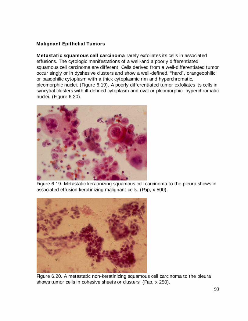
93
Malignant Epithelial Tumors
Metastatic squamous cell carcinoma rarely exfoliates its cells in associated effusions. The cytologic manifestations of a well-and a poorly differentiated squamous cell carcinoma are different. Cells derived from a well-differentiated tumor occur singly or in dyshesive clusters and show a well-defined, “hard”, orangeophilic or basophilic cytoplasm with a thick cytoplasmic rim and hyperchromatic, pleomorphic nuclei. (Figure 6.19). A poorly differentiated tumor exfoliates its cells in syncytial clusters with ill-defined cytoplasm and oval or pleomorphic, hyperchromatic nuclei. (Figure 6.20).
Figure 6.19. Metastatic keratinizing squamous cell carcinoma to the pleura shows in associated effusion keratinizing malignant cells. (Pap, x 500).
Figure 6.20. A metastatic non-keratinizing squamous cell carcinoma to the pleura shows tumor cells in cohesive sheets or clusters. (Pap, x 250).

94
Metastatic adenocarcinomas account for over 90% of malignant pleural
effusions. The exfoliated cancer cells, regardless of their primary tumors usually occur in tri-dimensional clusters of varying sizes with smooth contours, in small clusters and singly. The tumor cells may display a vacuolated cytoplasm, eccentrically located nuclei and prominent nucleoli. (Figures 6.21). A few additional cytologic findings may be seen in cell groups derived from adenocarcinomas arising from certain anatomic sites. Psammoma bodies may be seen in effusions associated with a metastatic ovarian serous cystadenocarcinoma, papillary adenocarcinoma of the lung and papillary carcinoma of the thyroid. A metastatic mammary duct carcinoma exfoliates small cancer cells with linear arrangement or “Indian files” pattern. A metastatic gastric signet-ring cell carcinoma exfoliates single cancer cells with large cytoplasmic vacuole and eccentrically located oval nuclei with prominent nucleoli. (Figure 6.22). A metastatic mucinous adenocarcinoma shows groups of cancer cells with vacuolated cytoplasm in a mucous background. Metastatic adenocarcinoma cells usually react positively with CEA, Ber-Ep4, Leu M1 and MOC-31 antibodies. (Figure 6.23). Tumor cells derived from a bronchogenic carcinoma are commonly TTF1 positive.
A

95
B
C Figure 6.21. Pleural effusion in metastatic adenocarcinoma to the pleura showing: A. Tumor cells in tridimensional ball-like clusters, in small clusters and singly. B. A cohesive cluster of malignant glandular cells. C. Tumor cells with “cell-embracing-cell” arrangement. (Pap, A x 100, B x 400, C x 500)

96
Figure 6.22. Metastatic signet-ring cell adenocarcinoma showing in pleural effusion single malignant cells with some displaying intracytoplasmic vacuoles. (Pap,x 500).
Figure 6.23. CB section from a pleural effusion secondary to a metastatic adenocarcinoma showing tumor cells displaying positive cytoplasmic reaction to CEA antibody (ABC, x 250).
Metastatic bronchogenic small cell carcinoma exfoliates small malignant cells with round nuclei showing a salt and pepper chromatin pattern and scant cytoplasm in small clusters displaying nuclear molding. (Figures 5.24-6.26). Micronucleoli may be present.

97
Figure 6.24. Metastatic small-cell carcinoma to the pleura shows in associated effusion a loose cluster of small malignant cells displaying focal nuclear molding. (Pap, x 400).
Figure 6.25. Metastatic small-cell carcinoma to the pleura shows in effusion a tight cluster of tumor cells with molding. (Pap, x 500).

98
A
B Figure 6.26. Metastatic small-cell carcinoma to the pleura showing tumor cells in large tridimensional clusters, smaller clusters and singly. (Pap, A x 100, B x 500). Metastatic large-cell carcinoma yields single and loosely clustered large malignant cells with abundant, well-defined cytoplasm, enlarged hyperchromatic nuclei and prominent nucleoli. (Figure 6.27).

99
Figure 6.27. Metastatic large-cell carcinoma to the pleura shows in associated effusion large pleomorphic malignant cells present predominantly singly. (Diff-Quik, x 500).
Metastatic neuroendocrine carcinoma shows dyshesive cell clusters with regular, round nuclei with chromatin clumping and conspicuous nucleoli. A positive cytoplasmic reaction with neuron-specific enolase or chromogranin antibody will confirm its neuroendocrine differentiation. Non-Epithelial Cancers
Lymphoma and Leukemia are the most common non-epithelial cancers associated with pleural effusions. A low-grade lymphoma or a lymphocytic leukemia exfoliates monomorphic benign-appearing lymphoid cells similar to mature lymphocytes. A high-grade lymphoma exfoliates monomorphic lymphoid cells with nuclear protrusion and indentation. Reed-Sternberg cells are seen in serous effusion secondary to Hodgkin disease. (Figures 6.28 and 6.29). A chronic myelogenous leukemia shows mature and immature myelogenous cells without “leukemic hiatus”. A multiple myeloma yields malignant plasma cells with bizarre forms. Multiple nuclei may be present.

100
Figure 6.28. Non-Hodgkin lymphoma involving the pleura showing in pleural effusion monomorphic malignant lymphoid cells. Some tumor cells display nuclear indentation or protrusion. (Pap, x 400).
Figure 6.29. Hodgkin disease involving the pleura showing in pleural effusion numerous benign lymphocytes, a Reed-Sternberg cells. (Pap, x 500). A metastatic melanoma shows single and clustered malignant cells dysplaying intranuclear cytoplasmic inclusions and prominent nucleoli. Intracytoplasmic melanin pigment granules may be present. A positive cytoplasmic reaction with HMB-45 or MART 1 antibody will confirm their melanocytic differentiation. A metastatic neuroblastoma yields small round cells with scant and fibrillary cytoplasm and tumor cells forming rosettes may be observed. The tumor cell cytoplasm stains positively with neuron-specific enolase and chromogranin antibodies. Bone and Soft Tissue Sarcomas show single and loosely clustered cancer

101
cells. The tumor cells usually loose their original shapes as seen in tumor tissue sections and tend to have a round configuration. Tumor typing is difficult without clinical data and immunocytochemical studies. Some tumors may show specific cytologic findings. A metastatic chondrosarcoma yields single chrondrocyte-like cells with abundant, well-defined cytoplasm and prominent nucleoli. (Figure 6.30). A metastatic Ewing sarcoma yields loosely clustered polygonal cells with oval nuclei and moderate amount of cytoplasm that stains positively with periodic acid- Schiff reagent. A metastatic biphasic synovial sarcoma shows small epithelial- like cells with oval nuclei and scant cytoplasm in clusters with focal gland-like arrangement and spindle cells with scant and ill-defined cytoplasm and oval nuclei. Cellular changes suggesting the transformation of spindle cells to epithelial-like cells have been observed. The tumor cells usually express cytokeratin and vimentin and stain negatively with CEA antibody.
Figure 6.30. Metastatic chondrosarcoma to the pleura showing in associated effusion single malignant cells with single or double nuclei and basophilic cytoplasm. (Pap,x 500). Diagnostic Accuracy
Effusion cytology is more sensitive than blind biopsy of the pleura in detecting pleural cancer (80% versus 45%) in experienced hands. False-negative diagnostic rates vary widely among reported series. The main reasons are inadequate cell sample, scantiness of malignant cells, faulty preparatory techniques and erroneous interpretations. False-positive diagnostic rates up to 3% have been reported. The most common error is the misinterpretation of highly reactive mesothelial cells in long-standing benign effusions as malignant glandular cells.
Cytologic, immunocytochemical and ultrastructural features of epithelial mesothelioma, reactive mesothelium and bronchogenic adenocarcinoma are tabulated in Table 6.1.

102
Table 6.1: Comparative Cytologic Manifestations of Reactive Mesothelium, Epithelial Mesothelioma and Bronchogenic Adenocarcinoma in Serous Effusions* CELLULAR FEATURES
REACTIVE MESOTHELIUM
EPITHELIAL MESOTHELIOMA
BRONCHOGENIC ADENOCARCINOMA
Architecture: -Singly, common -Monolayered sheets -Loose groups with “windows”
-Large cohesive clusters with lobulated borders -Small tight clusters with “windows” and “push-in” junctions
-Singly, rare -Tight multilayered, 3-dimensional clusters with smooth borders
Cells: -Configuration: -Cytoplasm: -Nucleus: -Anisonucleosis -Irregular contours -Molding -Chromatin -Nucleolus
-Polygonal, round -Well-defined -Foamy or homogenous - - - Fine Small
-Polygonal, round -Well-defined -Dense ectoplasm -Fuzzy periphery + + + Fine or coarse Small or large
-Polygonal, round -Ill-defined -Vacuolated + + + Coarse Large, single, multiple
Staining characteristics:
- Mucin - - + - Calretinin, CK5/6, D2-40, Mesothelin, WT-1
+
+
-
- CEA, MOC-31, Ber-Ep4, TTF-1
-
-
+
- Desmin + +/- - - EMA +, periphery ++, fuzzy, periphery +, periphery - GLUT-1 - + +/- Ultrastructure: - Long microvilli, with L:D >12 - Intracytoplasmic mucous granules
+ -
+ -
- +

103
* Adapted from Nguyen GK, Kline TK. Essentials of Cytology. An atlas. Igaku-Shoin, New York, 1993, p.88. C. OTHER PLEURAL TUMORS Benign pleural tumors are rarely encountered in pathology practice. Benign localized epithelial mesothelioma or adenomatoid tumor is an exceedingly rare neoplasm of the pleura. It is small and incidentally found in the lung resected for other conditions. Solitary fibrous tumor is a rare lesion that is most likely arising from the subpleural mesenchyme. It is not related to asbestos exposure and has no sex predilection. The tumor is asymptomatic and usually discovered incidentally by chest roentgenograms and it measures up to several cm in greatest dimension. The tumor is well-demarcated with pushing borders and is often pedunculated. Most solitary fibrous tumors of the pleura are benign but about 30% are malignant. About 80% of these tumors arise from the visceral pleura and the remainder originate from the parietal pleura. Histologically, it is characterized by benign fibroblastic cells arranged in a non-specific pattern and a collagenous, hyalinized stroma. Cellular atypia and mitoses are uncommon. The tumor yields in TTFNA a scanty cellular material showing bland spindle cells that react positively with CD34 and negatively with cytokeratin antibodies, in contrast to those of a fibrous mesothelioma. (Figure 6.31).
Figure 6.31. TTFNA from a localized fibrous tumor of the pleura shows benign, spindle cells with elongated nuclei in no specific pattern. (Pap, x 500).

104
BIBLIOGRAPHY Ali SZ, et al. Solitary fibrous tumor. A cytologic-histologic study with clinical, radiologic, and immunohistochemical correlations. Cancer (Cancer Cytopathol).1997;81:116. Battifora H, McCaughey WTE. Tumors of the serosa. In Atlas of tumor pathology, 3rd series, 1995. Washington DC, Armed Forces Institute of Pathology. Battifora H. The pleura. In Sternberg’s Diagnostic Surgical Pathology, 4th ed,2004, Mills SE et al, eds. Philadelphia, Lippincott Williams & Wilkins, p.1221. Bedrossian CWM. Malignant Effusions. A Multimodal Approach. New York, Igaku-Shoin, 1993. Cho EY, et al. Fine needle aspiration cytology of solitary fibrous tumours of the pleura. Cytopathology.2007;18:20. Churg A, et al. Tumors of the serosal membranes. In AFIP Atlas of Tumor Pathology, 4th series, 2006. Washington DC, Armed Forces Institutes of Pathology. DeLellis RA, Hoda RS. Immunochemistry and molecular biology in cytological diagnosis. In Koss’ Diagnostic Cytology and Its Histopathologic Bases. 5th ed, 2006. Philadelphia, Lippincott Williams & Wilkins. p.1635. DeMay RM. The art and science of cytopathology, Chicago, ASCP Press,1996. p 257. Fletsch PA, Abati A. Immunocytochemistry in effusion cytology. A contemporary review. Cancer (Cancer Cytopathol). 2001;93:293. Friedman MT, et al. Malignant mesothelioma.Immunohistochemistry and DNA ploidy analysis as methods to differentiate msothelioma from benign reactive mesothelial cell proliferation and adenocarcinoma in pleural and peritoneal effusions. Arch Pathol Lab Med. 1996; 120:957. Geisinger KR, et al. Effusions. In Modern Cytopathology. Geisinger KR, et al, eds. Philadelphia, Churchill Livingstone.2004, p 257. Ghadially FN. Diagnostic electron microscopic of tumours, 2nd ed, 1985. London, Butterworths.

105
Hammar SP, Bolen JM. Pleural neoplasms. In Pulmonary pathology, Dail DH, Hammar SP, eds. New York, Spinger-Verlag,1988. p. 973. Mullick SS, et al. P53 gene product in pleural effusions. Practical use in distinguishing benign from malignant cells. Acta Cytol.1996;40:855. Nguyen GK. Cytopathology of pleural mesotheliomas. Am J Clin Pathol.2000;114 (Suppl 1):S68. Nguyen GK, et al. Cytodiagnosis of malignant mesothelioma of the pleura by fine-needle aspiration biopsy. Diagn Cytopathol. 1999;21:253. Nguyen GK, Jeannot A. Cytology of synovial sarcoma metastases in pleural fluid. Acta Cytol.1982;26:517. Obers VJ, et al. Primary pleural tumors (mesotheliomas) presenting as localized masses. Fine needle aspiration cytologic findings, clinical and radiological features and review of the literature. Acta Cytol.1988; 32:567. Ordonez NG. Immunohistochemical diagnosis of epithelioid mesotheliomas: a critical review of old markers, new markers. Hum Pathol. 2002; 33:935. Remeal M, et al. Immunoreactivity for p53 in malignant mesothelioma and non- neoplastic mesothelium. J Pathol. 1992;168:371. Rosai J. Ackerman’s surgical pathology, 8th ed, 1996. St. Louis, Mosby. p 289, 2021. Saleh H. et al. Value of Ki-67 immunostain in identification of malignancy in serous effusions. Diagn cytopathol. 1999; 20:24. Sherman ME, Mark EJ. Effusion cytology in the diagnosis of malignant epithelioid and biphasic pleural mesothelioma. Arch Pathol Lab Med. 1990; 114:845. Sprigg AI, Grunze H. An unusual cytologic presentation of mesothelioma in serous effusions. Acta Cytol. 1983; 27:288. Sterrett GF, et al. Fine needle aspiration cytology of malignant mesothelioma. Acta Cytol. 1987;31:185. Tao LC. Aspiration biopsy cytology of mesothelioma. Diagn Cytopathol 1989;5:14.

106
Travis WD, et al. Pathology and Genetics of Tumours of the Lung, Pleura, Thymus and Heart. In WHO Classification of Tumors. Lyon, IARCPress, 2004.
Triol JH, et al. Malignant mesothelioma. Cytopathology of 75 cases seen in a New Jersey community hospital. Acta Cytol. 1984;28:37. Whitaker D, et al. Mesothelioma. In Diagnostic Cytopathology, 2nd ed, Gray W, McKee GT, eds, 2003, London, Churchill Livingstone, p. 205. Whitaker D. The cytology of malignant mesothelioma. Cytopathology. 2000;11:139.
THE END