essentials of oral and - download.e-bookshelf.de · essentials of oral and maxillofacial surgery...
TRANSCRIPT


Essentials of Oral and Maxillofacial Surgery

Essentials of Oral and Maxillofacial SurgeryEdited by
M. Anthony (Tony) PogrelDDS, MD, FRCS, FACSProfessor and ChairAssociate Dean for Hospital AffairsDepartment of Oral and Maxillofacial SurgeryUniversity of California, San FranciscoUSA
Karl-Erik KahnbergDDS, PhD, DrOdontProfessor Emeritus, Oral and Maxillofacial SurgeryInstitute of OdontologyThe Sahlgrenska AcademyUniversity of GothenburgSweden
Lars AnderssonDDS, PhD, DrOdontProfessor, Oral and Maxillofacial SurgeryChairman, Department of Surgical SciencesFaculty of DentistryHealth Sciences CenterKuwait UniversityKuwait
This edition first published 2014© 2014 by John Wiley & Sons, Ltd
Registered office: John Wiley & Sons, Ltd, The Atrium, Southern Gate, Chichester, West Sussex, PO19 8SQ, UK
Editorial offices: 9600 Garsington Road, Oxford, OX4 2DQ, UKThe Atrium, Southern Gate, Chichester, West Sussex, PO19 8SQ, UK111 River Street, Hoboken, NJ 07030-5774, USA
For details of our global editorial offices, for customer services and for information about how to apply for permission to reuse the copyright material in this book please see our website at www.wiley.com/wiley-blackwell
The right of the author to be identified as the author of this work has been asserted in accordance with the UK Copyright, Designs and Patents Act 1988.
All rights reserved. No part of this publication may be reproduced, stored in a retrieval system, or transmitted, in any form or by any means, electronic, mechanical, photocopying, recording or otherwise, except as permitted by the UK Copyright, Designs and Patents Act 1988, without the prior permission of the publisher.
Designations used by companies to distinguish their products are often claimed as trademarks. All brand names and product names used in this book are trade names, service marks, trademarks or registered trademarks of their respective owners. The publisher is not associated with any product or vendor mentioned in this book. It is sold on the understanding that the publisher is not engaged in rendering professional services. If professional advice or other expert assistance is required, the services of a competent professional should be sought.
The contents of this work are intended to further general scientific research, understanding, and discussion only and are not intended and should not be relied upon as recommending or promoting a specific method, diagnosis, or treatment by health science practitioners for any particular patient. The publisher and the author make no representations or warranties with respect to the accuracy or completeness of the contents of this work and specifically disclaim all warranties, including without limitation any implied warranties of fitness for a particular purpose. In view of ongoing research, equipment modifications, changes in governmental regulations, and the constant flow of information relating to the use of medicines, equipment, and devices, the reader is urged to review and evaluate the information provided in the package insert or instructions for each medicine, equipment, or device for, among other things, any changes in the instructions or indication of usage and for added warnings and precautions. Readers should consult with a specialist where appropriate. The fact that an organization or Website is referred to in this work as a citation and/or a potential source of further information does not mean that the author or the publisher endorses the information the organization or Website may provide or recommendations it may make. Further, readers should be aware that Internet Websites listed in this work may have changed or disappeared between when this work was written and when it is read. No warranty may be created or extended by any promotional statements for this work. Neither the publisher nor the author shall be liable for any damages arising herefrom.
Library of Congress Cataloging-in-Publication DataEssentials of oral and maxillofacial surgery / edited by M. Anthony (Tony) Pogrel, Karl-Erik Kahnberg, Lars Andersson. p. ; cm.
Includes bibliographical references and index.ISBN 978-1-4051-7623-1 (pbk.)I. Pogrel, M. Anthony, editor of compilation. II. Kahnberg, Karl-Erik, editor of compilation. III. Andersson, L. (Lars), 1950- editor of
compilation. [DNLM: 1. Oral Surgical Procedures–methods. 2. Stomatognathic System–surgery. WU 600]RK501617.6’05–dc23
2014001169
A catalogue record for this book is available from the British Library.
Wiley also publishes its books in a variety of electronic formats. Some content that appears in print may not be available in electronic books.
Cover image: © Yegor Tsyba/iStockphotoCover design by Garth Stewart
Set in 9.5/12 pt Palatino LT Std by Toppan Best-set Premedia Limited, Hong Kong
1 2014
v
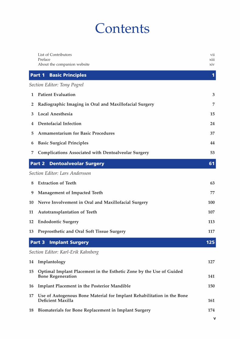
Contents
List of Contributors viiPreface xiiiAbout the companion website xiv
Part 1 Basic Principles 1
Section Editor: Tony Pogrel
1 PatientEvaluation 3
2 RadiographicImaginginOralandMaxillofacialSurgery 7
3 LocalAnesthesia 15
4 DentofacialInfection 24
5 ArmamentariumforBasicProcedures 37
6 BasicSurgicalPrinciples 44
7 ComplicationsAssociatedwithDentoalveolarSurgery 53
Part 2 Dentoalveolar Surgery 61
Section Editor: Lars Andersson
8 ExtractionofTeeth 63
9 ManagementofImpactedTeeth 77
10 NerveInvolvementinOralandMaxillofacialSurgery 100
11 AutotransplantationofTeeth 107
12 EndodonticSurgery 113
13 PreprostheticandOralSoftTissueSurgery 117
Part 3 Implant Surgery 125
Section Editor: Karl-Erik Kahnberg
14 Implantology 127
15 OptimalImplantPlacementintheEstheticZonebytheUseofGuidedBoneRegeneration 141
16 ImplantPlacementinthePosteriorMandible 150
17 UseofAutogenousBoneMaterialforImplantRehabilitationintheBoneDeficientMaxilla 161
18 BiomaterialsforBoneReplacementinImplantSurgery 174
vi Contents
Part 4 Oral Pathologic Lesions 185
Section Editor: Tony Pogrel
19 InitialEvaluationandManagementoftheOralandMaxillofacialPathologyPatient 187
20 CysticLesionsoftheJaws 195
21 OdontogenicandNon-odontogenicTumorsoftheJaws 201
22 PotentiallyMalignantDisordersoftheOralMucosa 229
23 PrinciplesofOralCancerManagement 235
24 ManagementofPatientsUndergoingRadiationandChemotherapy 240
25 SalivaryGlandDisorders 245
Part 5 Trauma 253
Section Editor: Lars Andersson
Introduction:OralandMaxillofacialTraumatology 255
26 TraumaticDentalInjuries 257
27 MaxillofacialBoneFractures 274
28 SoftTissueInjuries 288
Part 6 Dentofacial Deformities 295
Section Editor: Karl-Erik Kahnberg
29 CleftLipandPalate:AnOverview 297
30 CorrectionofDentofacialDeformities 315
31 MandibularReconstruction 331
Part 7 Temporomandibular Joint Disorders 345
Section Editor: Tony Pogrel
32 DiagnosisandNon-surgicalManagementofOrofacialPain 347
33 TemporomandibularJointSurgery(IncludingArthroscopy) 353
Index 371
List of ContributorsAdel Al-Asfour BDS, BA (Chapter 8)Associate ProfessorDepartment of Surgical SciencesFaculty of Dentistry, Health Sciences CenterKuwait UniversityKuwait
Ala Al-Musawi DDS, BA (Chapter 5)Assistant ProfessorDepartment of Surgical SciencesFaculty of Dentistry, Health Sciences CenterKuwait UniversityKuwait
Jens O. Andreasen DDS, Odont. Dr.h.c. (Chapters 11 and 28)ConsultantDepartment of Oral and Maxillofacial SurgerySection for Rare Oral DiseasesRigshospitaletCopenhagen, Denmark
Ashraf Ayoub PhD, FDS RCS, FDS RCPS, BDS, MDS (Chapter 4)Professor of Oral & Maxillofacial SurgeryGlasgow University Dental Hospital & SchoolGlasgow, UK
Selçuk Basa DDS, PhD (Chapter 13)Department of Oral and Maxillofacial SurgeryMarmara UniversityIstanbul, Turkey
Brian Bast DMD, MD (Chapter 27)Associate Clinical ProfessorResidency Program DirectorDepartment of Oral and Maxillofacial SurgeryUniversity of California, San FranciscoSan Francisco, California, USA
R. Bryan Bell DDS, MD, FACS (Chapter 21)Attending Surgeon and Director of Resident Education,OMFS ServiceACS Cancer Liaison PhysicianLegacy Emanuel Hospital and Health CenterClinical Associate Professor of Oral and MaxillofacialSurgeryOregon Health and Science UniversityHead and Neck Surgical AssociatesPortland, Oregon, USA
John Beumer III DDS, MSProfessor and ChairDivision of Advanced Prosthodontics, Biomaterials,and Hospital DentistryUniversity of California, Los Angeles – School ofDentistryLos Angeles, California, USA
Krishnamurthy Bonanthaya MBBS, MDS, FDSRCS, FFDRCS (Chapter 29)Professor, Oral and Maxillofacial SurgeryBangalore Institute of Dental SciencesandConsultant SurgeonBhagwan Mahavir Jain HospitalBangalore, India
Peter Carrotte BDS, MDS, LDS.RCS(Eng), MEd, MHEA (Chapter 12)Senior Clinical Teacher and Honorary AssociateSpecialistDepartment of Restorative DentistryUniversity of GlasgowGlasgow, UK
Allen Cheng DDS, MD (Chapter 19)Department of Oral and Maxillofacial SurgeryUniversity of California, San FranciscoSan Francisco, California, USA
Lim K. Cheung BDS, PhD, FFDRCS, FDSRCPS, FRACDS, FDSRCS(Edin), FRACDS(OMS), FHKAM(DS), FCDSHK(OMS), FFGDP(UK) (Chapter 30)Chair ProfessorDiscipline of Oral and Maxillofacial SurgeryFaculty of DentistryUniversity of Hong KongHong Kong, SAR, China
Radhika Chigurupati DMD, MS (Chapter 29)Associate Clinical Professor of Oral and MaxillofacialSurgeryDepartment of Oral and Maxillofacial SurgeryBoston UniversityBoston, Massachusetts, USA
Hannah Daile P. Chua DMD, MA, MDS (OMS), MOSRCS, PhDAssistant ProfessorDiscipline of Oral and Maxillofacial SurgeryFaculty of DentistryUniversity of Hong KongHong Kong, SAR, China
William Chung DDS, MD (Chapter 27)University of Pittsburgh Medical CenterPittsburgh, Pennsylvania, USA
Cameron M.L. Clokie DDS, PhD, FRCDCProfessor and Director of Graduate Program in Oraland Maxillofacial Surgery and AnaesthesiaUniversity of TorontoToronto, Ontario, Canada
Bernard J. Costello DMD, MD, FACS (Chapter 27)Chief, Division of Craniofacial and Cleft SurgeryAssociate Professor and Program DirectorDepartment of Oral and Maxillofacial SurgeryUniversity of Pittsburgh School of Dental MedicinePittsburgh, Pennsylvania, USA
Christer Dahlin DDS, PhD, Dr Odont (Chapter 15)Associate ProfessorDepartment of BiomaterialsInstitute for Clinical SciencesSahlgrenska Academy, University of GothenburgGothenburg, SwedenandDepartment of Oral and Maxillofacial SurgeryNÄL Medical Centre HospitalTrollhättan, Sweden
vii
viii List of Contributors
Nagi Demian DDS, MDAssistant ProfessorDepartment of Oral and Maxillofacial SurgeryUniversity of Texas Health Science Center – HoustonHouston, Texas, USA
Thomas B. Dodson DMD, MPH (Chapter 7)Professor and ChairDepartment of Oral and Maxillofacial SurgeryUniversity of WashingtonSeattle, Washington, USA
Carlo Ferretti BDS, MDent (MFOS), FCD (SA), MFOS (Chapter 30)Senior SpecialistDepartment of Maxillofacial and Oral SurgeryandChris Hani Baragwanath HospitalFaculty of Health Sciences,University of the WitwatersrandJohannesburg, South Africa
Earl G. Freymiller DMD, MDClinical ProfessorSection of Oral and Maxillofacial SurgeryUCLA School of DentistryLos Angeles, California, USA
Takashi Fujibayashi DDS, PhD (Chapter 22)Visiting ProfessorDepartment of Oral and Maxillofacial SurgeryKanagawa Dental CollegeYokosuka, Japan
Nicholas M. Goodger BSc, BDS, MBBS, PhD, FDSRCS(Eng),FFDRCSI, FRCSEd, DLORCS(Eng) (Chapter 20)Consultant Oral and Maxillofacial SurgeonEast Kent Hospitals University NHS Foundation TrustKent and Canterbury HospitalCanterbury, UK
Gosta Granstrom MD, DDS, PhDProfessor of Otolaryngology, Head and Neck SurgeryDepartment of Otolaryngology, Head and NeckSurgerySahlgrenska Academy, University of GothenburgGothenburg, Sweden
Firdaus Hariri BDS, MDS (OMS)Consultant and LecturerDepartment of Oral and Maxillofacial SurgeryFaculty of DentistryUniversity of MalayaKuala Lumpur, Malaysia
Richard H. Haug DDS (Chapter 28)Carolinas Center for Oral HealthCharlotte, North Carolina, USA
Andrew Heggie MBBS, MDSc, BDSc, FRACDS(OMS) (Chapter 29)Associate Professor, Oral and Maxillofacial SurgeryDepartment of Plastic and Maxillofacial SurgeryRoyal Children’s Hospital of MelbourneParkville, Australia
Christopher W. Hendy BDS, FDSRCS(Eng), LRCP, MRCS,FRCSEd (Chapter 20)East Kent Hospitals University NHS Foundation TrustKent and Canterbury HospitalCanterbury, UK
Alan S. Herford DDS, MD (Chapter 1)ChairmanDepartment of Oral and Maxillofacial SurgeryLoma Linda UniversityLoma Linda, California, USA
C. Michael Hill MDSc, FDSRCS, MSc, BDSConsultant and Honorary Senior Lecturer in Oral andMaxillofacial SurgeryCardiff Dental HospitalCardiff, UK
Anders Holmlund DDS, PhD Professor (Chapter 33)Department of Oral and Maxillofacial SurgeryInstitution of OdontologyKarolinska Institutet/Karolinska University HospitalHuddinge, Sweden
Mehran Hossaini DMD (Chapter 9)Health Sciences Associate Clinical ProfessorDepartment of Oral and Maxillofacial SurgeryUniversity of California, San FranciscoSan Francisco, California, USA
Vesa T. Kainulainen DDS, PhDAssistant ProfessorInstitute of DentistryUniversity of OuluOulu, Finland
Sanjiv Kanagaraja DDS, PhD (Chapter 8)Consultant/Assistant ProfessorDepartment of Oral and Maxillofacial SurgeryUniversity of GöteborgGöteborg, Sweden
Reha Kışnışçı DDS, PhD (Chapter 13)Department of Oral and Maxillofacial SurgeryAnkara UniversityAnkara, Turkey
Goran Kjeller PhD (Chapter 24)Associate ProfessorDepartment Oral and Maxillofacial SurgeryInstitute of OdontologySahlgren’s AcademyGothenburg UniversityGöteborg, Sweden
Paul Koshgerian DMD (Chapter 28)University of Louisville School of DentistryDepartment of Surgical and Hospital DentistryLouisville, Kentucky, USA
Fabio Kricheldorf DDS, MSc (Chapter 6)Professor of Oral and Maxillofacial Surgeryand ChairmanDepartment of Surgical Sciences, Faculty of DentistryUniversity of JoinvilleJoinville, Santa Catarina, Brazil
Tomoari Kuriyama DDS, PhD (Chapter 4)Honorary Clinical Instructor and Research FellowDepartment of Oral and Maxillofacial SurgeryGraduate School of Medical ScienceKanazawa UniversityKanazawa, JapanandPrivate practiceToyama, Japan
List of Contributors ix
David K. Lam DDS, MD, PhD, FRCDCAssistant ProfessorDepartment of Oral and Maxillofacial SurgeryUniversity of TorontoToronto, Ontario, Canada
Anh Le DDS, PhDProfessor and ChairDepartment of Oral and Maxillofacial Surgery University of PennsylvaniaPhiladelphia, Pennsylvania, USA
Michael A.O. Lewis PhD, BDS, FDSRCPSG, FRCPath,FDSRCS(Eng), FDSRCS(Ed), FFGDP(UK) (Chapter 4)Professor of Oral MedicineCardiff UniversityCardiff, UK
John Lo BDS, MDS (OMS), MOSRCS, FHKAM (DS), FCDSHK (OMS)Assistant ProfessorDiscipline of Oral and Maxillofacial SurgeryFaculty of DentistryUniversity of Hong KongHong Kong, SAR, China
Leif Lysell DDS, Odont Dr (Chapter 9)Associate ProfessorFaculty of OdontologyMalmö UniversityMalmö, SwedenandPrivate practiceKristianstad, Sweden
Carlo Maiorana MD, DDS (Chapter 18)Professor and ChairmanOral Surgery and ImplantologyDental Clinic Fondazione Cà GrandaUniversity of MilanMilan, Italy
Chantal Malevez MD, DDSSpecialist in Oral and Maxillofacial SurgeryProfessor EmeritusFree University of Brussels (ULB)andConsultant Department of Maxillofacial SurgeryChildren’s HospitalBrussels, BelgiumandConsultant at the EOICBrussels, Belgium
Joann Marruffo DDS, MSMaxillofacial ProsthodontistPrivate practiceHouston, Texas, USA
James McCaul PhD, FRCS(OMFS), FRCS(Glasg), FDSRCPSConsultant Oral and Maxillofacial/Head and NeckSurgeonBradford Teaching Hospitals NHS Foundation TrustBradford, UK
Mark McGurk BDS, MD, FDS RCSEng, FRCS Ed, DLO RCS (Chapter 25)ProfessorDepartment of Oral and Maxillofacial SurgeryKing’s CollegeLondon, UK
Charles McNeill DDS (Chapter 32)Professor Emeritus and DirectorUCSF Center for Orofacial PainDepartment of Oral and Maxillofacial SurgeryUniversity of California, San FranciscoSan Francisco, California, USA
John Gerard Meechan BSc, BDS, PhD, FDSRCS, FDSRCPS (Chapter 3)Senior Lecturer in Oral SurgerySchool of Dental SciencesNewcastle UniversityNewcastle upon Tyne, UK
Marc Christian Metzger MD, DMD, PhD (Chapter 27)Department of Craniomaxillofacial SurgeryUniversity Hospital FreiburgFreiburg, Germany
Colin Murray PhD, FDS RCS(Edin), BDS, FDS(Rest Dent) RCS(Edin), FDS RCPS(Glas) (Chapter 12)Professor in Restorative Dentistry/HonoraryConsultantHead of Clinical Dentistry Section and Head ofRestorative Group University of GlasgowGlasgow, UK
Joe NiamtuPrivate practiceCosmetic Facial SurgeryRichmond, Virginia, USA
Kyösti S. Oikarinen DDS, PhDProfessor and Head of Oral and Maxillofacial SurgeryInstitute of DentistryUniversity of OuluOulu, Finland
Andrew Ow BDS, MDS(OMS), FRACDS, MOSRCS, AdvDip(OMS)Assistant ProfessorDepartment of Oral and Maxillofacial SurgeryNational University of SingaporeSingapore
Zachary S. Peacock DMD, MDAssistant ProfessorDepartment of Oral and Maxillofacial SurgeryMassachusetts General Hospital and Harvard School of Dental MedicineBoston, Massachusetts, USA
Arne Petersson DDS, Odont Dr (Chapter 2)Department of Oral and Maxillofacial RadiologyFaculty of OdontologyMalmö UniversityMalmö, Sweden
Lars Rasmusson DDS, PhD (Chapter 14)Professor of Maxillofacial SurgeryHead of Department of Oral and Maxillofacial SurgeryThe Sahlgrenska AcademyUniversity of GothenburgGothenburg, Sweden
Tara Renton BDS, MDSc, PhD, FDS, RCS, FRACDS (OMS), ILTM (Chapter 9)Professor in Oral SurgeryKing’s College London Dental InstituteLondon, UK

x List of Contributors
Johan P. Reyneke B Ch D, M Ch D, FCMFOS (SA), PhD (Chapter 30)Honorary ProfessorDepartment of Maxillofacial and Oral SurgeryFaculty of Health SciencesUniversity of the WitwatersrandJohannesburgSouth AfricaandClinical ProfessorDepartment of Oral and Maxillofacial SurgeryUniversity of OklahomaOklahoma City, Oklahoma, USAandClinical ProfessorDepartment of Oral and Maxillofacial SurgeryUniversity of FloridaGainesville, Florida, USAandPrivate practice, Sunninghill HospitalSunninghill, Johannesburg, South Africa
Richard C. Robert DDS, MSClinical ProfessorOral and Maxillofacial SurgeryUniversity of California Medical CenterSan Francisco, California, USAandPrivate practice, Oral and Maxillofacial SurgerySouth San Francisco, California, USA
Simon N. Rogers BDS, MBChB, FDSRCS, FRCSProfessor, Regional Maxillofacial UnitUniversity Hospital AintreeLiverpool, UKandEvidence-based Practice Research Centre (EPRC)Faculty of HealthEdge Hill UniversityOrmskirk, UK
†Bo Rosenquist BSc, DDS, PhD (Chapter 16)Associate Professor of Oral and MaxillofacialSurgeryHead of the Head and Neck DivisionDepartment of Oral and Maxillofacial SurgeryUniversity Hospital of LundLund, Sweden
Patricia A. Rudd PT, DPT, CCTT (Chapter 32)Assistant Clinical ProfessorUCSF Center for Orofacial PainDepartment of Oral and Maxillofacial SurgeryUniversity of California, San FranciscoSan Francisco, California, USA
George K.B. Sándor MD, DDS, PhD, Dr. Habil, FRCDC,FRCSC, FACSProfessor of Tissue EngineeringRegea Institute for Regenerative MedicineUniversity of TampereTampere, FinlandandDosent in Oral and Maxillofacial SurgeryUniversity of OuluOulu, Finland
Henning Schliephake MD, DDS, PhDProfessor and ChairDepartment of Oral Maxillofacial SurgeryGeorge Augusta UniversityGöttingen, Germany
Rainer Schmelzeisen MD, DMD (Chapter 27)Professor and ChairmanDepartment of Craniomaxillofacial SurgeryUniversity Hospital FreiburgFreiburg, Germany
Brian L. Schmidt DDS, MD, PhD, FACS (Chapters 19, 23 and 31)ProfessorDepartment of Oral and Maxillofacial Surgery and Director of the Bluestone Center for Clinical ResearchNew York University School of DentistryNew York, NY, USA
Ralf Schon MD, DMD (Chapter 27)Associate ProfessorDepartment of Craniomaxillofacial SurgeryUniversity Hospital FreiburgFreiburg, Germany
Petr Schutz MD (Chapter 27)ConsultantHead of Oral and Maxillofacial Surgery UnitDental Center, Farwaniya HospitalMinistry of HealthKuwait
Lars Sennerby DDS, PhD (Chapter 14)Professor of Clinical and Experimental OralImplantologyDepartment of BiomaterialsInstitute for Clinical SciencesSahlgrenska AcademyUniversity of GothenburgGothenburg, SwedenandClinica FeltreFeltre, Italy
Bethany Serafin DMD (Chapter 28)Valley Village Oral Surgery AssociatesBaltimore, Maryland, USA
Arun B. Sharma BDS, MScHealth Sciences Clinical ProfessorDivision of ProsthodonticsUniversity of California, San Francisco – School ofDentistrySan Francisco, California, USA
Jeremy Sherman BDS, MBChB, FRCS, FDRCS, FRCS Ed (Chapter 25)Consultant Maxillofacial SurgeonDepartment of Oral and Maxillofacial SurgeryQueen Elizabeth II HospitalWelwyn Garden City, UK
Vivek Shetty DDS, Dr Med DentProfessorSection of Oral and Maxillofacial SurgeryUniversity of California, Los AngelesLos Angeles, California, USA
Ryan J. Smart DMD, MD (Chapter 7)ResidentDepartment of Oral and Maxillofacial SurgeryMassachusetts General HospitalBoston, Massachusetts, USA

List of Contributors xi
Srinivas M. Susarla DMD, MD, MPH (Chapter 7)ResidentDepartment of Oral and Maxillofacial SurgeryMassachusetts General HospitalBoston, Massachusetts, USA
Wayne K. Tanaka DDS, FACD, FICD (Chapter 1)Associate Professor, Predoctoral Program DirectorDepartment of Oral and Maxillofacial SurgeryLoma Linda UniversityLoma Linda, California, USA
Peter Tarnow MD, PhDChairman, The Craniofacial UnitDepartment of Plastic SurgerySahlgrenska University HospitalGothenburg, Sweden
Mitsuhiro Tsukiboshi DDS, PhD (Chapter 11)Private practice, general dentistryAichi, Japan
Sina Uçkan DDS, PhD (Chapter 13)Department of Oral and Maxillofacial SurgeryBaşkent UniversityAnkara, Turkey
Kalyan Voruganti BDSSenior House Offi cerRegional Maxillofacial UnitUniversity Hospital AintreeLiverpool, UK
Anders Westermark DDS PhD (Chapter 33)Associate ProfessorDepartment of Maxillofacial SurgeryKarolinska University HospitalStockholm, Sweden
Nils Weyer MD, DMD (Chapter 27)Department of Craniomaxillofacial SurgeryUniversity Hospital FreiburgFreiburg, Germany
David W. Williams BSc(Hons), PhD (Chapter 4)Reader in Oral MicrobiologySchool of DentistryCardiff UniversityCardiff, UK
Mark Eu-Kien Wong DDSChairman and Program DirectorDepartment of Oral and Maxillofacial SurgeryUniversity of Texas Health Science Center – HoustonHouston, Texas, USA
Leena P. Ylikontiola DDS, MD, PhDAssistant ProfessorInstitute of DentistryUniversity of OuluandCo-ordinator of Cleft Lip and Palate ProgramOulu University HospitalOulu, Finland
Li-wu Zheng DDS, MD, PhDAssistant ProfessorDiscipline of Oral and Maxillofacial SurgeryFaculty of DentistryUniversity of Hong KongHong Kong, SAR, China

Preface
xiii
M. Anthony (Tony) Pogrel Karl-Erik Kahnberg Lars Andersson
Editors at editorial board meeting, Gothenburg, Sweden, March 2013
Shortly after the successful launch of our interna-tional reference textbook Oral and Maxillofacial Surgery in 2010, we had the idea of abstracting and distilling the essential elements of the textbook and adding new sections to produce a textbook suitable for dental students and trainees worldwide. This textbook is the result of those efforts. It is designed to fulfill the cur-ricular needs in oral and maxillofacial surgery for all dental students and it will also fulfill most of the needs of trainees in oral and maxillofacial surgery and allied disciplines. We have maintained the same
team of international authors as in the larger text-book. We hope this textbook portrays the excitement we feel in the development of our specialty over the past 20 years and gives a flavor of some of the antici-pated achievements of the next few years.
This book is dedicated to our teachers and mentors (we stand on the shoulders of giants) as well as the dedication and sacrifices of our wives Ann, Ingrid, and Karin.

xiv
About the companion websiteThis book is accompanied by a companion website:
www.wiley.com/go/pogrel/oms
The website includes:
• 89 interactive multiple-choice questions• Powerpoints of all figures from the book for downloading

Basic PrinciplesPart 1:
(Section Editor: Tony Pogrel)

Chapter 1
Patient Evaluation
The goal of preoperative evaluation is to reduce patient risk and the morbidity of surgery, and is based on the premise that it will modify patient care and improve outcome.
The Joint Commission for the Accreditation of Healthcare Organizations (JCAHO) requires that all patients receive a preoperative anesthetic evaluation and the American Society of Anesthesiologists (ASA) has approved Basic Standards for Preoperative Care which outline the minimum requirements for a pre-operative evaluation. Preoperative patient assess-ment is important in order to develop a safe and appropriate surgical and anesthetic plan.
Obtaining a patient history
The importance of an accurate, detailed history cannot be overemphasized because it provides the framework on which the clinician builds an accurate diagnosis and treatment plan. An inaccurate or incomplete evaluation may lead to a delay in treat-ment, unnecessary testing, or misdiagnosis.
It is often helpful to review previous medical records. This can provide important information and save time during the interview process. The patient should be asked to describe the history of the present illness (HPI). Information should be gathered regard-ing onset, intensity, quality, location, duration, radia-tion, and any exacerbating or relieving factors. Constitutional symptoms that relate to the present illness should also be noted. Examples of pertinent positives and negatives with regard to the chief com-plaint may include fever, chills, loss of weight, weak-ness, etc.
The past medical history (PMH) alerts the clinician to any coexisting illnesses that may have an impact on any planned surgeries. A family history (FH) may
reveal risk factors for patients as well as the possibil-ity of inherited illnesses such as hemophilia or malig-nant hyperthermia.
The social history (SH) of a patient should include information regarding their social support system and also any habits such as tobacco, alcohol, or illicit drug use. These habits may adversely affect healing and also increase a patient’s risk for undergoing a planned surgical procedure.
A review of systems (ROS) is a comprehensive method of inquiring about a patient’s symptoms on an organ system basis. The review of systems may reveal undiagnosed medical conditions unknown to the patient.
Physical examination
During the physical exam the clinician further rein-forces or disproves impressions gained during the history-taking portion. Vital signs are recorded at the beginning of the physical exam. These include blood pressure, pulse rate, respiratory rate, and tem-perature. The patient’s general appearance should be noted.
For a complete description of examination tech-niques the reader is advised to consult textbooks on physical diagnosis.
Comorbidities/systemic diseases
The clinician needs to assess potential risk factors and understand their effect on treatment. Changes in heart rate, rhythm, blood pressure, preload, after-load, and inotropy may occur during surgery and these can have deleterious effects, especially in patients with comorbidities. The risks for complica-tions are greatest when caring for patients who are
3
Essentials of Oral and Maxillofacial Surgery, First Edition. Edited by M. Anthony Pogrel, Karl-Erik Kahnberg and Lars Andersson.© 2014 John Wiley & Sons, Ltd. Published 2014 by John Wiley & Sons, Ltd.Companion website: www.wiley.com/go/pogrel/oms
4 Basic Principles
severe hypertension are more prone to perioperative myocardial ischemia, ventricular dysrhythmias, and lability in blood pressure. For patients with blood pressures greater than 180/110 mmHg there is no absolute evidence that postponing surgery will decrease the cardiac risk. For patients without end-organ changes, such as renal insufficiency or left ven-tricular hypertrophy, it may be appropriate to proceed with surgery. However, patients with a markedly elevated blood pressure and new onset of a headache should have surgery delayed for further medical treatment. Patients with hypertension may have a contracted intravascular volume and therefore have an increased susceptibility to vasodilator effects of commonly used sedative and anesthetic agents. For elective surgery it is best to have the patient’s blood pressure optimized prior to surgery.
Risk factors for hypertension include smoking, hypercholesterolemia, increasing age, family history of cardiovascular disease, and diabetes. Untreated hypertension commonly causes coronary heart disease, cardiomegaly, congestive heart failure, and end-organ damage. When evaluating a patient with hypertension, it is important to determine the pres-ence of end-organ damage (heart, lung, and cerebrov-ascular systems). An elevated systolic blood pressure may be a better predictor of postoperative myocar-dial ischemia than elevated diastolic blood pressure.
Pulmonary systemPulmonary complications are a major cause of mor-bidity for patients undergoing a surgical procedure. They occur more frequently than cardiac complica-tions with an incidence of 5–10% in those having major non-cardiac surgeries. Perioperative pulmo-nary complications include atelectasis, pneumonia, bronchitis, bronchospasm, hypoxemia, and respira-tory complications. For patients with an upper respi-ratory illness, surgery should be delayed if possible for at least 2 weeks after resolution of the illness. Studies have indicated a 10% incidence of severe complications, respiratory as well as cardiac arrest, pneumonia, and prolonged intubation due to increased sputum, when surgery is performed on patients with an active upper respiratory tract infection.
During the presurgical evaluation, the clinician should obtain information about exercise tolerance, chronic cough, or unexplained dyspnea. On physical exam, findings of rhonchi, wheezing, decreased breath sounds, dullness to percussion, and a pro-longed expiratory phase are important. Preoperative pulmonary function tests are usually reserved for patients undergoing lung resection or those undergo-ing major surgery who have unexplained pulmonary signs and symptoms after a history and physical examination.
ObesityA patient is considered obese when their body weight is 20% or more above ideal weight. Obesity can be
already medically compromised. Many significant untoward events can be prevented by careful preop-erative assessment along with attentive intraopera-tive monitoring and support.
Cardiovascular system
Cardiac diseaseCardiac complications following non-cardiac surgery constitute an enormous burden of perioperative mor-bidity and mortality. More than one million operations annually are complicated by adverse cardiovascular events, such as perioperative myocardial infarction or death from cardiac causes. Common cardiac risk factors include diabetes, hypertension, family history of heart disease, hypercholesterolemia, and obesity. Certain populations of patients, such as the elderly, diabetics, or women, may present with more atypical features.
Methods for evaluating a patient’s cardiac risk preoperatively include a careful history, including exercise tolerance, physical examination, and electro-cardiogram (EKG). Based on this information, various risk indices, guidelines, and algorithms can assist the clinician in deciding which patients can undergo surgery without further testing and which patients may benefit from further cardiac evaluation or medical therapy prior to surgery. Risk assessment involves evaluating patients’ comorbidities and exer-cise tolerance, as well as the type of procedure to be performed to determine the overall risk of periopera-tive cardiac complications. Exercise tolerance is a major determinant of cardiac risk and need for further testing. Beta blockade has shown clear benefits in risk reduction whereas revascularization procedures, such as coronary artery bypass grafting, have not been shown to be useful in reducing non-cardiac sur-gical risk.
HypertensionHypertension is a common disease which can increase perioperative cardiac risk. Hypertension has been associated with an increase in the incidence of silent myocardial ischemia and infarction. The Joint National Committee on Prevention, Detection, Evalu-ation and Treatment of High Blood Pressure recently revised their definition. Hypertensive patients with left ventricular hypertrophy are at a higher periop-erative cardiac risk than non-hypertensive patients.
Controversy exists regarding whether to delay a surgical procedure in a patient with untreated or poorly controlled hypertension. Aggressive treat-ment of high blood pressure does diminish long-term risk. A study often quoted as the basis for delaying surgery for patients with a diastolic blood pressure greater than 110 mmHg actually demonstrated no major morbidity in that group of patients. Other authors have found little association between blood pressures less than 180 mmHg systolic or 110 mmHg diastolic and postoperative outcomes. Patients with

Patient Evaluation 5
scans, and arteriography are helpful in various cir-cumstances. The risks associated with these studies should be weighed against the added benefit from them.
Laboratory studies
Some institutions have preadmission screening test algorithms based on factors such as age of the patient (Table 1.1). Preoperative laboratory tests should be ordered based on defined indications such as positive findings on a history and physical exam. A thorough history and physical examination can be used to identify those medical conditions that might affect perioperative management and direct further labora-tory testing. A study by Golub et al. reviewed the records of 325 patients who had undergone preadmis-sion testing prior to surgery. Of these 272 (84%) had at least one abnormal screening test, while only 28 surgeries were canceled or delayed. Only three patients potentially benefited from preadmission testing, including a new diagnosis of diabetes in one and non-specific EKG changes in two. Another study
measured by the body mass index (BMI) which is derived by dividing the weight in kilograms by the height in meters squared (BMI = Wt/ht2).
A BMI greater than 30 suggests increased morbid-ity due to stroke, heart disease and diabetes. At a minimum, these conditions indicate the need for close evaluation of the patient’s airway and cardiac and pulmonary status. Even with an adequate airway, ventilation may be difficult because of the patient’s size and a tendency toward hypoxemia. There may also be significant cardiovascular changes.
On the other hand, the clinician should not dismiss a low BMI, especially with evidence suggesting an eating disorder. Nutritional deficiency may be present along with significant cardiac changes, fluid and elec-trolyte imbalances, delayed gastric emptying, and severe endocrine abnormalities.
Imaging
A patient’s presentation will dictate which films are required. Radiographs such as plain films, cone beam or fan beam computed tomography (CT), nuclear
Table 1.1 Sample preadmission screening test algorithm. (EBL, estimated blood loss; HTN, hypertension; IVDA, intravenous drug abuse; LMP, last menstrual period; ABG, arterial blood gases; CBC, complete blood count; PT, prothrombin time; PTT, partial thromboplastin time; LFTs, liver function tests; CXR, chest X-ray; EKG, electrocardiogram; HCG, human chorionic gonadotropin; UA, urinalysis; PFTs, pulmonary function tests; T/S, type and screen.)
CIRCLE APPROPRIATE LABS TO ORDER
Preoperative condition ABGs CBC PT/PTT
Lytes BUN/Creat
Blood/Glucose or Accucheck
LFT CXR EKG Hcg preg/UA
PFTs T/S
Possible EBL >500 ml X X
Neonates X
Age: >40 yr X
Age: >75 yr X X X
Cardiovascular disease/chronic HTN X X X X
Use of diuretics, digoxin X* X X
Severe pulmonary disease/prethoracotomy
X X X X X
Malignancy/radiation/chemotherapy X, plt X X
Hepatic disease X, plt X
Chronic alcoholism X, plt X X X X
Renal disease (dialysis) X X* X* –/+ –/+
Bleeding disorder/anticoagulant therapy
X,plt* X*
Diabetes –/+ X X* >30 yr
Possible pregnancy/gyn surgery X*
Note: Not all diseases are included. Therefore, the physician should use own judgment regarding patients having diseases that are not listed.In patients with stable medical conditions, labs and EKGs within the last 3 months, and CXR within the last year, will be acceptable. X Items should be done within 72 hours of surgery.* Urine pregnancy test if LMP >21 days with possibility of pregnancy or menstruating females <18 years of age, all women under-going tubal ligation and all women having a hysterectomy who are in their reproductive years or who are experiencing the first year of menopause.
6 Basic Principles
based locations. Many variables are considered when deciding on whether to perform a surgery in the office or perform the surgery elsewhere, including the size and severity of the surgery.
Patient factors should also be an important part of the decision on where to perform the procedure. Patients with poorly controlled medical conditions such as morbid obesity or poorly controlled hyper-tension should be carefully evaluated, and appropri-ate preoperative testing should be performed to determine their surgical risk. Patient factors such as increased age, an operating time longer than 120 minutes, cardiac diagnoses, peripheral vascular disease, cerebrovascular disease, malignancy, and immunodeficiency can place patients at higher risk for immediate hospital admission.
Advantages of performing surgery in a hospital setting include the addition of another health care provider to administer anesthetic during the surgical procedure. Imaging techniques such as ultrasonogra-phy, CT, and chest radiographs are readily available, as are blood chemistries to rapidly diagnose and treat complications. Also, procedures such as interven-tional radiology, for such things as embolization, are available. Ultimately the decision on where to perform a surgery depends on both the surgeon and informed patient considering the type and length of the procedure, patient health factors, and safety.
Summary
The process of preoperative evaluation is essential in assessing the medical condition of patients, evaluat-ing their overall health status, determining risk factors, and educating them. The goal of preoperative evaluation is to reduce patient risk and the morbidity of surgery.
Recommended reading
Golub R, Cantu R, Sorrento JJ, et al. (1992). Efficacy of preadmis-sion testing in ambulatory surgical patients. The American Journal of Surgery, 163, 565.
Herford AS, Tanaka WK. (2010) Patient evaluation. In: Oral and Maxillofacial Surgery (eds, Andersson L, Kahnberg K-E, Pogrel MA). Oxford, Wiley-Blackwell.
Narr BJ, Hansen TR, Warner MA. (1991) Preoperative labora-tory screening in healthy Mayo patients: cost-effective elimi-nation of tests and unchanged outcomes. Mayo Clinic Proceedings, 66, 155.
by Narr et al. demonstrated minimal benefits from routine testing and proposed that routine laboratory screening tests were not required in healthy patients.
Assessing anesthetic/surgical risk
Once the clinician has gathered information by inter-viewing and examining the patient, they can classify them according to the American Society of Anesthe-siologists (ASA) Classification of Physical Status (Table 1.2). Patients with a lower ASA classification represent a lower surgical risk than do patients with severe systemic disease. This system is commonly used and is helpful in identifying risk factors so that modifications in the treatment plan can be under-taken. The surgical procedure influences the scope of preoperative evaluation required by determining the potential range of physiologic flux during the periop-erative period.
Office vs inpatient
Once the clinician has gathered pertinent information during the preoperative work-up, they must decide where best to perform the surgical procedure.
Safety continues to be the guiding factor in decid-ing where various types of procedures should be performed. Options available include office surgery, ambulatory surgery centers, and traditional hospital-
Table 1.2 American Society of Anesthesiologists physical status classification.
Status Disease state
ASA class 1 No organic, physiologic, biochemical, or psychiatric disturbance
ASA class 2 Mild to moderate systemic disturbance that may not be related to the reason for surgery
ASA class 3 Severe systemic disturbance that may or may not be related to the reason for surgery
ASA class 4 Severe systemic disturbance that is life threatening with or without surgery
ASA class 5 Moribund patient who has little chance of survival but is submitted to surgery as a last resort (resuscitative effort)
Emergency operation (E)
Any patient in whom an emergency operation is required

Chapter 2
Radiographic Imaging in Oral and Maxillofacial Surgery
Introduction
The most common radiographic examinations of oral and maxillofacial surgery patients are intraoral and panoramic radiographs. However, today computed tomography (CT) and magnetic resonance imaging (MRI) are common examinations in imaging of many different conditions. A useful investigation is one in which the result – positive or negative – will alter management or add confidence to the clinician’s diagnosis. It is important to try to minimize the radia-tion dose to the patient (particularly children). CT can potentially give significant absorbed doses to the patient. The trend today is to use a low-dose tech-nique for CT, but this can be at the expense of the image quality and its use depends on the clinical problem.
Computed tomography (CT)
CT is a digital technique providing images of thin slices of the patient with a variable thickness. The slice thickness can be less than 1 mm by use of very small X-ray detectors and a fan-shaped X-ray beam transmitted through the patient. By simultaneously scanning several slices of the body (multislice CT), the scan time can be reduced significantly and the smallest details can be imaged within short scan times. Multislice CT enables a wide range of clinical applications and, through the use of computer soft-ware, three-dimensional (3D) images can be pro-duced. Images can be viewed in the axial, coronal, or sagittal planes depending on the diagnostic task. This is referred to as multiplanar reformatted (MPR) imaging. Images can also be viewed in any other plane decided by the operator. CT has the advantage over other radiographic techniques that it eliminates
superimposition of images of structures outside the area of interest. It has an inherent high-contrast reso-lution and differences between tissues that differ in physical density by less than 1% can be distinguished. For image display, each pixel is assigned a CT number (Hounsfield units – HU) representing density. The density of air is defined as −1000 HU, water as 0 HU and bone tissue has more than +400 HU. To allow the observer to interpret the image, only a limited number of HU are displayed. A clinically useful gray scale is achieved by setting the window level and window width on the computer console to a suitable range of HU, depending on the tissue being studied. The term “window level” represents the central HU of all the numbers within the window width. The window width covers the HU of all the tissues of interest and these are displayed as various shades of gray.
Cone-beam computed tomography (cone-beam CT)
This technique has been commercially available since the early years of the present century. Cone-beam CT is based on volumetric tomography, in contrast to conventional fan-beam CT where slices are scanned. From this, volume slices can be reconstructed in various planes. One advantage with cone-beam CT compared to conventional CT is the lower radiation dose. The radiation dose is reduced by up to 98% compared with conventional CT and is comparable to 2–28 average panoramic radiographs. The dose varies substantially, however, depending on the device, imaging field and selected technique factors.
The scan time is relatively short (around 10 s) and the resolution is high (i.e. around 0.125 mm) and approaches that of fan-beam CT. The software is usually adapted to maxillofacial imaging and is real-time interactive, for example for implant planning.
7
Essentials of Oral and Maxillofacial Surgery, First Edition. Edited by M. Anthony Pogrel, Karl-Erik Kahnberg and Lars Andersson.© 2014 John Wiley & Sons, Ltd. Published 2014 by John Wiley & Sons, Ltd.Companion website: www.wiley.com/go/pogrel/oms
8 Basic Principles
tages are relatively long imaging times and patients who suffer from claustrophobia cannot be examined. Open MRI scanners are sometimes used for claustro-phobic patients but the images are of low resolution and are usually unsuitable for head and neck imaging.
MRI physics is complex and an understanding of the basic concepts is important in order to manipulate the scan parameters to improve the quality of the images.
Impacted teeth
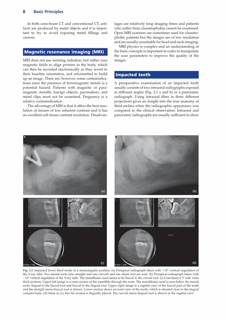
A preoperative examination of an impacted tooth usually consists of two intraoral radiographs exposed at different angles (Fig. 2.1 a and b) or a panoramic radiograph. Using intraoral films in three different projections gives an insight into the true anatomy of third molars when the radiographic appearance was compared to the clinical observation. Intraoral and panoramic radiographs are usually sufficient to show
In both cone-beam CT and conventional CT, arti-facts are produced by metal objects and it is impor-tant to try to avoid exposing metal fillings and crowns.
Magnetic resonance imaging (MRI)
MRI does not use ionizing radiation, but rather uses magnetic fields to align protons in the body, which can then be recorded electronically as they revert to their baseline orientation, and reformatted to build up an image. There are, however, some contraindica-tions since the presence of ferromagnetic metals is a potential hazard. Patients with magnetic or para-magnetic metallic foreign objects, pacemakers, and metal clips must not be examined. Pregnancy is a relative contraindication.
The advantage of MRI is that it offers the best reso-lution of tissues of low inherent contrast and it has an excellent soft tissue contrast resolution. Disadvan-
Fig. 2.1 Impacted lower third molar in a mesioangular position. (a) Periapical radiograph taken with +10° vertical angulation of the X-ray tube. Two mesial roots (one straight and one curved) and one distal root are seen. (b) Periapical radiograph taken with −10° vertical angulation of the X-ray tube. The mandibular canal seems to be buccal to the curved root. (c) Cone-beam CT with 1 mm thick sections. Upper left image is a cross-section of the mandible through the roots. The mandibular canal is seen below the mesial roots, lingual to the buccal root and buccal to the lingual root. Upper right image is a sagittal view of the buccal part of the tooth and the straight mesio-buccal root is shown. Lower section shows an axial view of the tooth, which is situated close to the lingual compact bone. (d) Same as (c), but the section is lingually placed. The curved mesio-lingual root is shown in the sagittal view.
(a) (b)
(c) (d)
Radiographic Imaging in Oral and Maxillofacial Surgery 9
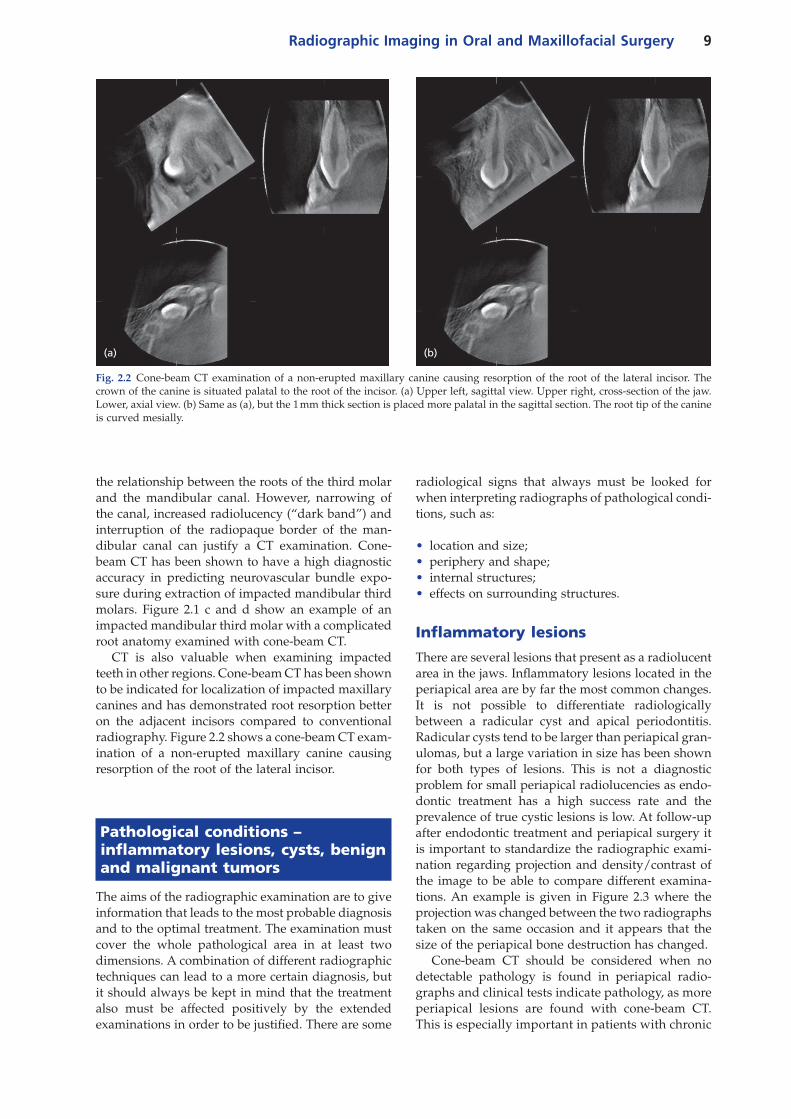
Fig. 2.2 Cone-beam CT examination of a non-erupted maxillary canine causing resorption of the root of the lateral incisor. The crown of the canine is situated palatal to the root of the incisor. (a) Upper left, sagittal view. Upper right, cross-section of the jaw. Lower, axial view. (b) Same as (a), but the 1 mm thick section is placed more palatal in the sagittal section. The root tip of the canine is curved mesially.
(a) (b)
the relationship between the roots of the third molar and the mandibular canal. However, narrowing of the canal, increased radiolucency (“dark band”) and interruption of the radiopaque border of the man-dibular canal can justify a CT examination. Cone-beam CT has been shown to have a high diagnostic accuracy in predicting neurovascular bundle expo-sure during extraction of impacted mandibular third molars. Figure 2.1 c and d show an example of an impacted mandibular third molar with a complicated root anatomy examined with cone-beam CT.
CT is also valuable when examining impacted teeth in other regions. Cone-beam CT has been shown to be indicated for localization of impacted maxillary canines and has demonstrated root resorption better on the adjacent incisors compared to conventional radiography. Figure 2.2 shows a cone-beam CT exam-ination of a non-erupted maxillary canine causing resorption of the root of the lateral incisor.
Pathological conditions – inflammatory lesions, cysts, benign and malignant tumors
The aims of the radiographic examination are to give information that leads to the most probable diagnosis and to the optimal treatment. The examination must cover the whole pathological area in at least two dimensions. A combination of different radiographic techniques can lead to a more certain diagnosis, but it should always be kept in mind that the treatment also must be affected positively by the extended examinations in order to be justified. There are some
radiological signs that always must be looked for when interpreting radiographs of pathological condi-tions, such as:
• location and size;• periphery and shape;• internal structures;• effects on surrounding structures.
Inflammatory lesions
There are several lesions that present as a radiolucent area in the jaws. Inflammatory lesions located in the periapical area are by far the most common changes. It is not possible to differentiate radiologically between a radicular cyst and apical periodontitis. Radicular cysts tend to be larger than periapical gran-ulomas, but a large variation in size has been shown for both types of lesions. This is not a diagnostic problem for small periapical radiolucencies as endo-dontic treatment has a high success rate and the prevalence of true cystic lesions is low. At follow-up after endodontic treatment and periapical surgery it is important to standardize the radiographic exami-nation regarding projection and density/contrast of the image to be able to compare different examina-tions. An example is given in Figure 2.3 where the projection was changed between the two radiographs taken on the same occasion and it appears that the size of the periapical bone destruction has changed.
Cone-beam CT should be considered when no detectable pathology is found in periapical radio-graphs and clinical tests indicate pathology, as more periapical lesions are found with cone-beam CT. This is especially important in patients with chronic
10 Basic Principles
of the maxillary sinus involving the upper jaw detected on a panoramic radiograph. Rapidly growing malig-nant lesions destroy the alveolar bone but usually no root resorption is present. A typical sign is that the teeth may appear to be floating in space: “floating teeth”.
The radiographic examination of malignant tumors often comprises CT and MRI to determine the extent of the tumor and to evaluate cervical lymphad-enopathy. Post-treatment examinations are usually performed to evaluate the effect of treatment. A com-bination of CT and positron emission tomography (PET) has been introduced and PET/CT is now widely used as an advanced clinical tool for the diag-nosis, staging, and restaging of cancer, and for the assessment of tumor therapy. A combination of MRI and PET is also becoming available. PET is a func-tional study where a radiolabelled isotope of glucose is given intravenously and areas of high metabolic activity can be recorded. The uptake is recorded by a nuclear imaging system and is normally merged with CT or MRI imaging for improved localization.
Temporomandibular joint (TMJ)
Imaging of TMD patients plays a minor role in the management of these patients as it has been shown that the treatment outcome is not affected by the radiological findings. Despite the success of conserv-ative care, however, some patients do not improve and TMJ surgery may be indicated. In these cases
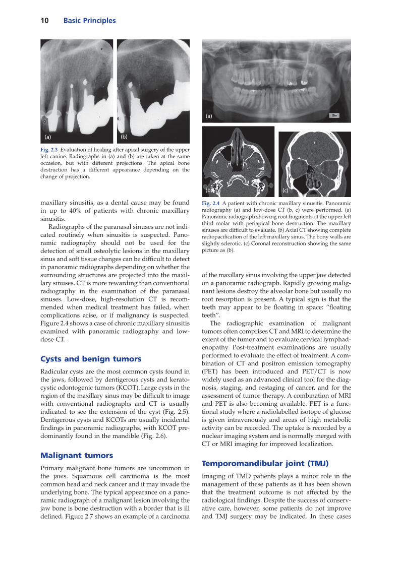
maxillary sinusitis, as a dental cause may be found in up to 40% of patients with chronic maxillary sinusitis.
Radiographs of the paranasal sinuses are not indi-cated routinely when sinusitis is suspected. Pano-ramic radiography should not be used for the detection of small osteolytic lesions in the maxillary sinus and soft tissue changes can be difficult to detect in panoramic radiographs depending on whether the surrounding structures are projected into the maxil-lary sinuses. CT is more rewarding than conventional radiography in the examination of the paranasal sinuses. Low-dose, high-resolution CT is recom-mended when medical treatment has failed, when complications arise, or if malignancy is suspected. Figure 2.4 shows a case of chronic maxillary sinusitis examined with panoramic radiography and low-dose CT.
Cysts and benign tumors
Radicular cysts are the most common cysts found in the jaws, followed by dentigerous cysts and kerato-cystic odontogenic tumors (KCOT). Large cysts in the region of the maxillary sinus may be difficult to image with conventional radiographs and CT is usually indicated to see the extension of the cyst (Fig. 2.5). Dentigerous cysts and KCOTs are usually incidental findings in panoramic radiographs, with KCOT pre-dominantly found in the mandible (Fig. 2.6).
Malignant tumors
Primary malignant bone tumors are uncommon in the jaws. Squamous cell carcinoma is the most common head and neck cancer and it may invade the underlying bone. The typical appearance on a pano-ramic radiograph of a malignant lesion involving the jaw bone is bone destruction with a border that is ill defined. Figure 2.7 shows an example of a carcinoma
Fig. 2.4 A patient with chronic maxillary sinusitis. Panoramic radiography (a) and low-dose CT (b, c) were performed. (a) Panoramic radiograph showing root fragments of the upper left third molar with periapical bone destruction. The maxillary sinuses are difficult to evaluate. (b) Axial CT showing complete radiopacification of the left maxillary sinus. The bony walls are slightly sclerotic. (c) Coronal reconstruction showing the same picture as (b).
(a)
(c)(b)
Fig. 2.3 Evaluation of healing after apical surgery of the upper left canine. Radiographs in (a) and (b) are taken at the same occasion, but with different projections. The apical bone destruction has a different appearance depending on the change of projection.
(a) (b)

Radiographic Imaging in Oral and Maxillofacial Surgery 11
radiography is indicated, as well as in patients with trauma, tumors, ankylosis and developmental anom-alies. Further, radiographic examination of patients with polyarthritic conditions, such as rheumatoid arthritis, can be recommended to evaluate the degree of joint destruction. Bone scanning with 99mtechne-tium phosphate isotopes might be indicated to determine the level of growth activity in condylar hyperplasia.
There are different techniques for imaging the TMJ: panoramic radiography, plain radiography, con-ventional and computed tomography, arthrography, and MRI. Panoramic radiography is not a reliable method for accurately showing the shape of the man-dibular condyle and the temporal component is poorly visualized. Plain radiography of the TMJ
Fig. 2.5 Patient with a fistula in the maxillary right canine region. Buccal swelling and symptoms of sinusitis. He mentions that a tooth was extracted in the region about 10 years ago when he had similar symptoms. The final diagnosis was proved to be residual cyst. (a) Panoramic radiograph which is difficult to interpret. (b) CT with an axial section showing well-defined bone destruction in the right maxillary canine region. (c) Coronal section showing the cystic lesion with thickened bone around the cyst. Soft tissue swellings are seen in the maxillary sinus. (d) Axial CT taken 10 years earlier, when the patient had symptoms of sinusitis. A cystic lesion is seen around the root tip of the right maxillary canine. The tooth was later extracted.
(a)
(b)
(c)
(d)
Fig. 2.6 Panoramic radiograph showing a multilocular bone destruction in the right mandibular ramus area. The patient had no symptoms and the cyst was detected in bitewing radio-graphs taken by his dentist. The tentative radiological diag-noses were ameloblastoma or keratocystic odontogenic tumor (KCOT). The diagnosis from the pathologist’s report was KCOT.

12 Basic Principles
depicts the mineralized part of the joint, but super-imposition of adjacent anatomic structures can make interpretation difficult. Conventional tomogra-phy improves the depiction of the bone structures.
However, minor bony changes will not be shown in conventional tomography. CT imaging provides exqui-site detail for bony abnormalities, such as ankylosis, fractures (Fig. 2.8), osseous tumors and arthrosis and 3D images can be produced; 3D reconstructions of a patient with condylar aplasia are shown in Figure 2.9.
MRI has replaced arthrography and can provide information about disk position, joint fluid, bone marrow changes, and bone structure at multiple levels of the joint. MRI is the prime diagnostic imaging technique in TMD patients. The technique, however,
Fig. 2.7 The patient complains of pain in the upper left jaw. A swelling of the left cheek is noticed. (a) Panoramic radiography shows an ill defined bone destruction in the area of the left maxillary sinus and the edentulous jaw seems to be involved in the bone destruction. (b, c) Contrast-enhanced axial CT sections showing bone destruction of the anterior and medial walls of the maxillary sinus and of the alveolar bone. The tumor is expanding buccally into the cheek. (d) Coronal section showing complete destruction of the jaw. The superior bony wall to the orbit seems intact.
(a)
(b)
(c)
(d)
is expensive and there are no studies showing when the results of the MRI examination will result in a better treatment outcome for the typical TMD patient. Imaging of the TMJ is definitely indicated prior to TMJ surgery and the preferred method is MRI if the soft tissue should be shown and CT if the hard tissue is of prime interest. In cases of tumors the methods often are combined.
Implant treatment
Panoramic radiography is the first choice for the radio-logical appraisal before implant treatment. The tech-nique is, however, dependent on proper positioning of the patient during exposure and objects located

Radiographic Imaging in Oral and Maxillofacial Surgery 13
outside the center of the sharply depicted plane are reproduced with distortions. Reliable measurements have been found for digital panoramic radiography, but both over and underestimation of vertical linear measurements have been found in other studies of panoramic radiography. The inherent errors in pano-ramic radiography should always be kept in mind whenever an exact assessment of a distance is required. Panoramic radiography is inferior to CT in visualization of the mandibular canal and in meas-urements related to the mandibular canal. Today, CT has almost totally replaced conventional tomogra-phy. There is equal accuracy so cone-beam CT should be preferred as this technique gives lower radiation dose compared to multislice CT and has a superior array of software for dental implant and related plan-
ning procedures. However, when a completely eden-tulous patient is examined several exposures with cone-beam CT with a narrow field size are necessary (Fig. 2.10). CT is indicated whenever the bone volume
Fig. 2.8 Cone-beam CT of a fracture of the condylar neck after a bicycle accident. Upper left, sagittal view; upper right, coronal view; lower right, axial view. The condylar fragment has been dislocated medially and inferiorly. The images are taken with 3D Accuitomo (Morita Corp.)
Fig. 2.9 CT 3D reconstructions of a patient with hemifacial microsomia and aplasia of the right condyle. The patient is missing the ear and a defective zygomatic arch is seen on the right side. The left side has developed normally.
Fig. 2.10 Multislice CT examination for planning of implant treatment of an edentulous maxilla. (a) Panoramic radiograph. The bucco-palatal bone width of the maxilla was judged to be questionable. (b) 3 mm thick paraxial reconstructions made per-pendicular to the alveolar bone (cross-sections) of the left side from the incisor to the premolar region. The images are pro-duced in scale 1 : 1. B = buccal, L = lingual. (c) 3 mm thick panoramic reconstructions. The number of the vertical lines can be identified in the paraxial sections in order to locate the section.
(a)
(c)(b)
14 Basic Principles
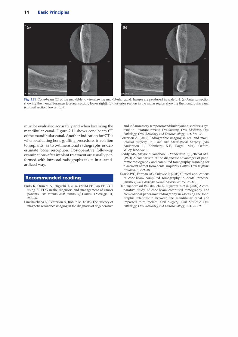
Fig. 2.11 Cone-beam CT of the mandible to visualize the mandibular canal. Images are produced in scale 1 : 1. (a) Anterior section showing the mental foramen (coronal section, lower right). (b) Posterior section in the molar region showing the mandibular canal (coronal section, lower right).
(b)(a)
must be evaluated accurately and when localizing the mandibular canal. Figure 2.11 shows cone-beam CT of the mandibular canal. Another indication for CT is when evaluating bone grafting procedures in relation to implants, as two-dimensional radiographs under-estimate bone resorption. Postoperative follow-up examinations after implant treatment are usually per-formed with intraoral radiographs taken in a stand-ardized way.
Recommended reading
Endo K, Oriuchi N, Higuchi T, et al. (2006) PET an PET/CT using 18F-FDG in the diagnosis and management of cancer patients. The International Journal of Clinical Oncology, 11, 286–96.
Limchaichana N, Petersson A, Rohlin M. (2006) The efficacy of magnetic resonance imaging in the diagnosis of degenerative
and inflammatory temporomandibular joint disorders: a sys-tematic literature review. OralSurgery, Oral Medicine, Oral Pathology, Oral Radiology and Endodontology, 102, 521–36.
Petersson A. (2010) Radiographic imaging in oral and maxil-lofacial surgery. In: Oral and Maxillofacial Surgery (eds, Andersson L, Kahnberg K-E, Pogrel MA). Oxford, Wiley-Blackwell.
Reddy MS, Mayfield-Donahoo T, Vanderven FJ, Jeffcoat MK. (1994) A comparison of the diagnostic advantages of pano-ramic radiography and computed tomography scanning for placement of root form dental implants. Clinical Oral Implants Research, 5, 229–38.
Scarfe WC, Farman AG, Sukovic P. (2006) Clinical applications of cone-beam computed tomography in dental practice. Journal of the Canadian Dental Association, 72, 75–80.
Tantanapornkul W, Okouchi K, Fujiwara Y, et al. (2007) A com-parative study of cone-beam computed tomography and conventional panoramic radiography in assessing the topo-graphic relationship between the mandibular canal and impacted third molars. Oral Surgery, Oral Medicine, Oral Pathology, Oral Radiology and Endodontology, 103, 253–9.