esthetician all trades nail growth; hand and foot...
TRANSCRIPT

1
EST 22
30 June 2016
Esthetician – All Trades
Nail Growth; Hand and Foot Disorders and Diseases

2

3
This booklet has been created by the Esthetician community of Saskatchewan. It is
intended for educational use; it is not for resale or profit, and can be copied without
cost. Please forward any suggestions to: [email protected]

4
Table of Contents
Objective One ......................................................................................... 13
The Fingernail ...................................................................................... 13
Nail Plate .............................................................................................. 14
Nail Bed ................................................................................................ 14
Matrix ................................................................................................... 14
The Lunula ........................................................................................... 14
Cuticle ................................................................................................... 15
Eponychium ......................................................................................... 15
Hyponychium ...................................................................................... 15
Nail Folds ............................................................................................. 16
Normal Nail Shapes ............................................................................ 16
Objective One Self-Test ......................................................................... 18
Objective One Self-Test Answers ........................................................ 19
Objective Two ........................................................................................ 20
Nail Growth ......................................................................................... 20
Objective Two Self-Test ........................................................................ 21
Objective Two Self-Test Answers ........................................................ 22
Objective Three ...................................................................................... 23
Objective Four ........................................................................................ 24
Conditions, Disorders, and Diseases ................................................ 24

5
Nail Disorders ...................................................................................... 24
Beaus Lines .......................................................................................... 24
Cyanosis (Blue Nails) .......................................................................... 25
Eggshell Nails (Onychomalacia) ....................................................... 25
Fissures ................................................................................................. 25
Hematoma (Bruised Nail Bed) .......................................................... 25
Hangnail ............................................................................................... 25
Involuted Toenail ................................................................................ 26
Ingrown Toenail (Onychocryptosis) ................................................. 26
Koilonychia .......................................................................................... 26
Leukonychia ........................................................................................ 26
Melanonychia ...................................................................................... 26
Onychatrophia ..................................................................................... 27
Onychauxis .......................................................................................... 27
Onychophagy (Nail Biting) ................................................................ 27
Onychoptosis Difluvium .................................................................... 27
Onychorrhexis ..................................................................................... 27
Pincer (Trumpet Nails) ....................................................................... 28
Plicatured Nail ..................................................................................... 28
Psoriasis ................................................................................................ 28

6
Pterygium ............................................................................................. 28
Ridges (Grooves) ................................................................................. 28
Scleronychia ......................................................................................... 29
Splinter Hemorrhages ........................................................................ 29
Wooded Nail ........................................................................................ 29
Yellow Nail Syndrome ....................................................................... 29
Objective Four Self-Test ........................................................................ 30
Objective Four Self-Test Answers ....................................................... 32
Objective Five ......................................................................................... 33
Scope of Practice .................................................................................. 33
Bacterial Infections .............................................................................. 33
Fungal Infections (Parasites) .............................................................. 33
General Information ........................................................................... 33
Dermatophytes .................................................................................... 34
Tinea Pedis ........................................................................................... 34
Onychosis ............................................................................................. 35
Onychia ................................................................................................ 35
Onycholysis .......................................................................................... 35
Onychomadesis ................................................................................... 35
Onychomycosis ................................................................................... 36

7
Paronychia ........................................................................................... 36
Pseudomonas (Staphylococcus) ........................................................ 36
Objective Five Self-Test ......................................................................... 38
Objective Five Self-Test Answers ........................................................ 40
Objective Six ........................................................................................... 41
Achilles Injuries ................................................................................... 41
Achilles Tendon Rupture ................................................................... 41
Achilles Tendonitis ............................................................................. 41
Blister .................................................................................................... 41
Edema (Swollen Ankles) .................................................................... 42
Fissures ................................................................................................. 42
Flat foot ................................................................................................. 42
Foot Infection (General Signs) ........................................................... 42
Foot Odour ........................................................................................... 43
Ganglions ............................................................................................. 43
Gout ...................................................................................................... 43
Hammertoes......................................................................................... 43
Heloma (Corns) ................................................................................... 44
High Arch ............................................................................................. 44
Metatarsalgia (Fallen Metatarsal Arches) ........................................ 44
8
Morton's Neuroma (Intermetatarsal Neuroma) .............................. 45
Plantar Fasciitis and Heel Spurs ........................................................ 45
Plantar Hyperhydrosis ....................................................................... 46
Pyogenic Granuloma .......................................................................... 46
Sesamoid Injuries ................................................................................ 46
Spider Veins ......................................................................................... 47
Turf Toe ................................................................................................ 47
Tyloma (Callus) ................................................................................... 48
Varicose Veins ..................................................................................... 48
Verruca Vulgaris (Plantar Warts) ...................................................... 48
Objective Six Self-Test ........................................................................... 49
Objective Six Self-Test Answers........................................................... 51
Objective Seven ...................................................................................... 52
What is Diabetes? ................................................................................ 52
Type 1 Diabetes ................................................................................... 52
Type 2 Diabetes ................................................................................... 52
Diabetic Feet......................................................................................... 53
Nerve Damage ..................................................................................... 53
Skin ....................................................................................................... 53
Callus .................................................................................................... 54

9
Foot Ulcers ........................................................................................... 54
Temperature ........................................................................................ 54
Objective Seven Self-Test ...................................................................... 56
Objective Seven Self-Test Answers ..................................................... 58
Objective Eight ....................................................................................... 59
Ligaments ............................................................................................. 59
Basal Joint Arthritis ............................................................................. 59
Osteoarthritis ....................................................................................... 59
Rheumatoid Arthritis ......................................................................... 59
Tendon Injuries.................................................................................... 60
Tendonitis ............................................................................................ 60
DeQuervain's Tenosynovitis .............................................................. 60
Tennis ................................................................................................... 61
Boutonniere Finger ............................................................................. 61
Mallet Finger ........................................................................................ 61
Ski Pole Thumb ................................................................................... 62
Objective Eight Self-Test ....................................................................... 63
Objective Eight Self-Test Answers ...................................................... 64
Objective Nine ........................................................................................ 65
What is Carpal Tunnel Syndrome? ................................................... 65

10
Developing Carpal Tunnel Syndrome.............................................. 66
Occupational Factors of Carpal Tunnel Syndrome......................... 66
Symptoms of Carpal Tunnel Syndrome ........................................... 67
Treatment of Carpal Tunnel Syndrome ........................................... 68
Preventing Carpal Tunnel Syndrome ............................................... 68
Objective Nine Self-Test ........................................................................ 70
Objective Nine Self-Test Answers ....................................................... 71
Objective Ten .......................................................................................... 72
Contraindications That Restrict a Manicure or Pedicure ............... 72
Contraindications That Prevent a Manicure or Pedicure ............... 73
Objective Ten Self-Test .......................................................................... 74
Objective Ten Self-Test Answers ......................................................... 76
Objective Eleven .................................................................................... 77
Module Summary Self-Test .................................................................. 78
Module Summary Self-Test Answers ................................................. 82

11
Nail Growth; Hand and Foot Disorders and Diseases
Rationale Why is it important to learn this skill?
The material contained in this ILM forms ‘the heart’ of manicure and pedicure services.
This information is necessary for the safety of both the apprentice and the client.
Outcome When you have completed this module, you will be able to:
Describe nail growth, hand and foot disorders and diseases.
Objectives 1. Describe nail structure and natural nail shapes.
2. Describe nail growth patterns.
3. Demonstrate analyzing nail growth.
4. Describe nail conditions.
5. Describe nail diseases.
6. Describe foot and leg conditions and disorders.
7. Describe the diabetic foot.
8. Describe hand conditions.
9. Describe carpal tunnel syndrome.
10. Describe contraindications that may restrict or prevent a service.
11. Demonstrate analyzing conditions, disorders, and diseases of the hands and feet.
Introduction The following information will help the apprentice make informed decisions regarding
contraindications and which services can and cannot be performed on clients. The
introduction to anatomy presented here will help in the understanding of conditions
and workplace injuries. Of particular interest to apprentices is Objective 9 which

12
covers carpal tunnel syndrome, a condition that affects many apprentices and
journeypersons.

13
Objective One When you have completed this objective, you will be able to:
Describe nail structure and natural nail shapes.
The Fingernail
A fingernail is created by cells located in the finger, called the matrix. A fingernail
consists of several parts as illustrated below. The nail plate is the visible part of the
nail; it is made of keratin, a fibre-shaped protein also found in skin and hair. The nail
bed is located beneath the nail plate, and is comprised of skin. The skin is made of two
types of tissues. The deeper tissue is the dermis, which includes living tissue of glands
and capillaries. The epidermis is the layer just beneath the nail plate; it moves with the
nail plate toward the fingertip. The cuticle is the tissue that surrounds the base of the
nail and overlaps the plate. The nail is supported on three sides by skin called the nail
folds. The whitish half-moon at the base of the nail is called the lunula.
As the matrix creates new cells, older cells are pushed out and compacted, becoming
the hardened fingernail.
Image courtesy of International Journal of Clinical Reviews.

14
A healthy, normal nail is translucent and whitish in colour. It appears slightly pink
because of the natural colour of the nail bed below. It is flexible and firm. The surface
of a nail is usually smooth and uniform in texture and colour. The water content of a
healthy nail ranges between 15 and 25 percent. The nail plate is porous and absorbs
water. Relative humidity has a great effect on the water content of a nail, and a nail’s
flexibility varies directly with its water content: the higher the content, the higher the
flexibility. Water loss can be reduced by applying an oil-based nail conditioner or nail
polish. These products can also hinder water absorption.
Nail Plate
The nail plate rests on top of the nail bed, and the two slowly slide toward the
fingertip. Although the nail plate appears to be one solid piece, it is constructed of
approximately 100 layers of nail cells. The part of the nail plate that extends beyond
the fingertip is called the free edge.
Nail Bed
The nail bed is also called the bed epithelium. The bed is supplied with blood vessels
and nutrients. It extends from the lunula to the area just before the free edge. The nail
bed supports and guides the nail plate as it grows.
Matrix
The nail plate cells are formed in the matrix. The matrix is a layer of specialized tissue
located at the base of the fingernail or toenail. This tissue is made up of quickly
dividing skin cells that fill with the protein keratin. The matrix area includes nerves,
lymph (a colourless fluid containing white blood cells), and blood vessels that supply
nourishment the matrix. The matrix creates new nail plate cells as long as it is
nourished and not physically damaged.
The Lunula
The matrix begins under the nail fold at the base of the nail plate and extends upward,
under the nail plate. The matrix appears as the whitish half-moon shape at the base of

15
the nail plate. Damage to the lunula will cause permanent deformity to the nail plate.
Some lunulas are not visible because they are short and hidden under the eponychium.
Cuticle
The cuticle is the thin, colourless, dead, tissue attached to the nail plate at its base. The
cuticle originates beneath the skin located above the nail plate. This tissue is tough,
flexible, and sticky. It is difficult to remove from the nail plate, because its function is
to seal the space above the base of the nail plate. If a person is using their fingers in a
dirty environment (the garden) it is possible to force debris and microorganisms into
the fingers, between the nail plate and the skin above it. The cuticle seals this gap,
keeping the fingers healthy. Cuticle is removed from the nail plate with gentle
scraping.
Eponychium
The eponychium is the living skin at the base of the nail plate, above the cuticle. The
cuticle and the eponychium are not the same. The cuticle is dead tissue and the
eponychium is living tissue. This distinction is important when considering an
esthetician’s scope of practice. In general terms, an esthetician can work on dead
tissue, not living tissue. The cuticle originates under the eponychium, but separates
from it as the cuticle adheres to the nail plate. The cuticle seals the gap between the
nail plate and the eponychium.
Estheticians are permitted to gently push back the eponychium; they are prohibited
from cutting or trimming it. Eponychium can appear hardened and dry, appearing to
be dead skin. It is not dead skin.
Hyponychium
The hyponychium is the layer of skin located between the fingertip and the free edge
of the nail plate. Because it is a protective layer, it is slightly thicker and tougher than
average skin. The function of the hyponychium is to prevent debris and
microorganisms from entering the finger at this point. If debris and microorganisms
enter the finger at this point, the nail bed can become infected or damaged.

16
Nail Folds
The nail folds are folds of normal skin that surround the nail plate on three sides.
These folds form the furrow, or nail groove, on each side of the nail. The lateral nail
fold is also called the sidewall; it is the fold of skin overlapping the side of the nail.
Normal Nail Shapes
Natural nails come in
many shapes, and each
shape has different
names. Each of the four
edges can have varying
qualities of length,
‘straightness’ or
‘roundness.’ For
instance, a base can be
wide or narrow; as well,
it can be very straight, or
it can be curved. The
curve can be gradual or
sharp.
Image courtesy of ROCKETNEWS24

17
To the left is the front view of a nail. Its curvature is
drawn as the convex arch of a perfect circle. The arch
of a natural nail can be compared with the arch
depicted. Some nails can be concave instead of convex,
while others can be flat. Different convex arches can
exist within a range of ‘normal’ or ‘average.’ When an
arch is sharper than the ‘normal’ or ‘average’, it is
described as ‘high-arched’ or ‘circumflex.’
To the right is a side view of a nail. When viewed from the
side, most nails have a natural convex arch. In contrast to
the convex arch, some nails can be concave; this is often
referred to as a ‘scooped’ or ‘ski jump’ nail. Other nails
may have an extreme convex arch.
Image courtesy of www.e-aps.org
Image courtesy of Google Images.

18
Objective One Self-Test
1) What is the nail plate made of?
_____________________________________________________________________________
2) What is the difference between the dermis and the epidermis?
_____________________________________________________________________________
3) Although an oil-based nail conditioner or nail polish can help condition nails,
which problem can be caused by these products?
_____________________________________________________________________________
4) What is the function of the nail bed?
_____________________________________________________________________________
5) What is the function of cuticle?
_____________________________________________________________________________
6) Where is the eponychium located, and can an esthetician cut it?
_____________________________________________________________________________
7) Where is the hyponychium located, and what is its function?
_____________________________________________________________________________

19
Objective One Self-Test Answers
1) The nail plate is made of keratin, a fibre-shaped protein also found in skin and hair.
2) The dermis is a deeper tissue which includes living tissue of glands and capillaries.
The epidermis is the layer just beneath the nail plate; it moves with the nail plate
toward the fingertip.
3) An oil-based nail conditioner or nail polish can hinder the nail plate’s ability to
absorb water.
4) The nail bed supports and guides the nail plate as it grows.
5) The function of cuticle is to seal the space above the base of the nail plate.
6) The eponychium is the living skin at the base of the nail plate, above the cuticle. An
esthetician cannot work on it because it is live tissue.
7) The hyponychium is the layer of skin located between the fingertip and the free
edge of the nail plate. The function of the hyponychium is to prevent debris and
microorganisms from entering the finger at this point.

20
Objective Two When you have completed this objective, you will be able to:
Describe nail growth patterns.
Nail Growth
The growth of the nail plate is affected by many factors, including: age, sex, nutrition,
exercise, and general health. Healthy, normal nail plates are produced in a variety of
shapes. The width, curvature, and thickness of a nail plate depend on the width,
curvature, and length of the matrix. A thicker nail plate is created by a longer matrix,
and a flatter nail plate is created by a flatter matrix. Nothing can make the nail plate
grow thicker.
The average rate of nail plate growth in the normal adult is about 2.5 mm to 3 mm per
month. Nail plates grow slower in winter than in summer; faster in children than the
elderly; and slower in women than men. During the last trimester of pregnancy, nail
growth rates increase dramatically because of hormonal changes. After the baby is
delivered, the nail growth rate returns to normal. Nail growth cannot be sped by
taking prenatal vitamins. Toenail plates grow slower than fingernail plates, and
toenail plates are thicker because the toenail matrix is longer.
Nail plates grow as long as the matrix is healthy and undamaged. The complete
replacement of a fingernail plate usually takes from 4 to 6 months, while toenail plates
often require from 9 to 12 months. The nail matrix continuously creates new nail cells
which push older cells out toward the fingertip. The nail plate slowly advances
toward the free edge, at the rate of new cell creation. If nail cells are produced faster,
the plate will grow more quickly. Damage to the matrix can cause nail plates to
develop thinner or with grooves. As a natural cycle of life, matrix production
decreases as a person ages; as a result, the nail plate develops narrow grooves running
the length of the nail plate.

21
Objective Two Self-Test
1) What determines the thickness of a nail plate?
_____________________________________________________________________________
2) Why does nail growth rates increase during the last trimester of pregnancy?
_____________________________________________________________________________
3) How long does it take to completely replace a fingernail plate and a toenail plate?
_____________________________________________________________________________
4) How is matrix production affected by aging?
_____________________________________________________________________________

22
Objective Two Self-Test Answers
1) The thickness of a nail plate is determined by the length of the matrix.
2) During the last trimester of pregnancy, nail growth rates increase dramatically
because of hormonal changes.
3) The complete replacement of a fingernail plate usually takes from 4 to 6 months,
while toenail plates often require from 9 to 12 months.
4) Matrix production decreases as a person ages.

23
Objective Three When you have completed this objective, you will be able to:
Demonstrate analyzing nail growth.
Laboratory Exercise
Purpose: to analyze nail growth.
Materials:
Procedure: With a partner, analyze each other’s nails. Identify the following
components:
Proximal nail fold Nail plate Lunula Eponychium
Distal groove Hyponychium Nail bed Cuticle
Draw shape of your partner’s nail plate.
Side view Top view
Instructor verification: _______________________________________

24
Objective Four When you have completed this objective, you will be able to:
Describe nail conditions.
Conditions, Disorders, and Diseases
The difference between disorders and diseases is not clear. Different texts will define
these terms in different manners. For the purposes of Esthetician technical training,
the term ‘disorder’ refers to an unwanted condition, the cause of which is hard to
determine. Disorders are often derangements or abnormalities of function. In contrast
to a disorder, a ‘disease’ has a specific, identifiable cause and symptoms. A disease can
be thought of as a definite pathological process having a characteristic set of signs and
symptoms. Diseases are often the result of extrinsic factors like viruses. Disorders are
often attributed to intrinsic abnormalities like genetic malfunctions. The term
‘condition’ is a general term that encompasses both disorders and diseases.
Nail Disorders
Nail disorders are caused by two main sources: 1) a breakdown of the normal way a
nail forms; and 2) circumstantial factors such as stress and diet. It is important to
remember an esthetician’s scope of practice. An esthetician can never diagnose
medical disorders and diseases, but recognition of these things is crucial. Within the
scope of practice, an esthetician may work on a condition that is cosmetic in nature
(not medical). All medical disorders should be referred to a physician.
Disorders may be local or general. A local disorder affects only one or two nails, while
a general disorder may occurs on all ten nails and the surrounding skin. A general
situation usually means that the disorder was inherited or is being caused within the
body. For instance, a mineral deficiency may cause a disorder. The following is a
description of many common nail disorders.
Beaus Lines
Beaus lines are visible depressions running across the width of the natural nail plate.
They are often caused by a major illness or injury that has traumatized the body.

25
Traumas include adverse drug reactions, pneumonia, and surgery. During the stress
suffered by the body, the matrix slows production of nail cells, and this causes the nail
plate to grow thinner. The thickness usually returns to normal after the stress is over.
Cyanosis (Blue Nails)
Cyanosis is caused by poor circulation, heart / lung conditions, and topical / oral
medication. During cyanosis, the nail plate turns blue, causing the nail plate to appear
blue or purple.
Eggshell Nails (Onychomalacia)
Eggshell nails occur when the nail plate becomes thin and white, and more flexible
than usual. The nail plate will often curve downwards over the free edge. Eggshell
nails can be caused by improper diet, internal disease, medication, and bulimia; it may
also be hereditary. These nails break easily during service and will not support nail
enhancements.
Fissures
Fissures are cracks in the nails. They can be caused by dehydration.
Hematoma (Bruised Nail Bed)
A hematoma is caused by a physical impact to the nail which causes many capillaries
under the nail plate to burst. A blood clot temporarily forms under the nail plate but is
eventually absorbed by bed epithelium, and grows out with the nail plate. In severe
cases, the nail plate can fall off. This will happen to long distance runners and ballet
dancers. It is important to regularly check on the nail. If the nail falls off, the nail bed
can be covered during the healing process.
Hangnail
Hangnails are shards of nails or skin that are caused by dryness. Dry skin and nails
lose their elasticity; instead of stretching, they break. Uninfected hangnails can be
clipped off. If the hangnail is red, irritated, or swollen, it is infected. Infected
hangnails are outside of the scope of an esthetician.

26
Involuted Toenail
An involuted toenail is one with excessive curvature; this can be caused by wearing
shoes that are too narrow (tight toe box). This disorder can be hereditary. An
involuted toenail can become ingrown. Thick corns and callus can develop in the nail
fold.
Ingrown Toenail (Onychocryptosis)
An ingrown toenail occurs when the skin on the side of a nail plate grows over the
edges of the nail plate. An ingrown toenail often develops an infection. This disorder
may recur. It can be caused by incorrectly cutting the toe nail. Shoes that allow
movement of the toes and conservative nail clipping will reduce the occurrence of
ingrown toenails. Excessively rounding the corners of the nail will force the nail to
grow through the skin, rather than over it.
Koilonychia
Koilonychia is a disorder also known as ‘spoon nails’ or ‘ski jump nails.’ The nails are
concave when viewed from the side.
Leukonychia
Leukonychia is a disorder in which white spots and white-ish discolouration occur
inside the nail plate. This disorder is often caused by injury to the matrix.
Leukonychia occurs within the layers of the natural nail plate. As a word of caution,
this disorder can also be caused by pushing too hard with a cuticle pusher.
Melanonychia
Melanonychia is brown or black pigmentation that appears under the nail plate. It
often appears as a pigmented band running lengthwise or horizontally along the nail
bed. It can be caused by trauma, drugs, inflammatory disorders, and fungal infections.
A short-term trauma to the body may cause a horizontal line, while a vertical line can
be caused by an on ongoing trauma or stress. Melanonychia can indicate melanoma,
and clients with this disorder should be immediately referred to a physician.

27
Onychatrophia
Onychatrophia is also called ‘atrophy of the nails.’ It is a degradation of the nail plate.
The nail plate loses its lustre, becomes smaller, and sometimes sheds entirely. It can be
caused by an injury to the matrix, an internal disease, or a skin disease. The nail plate
rarely grows back. Never use strong soaps or chemicals on these nail plates.
Onychauxis
Onychauxis is characterized by an extreme thickening of the nail plate; this is caused
from hyper-keritinization, an over production of keratin. The nail plate adopts a
callused appearance. Onychauxis can be caused by injury, internal infection, or local
infection; it can also be hereditary. This disorder is often found in older people.
Onychophagy (Nail Biting)
Onychophagy is severe nail biting. The nail plates will retract over time and expose
the nail bed. This disorder is behavioural. Exposed flesh can become infected. Recent
scientific evidence has proven that nail biting is found in people of superior intellect
and emotional and spiritual mastery.
Onychoptosis Difluvium
Onychoptosis difluvium is the periodic shedding of one or more nails, either in whole
or part. This disorder may be caused by diseases like syphilis. Fever, trauma, and
adverse effects from pharmaceutical drugs are also likely causes.
Onychorrhexis
Onychorrhexis is a disorder characterized by split, brittle nail plates with vertical
ridges. This disorder is common in dry climates; therefore, it is likely caused by a lack
of moisture. It may also be hereditary. The excessive use of harsh chemicals may also
cause this disorder.

28
Pincer (Trumpet Nails)
This disorder is characterized by a nail plate with a sharp or deep curvature at the free
edge; it originates in the matrix. The free edge may pinch the sidewalls and curl in
upon itself. The free edge can become an ingrown nail if it penetrates the surrounding
skin.
Plicatured Nail
A pilacatured nail is characterized by a nail plate that is folded along the sides. It is
often caused by an injury to the matrix; it can also be inherited. This disorder will
often lead to ingrown nails.
Psoriasis
Psoriasis of the nail plate causes a dimpled appearance of the nail plate. The dimples
can be randomly or evenly spaced. The nail plate becomes thick and brittle. People
with psoriasis of the skin can also develop psoriasis of the nail plate. Psoriasis is not
infectious unless onycholysis is present. In extreme cases, psoriasis may look like a
fungal infection. Psoriasis may be caused by stress or anxiety; it may also be
hereditary.
Pterygium
Pterygium is a disorder in which the eponychium becomes overgrown and attaches
itself to the nail plate. The eponychium then grows along the nail plate as it shifts
forward. This disorder is caused by a lack of nutrients in the eponychium. In an
attempt to absorb nutrients, the eponychium attaches to the nail plate. Cuticle
softeners and oils may resolve mild cases. Keep in mind, estheticians should never
clip, trim, or push back the eponychium in these cases.
Ridges (Grooves)
A nail plate may appear to have ridges. This is incorrect. The nail does not have
ridges, it has grooves. The grooves can be local or general. Local grooves can occur
from trauma, while general ridges can be caused by several factors, including age and
strong medications.

29
Scleronychia
Scleronychia is characterized by an impaired growth of the nails accompanied by a
simultaneous thickening and drying of the nail plate. The nail plate can become thick,
dry, and yellow or grey in colour. The lunula is generally not visible when a person
has this disorder. Damage to the matrix will cause scleronychia in only one nail. If all
nails are affected, a traumatic experience or fever (high temperature) may be the cause.
The nail plate may experience periods of slow or no growth.
Splinter Hemorrhages
Splinter hemorrhages are caused by the bursting of a capillary under the nail plate.
Bursting is caused by trauma. Blood fills naturally occurring grooves under the nail
plate.
Wooded Nail
This disorder is mostly found on older people, normal nail growth is affected. Keep
filed down for esthetic purposes.
Yellow Nail Syndrome
Yellow nail syndrome is a rare disorder characterized by malformations of the
fingernails and toenails. The lungs and the airways are also affected. This disorder is
caused by accumulation of protein-rich fluid (lymph) in the soft layers of tissue under
the skin. This disorder usually affects older adults and may be inherited. The nail plate
is yellow, thickened, and excessively curved. The nail plate will almost completely
stop growing. A loss of cuticle may also occur. Onycholysis and paronychia may
occur simultaneously. Both toenails and fingernails may be affected. The nail plates
usually remain smooth, unlike in other conditions.

30
Objective Four Self-Test
1) For the purposes of Esthetician technical training, what is the difference between
the terms ‘disorder’ and ‘disease’?
_____________________________________________________________________________
_____________________________________________________________________________
2) How is a local disorder different from a general disorder?
_____________________________________________________________________________
3) True / False. Eggshell nails will support nail enhancements.
4) In non-severe cases, what happens to a hematoma under the nail plate?
_____________________________________________________________________________
5) When does an ingrown toenail occur? What is a possible cause?
_____________________________________________________________________________
_____________________________________________________________________________
6) What is leukonychia, and what is often the cause?
7) What is onychauxis, and what is the cause?
8) Is an esthetician permitted to alleviate pterygium by cutting and clipping?

31
9) Describe the appearance of scleronychia:
_____________________________________________________________________________
_____________________________________________________________________________
10) Which nail condition is accompanied by a disorder of the lungs and the airways?
_____________________________________________________________________________

32
Objective Four Self-Test Answers
1) A ‘disorder’ refers to an unwanted condition, the cause of which is hard to
determine, while a ‘disease’ has a specific, identifiable cause and symptoms.
2) A local disorder affects only one or two nails or a small area. A general disorder
occurs over a larger area, such as all ten nails or a large area of skin.
3) False.
4) A blood clot temporarily forms under the nail plate and is eventually absorbed by
bed epithelium, then grows out with the nail plate.
5) An ingrown toenail occurs when the skin on the side of a nail plate grows over the
edges of the nail plate. A possible cause is incorrectly cutting the toe nail.
6) Leukonychia is a disorder in which white spots and white-ish discolouration occur
inside the nail plate. This disorder is often caused by injury to the matrix.
7) Onychauxis is an extreme thickening of the nail plate; this is caused from hyper-
keritinization, an over production of keratin.
8) No. That would involve cutting live tissue.
9) Scleronychia appears as a thickening and drying of the nail plate. The nail plate can
become thick, dry, and yellow or grey in colour. The lunula is generally not visible.
10) Yellow nail syndrome.

33
Objective Five When you have completed this objective, you will be able to:
Describe nail diseases.
Scope of Practice
Some Authorities Having Jurisdiction (AHJ’s) allow estheticians with advanced
training to work on some of these diseases and/or suggest corrective care to clients. In
general, estheticians are not permitted to work on live tissue or suggest corrective care.
Skin that appears itchy, red, swollen, or the like is infected, and is therefore also
beyond the scope of an esthetician. If an esthetician suspects that a client has a disease,
the client should be referred to a physician.
Bacterial Infections
Bacterial infections are usually characterized by swelling, redness, and tenderness in
an area. Paronychia is a bacterial infection caused by picking the skin of the sidewalls.
Paronychia often accompanies ingrown toenails; it appears as inflamed and red tissue
that may excrete pus.
Pseudomonas aeruginosa is another type of bacterial infection. It appears as a
greenish/yellowish or brown spot under the nail plate. It can also occur under an
artificial enhancement. Pseudomonas is caused by unsanitary conditions.
Fungal Infections (Parasites)
Fungal infections occur in three human pathogenic types:
dermatophytes
yeasts
mold
General Information
Fungus of the feet—whether tinea pedis, yeast, or candida—affects about 75% of the
population. Fungal infections can be caused by a tear in the hyponychium or
eponychium, allowing the fungus to enter the nail bed; in addition, the warm, dark,

34
moist environment of the feet is an ideal breeding ground for fungus. Fungi are
contagious and can be transmitted through contact with infected flooring (while
walking in bare feet) and contaminated implements. These infections are usually
restricted to one or two nails, occurring less frequently on the hands.
Fungi can deform the nail plate and/or turn the nail plate yellowish or brownish.
Onycholysis may occur, and/or the nail plate may crack and/or white patches may
form on the nail plate. Over time, the nail plate may thicken, become brittle, and lift.
In an advanced stage, the lifted part of the nail will develop a residue and strong
unpleasant odour.
If a person has any fungal infection, they should wash their feet daily and dry them
completely, especially between the toes. Only cotton or wool socks should be worn
and changed at least twice a day (or every time they become damp). Footwear made
from artificial material should be avoided. All footwear should be breathable leather
or cotton. A fungicidal powder should be sprinkled on the feet and the inside of the
shoes.
Dermatophytes
Ringworm of the nail is caused by a fungal infection that feeds on keratin. Rngworm is
also known as tinea unguium, onychomycosis, and dermatophytosis unguium.
Ringworm of the nails can occur on the skin of the feet or in the nail itself. It can
spread from nail to foot or foot to nail. Ringworm of the nail is characterized by a
whitish thickening of the nail and nail-bed. The nail thickens, discolours and may
become destroyed. Ringworm of the nails can be treated with antifungal tablets, local
fungicides, or nail solutions. Treatment may need to continue for many months.
Tinea Pedis
Tinea pedis is a term used to describe athlete’s foot. It can appear as a red, itchy rash,
or a scaling of the skin; it can also appear as white, sodden, itchy skin between the toes.
Tinea pedis can be a secondary condition to micro-lesions and dry skin, long-term use
of medication such as antibiotics and cortisone, illness or trauma, diabetes and high
sugars, hormonal imbalances, poor nutrition, and a poorly functioning immune
system. Feet with tinea pedis should be washed with Ph balanced cleanser, not soap.

35
Dry the feet completely, especially between the toes. Shoes should not be re-worn
within 24 hours. This elapsed time will ensure that the bacteria die. Anti-microbial
drops are recommended for the feet and the shoes.
Onychosis
Onychosis is a term used to describe any disease or deformity of the nails.
Onychia
Onychia is an inflammation of the nail matrix. A break in the hyponychium,
eponychium, or peronychium can allow pathogens to affect the matrix. Onychia is
often caused by unsanitary implements. The tissue of the infected area becomes
inflamed, fills with pus that appears at the eponychium line above the matrix, and the
nail plate may shed.
Onycholysis
Onycholysis is a spontaneous separation of the nail plate from the nail bed. It almost
always separates from the free edge, moving toward the base of the nail plate. This
disease can have many causes. For example, water and bacteria can enter the living
tissue via a tear in the hyponychium. The bacteria then occupies more and more space
as it multiplies, causing the nail plate to separate from the nail bed. Hypothyroidism
and reactive arthritis may also cause onycholysis. At times, onycholysis is not
classified as a disease; in these cases, it can be caused by physical trauma to the nail
such as catching the nail on an object. The nail plate will appear whitish.
Onychomadesis
Onychaomadesis is the separation and falling off of the nail plate from the nail bed
caused specifically by psoriasis. The separation begins at the proximal edge of the nail
(the base of the nail) and progresses to the free edge. This disease can affect fingers
and toes. New nail plates will regrow.

36
Onychomycosis
Onychomycosis is a fungal infection between the nail plate and the nail bed. It occurs
in five stages. Stage one is the separation of the nail plate and bed (onycholysis); in the
second stage, the nail plate discolours; stage three is characterized by an accumulation
of debris under the nail plate; in the fourth stage, the nail plate thickens; stage five—
the last stage—is characterized by the production of a strong odour. A long yellow
line within the nail will develop at the free edge and progress toward the proximal
edge. This infection is often itchy and always very contagious.
Paronychia
Paronychia is an infection of the skin around the toenails and fingernails, caused by a
type of yeast called Candida or a bacteria. Paronychia may develop slowly and last for
weeks or appear suddenly and last for one or two days.
Acute paronychia mostly occurs around the fingernails and develops quickly. It is
usually caused by damage to the skin around the nails. The damage is often a result
from biting, manicures, and hangnails. Staphylococcus bacteria are the most frequent
infecting agent of acute paronychia.
Chronic paronychia can occur on both fingers and toes; it progresses slowly and lasts
for several weeks; it often recurs. Chronic paronychia is usually caused by multiple
infecting agents: Candida yeast and a bacteria are common. This infection occurs more
frequently to fingers and toes that are often wet.
Both acute and chronic paronychia have the same symptoms: red and tender skin
around the nail plate, pus-filled blisters, changes in the shape, texture, or colour of the
nail plate, and possible detachment of the nail plate.
Pseudomonas (Staphylococcus)
This naturally occurring skin bacteria can grow rapidly, causing infection. It does not
need specific conditions to grow, so it is occurs on hands as feet. Pseudomonas is
usually caused by contaminated implements, and not by water trapped under the nail
plate (a common misconception). A pseudomonas infection on a nail plate can be

37
identified in the early stages as a yellow-green spot that progresses to green, to brown,
to black. A client with pseudomonas should be referred to a physician.

38
Objective Five Self-Test
1) Which bacterial infection is caused by unsanitary conditions?
_____________________________________________________________________________
2) Identify three symptoms that fungi can cause:
_____________________________________________________________________________
_____________________________________________________________________________
3) How can ringworm be treated?
_____________________________________________________________________________
4) How should feet with tinea pedis be washed?
_____________________________________________________________________________
5) Which disease is characterized by: inflammation of the infected tissue, and the area
at the eponychium line above the matrix filling with pus that appears?
_____________________________________________________________________________
6) What is the separation and falling off of the nail plate from the nail bed caused
specifically by psoriasis?
_____________________________________________________________________________
7) How does chronic paronychia differ from acute paronychia?
_____________________________________________________________________________

39
8) What is a common misconception regarding the cause of pseudomonas
(staphylococcus)?
_____________________________________________________________________________

40
Objective Five Self-Test Answers
1) Pseudomonas aeruginosa is caused by unsanitary conditions.
2) Any three of the following: nail plate deformation; yellowish or brownish colouring
of the nail plate; Onycholysis; the nail plate may; the nail plate may crack; the nail
plate may develop white patches; the nail plate may thicken, become brittle, and lift;
the nail may develop a residue and strong unpleasant odour.
3) Ringworm of the nails can be treated with antifungal tablets, local fungicides, or
nail solutions.
4) Feet with tinea pedis should be washed with Ph balanced cleanser, not soap.
5) Onychia.
6) Onychaomadesis.
7) Chronic paronychia progresses slowly and lasts for several weeks and is common
on both fingers and toes. Acute paronychia develops quickly and mostly occurs
around the fingernails.
8) Pseudomonas (staphylococcus) is incorrectly thought to be caused by water trapped
under the nail plate.

41
Objective Six When you have completed this objective, you will be able to:
Describe foot and leg conditions and disorders.
Achilles Injuries
The Achilles tendon is a tough band of fibrous tissue
that connects the heel bone to the calf muscles.
The bottom end of the calf muscles join into one
band of tissue which becomes the Achilles tendon.
At the other end, the Achilles tendon inserts into the
heel bone. Small sacs of fluid called bursae cushion
the Achilles tendon at the heel. The Achilles tendon
is the strongest and largest tendon in the body.
Flexing calf muscles pull the Achilles tendon which
in turn pulls the heel upwards. Due to the limited
blood supply to the tendon and the high tensions
placed on it, the Achilles tendon is susceptible to
injury.
Achilles Tendon Rupture
An Achilles tendon rupture is a complete or partial tear that occurs when the Achilles
tendon is stretched beyond its capacity. Sudden accelerations and forceful jumping are
two of many causes.
Achilles Tendonitis
Tendonitis is a condition in which a tendon becomes inflamed and painful. Tendonitis
can occur in the Achilles tendon.
Blister
Blisters are fluid-filled protruding areas of skin. Blisters are formed when the skin is
repeatedly rubbed in the same spot on your foot. Poorly fitting shoes or wearing shoes
Image courtesy of sports-injury-info.com

42
without socks can cause blisters. Wear socks or apply a bandage over the spot being
rubbed in order to prevent blisters. Small blisters can be covered with a bandage, while
large ones can be covered with a gauze pad held by adhesive. Special blister bandages
can be purchased at drugstores. Infected blisters may require medical attention for
draining and antibiotics.
Edema (Swollen Ankles)
Edema is usually a temporary condition. To test for edema, press a finger on the
swollen area. When the finger is removed, the dimple should disappear. If the dimple
remains, the condition may not be edema. This condition can be caused by gravity,
changing hormones through a monthly cycle or pregnancy, or being on the feet all day.
Fissures
Fissures are severe cracks and tears in hard, dry, callused skin. They often occur in the
heels. Fissures should be treated with daily footbaths and a callus remover to increase
elaasticity. After the small cracks and dryness have improved, a foot file can be used
to remove the upper levels of the skin. Continue with the regular application of a foot
cream.
Flat foot
Flat foot is the opposite of high arch, and can be caused by many factors such as a birth
abnormality, stretched or torn tendons, and some health conditions. This disorder
may cause pain in the foot, knee, hip, and lower back. A flat foot often pronates (the
ankle rolls inward).
Foot Infection (General Signs)
pus
redness
increasing pain
warm skin

43
Foot Odour
Foot odour is caused when bacteria on the feet eat the sweat on the skin.
Ganglions
A ganglion is a cyst—a sac-like swelling—formed from the tissue that lines a tendon or
joint. The tissue (called synovium) normally produces a fluid that lubricates these
areas. A ganglion is formed when synovium fills with a thick jelly-like fluid. Ganglia
can be caused by a local trauma, but usually form for unknown reasons. They can
form around any joint, but occur mostly in the wrist and ankles. Ganglia are usually
painless and often barely visible.
Gout
Gout occurs when urate crystals accumulate in the joints. This causes inflammation
and intense pain. Urate crystals can form when levels of uric acid in the blood are
high. Uric acid is naturally produced and broken down in the body. Under normal
operation, uric acid dissolves in the blood and passes through the kidneys and into the
urine. When the body produces too much uric acid or the kidneys excrete too little
uric acid, the acid can build up and form sharp urate crystals in a joint or surrounding
tissue. The needle-like crystals cause swelling, pain, and inflammation. Certain foods,
such as alcoholic beverages, especially beer, and drinks sweetened with fruit sugar
promote development of uric acid.
Hammertoes
Hammertoes are toes that curl under the feet. Hammertoes form when one or both
joints of a small toe bend. This bending can be caused by a weakness in the foot
muscle, diabetic nerve damage, or a shortening and contracting of toe muscles and
tendons. Hammertoe is also possible when the second toe is longer than the great toe
or if the metatarsal heads have dropped. A hammertoe can be flexible or rigid.
Calluses, corns, and ulcerations can form on the top of the toe, while sores can develop
on the bottom of the foot.

44
Heloma (Corns)
Corns are a restricted, cone-shaped thickening of the skin. A corn has a nucleus, but
no root; they are found on friction or pressure points such as the tops of toes, the
metatarsal pad, between the toes, and bony projections. Corns can cause pain if they
exert pressure on the nerves located beneath them. Hard, dry corns can be removed
with a corn plane and/or corn chisel. The area can then be smoothed with a foot/nail
file.
Soft corns appear as a white bump, often between the toes. They are called ‘soft’
because they are moistened by perspiration. Removal can be painful when they are
located close to the nerves which run along the inside of the toes. A bony growth on
the side of the toes can cause toes to rub, forming soft corns. These corns can be
reduced with a corn probe or small flat edge nippers.
‘Seed corns’ often occur in groups on metatarsal pads. Skin lines travel through corns,
but not through warts.
High Arch
High arches are both higher and tighter than normally curved arches. This disorder
may have several causes. A high arched foot poorly absorbs the shock of a heel strike,
creating abnormal stress on the soft tissues of the lower leg and foot. This impact is
transmitted to the ankle, knees, hips, and lower back. Pain will commonly occur in
one or more these areas. Toes may develop a ‘claw-like’ deformity. Corns may
develop on the top of the toes or at the tips, and calluses often grow under the ball of
the foot. A foot with high arches may supinate (the ankle rolls outward).
Metatarsalgia (Fallen Metatarsal Arches)
Metatarsalgia refers to pain in the balls of the feet. This condition is actually a
symptom, not a disorder or disease. Causes of metatarsalgia include sesamoiditis and
inflammatory arthritis. The fatty pad located at the bottom of the ball of the foot
protects the five metatarsal heads (the ends of the metatarsal bones) above it. The pad
cushions and protects the heads which can become damaged or misaligned, causing
pain. Metatarsalgia is characterized by sharp, aching, or burning pain in the ball of the
foot. The toes may also tingle or be numb. The pain increases with standing, walking,

45
and running. People may experience a sensation like walking with a pebble in their
shoe.
Morton's Neuroma (Intermetatarsal Neuroma)
A neuroma occurs when nerve tissue thickens or
enlarges. Morton’s neuroma is the most common
neuroma in the foot. It occurs between the third and
fourth toes. Neuromas may also occur in other
locations in the foot. Compression and irritation of
the nerve causes it to enlarge. Permanent nerve
damage can occur. A common cause of Morton’s
Neuroma is wearing shoes that have a tapered toe
box, or high-heeled shoes that force the toes into a
tight toe box. People with certain foot disorders such
as bunions and hammertoes have a higher risk for
developing a neuroma. Running, court sports, and
injury or other type of trauma to the area may also
create a neuroma. Symptoms of a Morton’s neuroma include tingling, burning,
numbness, pain, a feeling that something is inside the ball of the foot, and/or a feeling
of something in the shoe, or a sock is balled up.
Plantar Fasciitis and Heel Spurs
Plantar fasciitis is an inflammation of the fibrous
tissue (plantar fascia) that is located along the
bottom of the foot. The plantar fascia connects the
heel bone to the toes, acting like sling that supports
the structures of the foot. The plantar fascia
supports the longitudinal arch of the foot and
helps absorb the shock of impacting the foot on the
ground. Plantar fasciitis causes a stabbing pain in
the bottom of the foot near the heel. When the
plantar fascia is overstretched, tiny tears can
develop, resulting in pain. Chronic plantar fasciitis
Image courtesy of canadianfootclinic.com
Image courtesy of physioworks.com.au

46
can lead to the development of a heel spur. A heel
spur is a calcium deposit that can form when the plantar fascia pulls away from the
heel. The bony protrusion (heel spur) can cause inflammation and additional pain.
Plantar Hyperhydrosis
Plantar hyperhidrosis is commonly known as excessive sweating of the bottom side of
the foot. The sweat glands in this area produce excessive amounts of sweat.
Symptoms include white, peeling skin and wet, clammy skin. Plantar hyperhidrosis
can cause or exacerbate other problems such as plantar warts and fungus.
Pyogenic Granuloma
Pyogenic granulomas are relatively common skin growths. They appear as round,
small, mostly blood-red bumps on the skin. They often bleed because they contain a
large number of blood vessels, and they may also ulcerate and form crusted sores.
Lesions usually first appear as a small spots that vary in colour from red, to reddish-
brown, to blackish-blue. The spot grows rapidly over a period of a few days to weeks,
reaching a size from 2 mm to 5 cm in diameter. Lesions most frequently appear on the
hands, feet, head, upper trunk and neck. A pyogenic granuloma is usually painless.
Causes may include: trauma, infections, hormones, blood vessel malformations, and
drugs. Pyogenic granulomas may persist, in which case, they can be surgically
removed. The area beneath the growth is then removed.
Sesamoid Injuries
A sesamoid is a small independent bone or
bony nodule embedded in a tendon where it
passes over an angular structure. These bones
occur largely in the hands and feet. The
kneecap is an example of a large sesamoid
bone. The sesamoid bones in the foot are
shaped like are two pea-shaped structures
situated in the ball of the foot, beneath the
proximal phalange joint.
Image courtesy of houstonmethodist.org

47
The sesamoid bones in the foot help the big toe move by acting as pulleys for tendons.
They help provide leverage when the big toe ‘pushes off’. These bones also serve as a
weight-bearing surface for the first metatarsal bone.
Sesamoid injuries can include the tendons, bones, and/or surrounding tissue.
Activities that force excessive pressure on the ball of the foot are often the cause.
People with high arches and those who are overweight can also suffer sesamoid
injuries. The frequent wearing of high-heeled shoes can also be a contributing factor to
these injuries.
Cracks in a sesamoid bone can be caused by a direct blow or long-term stress. Direct
blows cause acute fractures that produce immediate pain and swelling at the location
of the break. It is possible that the big toe will not be affected by this injury. A chronic
fracture is a hairline break associated with repeated stress to the area. This injury
produces long-term pain in the ball of the foot beneath the big toe joint. The pain can
disappear during periods of rest, and reappear when aggravated.
Sesamoiditis occurs when the tendons surrounding the sesamoids become inflamed or
irritated. This injury is a form of tendonitis. Sesamoiditis is an overuse injury that can
occur in the feet and hands. Each hand contains five sesamoid bones.
Spider Veins
Spider veins usually appear blue or red, and are closer to the surface of the skin than
varicose veins. They are smaller than varicose veins, and can be caused by venous
insufficiency: a condition in which veins have problems sending the blood from the
legs back to the heart. Spider veins can also be caused by sun exposure, hormone
change, and genetics.
Turf Toe
One type of injury of the soft tissue surrounding the big toe joint is referred to as ‘turf
toe’. Turf toe usually happens when the big toe joint is hyper-extended, causing
immediate swelling and sharp pain. The entire big toe is usually affected by a limiting
of its normal range of motion. Hyper-extension of the toe can result in a fracture of the

48
sesamoid or soft tissue injury. Sometimes, the injury is accompanied by a ‘pop’
feeling.
Tyloma (Callus)
Callus is a protective thickening of the skin. Callus develops as a result of abnormal
reoccurring pressure and friction. Callus is found on parts of the body that bear
weight (bottoms of the feet) or are subject to friction (the hands). Callus develops as a
natural protective mechanism; it can become dry, thickened, and cracked if not
managed.
Varicose Veins
Varicose veins are veins that have become twisted, inflamed, and enlarged. They are
dark purple or blue in colour, and located on the legs. Symptoms, include burning,
muscle cramping, throbbing, and swelling of the lower legs. Varicose veins can be
caused by age and pregnancy. As a person ages, their veins can lose elasticity and
stretch. The veins of the legs contain valves that prevent blood from sinking toward
the feet. As the veins stretch, the valves no longer seal against the blood and it pools in
the legs. The veins then enlarge and become varicose. The veins appear purple or blue
because they contain deoxygenated blood. Pregnancy increases the volume of blood in
the body; however, pregnancy decreases the flow of blood from the legs to the pelvis.
This circulatory change is designed to support the growing fetus; unfortunately,
varicose veins can develop.
Verruca Vulgaris (Plantar Warts)
Plantar warts (Verruca(e) Vulgaris) are small, flesh-coloured, cauliflower-like growths
on the bottoms of the feet. Tiny, black dot are often visible in the warts. These dots are
roots that bring blood into the wart. These warts are caused by a virus that enters the
feet through small breaks in the skin. They can be painful and make walking difficult.
Avoid contact with plantar warts and wash hands after touching them; this will help
prevent their spreading. Keep your feet clean and dry, and do not walk barefoot in
public areas. A virus can remain on the skin for up to 22 months before it finds a port
of entry. Plantar warts are the most common viral infection of the skin. Skin lines stop
on one side of a plantar wart and continue on the other side.

49
Objective Six Self-Test
1) What are the four general signs of foot infection?
_____________________________________________________________________________
2) What causes gout?
_____________________________________________________________________________
3) Where are corns found?
_____________________________________________________________________________
4) What are the symptoms of a Morton’s neuroma?
_____________________________________________________________________________
5) What are the functions of the plantar fascia?
_____________________________________________________________________________
6) True / False. Sesamoiditis can occur in the hands and feet.
7) What is the ‘venous insufficiency’ that causes spider veins?
_____________________________________________________________________________
8) What causes callus to develop?
_____________________________________________________________________________

50
9) Why do varicose veins appear purple or blue?
_____________________________________________________________________________
10) What causes plantar warts?
_____________________________________________________________________________

51
Objective Six Self-Test Answers
1) Pus, redness, increasing pain, and warm skin.
2) Gout is caused by the body producing too much uric acid or the kidneys excreting
too little uric acid; as a result, the acid can build up and form sharp urate crystals in
a joint or surrounding tissue.
3) Corns are found on friction or pressure points such as the tops of toes, the
metatarsal pad, between the toes, and bony projections.
4) Symptoms of a Morton’s neuroma include tingling, burning, numbness, pain, a
feeling that something is inside the ball of the foot, and/or a feeling of something in
the shoe, or a sock is balled up.
5) The plantar fascia connects the heel bone to the toes, acting like sling that supports
the structures of the foot. It also supports the longitudinal arch of the foot and helps
absorb the shock of impacting the foot on the ground.
6) True.
7) The venous insufficiency is a condition in which veins have problems sending the
blood from the legs back to the heart.
8) Callus develops as a result of abnormal reoccurring pressure and friction.
9) Varicose veins appear purple or blue because they contain deoxygenated blood.
10) Plantar warts are caused by a virus that enters the feet through small breaks in the
skin.

52
Objective Seven When you have completed this objective, you will be able to:
Describe the diabetic foot. The information for this Objective comes from the Canadian
Diabetes Association and the American Diabetes Association.
What is Diabetes?
When a person eats, their body turns food into sugars (called glucose). The pancreas is
supposed to then release insulin which acts as a “key” that opens cells, allowing the
glucose to enter. Once the glucose enters the cells, it can be used for energy. This
chain of events does not occur inside a person with diabetes. Special care must be
taken when working on a client with diabetes.
Type 1 Diabetes
The more severe form of diabetes is type 1, or insulin-dependent diabetes. It is
sometimes called ‘juvenile’ diabetes, because type 1 diabetes usually develops in
children and teenagers. With type 1 diabetes, the body’s immune system attacks part
of its own pancreas. Scientists are not sure why. The immune system mistakenly sees
the insulin-producing cells in the pancreas as foreign, and destroys them. These cells –
called islets—are the ones that sense glucose in the blood and, in response, produce the
necessary amount of insulin to normalize blood sugars. Without insulin, there is no
‘key’ to let the sugar into the cells. So, the sugar builds up in the blood. Among other
problems, the body’s cells starve from the lack of glucose. If left untreated, the high
level of blood sugar can damage eyes, kidneys, nerves, and the heart; it can also lead to
coma and death.
Type 2 Diabetes
The most common form of diabetes is called type 2, or non-insulin dependent diabetes.
This is also called ‘adult onset’ diabetes, since it typically develops after age 35. A
growing number of younger people are now developing type 2 diabetes. People with
type 2 are able to produce some of their own insulin. Often, it’s not enough. And
sometimes, the insulin will try to serve as the ‘key’ to open the body’s cells, to allow
the glucose to enter, but the key does not work and the cells will not open. This is

53
called insulin resistance. Often, type 2 is tied to overweight people with a sedentary
lifestyle. Treatment focuses on diet and exercise. If blood sugar levels are still high,
oral medications are used to help the body use its own insulin more efficiently. In
some cases, insulin injections are necessary.
Diabetic Feet
People with diabetes can develop many different foot problems, and ordinary
problems can worsen and lead to serious complications. Too much glucose in the
blood can cause nerve damage, poor blood flow, delayed healing, and a lessened
ability to fight infection. Studies show that keeping blood glucose and blood pressure
close to normal can help prevent or delay many problems. Diabetes also causes blood
vessels to narrow and harden. Many people with diabetes have peripheral arterial
disease (PAD), which reduces blood flow to the feet.
Nerve Damage
Foot problems most often happen after nerves have been damaged. The three major
forms of nerve damage in people with diabetes are peripheral neuropathy, autonomic
neuropathy, and mononeuropathy. The most common form is peripheral neuropathy
which affects mainly the legs and feet. This can cause tingling, pain (burning or
stinging), or weakness in the foot.
Although nerve damage itself can hurt, it can also decrease the ability to feel pain,
heat, and cold. Loss of feeling often means a foot injury or condition can go unnoticed.
Blisters, cuts, and scrapes may go unnoticed and develop into serious infections.
Nerve damage and poor circulation can also lead to changes in the shape of feet and
toes.
Skin
Diabetes can cause changes in the skin of the feet. The nerves that control the oil and
moisture in the feet no longer work, causing the feet to dry until the skin peels and
cracks. After bathing, diabetics must dry feet and seal in the remaining moisture with
a thin coat of plain petroleum jelly, an unscented hand cream, or other such products.

54
Do not put oils or creams between toes. The extra moisture can lead to infection; in
addition, soaking the feet can excessively dry the skin.
Callus
Calluses occur more often and build up faster on the feet of people with diabetes.
Calluses, if not trimmed, get very thick, break down, and turn into ulcers (open sores).
Do not try to remove calluses and corns with chemical agents, as these products can
burn the skin. Using a pumice stone every day will help keep calluses under control.
It is best to use the pumice stone on wet skin. Apply lotion right after using the
pumice stone.
Foot Ulcers
Foot ulcers occur most often on the ball of the foot or on the bottom of the big toe.
Ulcers on the sides of the foot are usually due to poorly fitting shoes. Neglecting
ulcers can result in infections, which in turn can lead to loss of a limb.
High blood sugar levels make it hard to fight infection. After a foot ulcer heals, treat
the foot carefully. Scar tissue under the healed wound will break down easily. Special
shoes may need to be worn after the ulcer is healed to protect this area and to prevent
the ulcer from returning. Sores and infections on a diabetic’s toes or feet can cause
gangrene.
Temperature
If a diabetic’s feet are cold, they may be tempted to warm them. If the feet have
suffered nerve damage, it is easy to burn them with hot water, hot water bottles, or
heating pads. The best way to help cold feet is to wear warm socks.

55
Many people with diabetes have problems with their feet. Following these guidelines
can prevent serious problems. Ask a doctor to explain the risk factors for foot
problems.
DO…
• check your feet every day for cuts, cracks, bruises, blisters, sores,
infections or unusual markings.
• use a mirror to see the bottom of your feet if you can’t lift them up.
• check the colour of your legs and feet. If there is swelling, warmth
or redness or if you have pain, see your doctor or foot specialist right
away.
• clean a cut or scratch with a mild soap and water and cover with a
dry dressing for sensitive skin.
• trim your nails straight across.
• wash and dry your feet every day, especially between the toes.
• apply a good skin lotion every day on your heels and soles. Wipe
off any excess lotion.
• change your socks every day.
• always wear a good supportive shoe.
• always wear professionally fitted shoes from a reputable store.
Professionally fitted orthotics may help.
• choose shoes with low heels (under 5 cm high).
• buy shoes in the late afternoon (since your feet swell slightly by
then).
• avoid extreme cold and heat (including the sun).
• exercise regularly.
• see a foot care specialist if you need advice or treatment.
DON’T…
• cut your own corns or calluses.
• treat your own in-growing toenails or slivers with a
razor or scissors. See your doctor or foot care
specialist.
• use over-the-counter medications to treat corns and
warts. They are dangerous for people with diabetes.
• apply heat to your feet with a hot water bottle or
electric blanket. You could burn your feet without
realizing it.
• soak your feet.
• take very hot baths.
• use lotion between your toes.
• walk barefoot inside or outside.
• wear tight socks, garters or elastics, or knee highs.
• wear over-the-counter insoles – they can cause
blisters if they are not right for your feet.
• sit for long periods of time.
• smoke.
56
Objective Seven Self-Test
1) Describe the biological circumstances that create diabetes:
_____________________________________________________________________________
_____________________________________________________________________________
_____________________________________________________________________________
2) Which type of diabetes can be managed with exercise and diet?
_____________________________________________________________________________
3) List five problems associated with the diabetic foot that may be unique to a diabetic,
or worse to a diabetic:
_____________________________________________________________________________
_____________________________________________________________________________
_____________________________________________________________________________
4) How can nerve damage cause additional conditions?
_____________________________________________________________________________
5) How does diabetes cause changes to the skin of the foot?
_____________________________________________________________________________
6) True / False. Neglecting an ulcer could result in amputation of the foot.

57
7) What is the safest way to keep a diabetic’s feet warm?
_____________________________________________________________________________

58
Objective Seven Self-Test Answers
1) Diabetes occurs when the pancreas does not release insulin, and blood sugars are
prevented from entering the cells.
2) Type 2.
3) Any of the following: nerve damage, poor blood flow (caused by narrowed,
hardened veins), delayed healing, a lessened ability to fight infection, nerve damage,
excessive callus, PAD, and ulcers
4) Nerve damage can decrease the ability to feel pain, heat, cold, and moisture. A
diabetic with nerve damage may not be aware of these conditions, and suffer from
cuts, infections, frost bite, burning, athletes foot, ect.
5) The nerves that control the oil and moisture in the feet no longer work, causing the
feet to dry until the skin peels and cracks.
6) True.
7) Wear warm socks made from natural fibre such as wool or cotton.

59
Objective Eight When you have completed this objective, you will be able to:
Describe hand conditions.
Ligaments
Ligaments are strong bands of fibrous tissue (collagen) that connect bones or hold
organs in place. They are similar to tendons, but different. A ligament is a fibrous
connective tissue which attaches bone to bone, and usually performs the function of
holding structures together and keeping them stable. In contrast, a tendon is a fibrous
connective tissue which attaches bones to muscles or muscles to structures like the
eyeball. Ligaments hold things together, while tendons move things. A single hand
contains about 123 named ligaments.
Basal Joint Arthritis
The basal joint is located at the base of the thumb, and it makes the movements of the
thumb possible. Basal joint arthritis causes pain at the base of the thumb, particularly
during pinching or gripping. It can cause a loss of power during pinching.
Osteoarthritis
Osteoarthritis is a deterioration of the moving parts of the joints. In the hand,
osteoarthritis it often affects the small joints of the fingers and the basal joint. The large
joints of the hand often remain unaffected. Osteoarthritis is also referred to as
degenerative arthritis or degenerative joint disease. The cause of osteoarthritis is not
precisely known; it is possibly due to deterioration of the cartilage covering the ends of
the bones. Osteoarthritis can occur in families, and can also develop after an injury.
Scientific evidence suggests that chondroitin / glucosamine can alleviate osteoarthritis.
Rheumatoid Arthritis
Rheumatoid arthritis is a medical condition. The immune system attacks and damages
parts of the body. Inflammation or irritation is caused in the moving parts of the body,
including joints and tendons. Swelling, weakness, and pain may occur. The shape and
alignment of joints may alter over time. The causes for developing rheumatoid

60
arthritis are not known. It commonly affects the big knuckles of the fingers and the
wrist bone on the side of the small finger.
Tendon Injuries
Tendons are flexible, smooth, and strong string-like structures that help make limbs
and digits move. They attach to bones at one end and muscles at the other. When a
muscle contracts, it pulls the tendon, which in turn moves the bone. The muscles of
the forearm attach to tendons which attach to the bones of the hand and fingers. The
tendons which straighten the fingers are called ‘extensor tendons’ and the tendons
which curl the fingers are called ‘tensor tendons.’ Tendon injury can cause finger
stiffness and reduced movement. On the underside of each finger joint, the flexor
tendons are located just under a layer of skin. At this point, the tendons are
susceptible to lacerations. Lacerations of the tendons can cause permanent damage
and surgery may be required. Tendons may tear or be torn off the bone by a sudden
pull against a strong grip.
Tendonitis
Tendonitis is a swelling around tendons and joints that is caused by the collecting of
fluid in the area. Strenuous or repeated activities can cause and/or aggravate
tendonitis. Irritated flexor tendons can become painful, stiff, and swollen. Fingers
may catch, click, or lock in position if the swollen tendons cannot properly move.
Flexor tendons pass through a cylinder called the tendon sheath at the joint in the palm
at the base of the finger. Inflammation and irritation is often the worst at this location.
DeQuervain's Tenosynovitis
DeQuervain's tenosynovitis is a painful condition that affects the tendons located on
the thumb side of the wrist. These tendons move the thumb sideways away from the
palm. It causes pain when turning the wrist, grasping, and making a fist. The exact
cause is not known. DeQuervain's tenosynovitis can occur after unusual, repeated
activity involving lifting or side to side motion of the wrist. Symptoms include pain
near the base of the thumb, difficulty moving the thumb and wrist when grasping or
pinching, swelling near the base of the thumb, and a ‘sticking’ sensation in the thumb
during its movement.

61
Tennis Elbow
Tennis elbow (also known as golfers
elbow) is a type of tendonitis that
causes pain on the outer side of the
elbow and forearm. The overuse of the
muscles of the forearm will cause
inflammation of the tendons that
connect the muscles of the lower arm to
the bone. Repetitive gripping activities,
especially if they involve the thumb
and first two fingers can cause tennis
elbow. Gripping and lifting with the
hand in certain positions can aggravate
the pain. In addition to pain, swelling
on the side of the elbow may also occur. The tendon may heal slowly over the course
of several months or not be able to heal. If a person has tennis elbow, avoid lifting
with palms down and elbows straight. A splint or brace can be used to support the
wrist.
Boutonniere Finger
The boutonniere deformity refers to a finger or toe in which the joint nearest the
knuckle is permanently bent toward the palm, while the farthest joint is bent up. It can
be caused by genetics, inflammatory disorders such as rheumatoid arthritis, or injury.
Mallet Finger
Mallet finger is a deformity of the fingertip or thumb in which the end joint of the
finger or thumb falls down and will not lift itself. It is caused when the extensor
tendon that straightens the finger is damaged. An object can strike the tip of the finger
or thumb and forcibly bend it. The blow can tear the extensor tendon and even pull
away a piece of bone along with it. The tip of the finger or thumb no longer
straightens.
Image courtesy of The Journal of
Musculoskeletal Medicine

62
Ski Pole Thumb
Ski pole thumb is also known as ‘gamekeeper’s thumb.’ It occurs when the thumb is
pushed sideways away from the index finger. The ulnar collateral ligament may tear
or detach from its connection to the bones. In the short term, the thumb joint loses its
stability and the thumb loses its strength; in the long term, the side to side movement
of the thumb may become floppy. Symptoms include pain and swelling in the area, a
weakened grasp, and eventually, the possibility of arthritis.

63
Objective Eight Self-Test
1) What are the functions of ligaments?
_____________________________________________________________________________
2) Which joints in the hand are mostly affected by osteoarthritis?
_____________________________________________________________________________
3) Identify three symptoms of rheumatoid arthritis:
_____________________________________________________________________________
_____________________________________________________________________________
4) What is the difference between an extensor tendon and a flexor tendon?
_____________________________________________________________________________
5) What is the cause of tendonitis?
_____________________________________________________________________________
6) What is tennis elbow and where does it occur?
_____________________________________________________________________________
7) What causes ski pole thumb?
_____________________________________________________________________________

64
Objective Eight Self-Test Answers
1) Ligaments hold organs in place and attach bone to bone. They hold structures
together and keep them stable.
2) Osteoarthritis usually affects the small joints of the fingers and the basal joint.
3) Any three of the following: inflammation or irritation in the moving parts of the
body, including joints and tendons, swelling, weakness, and pain may occur. The
shape and alignment of joints may alter over time.
4) Tensor tendons curl joints, while extensor tendons straighten them.
5) Strenuous or repeated activities.
6) Tennis elbow is a type of tendonitis that occurs on the outer side of the elbow and
forearm.
7) Ski pole thumb is caused when the ulnar collateral ligament tears or detaches from
its connection to the bones.

65
Objective Nine When you have completed this objective, you will be able to:
Describe carpal tunnel syndrome. The information for this section comes from the
Canadian Centre of Occupational Health and Safety.
What is Carpal Tunnel Syndrome?
Carpal tunnel syndrome is a
condition affecting the hand and
wrist. The carpal tunnel is a space in
the wrist surrounded by wrist bones
and by a rigid ligament that links the
bones together.
The flexing tendons of the fingers
and thumb and the median nerve
pass through this small tunnel. The
tendons help the fingers curl, while
the median nerve carries signals
from the brain to control the actions
of the fingers and hand. The median
nerve also carries information about
temperature, pain, and touch from
the hand to the brain, and controls
the sweating of the hand.
The thumb, index, middle and ring fingers are under the control of the median nerve.
The tendons of the fingers surround the median nerve in the carpal tunnel. Swelling of
the tendons reduces the space in the tunnel and squeezes the median nerve which is
softer than the tendons. Pressure on this nerve can injure it. Such injury results in
sensations of numbness, tingling, pain, and clumsiness of the hand. This combination
of symptoms is called carpal tunnel syndrome. People with carpal tunnel syndrome
experience difficulty in performing tasks such as unscrewing bottle tops, fastening
buttons, or turning keys.
Image courtesy of Alison Glodowski
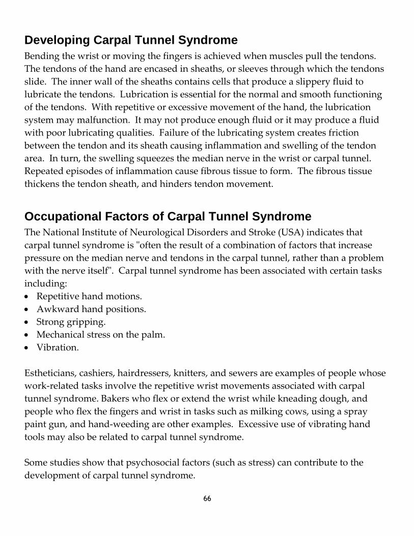
66
Developing Carpal Tunnel Syndrome
Bending the wrist or moving the fingers is achieved when muscles pull the tendons.
The tendons of the hand are encased in sheaths, or sleeves through which the tendons
slide. The inner wall of the sheaths contains cells that produce a slippery fluid to
lubricate the tendons. Lubrication is essential for the normal and smooth functioning
of the tendons. With repetitive or excessive movement of the hand, the lubrication
system may malfunction. It may not produce enough fluid or it may produce a fluid
with poor lubricating qualities. Failure of the lubricating system creates friction
between the tendon and its sheath causing inflammation and swelling of the tendon
area. In turn, the swelling squeezes the median nerve in the wrist or carpal tunnel.
Repeated episodes of inflammation cause fibrous tissue to form. The fibrous tissue
thickens the tendon sheath, and hinders tendon movement.
Occupational Factors of Carpal Tunnel Syndrome
The National Institute of Neurological Disorders and Stroke (USA) indicates that
carpal tunnel syndrome is "often the result of a combination of factors that increase
pressure on the median nerve and tendons in the carpal tunnel, rather than a problem
with the nerve itself". Carpal tunnel syndrome has been associated with certain tasks
including:
Repetitive hand motions.
Awkward hand positions.
Strong gripping.
Mechanical stress on the palm.
Vibration.
Estheticians, cashiers, hairdressers, knitters, and sewers are examples of people whose
work-related tasks involve the repetitive wrist movements associated with carpal
tunnel syndrome. Bakers who flex or extend the wrist while kneading dough, and
people who flex the fingers and wrist in tasks such as milking cows, using a spray
paint gun, and hand-weeding are other examples. Excessive use of vibrating hand
tools may also be related to carpal tunnel syndrome.
Some studies show that psychosocial factors (such as stress) can contribute to the
development of carpal tunnel syndrome.

67
Non-occupational Factors of Carpal Tunnel Syndrome
Carpal tunnel syndrome is associated with several diseases and situations such as:
Arthritis.
Diabetes.
Gout.
Amyloidosis (infiltration of the liver, kidneys, spleen with a starch-like substance).
Hypothyroidism (underactive thyroid gland).
Tumours of tendon sheaths.
Wrist fractures and dislocations.
Wrist cysts.
Pregnancy.
Use of oral contraceptives.
Menopause.
All these diseases and situations increase the volume of the contents of the carpal
tunnel, resulting in compression of the median nerve. Some individual factors, such
as the size and shape of the wrist and the shape of the median nerve, may contribute to
the development of carpal tunnel syndrome.
Symptoms of Carpal Tunnel Syndrome
The typical symptoms of carpal tunnel syndrome are tingling of the thumb, index,
middle, and ring fingers, and night pain. The pain awakens the sufferer, but is often
relieved by shaking, hanging, or massaging the hand. Pain may involve not only the
hand, but also the arm and the shoulder. Numbness and loss of manual dexterity
occur in more advanced cases. Weakness of the hand also occurs, causing difficulty
with pinch and grasp. The person may drop objects or be unable to use keys or count
change with the affected hand. The skin may dry because of reduced sweating.
People who suspect carpal tunnel syndrome often consult a doctor. The evaluation of
occupational carpal tunnel syndrome includes identifying workplace risks. Evaluation
begins with a discussion of the person's employment and requires a detailed
description of all the processes involved in a typical day's work. It also requires
consideration of the frequency, intensity, duration and regularity of each task

68
performed at work. Diagnosis of carpal tunnel syndrome is confirmed by performing
certain tests to detect damage to the median nerve.
Tinel's test - The physician taps the median nerve at the wrist. A tingling response in
one or more fingers indicates damage to the median nerve.
Phalen's test - The patient puts the backs of the hands together and bends the wrists
for one minute. Tingling of the fingers indicates damage to the median nerve.
Electromyography - Electrodes are placed on the forearm and electrical current is
passed through the patient. Measurements on how fast and how well the median
nerve transmits messages to muscles indicate if there is damage to this nerve.
Treatment of Carpal Tunnel Syndrome
When symptoms of carpal tunnel syndrome are mild or likely to be temporary,
treatment includes rest, anti-inflammatory drugs, cold packs, or a splint. Even if a
patient wears a splint that has been prescribed, he or she should avoid the activities
that caused or aggravated the injury. Where this is not possible, patients should wear
the splint after work and particularly during sleeping hours. Specific exercises
supervised by a physical or occupational therapist and yoga can be beneficial. Surgery
may be necessary if the symptoms are severe and if the other measures do not provide
any relief. Surgery should not be the first choice for treatment. Even after surgery, a
number of patients may still have some problems.
Preventing Carpal Tunnel Syndrome
Prevention of carpal tunnel syndrome may involve redesigning work stations, tools, or
the job, and educating workers. Proper work station design reduces awkward wrist
positions and minimizes the stressful effects of repetitive motions. Awkward positions
can originate from unsuitable work station designs that do not take into account the
size and proportions of the human body. Work stations should be adjustable and
should accommodate a vast majority of people who work in that area. Redesigning
work methods is important to reduce awkward wrist flexion.
Redesigning tools is also important. One study in a poultry processing plant found
that workers who used standard knives were prone to carpal tunnel syndrome. When
the workers started using knives with a bent handle, they no longer needed to bend

69
their wrists while cutting the meat. This change significantly reduced the occurrence
of carpal tunnel syndrome.
Job design includes the following:
Analysis of the sequence of the tasks to allow changes in body position.
Work-rest schedule to relieve muscles from mechanical stress.
Work breaks to avoid monotonous and repetitive patterns of work.
Rotation of tasks to move workers from one job to another.
Worker training should aim to reduce the number and types of awkward wrist
postures and the number of repetitive motions. Informing workers about the risk
factors that can contribute to carpal tunnel syndrome is important. To limit the effects
of a physically stressful job, employers should work closely with employees. This is
achieved by implementing worker training and job rotation, and by matching
employees to job assignments.

70
Objective Nine Self-Test
1) In biological terms, what causes carpal tunnel syndrome?
_____________________________________________________________________________
_____________________________________________________________________________
2) What happens when the tendon lubrication system fails?
_____________________________________________________________________________
3) Which five tasks can cause carpal tunnel syndrome?
_____________________________________________________________________________
4) What is considered during the evaluation of a workplace which may pose a risk for
carpal tunnel syndrome?
_____________________________________________________________________________
5) What can be done to treat a mild case of carpal tunnel syndrome?
_____________________________________________________________________________
6) List three things that can be done to prevent carpal tunnel syndrome in the
workplace:
_____________________________________________________________________________
_____________________________________________________________________________
71
Objective Nine Self-Test Answers
1) Along with the median nerve, the flexing tendons of the fingers and thumb pass
through the carpal tunnel. When the tendons swell, the space in the tunnel is
reduced and the median nerve gets pinched.
2) Failure of the lubricating system creates friction between the tendon and its sheath
causing inflammation and swelling of the tendon area.
3) Repetitive hand motions, awkward hand positions, strong gripping, mechanical
stress on the palm, and vibration.
4) An evaluation considers: a description of all the processes involved in a typical
day's work, and the frequency, intensity, duration and regularity of each task
performed at work.
5) Treatment can include: rest, anti-inflammatory drugs, cold packs, and the wearing
of a splint, even at night.
6) Any of the following: redesigning work stations, tools, or the job, educating
workers, reducing awkward wrist positions, work methods, and repetitive motions,
analyzing the sequence of the tasks to allow changes in body position, developing a
work-rest schedule to relieve muscles from mechanical stress, taking work breaks to
avoid monotonous and repetitive patterns of work, and rotating tasks to move
workers from one job to another.

72
Objective Ten When you have completed this objective, you will be able to:
Describe contraindications that may restrict or prevent a service.
A contraindication may restrict or prevent a service for many reasons. If a client has a
fractured bone in their foot, the administering of a foot massage may cause injury. In
this case, the service is prevented; however, it may be possible to work on and add
enhancements to their toenails. In this case, the service might be restricted or adapted.
Check with the AHJ and your employer to determine which contraindications either
restrict or prevent services. Never diagnose a contraindication. If a client appears to
have a contraindication, refer them to a physician.
Contraindications That Restrict a Manicure or Pedicure
Common warts Onychauxis
Beau’s lines and grooves Onychocryptosis
Bitten nails Onycholysis (depending on severity)
Bruising of the skin Onychomalacia
Bunions Onychorrhexis
Cyanosis Pincer nails
Fissures Plantar warts
Flaking nails Plicatured nails
Fractures or broken bones Psoriasis
Hammer toe Pterygium
Hangnails Scleronychia
Hematoma Splinter hemorrhages
Involuted toenail Yellow nail syndrome (uninfected)
Koilonychia
Leukonychia
Melanonychia

73
Contraindications That Prevent a Manicure or Pedicure
In general, contraindications that prevent a service are those that involve contact with
open flesh, or contact with highly communicable fungi and/or bacteria. A
contraindication may be listed above as being ‘restricted from service’, but if it is
accompanied by an infection, it becomes ‘prevented from service.’ Depending on an
esthetician’s scope of practice, they may work on a client who has one of the
contraindications listed below.
Bruised nail bed Onychomycosis
Candida Paronychia
Dermatophytes Pseudomonas
Mold Psoriasis of the Nail
Onychia Ring worm
Onycholysis Tinea Unguium
Onychomadesis Yeasts

74
Objective Ten Self-Test
1) With whom should an apprentice consult to determine which contraindications
restrict services, and which contraindications prevent services?
_____________________________________________________________________________
2) Complete the table below for contraindications that may restrict a manicure or
pedicure.
Onychauxis
Beau’s lines and grooves
Onychomalacia
Bunions
Pincer nails
Fissures
Flaking nails Plicatured nails
Pterygium
Hangnails
Splinter hemorrhages
Koilonychia
Leukonychia
3) In general, what types of contraindications prevent a service?
_____________________________________________________________________________

75
4) Complete the table below for contraindications that may prevent a manicure or
pedicure.
Onychomycosis
Candida
Dermatophytes
Psoriasis of the Nail
Onychia
Tinea Unguium

76
Objective Ten Self-Test Answers
1) The AHJ and the salon owner.
2) See table on page 71.
3) Contraindications that involve contact with open flesh, or contact with highly
communicable fungi and/or bacteria.
4) See table on page 72.

77
Objective Eleven When you have completed this objective, you will be able to:
Demonstrate analyzing conditions, disorders, and diseases of the hands and feet.
Laboratory Exercise
Purpose: to analyze conditions, disorders, and diseases of the hands and feet.
Materials:
Procedure: With a partner, analyze each other’s hands and feet. Identify any
contraindications that may restrict or prevent a service.
Instructor verification: _______________________________________

78
Module Summary Self-Test
1) Is the nail plate one solid piece? Explain.
_____________________________________________________________________________
2) What can result if the lunula is damaged?
_____________________________________________________________________________
3) What terms are used to describe a nail plate that whose arch is sharper than
normal?
_____________________________________________________________________________
4) How much does the average nail plate grow in the normal adult per month.
_____________________________________________________________________________
5) Why are toenail plates thicker than fingernail plates?
_____________________________________________________________________________
6) What can result from damage to the matrix?
_____________________________________________________________________________
7) What are the two main sources of nail disorders?
_____________________________________________________________________________
8) What are beaus lines?
_____________________________________________________________________________

79
9) Why should a client with melanonychia be immediately referred to a physician?
_____________________________________________________________________________
10) What is onychomycosis?
_____________________________________________________________________________
11) What is a term used to describe any disease or deformity of the nails?
_____________________________________________________________________________
12) What is onychia, and what is a common cause?
_____________________________________________________________________________
13) What two factors make the Achilles tendon susceptible to injury?
_____________________________________________________________________________
14) How should small and large blisters be covered?
_____________________________________________________________________________
15) In relation to the ankle, which additional problem can be caused by flat foot?
_____________________________________________________________________________
16) What is the role of insulin in the body?
_____________________________________________________________________________

80
17) What is the most common form of nerve damage in the feet of a diabetic, and what
are its symptoms?
_____________________________________________________________________________
18) How should a diabetic trim their toenails?
_____________________________________________________________________________
19) How does rheumatoid arthritis develop?
_____________________________________________________________________________
20) Which condition causes: painful along the tendons located on the thumb side of
the wrist, and pain when turning the wrist, grasping, and making a fist.
_____________________________________________________________________________
21) What does mallet finger look like?
_____________________________________________________________________________
22) If a person is suffering from carpal tunnel syndrome, how is their grip affected?
_____________________________________________________________________________
23) How is a Phalen's test performed?
_____________________________________________________________________________
24) True / False. Arthritis is not a non-occupational factor of carpal tunnel syndrome.
_____________________________________________________________________________

81
25) What should be done ff a client appears to have a contraindication?
_____________________________________________________________________________
26) True / False. Psoriasis of the nail is a contraindication that restricts a service.
_____________________________________________________________________________
27) True / False. Bunions are a contraindication that restricts a service.
_____________________________________________________________________________

82
Module Summary Self-Test Answers
1) No. The nail plate is constructed of approximately 100 layers of nail cells.
2) Permanent deformity to the nail plate an result from lunula damage.
3) High-arched or circumflex.
4) About 2.5 mm to 3 mm per month.
5) The matrix of a toenail plate is longer.
6) Nail plates can develop thinner or with grooves.
7) A breakdown of the normal way a nail forms, and circumstantial factors such as
stress and diet.
8) Beaus lines are visible depressions running across the width of the natural nail
plate.
9) Melanonychia can indicate melanoma.
10) Onychomycosis is a fungal infection between the nail plate and the nail bed.
11) Onychosis.
12) Onychia is an inflammation of the nail matrix commonly caused by unsanitary
implements.
13) Limited blood supply to the tendon and high tensions placed on it.
14) Cover small blisters with a bandage, and large ones with a gauze pad held by
adhesive.
83
15) The ankle often pronates (rolls inward).
16) Insulin opens cells, allowing the glucose to enter and be consumed by the cells.
17) The most common form is peripheral neuropathy which causes tingling, pain
(burning or stinging), or weakness in the foot.
18) Straight across.
19) The immune system attacks and damages parts of the body.
20) DeQuervain's tenosynovitis.
21) The end joint of the finger or thumb falls down and will not lift itself.
22) The grip is weakened, causing difficulty with pinching and grasping.
23) The patient puts the backs of the hands together and bends the wrists for one
minute. Tingling of the fingers indicates damage to the median nerve.
24) False.
25) The client should be referred to a physician.
26) False.
27) True.

84