evaluation of phytotoxic and mutagenic effects of some
TRANSCRIPT

748
http://journals.tubitak.gov.tr/biology/
Turkish Journal of Biology Turk J Biol(2013) 37: 748-756© TÜBİTAKdoi:10.3906/biy-1304-39
Evaluation of phytotoxic and mutagenic effects of some cinnamic acidderivatives using the Triticum test
Alexandra JITĂREANU1, Silvica PĂDUREANU2,*, Gabriela TĂTĂRÎNGĂ1, Cristina TUCHILUȘ1, Ursula STĂNESCU1
1Faculty of Pharmacy, University of Medicine and Pharmacy Grigore T. Popa Iasi, Iasi, Romania2Department of Cell Biology, Faculty of Agriculture, University of Agronomy Sciences and Veterinary Medicine Iasi, Iasi, Romania
* Correspondence: [email protected]
1. IntroductionPlant tests are widely used to assess the toxicity of different chemicals because they are very easy to perform, are inexpensive, and present good correlations with other test systems (Fiskesjo, 1985; Yüzbaşıoğlu et al., 2009; Palanikumar et al., 2011; Sharma et al., 2012). Triticum aestivum is frequently used in routine phytotoxicity tests (Lee et al., 2008; Pădureanu, 2008; Kalcheva et al., 2009; Kumar et al., 2010). The Triticum test has 2 targets: toxicity and mutagenicity assessment. Toxicity is associated with growth inhibition, while mutagenicity is correlated with the rate of chromosome disturbances (Fiskesjo, 1985).
Cinnamic acid derivatives are important compounds with a wide range of biological activities: antibacterial, antifungal, antioxidant, antiinflammatory, and antitumoral (Da Cunha et al., 2004; Narasimhan et al., 2004; Xu et al., 2005; Sharma, 2011). Some cinnamic acid derivatives are naturally occurring substances found in various plants. Cinnamic acid can also be found in free form, but it is especially common in the form of esters. Cinnamic esters are obtained from various plant sources and are
very important in perfumery, the cosmetic industry, and pharmaceutics. Methyl caffeate is found in Gaillardia pulchella, Gochnatia rusbyana, Notopterygium incisum, and the fruits of Linum usitatissimum and is reported to possess both antitumor and antimicrobial activities. Ethyl 3,4,5-trimethoxycinnamate, found in Piper longum, has an important role in controlling inflammatory diseases (Sharma, 2011).
Hydroxycinnamic acids (caffeic, ferulic, and coumaric) and their derivatives are extremely potent natural antitumor, antioxidant, and antimicrobial agents. Caffeic acid phenethyl ester (CAPE) from propolis is only one example of an extremely potent biologically active natural cinnamic acid derivative (Bankova, 2009).
The aim of this paper was to evaluate the phytotoxic and mutagenic effects of 5 cinnamic acid derivatives [cinnamic acid (I), 2,3-dibromo-3-phenyl-propanoic acid (II), 2,3-dibromo-3-(3-bromophenyl)-propanoic acid (III), 2,3-dibromo-3-(4-hydroxy-3-methoxyphenyl)-propanoic acid (IV), and 2,3-dibromo-3-(3-bromo-4-hydroxy-5-methoxyphenyl)-propanoic acid (V)] in order to obtain
Abstract: Five cinnamic acid derivatives [cinnamic acid, 2,3-dibromo-3-phenyl-propanoic acid, 2,3-dibromo-3-(3-bromophenyl)-propanoic acid, 2,3-dibromo-3-(4-hydroxy-3-methoxyphenyl)-propanoic acid, and 2,3-dibromo-3-(3-bromo-4-hydroxy-5-methoxyphenyl)-propanoic acid] were found to be active against Staphylococcus aureus ATCC 25923, and their minimal bactericidal concentrations were determined (100 µg/mL). The first step in assessing their toxicological potential was the phytotoxicity and genotoxicity evaluation on Triticum aestivum. Wheat seeds were exposed to solutions of the tested compounds (100 µg/mL) for 24 and 48 h. The development of roots and seedlings, germination percentage, mitotic index, chromosomal aberrations, and total polyphenol content were analyzed. The substances caused in most experimental cases a slight inhibition in the growth of the tested plantlets in comparison to the control, with the exception of 2,3-dibromo-3-(3-bromo-4-hydroxy-5-methoxyphenyl)-propanoic acid (48 h of exposure). All compounds inhibited the germination process and mitotic activity. No aberrant metaphases were generated, but abnormal anatelophases appeared, and 4 types of chromosomal aberrations were identified: chromosome bridges, chromosome fragments, micronuclei, and multipolar anatelophases. Wheat plantlet metabolism was also affected; the total polyphenol content decreased in the treated plantlets.
Key words: Chromosomal aberrations, micronucleus, mitotic division, phytotoxicity, polyphenols, Triticum test
Received: 18.04.2013 Accepted: 02.08.2013 Published Online: 08.10.2013 Printed: 04.11.2013
Research Article
JITĂREANU et al. / Turk J Biol
749
some useful information about the toxic potential of the chemicals for living organisms. Although phytotoxicity tests are usually used in environmental monitoring, they can also represent useful screening tools for determining the risk that tested chemicals pose to the health of living organisms before resorting to more complex toxicity tests. This preliminary testing during the early stages of drug development can prevent the loss of both time and money.
The selected compounds presented good antistaphylococcal activity during previous preliminary qualitative antimicrobial testing (Jităreanu et al., 2011), justifying the determination of their minimum inhibitory concentrations (MICs) and minimum bactericidal concentrations (MBCs) on Staphylococcus aureus ATCC 25923, and the values found for MBC were later used in the phytotoxicity assay. Staphylococcus aureus is a very common pathogen responsible for causing several disease processes (skin and soft tissue infections, sepsis, osteomyelitis, and pneumonia). S. aureus is also associated with toxin-mediated food poisoning. Over the past decade, increasing rates of methicillin-resistant S. aureus colonization and infection were observed, and this matter is a growing concern of the global health care system (Crago et al., 2012). In this context, the development of new active drugs is of great importance.
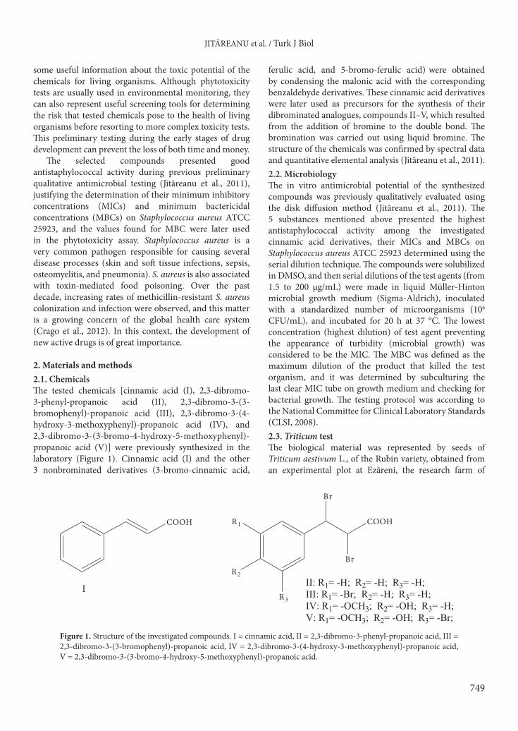
2. Materials and methods2.1. Chemicals The tested chemicals [cinnamic acid (I), 2,3-dibromo-3-phenyl-propanoic acid (II), 2,3-dibromo-3-(3-bromophenyl)-propanoic acid (III), 2,3-dibromo-3-(4-hydroxy-3-methoxyphenyl)-propanoic acid (IV), and 2,3-dibromo-3-(3-bromo-4-hydroxy-5-methoxyphenyl)-propanoic acid (V)] were previously synthesized in the laboratory (Figure 1). Cinnamic acid (I) and the other 3 nonbrominated derivatives (3-bromo-cinnamic acid,
ferulic acid, and 5-bromo-ferulic acid) were obtained by condensing the malonic acid with the corresponding benzaldehyde derivatives. These cinnamic acid derivatives were later used as precursors for the synthesis of their dibrominated analogues, compounds II–V, which resulted from the addition of bromine to the double bond. The bromination was carried out using liquid bromine. The structure of the chemicals was confirmed by spectral data and quantitative elemental analysis (Jităreanu et al., 2011). 2.2. MicrobiologyThe in vitro antimicrobial potential of the synthesized compounds was previously qualitatively evaluated using the disk diffusion method (Jităreanu et al., 2011). The 5 substances mentioned above presented the highest antistaphylococcal activity among the investigated cinnamic acid derivatives, their MICs and MBCs on Staphylococcus aureus ATCC 25923 determined using the serial dilution technique. The compounds were solubilized in DMSO, and then serial dilutions of the test agents (from 1.5 to 200 µg/mL) were made in liquid Müller-Hinton microbial growth medium (Sigma-Aldrich), inoculated with a standardized number of microorganisms (106 CFU/mL), and incubated for 20 h at 37 °C. The lowest concentration (highest dilution) of test agent preventing the appearance of turbidity (microbial growth) was considered to be the MIC. The MBC was defined as the maximum dilution of the product that killed the test organism, and it was determined by subculturing the last clear MIC tube on growth medium and checking for bacterial growth. The testing protocol was according to the National Committee for Clinical Laboratory Standards (CLSI, 2008).2.3. Triticum testThe biological material was represented by seeds of Triticum aestivum L., of the Rubin variety, obtained from an experimental plot at Ezăreni, the research farm of
COOH COOH
R3
R2
R1
I II: R1= -H; R2= -H; R3= -H;III: R1= -Br; R2= -H; R3= -H;IV: R1= -OCH3; R2= -OH; R3= -H;V: R1= -OCH3; R2= -OH; R3= -Br;
Br
Br
Figure 1. Structure of the investigated compounds. I = cinnamic acid, II = 2,3-dibromo-3-phenyl-propanoic acid, III = 2,3-dibromo-3-(3-bromophenyl)-propanoic acid, IV = 2,3-dibromo-3-(4-hydroxy-3-methoxyphenyl)-propanoic acid, V = 2,3-dibromo-3-(3-bromo-4-hydroxy-5-methoxyphenyl)-propanoic acid.

JITĂREANU et al. / Turk J Biol
750
the University of Agricultural Sciences and Veterinary Medicine, Iasi, Romania. The experiment was conducted in petri dishes using 100 seeds per dish. The seeds were soaked in 100 µg/mL solution of the tested compounds for 24 and 48 h, assessing also the influence of exposure time on the development of Triticum aestivum plantlets. This concentration was chosen because it was the value of the MBC determined for all 5 investigated cinnamic acid derivatives. After 24 or 48 h of exposure to the tested chemicals, the seeds were put to germinate in laboratory conditions. The control group was identical in every aspect to the test group, except for the exposure to the tested substances. The experiments included 3 replicates per treatment.
When the embryonic roots reached 15–17 mm in length, half of the germinated seeds were used for cytogenetic investigations, while the rest were left to develop further until the ninth day of the experiment, when the roots and seedlings were measured.2.4. Cytogenetic analysisTreated and control roots were fixed in Carnoy’s fixative solution for 24 h at 4 °C, then hydrolyzed with HCl and stained with carbol-fuchsin solution (Carr reagent) (Gamborg and Wetter, 1975). Ten slides were prepared for each variant using the squash technique, and 20 microscopic fields per slide were examined. Different mitotic stages were counted in at least 5000 cells in order to determine the mitotic index. The microscopic examination was performed with a Hund Wetzlar optic microscope equipped with a digital camera that was used to take the photographs (Pădureanu, 2008).
The equations used to calculate the mitotic index, phase indices, and percentages of metaphase (Mab) and anatelophase (A – Tab) abnormalities are shown below (Truță et al., 2011):mitotic index = total dividing cells × 100/total cells
(dividing and nondividing),prophase (%) = prophase cells × 100/total dividing cells,metaphase (%) = metaphase cells × 100/total dividing cells,anaphase (%) = anaphase cells × 100/total dividing cells,telophase (%) = telophase cells × 100/total dividing cells,Mab (%) = Mab × 100/total dividing cells,A – Tab (%) = A – Tab × 100/total dividing cells.2.5. Extraction and determination of polyphenolsA 1.5-g mixture of chopped and homogenized fresh seedlings was extracted with 50 mL of methanol under reflux conditions for 30 min. The total content of polyphenols was determined with the Folin–Ciocalteu method using gallic acid for calibration: 3040 µL of distilled water and 200 µL of Folin–Ciocalteu reagent were added to 160 µL of extract; 600 µL of 20% sodium
carbonate solution was added to the mixture after 20 min; and the absorbance was read after 2 h at 765 nm (Anesini et al., 2008). The total polyphenol content was expressed as gallic acid equivalent (GAE) in mg/100 g.2.6. Statistical analysesResults were given as mean ± standard deviation. The experimental data were processed using analysis of variance (ANOVA) to assess the significant differences in seedling and root length between the control and each treatment, and Student’s t-test was applied in the case of cytogenetic analyses.
3. Results and discussionThe 5 tested cinnamic acid derivatives proved to be good antistaphylococcal agents, and the values obtained for MIC and MBC are presented in Table 1. The MBC determined for all 5 investigated substances was 100 µg/mL, while the MIC varied between 25 and 50 µg/mL. The MIC values were much higher than for ampicillin (0.25 µg/mL), similar to values obtained for 2 antibacterial sulfonamides (sulfapyridine and sulfachloropyridazine, 16 µg/mL) on the same S. aureus strain (Lupașcu et al., 2012); however, the antibacterial potential of the tested compounds was much higher than that of propyl 4-hydroxybenzoate, a paraben used as a food preservative (Gutiérrez-Larraínzar et al., 2013).
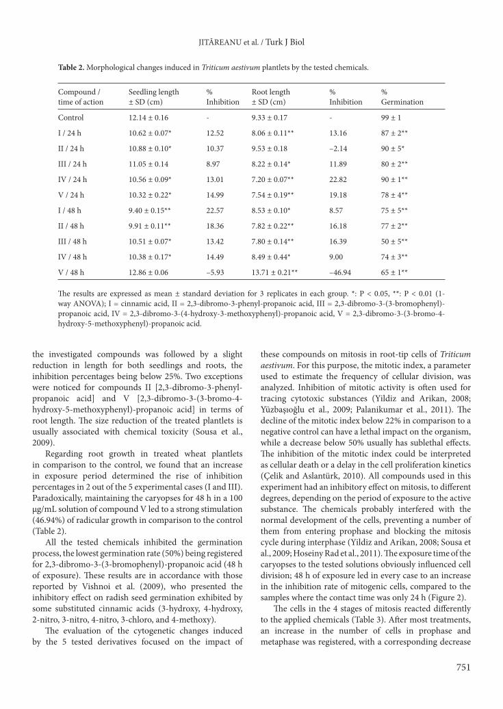
One way to assess the toxicity of the chemicals was by analyzing their impact on the germination and development of wheat plantlets. The seedlings and roots were measured and percentages of growth inhibition in comparison to the control were calculated, the results being presented in Table 2. In most cases, exposure to
Table 1. Minimal inhibitory and bactericidal concentrations of the tested compounds against Staphylococcus aureus ATCC 25923.
Compound MIC (µg/mL) MBC (µg/mL)
I 50 100
II 25 100
III 50 100
IV 50 100
V 25 100
Ampicillin 0.25 0.5
I = cinnamic acid, II = 2,3-dibromo-3-phenyl-propanoic acid, III = 2,3-dibromo-3-(3-bromophenyl)-propanoic acid, IV = 2,3-dibromo-3-(4-hydroxy-3-methoxyphenyl)-propanoic acid, V = 2,3-dibromo-3-(3-bromo-4-hydroxy-5-methoxyphenyl)-propanoic acid.
JITĂREANU et al. / Turk J Biol
751
the investigated compounds was followed by a slight reduction in length for both seedlings and roots, the inhibition percentages being below 25%. Two exceptions were noticed for compounds II [2,3-dibromo-3-phenyl-propanoic acid] and V [2,3-dibromo-3-(3-bromo-4-hydroxy-5-methoxyphenyl)-propanoic acid] in terms of root length. The size reduction of the treated plantlets is usually associated with chemical toxicity (Sousa et al., 2009).
Regarding root growth in treated wheat plantlets in comparison to the control, we found that an increase in exposure period determined the rise of inhibition percentages in 2 out of the 5 experimental cases (I and III). Paradoxically, maintaining the caryopses for 48 h in a 100 µg/mL solution of compound V led to a strong stimulation (46.94%) of radicular growth in comparison to the control (Table 2).
All the tested chemicals inhibited the germination process, the lowest germination rate (50%) being registered for 2,3-dibromo-3-(3-bromophenyl)-propanoic acid (48 h of exposure). These results are in accordance with those reported by Vishnoi et al. (2009), who presented the inhibitory effect on radish seed germination exhibited by some substituted cinnamic acids (3-hydroxy, 4-hydroxy, 2-nitro, 3-nitro, 4-nitro, 3-chloro, and 4-methoxy).
The evaluation of the cytogenetic changes induced by the 5 tested derivatives focused on the impact of
these compounds on mitosis in root-tip cells of Triticum aestivum. For this purpose, the mitotic index, a parameter used to estimate the frequency of cellular division, was analyzed. Inhibition of mitotic activity is often used for tracing cytotoxic substances (Yildiz and Arikan, 2008; Yüzbaşıoğlu et al., 2009; Palanikumar et al., 2011). The decline of the mitotic index below 22% in comparison to a negative control can have a lethal impact on the organism, while a decrease below 50% usually has sublethal effects. The inhibition of the mitotic index could be interpreted as cellular death or a delay in the cell proliferation kinetics (Çelik and Aslantürk, 2010). All compounds used in this experiment had an inhibitory effect on mitosis, to different degrees, depending on the period of exposure to the active substance. The chemicals probably interfered with the normal development of the cells, preventing a number of them from entering prophase and blocking the mitosis cycle during interphase (Yildiz and Arikan, 2008; Sousa et al., 2009; Hoseiny Rad et al., 2011). The exposure time of the caryopses to the tested solutions obviously influenced cell division; 48 h of exposure led in every case to an increase in the inhibition rate of mitogenic cells, compared to the samples where the contact time was only 24 h (Figure 2).
The cells in the 4 stages of mitosis reacted differently to the applied chemicals (Table 3). After most treatments, an increase in the number of cells in prophase and metaphase was registered, with a corresponding decrease
Table 2. Morphological changes induced in Triticum aestivum plantlets by the tested chemicals.
Compound /time of action
Seedling length± SD (cm)
% Inhibition
Root length± SD (cm)
% Inhibition
% Germination
Control 12.14 ± 0.16 - 9.33 ± 0.17 - 99 ± 1
I / 24 h 10.62 ± 0.07* 12.52 8.06 ± 0.11** 13.16 87 ± 2**
II / 24 h 10.88 ± 0.10* 10.37 9.53 ± 0.18 –2.14 90 ± 5*
III / 24 h 11.05 ± 0.14 8.97 8.22 ± 0.14* 11.89 80 ± 2**
IV / 24 h 10.56 ± 0.09* 13.01 7.20 ± 0.07** 22.82 90 ± 1**
V / 24 h 10.32 ± 0.22* 14.99 7.54 ± 0.19** 19.18 78 ± 4**
I / 48 h 9.40 ± 0.15** 22.57 8.53 ± 0.10* 8.57 75 ± 5**
II / 48 h 9.91 ± 0.11** 18.36 7.82 ± 0.22** 16.18 77 ± 2**
III / 48 h 10.51 ± 0.07* 13.42 7.80 ± 0.14** 16.39 50 ± 5**
IV / 48 h 10.38 ± 0.17* 14.49 8.49 ± 0.44* 9.00 74 ± 3**
V / 48 h 12.86 ± 0.06 –5.93 13.71 ± 0.21** –46.94 65 ± 1**
The results are expressed as mean ± standard deviation for 3 replicates in each group. *: P < 0.05, **: P < 0.01 (1-way ANOVA); I = cinnamic acid, II = 2,3-dibromo-3-phenyl-propanoic acid, III = 2,3-dibromo-3-(3-bromophenyl)-propanoic acid, IV = 2,3-dibromo-3-(4-hydroxy-3-methoxyphenyl)-propanoic acid, V = 2,3-dibromo-3-(3-bromo-4-hydroxy-5-methoxyphenyl)-propanoic acid.

JITĂREANU et al. / Turk J Biol
752
in the anaphase and telophase indices as compared to the control. The higher frequency of prophase cells was previously associated with cytotoxicity; some of the cells entering into mitosis were arrested during prophase, thus preventing cell division finalization (Panneerselvam et al., 2012).
In this study we also investigated the chromosomal aberrations that appeared in the root meristem cells of Triticum aestivum, focusing on parameters that we considered important: the proportion of cells in aberrant metaphases and anatelophases and the type and frequency of chromosomal aberrations. The chromosome aberration
38.2
9
80.1
1
88.4
9
76.3
8 87.4
90.1
1
91.1
9
88.2
5
91.7
1
89.4
92.3
61.7
1
19.8
9
11.5
1
23.6
2 12.6
9.89
8.81
11.7
5
8.29
10.6
7.7
0
20
40
60
80
100
120
Control I/24 h I/48 h II/24 h II/48 h III/24 h III/48 h IV/24 h IV/48 h V/24 h V/48 h
Cells in division Cells in interphase
% ce
lls
Figure 2. Proportion of cells in interphase and division in Triticum aestivum root meristem cells treated with cinnamic acid derivatives.I = cinnamic acid, II = 2,3-dibromo-3-phenyl-propanoic acid, III = 2,3-dibromo-3-(3-bromophenyl)-propanoic acid, IV = 2,3-dibromo-3-(4-hydroxy-3-methoxyphenyl)-propanoic acid, V = 2,3-dibromo-3-(3-bromo-4-hydroxy-5-methoxyphenyl)-propanoic acid.
Table 3. Effects of cinnamic acid derivatives on mitotic index and phase indices in root meristem cells of Triticum aestivum L.
Compound /time of action
Total analyzedcells
Total dividingcells
Mitotic index%
Prophase%
Metaphase%
Anaphase%
Telophase%
Control 5960 3678 61.71 ± 3.61 45.84 ± 2.93 16.59 ± 1.37 16.04 ± 0.25 21.53 ± 1.52
I / 24 h 5632 1120 19.89 ± 1.75*** 60.72 ± 4.75*** 17.14 ± 1.08 7.14 ± 0.37*** 15.00 ± 1.28***
II / 24 h 5868 1386 23.62 ± 1.83*** 74.03 ± 5.93*** 11.69 ± 0.95*** 3.25 ± 0.11*** 11.03 ± 0.97***
III / 24 h 5340 528 9.89 ± 0.73*** 44.32 ± 3.85 30.68 ± 2.77*** 12.5 ± 1.09** 12.5 ± 0.82***
IV / 24 h 5517 585 10.60 ± 1.15*** 53.85 ± 3.66*** 23.08 ± 1.54*** 6.15 ± 0.34*** 16.92 ± 1.53***
V / 24 h 5312 624 11.75 ± 0.83*** 35.90 ± 2.11*** 24.36 ± 2.08*** 7.69 ± 0.67*** 32.05 ± 2.95***
I / 48 h 5319 612 11.51 ± 1.05*** 48.53 ± 3.17* 20.59 ± 1.57*** 10.29 ± 0.79*** 20.59 ± 2.10
II / 48 h 5240 660 12.60 ± 2.01*** 66.67 ± 5.06*** 19.70 ± 0.82*** 3.03 ± 0.25*** 10.60 ± 0.69***
III / 48 h 5310 468 8.81 ± 0.81*** 47.44 ± 2.97 20.51 ± 2.19*** 16.67 ± 1.12 15.38 ± 1.10***
IV / 48 h 5040 388 7.70 ± 0.31*** 56.70 ± 5.09*** 15.46 ± 1.30 15.47 ± 0.95 12.37 ± 0.73***
V / 48 h 5236 434 8.29 ± 0.52*** 48.39 ± 2.15* 19.35 ± 1.77** 9.68 ± 0.38*** 22.58 ± 2.77
*: P < 0.05, **: P < 0.01; ***: P < 0.001 (Student’s t-test); I = cinnamic acid, II = 2,3-dibromo-3-phenyl-propanoic acid, III = 2,3-dibromo-3-(3-bromophenyl)-propanoic acid, IV = 2,3-dibromo-3-(4-hydroxy-3-methoxyphenyl)-propanoic acid, V = 2,3-dibromo-3-(3-bromo-4-hydroxy-5-methoxyphenyl)-propanoic acid.

JITĂREANU et al. / Turk J Biol
753
assay is a useful and sensitive test for detection of genotoxins (Galloway, 2000; Sousa et al., 2009; Yüzbaşıoğlu et al., 2009; Palanikumar et al., 2011).
We noticed that no aberrant metaphases were generated during the treatment with the tested substances, regardless of contact time.
As far as anatelophases were concerned, the 5 investigated cinnamic acid derivatives induced chromosomal aberrations in all experimental cases. The low percentage of abnormal cells (0.05%) found in the control was probably caused by an automutagenic effect (Dragoeva et al., 2012), but the clastogenic potential of the investigated chemicals was evident, with percentages of anatelophase abnormalities varying in the treated plantlets from 1.54% to 17.95%. The highest mutagenic potential (% anatelophase abnormalities > 10%) was recorded for compounds I (48 h) and III (48 h), while compound IV [2,3-dibromo-3-(4-hydroxy-3-methoxyphenyl)-propa-noic acid] presented the lowest genotoxicity.
The genotoxicity of cinnamic, caffeic, and ferulic acids was previously tested by Maistro et al. (2011) on rat hepatoma tissue cells using 2 methods: the comet and micronucleus assays. Caffeic, cinnamic, and ferulic acids were not genotoxic according to the comet assay, but a certain clastogenic potential was observed for all compounds in the micronucleus test.
Four types of chromosomal aberrations were identified: chromosome bridges, chromosome fragments, micronuclei, and multipolar anatelophases (Table 4).
Chromosomal bridges are the result of the breakage and fusion of 2 chromosomes that possibly supported terminal deletions, being visible between the 2 chromatid groups separated to the cell poles (Kalcheva et al., 2009; Truță et al., 2011). Both chromosome bridges and fragments are signs of clastogenic effects (Kumar et al., 2010; Akaneme and Amaefule, 2012).
Micronuclei are structures found in the cytoplasm that contain chromatin and are surrounded by a membrane. They result from chromosome fragments or whole chromosomes lagging at anaphase (Fenech, 2000), and their formation is an effective indicator of cytological damage, confirming the clastogenic potential of the chemicals (Hoseiny Rad et al., 2011; Dragoeva et al., 2012). Micronuclei evaluation provides a reliable image of both chromosome breakage and chromosome loss.
Multipolar anatelophases represent complex aberrations, and they have more severe repercussions at the genetic level and on plant growth and development (Truță et al., 2011). The highest number of multipolar anatelophases was recorded in the meristem root cells of seeds exposed to 2,3-dibromo-3-(3-bromophenyl)-propanoic acid (III) for 48 h, confirming the high genotoxicity of the compound.
Table 4. Effect of cinnamic acid derivatives on abnormalities in mitotic cells in root meristems of Triticum aestivum L.
Compound /time of action
Mab%
A – Tab%
Anatelophase types of chromosome aberrations
Bridges Fragments Micronuclei Multipolar anatelophases
No. % No. % No. % No. %
Control - 0.05 ± 0.01 2 0.05 - - - - - -
I / 24 h - 5.71 ± 0.25*** 21 1.87 - - 43 3.84 - -
II / 24 h - 7.14 ± 0.37*** 90 6.49 - - 9 0.65 - -
III / 24 h - 3.41 ± 0.19*** 15 2.84 - - - - 3 0.57
IV / 24 h - 1.54 ± 0.35 9 1.54 - - - - - -
V / 24 h - 3.85 ± 0.63*** 8 1.28 16 2.57 - - - -
I / 48 h - 13.24 ± 0.58*** 72 11.76 - - - - 9 1.48
II / 48 h - 3.03 ± 0.11*** 15 2.27 - - 5 0.76 - -
III / 48 h - 17.95 ± 0.95*** 41 8.76 7 1.50 - - 36 7.69
IV / 48 h - 2.06 ± 0.28* 8 2.06 - - - - - -
V / 48 h - 1.61 ± 0.17* 4 0.92 - - 3 0.69 - -
*: P < 0.05, ***: P < 0.001 (Student’s t-test); I = cinnamic acid, II = 2,3-dibromo-3-phenyl-propanoic acid, III = 2,3-dibromo-3-(3-bromophenyl)-propanoic acid, IV = 2,3-dibromo-3-(4-hydroxy-3-methoxyphenyl)- propanoic acid, V = 2,3-dibromo-3-(3-bromo-4-hydroxy-5-methoxyphenyl)-propanoic acid

JITĂREANU et al. / Turk J Biol
754
Some of the observed chromosomal aberrations are presented in Figure 3.
Recent advances in molecular biology led to the development of some DNA analysis techniques that can be used in genotoxicology. Azimi et al. (2013) observed the DNA changes that appeared in Triticum aestivum plants after exposure to cadmium using random amplified polymorphic DNA (RAPD) analysis.
In this study we only investigated the cytogenetic changes (cellular division rates and chromosomal aberrations)
induced by some cinnamic acid derivatives in wheat plantlets; however, because the tested compounds displayed a certain degree of genotoxicity, our future research will also focus on measuring the effect of the chemicals directly on DNA, using different molecular markers.
Another investigated parameter was the capacity for polyphenol biosynthesis in the treated wheat plantlets in comparison to the control, in this way assessing the impact of the tested substances on plantlets’ metabolism (Wronka et al., 1995).
G
A B C
D E F
Figure 3. Chromosomal aberrations induced by cinnamic acid (I), 2,3-dibromo-3-phenyl-propanoic acid (II), 2,3-dibromo-3-(3-bromophenyl)-propanoic acid (III), 2,3-dibromo-3-(4-hydroxy-3-methoxyphenyl)-propanoic acid (IV), 2,3-dibromo-3-(3-bromo-4-hydroxy-5-methoxyphenyl)-propanoic acid (V) in root meristem cells of Triticum aestivum. A) Anatelophase with multiple bridges (I / 24 h; 1000×); B) anaphase with multiple bridges (I / 48 h; 1000×); C) anatelophase with broken bridges (I / 48 h; 1000×); D) anatelophase with broken bridges (V / 48 h; 1000×); E) micronucleus in late telophase (II / 24 h; 1000×); F) anatelophase with broken and continuous bridges (II / 48 h; 1000×); G) multipolar anaphase (III / 48 h; 1000×).

JITĂREANU et al. / Turk J Biol
755
According to the quantitative determination, a decrease in the biosynthesis of polyphenols was directly influenced by the time of exposure. There were also differences in the biosynthetic inhibitory activity between the 5 analyzed substances, the closest to the control being sample IV [2,3-dibromo-3-(4-hydroxy-3-methoxyphenyl)-propa-noic acid], while the lowest total polyphenol content was found in III [2,3-dibromo-3-(3-bromophenyl)-propanoic acid] (Figure 4). In an attempt to correlate the inhibitory effect of the compounds on the biosynthetic capacity of the plantlets with the other investigated aspects, a connection was observed between the genotoxic potential of the
compounds and the reduction of the total polyphenol content (compound III displayed a high genotoxic potential, while compound IV presented the lowest genotoxicity); however, in order to establish a strong correlation, further analyses are necessary.
Toxicity assessment is of great importance for pharmaceutical substances. We considered it necessary to subject the biologically active substances to a phytobiological screening in order to evaluate their influence on living organisms. By using the Triticum test, both the toxicity and mutagenity of the compounds were evaluated.
In most cases, 100 µg/mL solutions of the tested substances (the value determined for the MBC in the microbiology assay) moderately influenced the morphology of Triticum aestivum plantlets, all the recorded percentages of growth inhibition being below 25% for both roots and seedlings. Despite this fact, all chemicals strongly inhibited germination and mitosis. The inhibition of mitotic activity, associated with the appearance of chromosomal aberrations, indicated a clastogenic potential for the investigated compounds. The lowest genotoxicity was exhibited by 2,3-dibromo-3-(3-bromo-4-hydroxy-5-methoxyphenyl)-propanoic acid, while 2,3-dibromo-3-(3-bromophenyl)-propanoic acid presented an elevated mutagenic potential and the highest number of complex chromosomal aberrations (multipolar anatelophases), with more severe repercussions at the genetic level.
Therefore, the genotoxic potential of the 5 cinnamic acid derivatives must not be ignored, and further investigation is compulsory if the compounds are to be used for their antimicrobial or antioxidant properties.
170.
66
165.
54
147.
52
227.
9
127.
38 148.
33
132.
94
130.
12 157.
13
141.
95
230.
75
0
50
100
150
200
250
I II III IV V Control
24 h 48 h Control
mg%
GA
E
Figure 4. Total polyphenol content in Triticum plantlets treated with cinnamic acid derivatives. I = cinnamic acid, II = 2,3-dibromo-3-phenyl-propanoic acid, III = 2,3-dibromo-3-(3-bromophenyl)-propanoic acid, IV = 2,3-dibromo-3-(4-hydroxy-3-methoxyphenyl)-propanoic acid, V = 2,3-dibromo-3-(3-bromo-4-hydroxy-5-methoxyphenyl)-propanoic acid.
References
Akaneme FI, Amaefule CC (2012). Evaluation of the cytotoxicity and genotoxicity of aqueous leaf extracts of Azadirachta indica A. Juss using the Allium test. J Med Plants Res 6: 3898–3907.
Anesini C, Ferraro G, Filip R (2008). Total polyphenol content and antioxidant capacity of commercially available tea (Camellia sinensis) in Argentina. J Agr Food Chem 56: 9225–9229.
Azimi A, Shahriari F, Fotovat A, Qale RK, Agje KH (2013). Investigation of DNA changes in wheat (Triticum aestivum L.) induced by cadmium using random amplified polymorphic DNA (RAPD) analysis. Afr J Biotechnol 12: 1921–1929.
Bankova V (2009). Chemical diversity of propolis makes it a valuable source of new biologically active compounds. JAAS 1: 23–28.
Çelik TA, Aslantürk ÖS (2010). Evaluation of cytotoxicity and genotoxicity of Inula viscosa leaf extracts with Allium test. J Biomed Biotechnol 1: 1–8.
CLSI (2008). Clinical and Laboratory Standards Institute. Methods for Dilution Antimicrobial Susceptibility Tests for Bacteria that Grow Aerobically. Approved Standard M07-A8 and Informational Supplement M100-S19. Wayne, PA, USA: National Committee for Clinical Laboratory Standards.
Crago B, Ferrato C, Drews SJ, Svenson LW, Tyrrell G, Louie M (2012). Prevalence of Staphylococcus aureus and methicillin-resistant S. aureus (MRSA) in food samples associated with foodborne illness in Alberta, Canada from 2007 to 2010. Food Microbiol 32: 202–205.
Da Cunha FM, Duma D, Assreuy J, Buzzi FC, Niero R, Campos MM, Calixto JB (2004). Caffeic acid derivatives: in vitro and in vivo anti-inflammatory properties. Free Radical Res 38: 1241–1253.
Dragoeva A, Koleva V, Hasanova N, Slanev S (2012). Cytotoxic and genotoxic effects of diphenyl-ether herbicide GOAL (Oxyfluoren) using the Allium cepa test. Res J Mut 2: 1–9.

JITĂREANU et al. / Turk J Biol
756
Fenech M (2000). The in vitro micronucleus technique. Mutat Res 455: 81–95.
Fiskesjo G (1985). The Allium test as a standard in environmental monitoring. Hereditas 102: 99–112.
Galloway SM (2000). Cytotoxicity and chromosome aberrations in vitro: experience in industry and the case for an upper limit on toxicity in the aberration assay. Environ Mol Mutagen 35: 191–201.
Gamborg OL, Wetter LR (1975). Plant Tissue Culture Methods. Saskatoon, Canada: National Research Council of Canada.
Gutiérrez-Larraínzar M, Rúa J, De Arriaga D, Valle P, García-Armesto MR (2013). In vitro assessment of synthetic phenolic antioxidants for inhibition of foodborne Staphylococcus aureus, Bacillus cereus and Pseudomonas fluorescens. Food Control 30: 393–399.
Hoseiny Rad M, Aivazi AA, Jagannath S (2011). Cytogenetic and biochemical effects of imazethapyr in wheat (Triticum durum). Turk J Biol 35: 663–670.
Jităreanu A, Tătărîngă G, Zbancioc AM, Tuchiluș C, Stănescu U (2011). Antimicrobial activity of some cinnamic acid derivatives. Rev Med Chir Soc Med Nat Iasi 115: 965–971 (article in Romanian with an abstract in English).
Kalcheva V, Dragoeva A, Kalchev K, Enchev D (2009). Determination of cytotoxic effect of 4-bromo-N, N-diethyl-5,5-dimethyl-2,5-dihydro-1,2-oxaphosphol-2-amine 2-oxide. Biotechnol Biotec Eq 23: 414–417.
Kumar S, Arya SK, Roy BK, Singh AK (2010). The effects of 2,4-dichlorophenoxy acetic acid and isoproturon herbicides on the mitotic activity of wheat (Triticum aestivum L.) root tips. Turk J Biol 34: 55–66.
Lee WM, An YJ, Yoon H, Kweon HS (2008). Toxicity and bioavailability of copper nanoparticles to the terrestrial plants mung bean (Phaseolus radiates) and wheat (Triticum aestivum): plant agar test for water-insoluble nanoparticles. Environ Toxicol Chem 27: 1915–1921.
Lupașcu D, Tuchiluş C, Lupuşoru CE, Ghiciuc C, Şutu M, Neagu A, Profire L (2012). Synthesis and biological evaluation of some new rutin semisynthetic derivatives as antibacterial agents. Farmacia 60: 556–564.
Maistro EL, Angeli JPF, Andrade SF, Mantovani MS (2011). In vitro genotoxicity assessment of caffeic, cinnamic and ferulic acids. Genet Mol Res 10: 1130–1140.
Narasimhan B, Belsare D, Pharande D (2004). Esters, amides and substituted derivatives of cinnamic acid: synthesis, antimicrobial activity and QSAR investigations. Eur J Med Chem 39: 827–834.
Pădureanu S (2008). Citogenetic effects induced by sodium nitrite on mitotic division at Triticum aestivum L. Lucr St Seria Agronomie 51: 3–8.
Palanikumar L, Ragunathan I, Panneerselvam N (2011). Chromosome aberrations induced by curcumin and aloin in Allium cepa L. root meristem cells. Turk J Biol 35: 145–152.
Panneerselvam N, Palanikumar L, Gopinathan S (2012). Chromosomal aberrations induced by Glycidol in Allium cepa L. root meristem cells. IJPSR 3: 300–304.
Sharma P (2011). Cinnamic acid derivatives: a new chapter of various pharmacological activities. J Chem Pharm Res 3: 403–423.
Sharma S, Nagpal A, Vig AP (2012). Genoprotective potential of Brassica juncea (L.) Czern. against mercury-induced genotoxicity in Allium cepa L. Turk J Biol 36: 622–629.
Sousa SM, Silva PS, Campos JMS, Viccini FL (2009). Cytotoxic and genotoxic effects of two medicinal species of Verbenaceae. Caryologia 62: 326–333.
Truță E, Zamfirache MM, Olteanu Z (2011). Caffeine induced genotoxic effects in Phaseolus vulgaris L. and Raphanus sativus L. Bot Serb 35: 49–54.
Vishnoi S, Agrawal V, Kasana VK (2009). Synthesis and structure-activity relationships of substituted cinnamic acids and amide analogues: a new class of herbicides. J Agr Food Chem 57: 3261–3265.
Wronka M, Kuras M, Tykarska T, Podstolski A, Zobel AM (1995). Inhibition of the production of phenolic compounds in Brassica napus 2-amino-oxyacetic acid. Ann Bot-London 75: 319–324.
Xu F, Zhang S, Shao R, Zhen Y (2005). Anticancer activity of sodium caffeate and its mechanism. Acta Pharm Sinic 26: 1248–1252.
Yildiz M, Arikan EV (2008). Genotoxicity testing of quizalofop-P-ethyl herbicide using the Allium cepa anaphase-telophase chromosome aberration assay. Caryologia 61: 45–52.
Yüzbaşıoğlu D, Ünal F, Sancak C (2009). Genotoxic effects of herbicide Illoxan (Diclofop-Methyl) on Allium cepa L. Turk J Biol 33: 283–290.