evaluation of the kinematic parameters of normal-paced
TRANSCRIPT

Acta of Bioengineering and Biomechanics Original paperVol. 13, No. 3, 2011
Evaluation of the kinematic parameters of normal-paced gaitin subjects with gonarthrosis and the influence
of gonarthrosis on the function of the ankle joint and hip joint
KATARZYNA OGRODZKA1*, TADEUSZ NIEDŹWIEDZKI2, WIESŁAW CHWAŁA3
1 Department of Clinical Rehabilitation, Section of Rehabilitation in Traumatology,University School of Physical Education, Kraków, Poland.
2 Clinic of Orthopaedics and Traumatology of the Musculoskeletal System,Jagiellonian University Collegium Medicum, Kraków, Poland.3 Department of Anthropomotorics, Section of Biomechanics,
University School of Physical Education, Kraków, Poland.
The aim of the study was to assess the variability of parameters characterising the gait of persons suffering from degenerativechanges of the knee joint and their influence on the ankle and hip joints. The values of the angular changes in the knee, ankle and hipjoints in the three planes of motion were assessed.
Locomotion tests were performed on 27 persons, aged between 60 and 74, using Vicon 250, the three-dimensional analysis system.The sharpest deviations from the results of the control group were revealed in the transverse and frontal planes. Degenerative knee jointdisease has changed the gait stereotype causing a reduction in the economy of gonarthrosis patients’ locomotion, the influence of thedisease on the function of the neighbouring joints is also distinctly marked.
Key words: locomotion, three-dimensional motion analysis, gonarthrosis
1. Introduction
Degenerative knee joint disease is defined asa chronic, non-inflammatory disease with multi-factoretiology resulting from an imbalance between regen-erative and degenerative processes in the articularcartilage and the subcartilage layer [1]–[4].
The most frequent cause of degenerativechanges in the knee is the change of load on thearticular surfaces whilst walking [5], which iscaused by prior genu varum or genu valgum, jointinstability or injury, including damage to intra-articular structures: articular crescents, cruciateligaments, lateral ligaments, leading to disorders in
the knee content, and also by intra-articular frac-tures and articular cartilage injuries [4], [6]–[9].Changes in the neighbouring joints and obesity mayalso promote the development of degenerativechanges in knee joints [10].
The main clinical symptom is pain located in theanterior or medial part of the knee and in the upper-medial part of the shinbones [3], which becomes moreintense under load; with time the pain also begins tooccur at rest [10]. The disease is progressive andcauses motion to become restricted in the sagittalplane. Expansion of the joint outline, mostly atrophyof the medial head (vastus medialis) of the quadricepsfemoris muscle and deformation of the joint, occurstogether with its development.
______________________________
* Corresponding author: Katarzyna Ogrodzka, Department of Clinical Rehabilitation, Section of Rehabilitation in Traumatology, UniversitySchool of Physical Education, al. Jana Pawła II 78, 31-571 Kraków, Poland. Tel: +48 6000 443 384, e-mail: [email protected]
Received: December 6th, 2010Accepted for publication: June 2nd, 2011

K. OGRODZKA et al.48
Changes in the gait stereotype also occur as a resultof the disease development. Due to the progressivenature of the disease, locomotion function becomesimpaired, and, as a result, various compensationmechanisms are developed in the biokinetic chainconsisting of the wing of ilium and joints of the lowerextremities.
Lower extremity kinematics involving the move-ment of joints in the sagittal plane during movementhas been examined in a large number of researchstudies [11], [14]. However, three-dimensional gaitanalysis [15]–[18] conducted in persons with degen-erative changes of knee joints is still an area whichhas not been fully analysed.
This has become the basis for establishing as-sumptions and determining the aim of research in-volving an attempt to assess the variability of pa-rameters characterising the gait of persons sufferingfrom degenerative changes of the knee joint qualifiedfor an arthroplasty procedure.
The values of the angular changes in the knee, an-kle and hip joints in the three planes of motion wereassessed.
2. Material and methods
27 patients participated in the study, including20 women (74%) and 7 men (26%), aged between 60and 74, qualified for knee arthroplasty due to degen-erative changes of the joint. The criterion for qualifi-cation of treatment included, among other things,three and four degree arthrosis, and the duration ofarthrosis (from the diagnosis to the knee replacementprocedure) was approx. 5 years.
X-ray tests performed within the framework ofpre-operative diagnostics at the clinic did not revealdegenerative changes in hip joints. A description ofthe test group is presented in the table.
Before the procedure, the passive range of motionand the contracture in the knee joints were measuredgoniometrically for each of the patients. The meas-urement was performed in the forward horizontal po-sition with a straightened hip joint. The maximumflexion of the joint was on average 86.1° (±8.4) in theaffected leg and 94.7° (±12.2) in the healthy leg, andin each person with the contracture of the joints ob-served, the mean value in the healthy leg was 14.9°(±6.03) and in the affected leg 15.4° (±6.8). The sig-nificant limitation of motion both in the affected legand in the healthy leg may result from pathologicallesions and degenerative processes caused by the ag-
ing process in the joints – and, as ZEMBATY [19]states, muscle and tendon flexibility becomes reducedwith age, and mobility is reduced by 25% by the ageof 85.
The control group consisted of 30 healthy personsaged between 50 and 70 (18 women and 12 men) inwhom no significant neurological diseases or ortho-paedic injuries which might affect the individual gaitpattern were found.
Table. Characteristics of the examined and control groups
Feature Examined groupx ± SD
Control groupx ± SD
Age 66±5.2 62±7.5Mass (kg) 82.3±12.3 80±10.6
Height (cm) 159.0±7.8 163±8.6Speed (m/s) 0.6±0.2 1.2±0.3
Cadence (step/min) 87±18.8 112±15.4
Locomotion tests were conducted at the BiokineticsLaboratory, Department of Anthropomotorics, Univer-sity School of Physical Education in Cracow.
The research project was approved by the BioethicsCommittee at the Regional Chamber of Physicians.
The research was financed from the scientificfunds for the years 2008–2009 as a research project.
The gait was examined using the Vicon 250 com-puter system for three-dimensional gait analysis. Thissystem consists of five cameras with a set of luminescentdiodes and a data station. The cameras work in the infra-red band, and the speed of image recording depends onthe setting and type of camera. The frequency of cameraoperation is 120 images per second. The recorded two-dimensional image from one of the cameras is thentransmitted to the data station, where, after being com-bined with the images from the other cameras, it createsa three-dimensional representation of markers.
The data station is a specialised computer whichcollects and processes the data recorded by the cam-eras. Markers are plastic balls with a diameter of25 mm covered with fluorescent material. The sys-tem determines the three-dimensional location of themarkers in the form of points and registers theirchanges in space. The so-called passive markers areglued directly on the patient’s skin. Their arrange-ment reflects the pattern of the biomechanical model.They are glued along the joint axes at an appropriatedistance from the centre of the joints and at charac-teristic points on the head, chest and pelvis. In thisway, it is possible to create a spatial representationof these segments of the body and to measure the

Evaluation of the kinematic parameters of normal-paced gait in subjects with gonarthrosis 49
individual parameters – the dimensions of the pelvisand the span of the chest.
It is important to place the markers of the head,trunk and the lower half of the body in a precise man-ner. Anterior head markers define the beginning andthe scale of the head as a body part, and the posteriormarkers indicate its location in space. Trunk markers(C7, CLAV, TH10, STRN), together with head mark-ers, determine the axes of the coordinate system of thetrunk. The pelvic markers (LASI, RASI), togetherwith the sacrum marker, define the axes of the coordi-nate system of the pelvis. The marker of the sacrumshould be placed in the plane perpendicular to the linejoining the ASIS markers (LASI and RASI). It is veryimportant to place the knee markers and the thigh andcrus markers in the proper manner.
In the analysis of the patients’ locomotion, the ter-minology devised at the Rancho Los Amigos MedicalCenter [17] was used. It is assumed that one gait cycleamounts to 100%. This distinguishes the following gaitphases:
1. Initial contact – 0%.2. Loading response – 0–10%.3. Midstance – 10–30%.4. Terminal stance – 30–50%.5. Pressing – 50–60%.6. Initial swing – 60–70%.7. Midswing – 70–85%.8. Terminal swing – 85–100%.Both groups of subjects performed locomotion at
a self-selected speed along a 25-metre gait path. 30 stepscharacterised by a similar speed and frequency ofcyclical movements were selected from among theregistered gait cycles. The mean values of individuallocomotion patterns were devised on their basis andafterwards the mean values of angular changes in theindividual joints characterising both groups of sub-jects were calculated. The results of the mean valuesof the gait of patients suffering from degenerativedisease were presented in the diagrams against theresults, including the average and ±s – the grey ribbonof double standard deviation.
3. Results
3.1. Analysis of angular changes inthe hip joints in the sagittal plane
Figure 1 presents angular changes in hip joints inthe sagittal plane. The grey ribbon in each diagram
shows the variability of the results in the healthypopulation.
The amplitude of hip joints before knee arthro-plasty is slightly shifted in time during the gait cy-cle compared to the biomechanical norm – by ap-prox. 4%.
Before the procedure, the hip joints are exces-sively flexed – there is no physiological overextensionat the terminal stance and preswing phases. Themaximum extension in hip joints was approx. 10° ofthe flexion at the preswing phase, and the correct val-ues amount to approx. 10° of overextension. Thus, thedifference between the test results and the normamounts to 20° (figure 1).
Fig. 1. Angular changes in the hip joints in the sagittal plane:Ext – extension, Flex – flexion, Hip – hip motion
before knee arthroplasty, Angle (degrees) – angular degreesin the knee joint (in degrees), Normalised (percent)– normalised time of the gait cycle (in percentages),
Norm – angular changes during normal gaitin the control group (mean value±SD)
3.2. Analysis of angular changes inthe hip joints in the frontal plane
Slight abduction of the hip joints was maintainedthroughout the gait sample. Improper abduction at thestance phase amounted to approx. 2°. On the otherhand, the abduction of the hip joints compared to thenorm is insufficient at the swing phase – the differ-ence is approx. 2° (figure 2).
Normalised (percent)
Hip Flexion/Extension
Angl
e (d
egre
es)
K. OGRODZKA et al.50
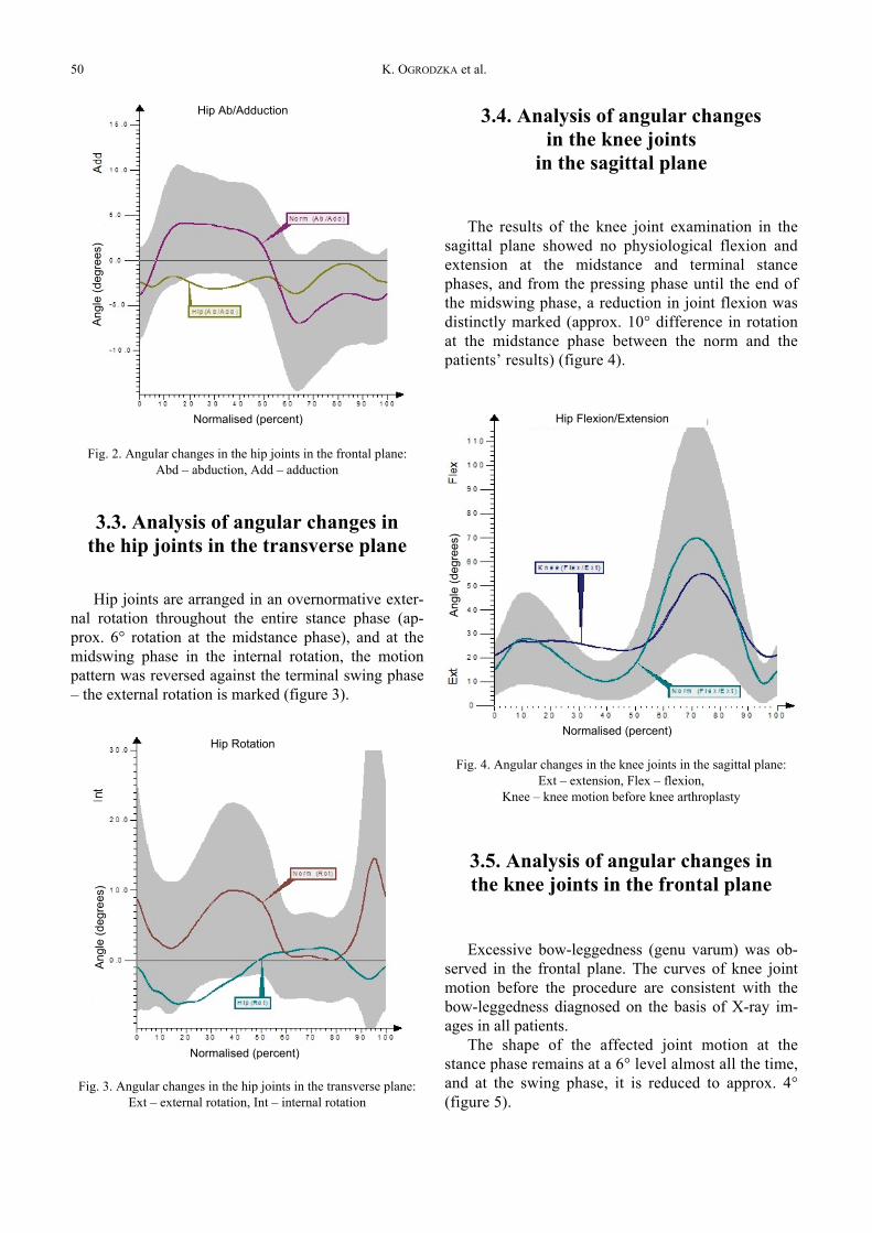
Fig. 2. Angular changes in the hip joints in the frontal plane:Abd – abduction, Add – adduction
3.3. Analysis of angular changes inthe hip joints in the transverse plane
Hip joints are arranged in an overnormative exter-nal rotation throughout the entire stance phase (ap-prox. 6° rotation at the midstance phase), and at themidswing phase in the internal rotation, the motionpattern was reversed against the terminal swing phase– the external rotation is marked (figure 3).
Fig. 3. Angular changes in the hip joints in the transverse plane:Ext – external rotation, Int – internal rotation
3.4. Analysis of angular changesin the knee joints
in the sagittal plane
The results of the knee joint examination in thesagittal plane showed no physiological flexion andextension at the midstance and terminal stancephases, and from the pressing phase until the end ofthe midswing phase, a reduction in joint flexion wasdistinctly marked (approx. 10° difference in rotationat the midstance phase between the norm and thepatients’ results) (figure 4).
Fig. 4. Angular changes in the knee joints in the sagittal plane:Ext – extension, Flex – flexion,
Knee – knee motion before knee arthroplasty
3.5. Analysis of angular changes inthe knee joints in the frontal plane
Excessive bow-leggedness (genu varum) was ob-served in the frontal plane. The curves of knee jointmotion before the procedure are consistent with thebow-leggedness diagnosed on the basis of X-ray im-ages in all patients.
The shape of the affected joint motion at thestance phase remains at a 6° level almost all the time,and at the swing phase, it is reduced to approx. 4°(figure 5).
Normalised (percent)
Hip Ab/Adduction
Normalised (percent)
Hip RotationNormalised (percent)
Hip Flexion/Extension
Ang
le (d
egre
es)
Ang
le (d
egre
es)
Ang
le (d
egre
es)

Evaluation of the kinematic parameters of normal-paced gait in subjects with gonarthrosis 51
Fig. 5. Angular changes in the knee joints in the frontal plane:Var – varus, Val – valgus
3.6. Analysis of angular changes inthe knee joints in the transverse plane
The crus motion in the transverse plane was char-acterised by a shift of the range of angular changes inthe direction of external rotation throughout the gaitcycle (figure 6).
Fig. 6. Angular changes in the knee joints in the transverse planeExt – external rotation, Int – internal rotation
3.7. Analysis of angular changes inthe ankle joints in the sagittal plane
Degenerative changes in knee joints also causeda change of the gait pattern in ankle joints. Up to themiddle of the preswing phase, the curve of changesfalls within the biomechanical norm (within its upper
limits), and excessive dorsiflexion occurs from thesecond half of the preswing phase to the end of theinitial swing phase. There is no natural plantar flexionat the gait transition phase (figure 7).
Fig. 7. Angular changes in the ankle joints in the sagittal planePlan – plantar flexion, Dors – dorsal flexion
3.8. Analysis of angular changes inthe ankle joints in the frontal plane
The results of the ankle joint examination in thefrontal plane showed adduction of the joints throughoutthe gait cycle. The ankle joints become slightly ab-ducted only at the end of the stance phase (figure 8).
Fig. 8. Angular changes in the ankle joints in the frontal plane:Abd – abduction, Add – adduction
Normalised (percent)
Knee Ab/Adduction
Normalised (percent)
Knee Rotation
Normalised (percent)
Ankle Dorsi/Plantar
Normalised (percent)
Ankle Ab/Adduction
Ang
le (d
egre
es)
Ang
le (d
egre
es)
Ang
le (d
egre
es)
Ang
le (d
egre
es)

K. OGRODZKA et al.52
3.9. Analysis of angular changes inthe ankle joints in the transverse plane
In the transverse plane, the amplitude of anklejoint movement corresponds to the upper limit ofthe biomechanical norm – the joints are at an ap-prox. 5° external rotation throughout the gait cycle(figure 9).
Fig. 9. Angular changes in the ankle joints in the transverse plane:Ext – external rotation, Int – internal rotation
4. Summary of the results
4.1. Analysis of angular changesin the hip joints
Due to knee joint disease, distinct changes in theposition of the hip joints in all planes of motion oc-curred. Hip joints are excessively flexed and ab-ducted. At the stance phase, the joints are externallyrotated, while internal rotation occurs at the swingphase.
4.2. Analysis of angular changesin the knee joints
Knee joints are excessively flexed at the stancephase, and this flexion is insufficient at the swingphases. Additionally, bow-leggedness and externalrotation of the joints are marked.
4.3. Analysis of angular changesin the ankle joints
Degenerative changes in knee joints also causeddysfunction of ankle joints. Ankle joints are dorsallyflexed, adducted and slight external rotation occurs.
5. Discussion
Advanced degenerative changes of knee joints sig-nificantly affect the gait stereotype, resulting in a pa-tient’s functional limitation. To assess the scope anddegree of dysfunction, an accurate and thorough gaitanalysis is necessary, as this shows and illustrates themotor disorders in these patients. Three-dimensionalmotion analysis, presenting spatial arrangement ofbody segments in three planes, offers such a possibility.Literature on the subject [20], [21] provides a compre-hensive description of the work of the knee joint beforeknee replacement, but these studies are mostly limitedto the presentation of joint movement in the sagittalplane without considering the other planes or the influ-ence of the disease on the ankle and hip joints.
In the authors’ own research, in accordance withthe adopted aims of the study, an assessment ofchanges in kinematic gait parameters was carried outin persons with degenerative changes of the knee jointbased on three-dimensional motion analysis using theVicon 250 system. This analysis included angularchanges of knee, ankle and hip joints in the threeplanes of motion.
The results of locomotion tests in patients suffer-ing from degenerative changes corroborate that thepattern of ankle, knee and hip joint movement is dis-tinctly influenced by the disease in the three planes ofmotion.
At the stance phase, the hip joints were flexed, ab-ducted and externally rotated at the same time. Flex-ion contracture and external rotation occur in kneejoints, while dorsiflexion, external rotation and ad-duction are characteristic of ankle joints.
At the swing phase, the hip joints are internallyrotated. Knee joints are not sufficiently flexed, and thefeet remain dorsally flexed.
A wrong range of motion in the knee joints in thesagittal plane while walking results in a lack of plan-tarflexion in the ankle joints at the initial swing phaseand persistent contracture of the hip joints. Thismakes it possible to move the lower extremity without“catching” the ground.
Normalised (percent)
Ankle Rotation
Ang
le (d
egre
es)

Evaluation of the kinematic parameters of normal-paced gait in subjects with gonarthrosis 53
Changes in the patients’ gait stereotype beforeknee arthroplasty may primarily result from their long-standing locomotor habits. Increasing pain makes thepatients change their way of walking – if the knee jointis straightened, the pressure on the joint surface in-creases and the pain becomes more intense. As a re-sult, these patients try to avoid the straightening of thejoint, which is characteristic of the beginning and endof the stance phase. Flexion contracture of knee jointsis the result of long-term knee flexion.
There are numerous reports in literature referringto the influence of knee joint degenerative disease onthe three-planar range of motion of the remaininglower extremity joints. They are mostly limited tostatements that the single-support phase becomesshortened as a result of the disease [11], [12].
ASTEPHEN et al. [22], on the basis of locomotiontests of persons with degenerative changes of kneejoints, concluded that changes in ankle and knee jointsare marked in the sagittal plane. However, they didnot determine the direction of the changes in thejoints.
Similar results for the sagittal plane were obtainedby BALIUNAS et al. [21], who discovered a restrictionof the range of motion throughout the gait cycle.
This is another one confirmation of the results ofthe authors’ own studies, where a clear restriction ofthe active range of motion was found in the knee jointin the sagittal plane.
MANETTA et al. [22] examined 10 persons suffer-ing from degenerative knee joint disease and com-pared the results with the control group. They deter-mined the kinetics and kinematics of the gait on thebasis of a motion analysis system. It turned out thatthe maximum flexion at the support stage was lowerin the patients. As the values of the maximum flexionat the support phase were similar among the patientscompared to the norm and no weakening of the quad-riceps femoris muscle was observed, they concludedthat this pattern was caused by a compensatory reduc-tion in gait speed.
These reports are not fully consistent with the re-sults of the authors’ own research, as extension limi-tation was observed in the patients examined (in-creased flexion) at the support phase.
Results similar to those reported by MANETTA etal. [22], and not fully consistent with the results of theauthors’ own research, were obtained by CHILDS et al.[23], who found that flexion during loading responsewas limited in patients suffering from articular diseasecompared to the norm.
In view of the analysis of the locomotion test re-sults presented above and detailed interpretation of
angular changes in lower extremity joints in personsbefore knee arthroplasty, with reference to the entirebiomechanism, it seems justified to undertake re-search aimed at determining the pattern of knee jointmovement in patients with degenerative changes usingthe results of three-dimensional motion analysis, fo-cusing on presenting mutual conditioning of bodysegments in the three planes.
The evaluation of gait parameters during walkingis helpful in assessing abnormal gait, in quantifyingimprovements resulting from intervention, or in pre-dicting subsequent events such as ageing or falls [24].
Considering the controversial results obtained bysome authors, who determine the gait stereotype inpersons suffering from osteoarthrosis, a thorough veri-fication of changes occurring in the knee joint beforeknee arthroplasty seems necessary, as it will be sig-nificant in the planning and selection of appropriaterehabilitation treatment in these patients.
6. Conclusions
1. Degenerative knee joint disease significantly af-fects the correct gait stereotype, causing deviationsfrom the biomechanical norm as regards angularchanges in lower extremity joints in the three planesof motion.
2. A changed motion pattern was most distinct inthe frontal and transverse planes.
References
[1] LEE J.A., Choroba zwyrodnieniowa stawów kolanowychu dorosłych. Przywrócenie sprawności i utrzymanie zdro-wego stawu. Wytyczne Institute for Clinical Systems Inte-gration, Minneapolis, Medycyna po Dyplomie, 2000, 9 (4),115–128.
[2] WIDUCHOWSKI J., Kolano, endoprotezoplastyka – całkowitawymiana stawu, Sport & Med. s.c., 2001, Katowice.
[3] WIERUSZ-KOZŁOWSKA M., MARKUSZEWSKI J., Choroba zwy-rodnieniowa stawów, [w:] Wiktora Degi ortopedia i rehabili-tacja, Marciniak W., Szulc A. (red.), PZWL, Warszawa, 2004,tom 2.
[4] MESSIER S.P., DeVITA P., COWAN R.E., SEAY J., YOUNG H.C.,MARSH A.P., Do older adults with knee osteoarthritis placegreater loads on the knee during gait? A preliminary study,Arch. Phys. Med. Rehabil., 2005, 86, 703–709.
[5] ANDRIACCHI T.P., LANG P.L., ALEXANDER E.J., HURWITZ D.E.,Methods for evaluating the progression of osteoarthritis,J. Rehabil. Res. Dev., 2000, 37(2), 163–170.
[6] GOLEC E., CZABAŃSKI P., GOLEC J., Ocena wyników ruchowegousprawniania chorych z zaawansowanymi zmianami zwy-rodnieniowymi stawów kolanowych, Fizjoterapia, 1999, 7(3),20–23.

K. OGRODZKA et al.54
[7] LEE J.A., Choroba zwyrodnieniowa stawów kolanowychu dorosłych. Przywracanie czynności i zmniejszanie objawówchorobowych. Wytyczne Institute for Clinical Systems Inte-gration, Minneapolis, Medycyna po Dyplomie, 1998, 7(6),105–113.
[8] WOLNY T., SAULICZ E., MOLICKA D., RYNGIER P., Terapiawtórnych skutków zmian zwyrodnieniowych stawu kola-nowego – ocena skuteczności różnych sposobów postępo-wania, Medycyna Sportowa, 2002, 10, 40–43.
[9] BECKER R., BERTH A., NEHRING M., AWISZUS F., Neuromus-cular quadriceps dysfunction prior to osteoarthritis of theknee, J. Orthop. Res., 2004, 22, 768–773.
[10] JEZIERSKI C., Wpływ kriostymulacji i usprawniania na wy-dolność chodu w gonarthrosis, Fizjoterapia, 1996, 4(1–2),44–47.
[11] DELUZIO K.J., WYSS U.P., ZEE B., COSTIGAN A., SORBIE C.,Principal component models of knee kinematics and kinetics:Normal vs. pathological gait patterns, Hum. Mov. Sci., 1997,16, 201–217.
[12] YU S., STUART M.J., KIENBACHER T., GROWNEY E.S., AN K.-N.,Valgus–varus motion of the knee in normal level walking andstair climbing, Clin. Biomech., 1997, 12(5), 286–293.
[13] ROGIND H., BIBOW-NIELSEN B., JENSEN B., MOLLER H.C.,FRIMODT-MOLLER H., BLIDDAL H., The effect of a physicaltraining program on patients with osteoarthritis of the knee,Arch. Phys. Med. Rehabil., 1998, 79, 1421–1427.
[14] ROSSI M., BROWN L., WHITEHURST M., CHANI C., HANKINS J.,TAYLOR C., Comparison of knee extensor strength betweenlimbs in individuals with bilateral total knee replacement,Arch. Phys. Med. Rehabil., 2002, 83, 523–526.
[15] HOCHBERG M.C., ALTMAN R.D., BRAND K.D., CLARK B.M.,DIEPPE P.A., GRIFFIN M.R., MOSKOWITZ R.W., SCHNITZER T.J.,Guidelines for medical management of osteoarthrosis. Part II.Osteoarthrosis of the knee, Arthritis Rheumatism, 1995, 11,1541–1546.
[16] ANDRIACCHI T.P., ALEXANDER E.J., Studies of human loco-motion: past, present and future, J. Biomech., 2000, 33,1217–1224.
[17] PERRY J., Gait analysis, Thorofare, SLACK, 1992.[18] ALLARD P., CAPPOZZO A., LUNDBERG A., VAUGHAN C.L.,
Three-dimensional analysis of Human Locomotion, Wileyand Sons, New York, 1997.
[19] ZEMBATY A., Fizjoterapia, PZWL, Warszawa, 1987.[20] MANETTA J., FRANZ L.H., MOON C., PERELL K.L., FANG M.,
Comparison of hip and knee muscle moment in subjects withand without knee pain, Gait Posture, 2002, 16, 249–254.
[21] BALIUNAS A.J., HURWITZ A.B., RYAL A.B., KARRAR A.,CASE J.P., BLOCK J.A., ANDRIACCHI T.P., Increased knee jointloads during walking are present in subjects with knee osteo-arthritis, Osteoarthritis and Cartilage, 2002, 10, 573–579.
[22] ASTEPHEN J.L., DELUZIO K.J., CALDWELL G.E., HUBLEY-KOZEY C.L., DUNBAR M.J., Gait and neuromuscular changesassociated with knee OA severity, J. Biomech., 2007, 40(S2),287–294.
[23] CHILDS J., SPARTO P., FITZGERALD K., BIZZINI M., IRRGANG J.,Alterations in lower extremity movement and muscle activa-tion patterns in individuals with knee osteoarthritis, ClinicalBiomech., 2004, 19, 44–49.
[24] KISS R., Variability of gait characterized by normalizeddeviation, Acta Bioeng. Biomech., 2010, 12(1), 19–23.