evolution of developmental potential and the multiple...
TRANSCRIPT
q 2002 The Paleontological Society. All rights reserved. 0094-8373/02/2801-0006/$1.00
Paleobiology, 28(1), 2002, pp. 70–100
Evolution of developmental potential and the multipleindependent origins of leaves in Paleozoic vascular plants
C. Kevin Boyce and Andrew H. Knoll
Abstract.—Four vascular plant lineages, the ferns, sphenopsids, progymnosperms, and seed plants,evolved laminated leaves in the Paleozoic. A principal coordinate analysis of 641 leaf species fromNorth American and European floras ranging in age from Middle Devonian through the end of thePermian shows that the clades followed parallel trajectories of evolution: each clade exhibits rapidradiation of leaf morphologies from simple (and similar) forms in the Late Devonian/Early Car-boniferous to diverse, differentiated leaf forms, with strong constraint on further diversificationbeginning in the mid Carboniferous. Similar morphospace trajectories have been documented instudies of morphological evolution in animals; however, plant fossils present unique opportunitiesfor understanding the developmental processes that underlie such patterns. Detailed comparisonof venation in Paleozoic leaves with that of modern leaves for which developmental mechanismsare known suggests developmental interpretations for the origination and early evolution of leaves.The parallel evolution of a marginal meristem by the modification of developmental mechanismsavailable in the common ancestor of all groups resulted in the pattern of leaf evolution repeatedby each clade. Early steps of leaf evolution were followed by constraint on further diversificationas the possible elaborations of marginal growth were exhausted. Hypotheses of development inPaleozoic leaves can be tested by the study of living plants with analogous leaf morphologies.
Charles Kevin Boyce and Andrew H. Knoll. Department of Organismic and Evolutionary Biology, HarvardUniversity, 26 Oxford Street, Cambridge, Massachusetts 02138. E-mail: [email protected]
Accepted: 15 August 2001
Introduction
Paleontology enjoys a rich tradition of re-search on the evolution of morphological di-versity. Beginning with Raup’s (1966) quanti-fication of molluscan morphospace based onthe geometry of coiled shells, paleontologistshave used mathematical descriptions of shape(MacLeod 1999; Smith and Bunje 1999); con-tinuous, quantitative measurements of dis-tances among morphological points deemedhomologous (Bookstein 1991); and discretequalitative characterizations of morphology(Foote 1995) to illuminate the history of mor-phological diversity in invertebrates and skel-etonized protists (see McGhee 1999 for re-view).
Developmental biology offers the prospectof understanding the genetic and physiologi-cal bases of morphology and, hence, of mor-phological evolution. To date, however, few at-tempts to integrate data from paleontologyand developmental biology (reviewed in Shu-bin et al. 1997; Valentine et al. 1999; Knoll andCarroll 1999) have taken advantage of the pos-sibilities afforded by morphometric analysesof the fossil record.
Vascular plants are particularly well suitedfor the integration of developmental biologyand paleontology. The presence in plants ofcell walls vastly increases the probability ofanatomical preservation. Cell walls also pro-hibit cell migration, constraining the types ofdevelopment that are possible in plants andfacilitating the recovery of developmental pat-tern from fossils. (In structures such as thevascular cambium of seed plants, the tips ofdeveloping cells can grow intrusively betweenother cells, and cell contacts can be establishedbetween cell files on either side of a cambialinitial that is lost. However, even in this spe-cial case of secondary growth, there is no ac-tual cell migration; cambial ontogeny can stillbe traced readily in the cambium-derivedwood [Barghoorn 1940]). This stands inmarked contrast to animals, where ontogenyinvolves complex patterns of cell movement,changing cell contacts, and cell death.
Additional advantages arise because landplants essentially all make their living in thesame way (Knoll and Niklas 1987; Niklas1994). There are various specializations to dealwith limitations of water, light, nutrients,

71EVOLUTION OF LEAF DEVELOPMENT IN THE PALEOZOIC
symbionts, and substrates, but, with the ex-ception of a few parasites, all plants gathersunlight, water, and carbon dioxide in order toconduct photosynthesis. As a result, there is,in general, far less uncertainty about the in-terpretation of functional morphology in fos-sil plants than there is with fossil animals.This uniformity of life strategy, in conjunctionwith developmental constraints, also increas-es the likelihood of evolutionary convergence.Roots, secondary growth, and laminate leaveseach evolved multiple times in different tra-cheophyte lineages. Such repeated instancesof convergent evolution permit developmentalcomparison of multiple independent originsof morphologically and functionally similarstructures.
This combination of developmental con-straint, cellular preservation, and convergentevolution makes plants unusually attractivesubjects for morphological analysis. Leavesare particularly advantageous for studies ofmorphological evolution. Leaf compressionsare abundant in fluviatile and lacustrine de-positional systems, the leaf fossil record iswell documented, and leaves are the one or-gan for which both overall morphology anddetails of vascularization are commonly avail-able in the same specimen. Furthermore, lam-inate leaves are known to have evolved inde-pendently in several Paleozoic tracheophyteclades, and the degree of morphological con-vergence among these early leaves is high.Leaves produced by early pteridophyte andseed plant lineages were in some cases so sim-ilar to modern fern leaves that only in the ear-ly twentieth century did paleontologists rec-ognize that some were borne by seed plants(reviewed in Scott 1909).
In this paper, we present a morphospaceanalysis of Paleozoic leaves and interpret theresults in light of developmental processes in-ferred from preserved morphologies.
Patterns of Morphological Evolution inPaleozoic Leaves
During the later Devonian and Early Car-boniferous, laminate leaves containing multi-ple veins evolved independently in seedplants, progymnosperms, ferns, and sphenop-sids. The leaves of ferns, seed plants, and pro-
gymnosperms have traditionally been termedmegaphylls and considered to be homolo-gous. By definition (Gifford and Foster 1989),megaphylls are associated with leaf gaps inthe stele of the supporting stem; they can befrondose or entire, and typically are laminateand contain more than one vein (unless sec-ondarily reduced as in most conifers). Al-though widely applied, this megaphyll typol-ogy is an artifact of the depauperate living flo-ra. Once fossils are included, no component ofthe megaphyll concept emerges as a synapo-morphy uniting these lineages. In particular,the central tenet of associated leaf gaps is notrelevant to the earliest fossil representatives ofthese lineages, all of which are protostelic(Taylor and Taylor 1993).
It is possible that some or all of these line-ages inherited a lateral branch system with abroadly frondlike architecture from theircommon ancestor (Kenrick and Crane 1997).The likelihood of this is dependent on the phy-logenetic placement of a few key taxa of am-biguous affinities. The traditional placementof the ferns and seed plants as sister taxa, withEquisetum as the closest outgroup, suggestedthat a frondose megaphyllous leaf was a syn-apomorphy shared by the ferns and the seedplants. However, the most recent phylogeneticwork based on living plants places Equisetumand the Psilotales along with eusporangiateferns as basal lineages in a clade containing allextant pteridophytes, save for lycopods (Pryeret al. 2001). Statements about last common an-cestors, then, depend critically on how keyDevonian plants without laminated leaves areadded to this phylogeny.
Even if certain frond characteristics turn outto be synapomorphies of the clade that in-cludes sphenophytes, ferns, progymno-sperms, and seed plants, however, the termi-nal units on any fronds inherited from a com-mon ancestor would have had little or no lam-ination. The earliest known leaves in each ofthe four clades are highly dissected structurescomposed of segments that were small, nar-row, and with a single vein. In light of thesefossils, our assessment of leaf evolution doesnot depend on any particular phylogenetic hy-pothesis.
A survey of the Paleozoic compression flora

72 C. KEVIN BOYCE AND ANDREW H. KNOLL
of North America and Western and CentralEurope was carried out to investigate patternsof morphological diversification in the earlyevolution of leaves within and among groups.Each species was described from a single pri-mary source, although stratigraphic rangesand taxonomic affinities were modified usingthe full list of sources (see Appendix 1). Tax-onomic affinity was assigned only to leaveswith documented connection to either fertilestructures or stems with diagnostic anatomi-cal features. Association of leaves and fertilestructures at the same localities was not con-sidered sufficient for taxonomic assignment.
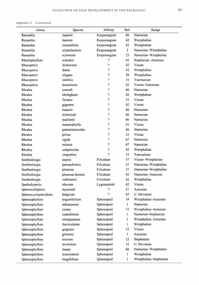
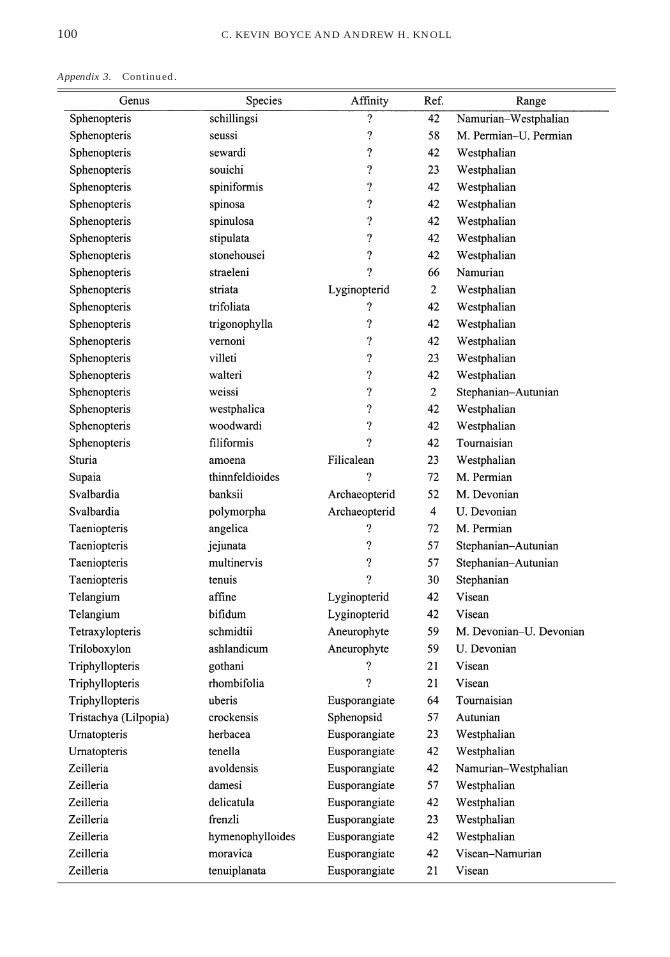
Morphological similarity of a species to oth-er leaf species of known taxonomic affinityalso was not considered. For example, manyNeuropteris species are listed as having un-known affinities despite the fact that someNeuropteris species are known to have beenborne by seed plants. An exception to this wasmade in the case of the gigantopterids. Seedplant identity has been documented only forAsian gigantopterid species, which are be-yond the scope of this study, but the uniqueconstruction of gigantopterids warrantsplacement of the two gigantopterid species in-cluded from North American localities as seedplants. Fossils that could not be identified tothe species level and taxa for which photoswith identifiable venation were not availablewere excluded from the analysis. Of the re-sulting list of 641 taxa, 52 are seed plants, 144are pteridophytes, and 445 are of unknown af-finity (many of these are probably but not de-monstrably seed plant remains). Among thepteridophytes, there are 15 progymnosperms,33 sphenopsids, 19 leptosporangiate ferns, 27marattialean ferns, 15 zygopterids, and 35eusporangiate species of other or unknown af-finities. See Appendix 3 for a list of species,their stratigraphic ranges, and their taxonomicaffinities.
Taxa were coded for 63 unordered binaryand multistate characters (see Appendix 2; acomplete data matrix of character codings foreach species is available from C. K. B.) describ-ing individual pinnules rather than entirefronds and concerned primarily with venationand laminar structure rather than overall pin-nule shape. Individual leaf species commonly
display considerable laminar variability, evenbetween the pinnules within a single fossilfrond. This variability was included in thecharacter codings by the use of ‘‘variable’’ asa state for many characters (see Appendix 2for examples). Coding for variability intro-duces two important, but potentially negativeeffects. First, it requires the use of charactersthat are inapplicable to large subsets of thetaxa. Second, taxa based on few or incompletefossil specimens can be miscoded becausethere is less opportunity for actual species var-iability to be demonstrated in available ex-amples. Despite these complications, inclu-sion of variability is preferable to its exclusion,because variation is such a common aspect ofplant morphology (e.g., Knauss and Gillespie2001) and because range of variation is poten-tially informative about development.
A principal coordinate analysis was used toprovide a more comprehensible summary ofthe information recorded in the study. Theoriginal matrix of character codings for the641 taxa was used to create a 641 by 641 matrixof the pairwise dissimilarities of all speciescalculated as the number of character mis-matches divided by the number of charactersthat are not missing or nonapplicable. (Dis-similarity matrix and related statistics werecalculated using software provided by R. Lu-pia; further details of methodology describedin Lupia 1999. Mathematica was used for theprincipal coordinates analysis and all othercalculations.) This matrix was then trans-formed to move the centroid of the dissimi-larity distribution to zero (Gower 1966). Ei-genvalues and eigenvectors of the trans-formed dissimilarity matrix were determined,and the component values of each eigenvectorwere used to position each taxon with respectto a particular principal coordinate axis(Sneath and Sokal 1973). The magnitude of theeigenvalue corresponding to each eigenvectorgives an indication of the relative contributionof that axis to the summary of informationfrom the original data matrix. The first twoprincipal coordinate axes were plotted as arepresentation of morphospace occupiedthrough time (Figs. 1, 2). These axes containabout 51% of the information in the originaldistance matrix, as extimated from the sum of

73EVOLUTION OF LEAF DEVELOPMENT IN THE PALEOZOIC
FIGURE 1. Principal coordinate analysis morphospace of Paleozoic leaf compression fossils, plotted by stratigraphicinterval (A); and showing the placement of taxa within important groups of ferns (B) and seed plants (C).
the two corresponding eigenvalues divided bythe sum of all eigenvalues of the transformeddistance matrix (Foote 1995).
The taxa span the time interval from MiddleDevonian until the end of the Permian. TheDevonian through Early Permian (Autunian)is divided into intervals averaging approxi-mately 15 million years and ranging from 8 to21 million years in duration. The later Permian
is not well represented for two reasons: (1)there are fewer productive localities withinthe geographic area covered, and (2) poorpreservation and coriaceous habit commonlyobscure venation pattern in specimens that areavailable (Kerp 2000).
Principal coordinate analyses provide aconvenient method for visualizing large quan-tities of morphological information by geo-

74 C. KEVIN BOYCE AND ANDREW H. KNOLL
FIGURE 2. Location within the morphospace of important form genera and other morphologically distinctivegroups.
FIGURE 3. Average pairwise dissimilarity between spe-cies for each interval with 95% confidence intervalsbased upon 1000 bootstrap resampling runs.
metrically summarizing as much of the vari-ability between taxa as possible on a few axesin the form of a morphospace. However, be-cause such an analysis is entirely dependentupon the overall composition of the data set,it can be highly influenced by taxonomic andmorphological decisions. In particular, the gi-gantopterids bore the most complex and mor-phologically distinctive leaves of any plants inthe Paleozoic, but there was no possibility oftheir leaves having coordinates distinct fromother taxa on axes 1 and 2 because gigantop-terids represented only about 0.3% of the in-cluded species and the characters that distin-guish them were invariant among all othertaxa. Placement of the diverse morphologiesof the Devonian and Carboniferous leaves is,however, more amenable to morphological in-
terpretation (Figs. 1, 2), and the overall patternof diversification seen in the 50% of the infor-mation summarized in the two-dimensionalmorphospace plot represents well the data setas a whole (Fig. 3).
Three interesting patterns emerge from theresulting Paleozoic leaf morphospace (Fig. 1).First, the areas of initial morphospace occu-pation in the Devonian and Early Carbonif-erous remain occupied in the later record, butthe taxonomic affinities of the plants exhibit-ing these leaf morphologies change over time.Second, ferns, seed plants, progymnosperms,and sphenopsids all share the same initial lo-cation in morphospace, diverging only withsubsequent evolution. This suggests that theindependent origins of leaves were based onmodifications of a common underlying devel-opmental system. Third, diversity and dispar-ity (total occupied morphospace) initially in-crease in tandem, but after the Namurian, fur-ther increases in taxonomic diversity do notchange the range of morphologies occupied,suggesting that the limits of a biologicallyconstrained system had been reached. Earlyleaf evolution thus appears to be constrainedat both ends of the radiation, first by initial ar-chitectures and later by the limits of leaf mor-phological variation.
Developmental Interpretations ofMorphological Patterns
Insights from living organisms have longbeen applied to paleontological studies of

75EVOLUTION OF LEAF DEVELOPMENT IN THE PALEOZOIC
plant function. Examples include biomechan-ical modeling of extinct species based on char-acteristics of living tissues (Roth and Mos-brugger 1996; Niklas 1997a,b; examples inBateman et al. 1998) and estimates of past car-bon dioxide levels based on stomatal indicesderived from present-day plants (McElwain etal. 1999). In similar ways, paleobotanical stud-ies of morphological evolution can benefitfrom advances in plant developmental biolo-gy. For example, following earlier suggestions(Scheckler 1976, 1978; Wight 1987), Stein(1993) modeled stem vasculature as a functionof auxin diffusion from the stem apex and lat-eral primordia, successfully reproducing theobserved stelar morphologies of some Devo-nian plant axes.
An understanding of leaf development inliving plants may similarly inform our under-standing of early leaf evolution. Comparativebiology suggests that mechanisms of meriste-matic growth will likely impose constraints onleaf development and, hence, potential leafmorphologies. For this reason, fossil leavesmay provide an indirect record of leaf meri-stematic capability through time. The study ofleaf evolution has traditionally relied upon in-terpretation of frond architecture and the po-sitional identity of laminar subunits of thefrond. These traits are important for whole-plant reconstruction and for systematics, butthey cannot illuminate the developmental ca-pacity of the foliar meristems of these plants.The emphasis here is on the meristematic po-tential present in early leaf-bearing plants, us-ing comparisons with living plants to con-strain models of lamina development and evo-lution. Rather than considering positional ho-mology and evolution, the focus is uponmeristematic homology and evolution (Stein1998).
Development can be considered in terms oftwo related processes: (1) growth, includingcell division and the differentiation of individ-ual cells, and (2) the patterning of differenti-ated cells to form functional tissues (Wolpert1971; Sachs 1991). The leaves of extant plantsdisplay a number of different growth mecha-nisms. Anatomical studies of morphologicallydiverse ferns indicate leaf growth by meri-stems located at the margin of the developing
organ (Pray 1960, 1962; Zurakowski and Gif-ford 1988; White and Turner 1995; Korn 1998).Marginal meristems consist of a peripheralrow of dividing initials, which are the ulti-mate source of all cells in the leaf. Additionalsubmarginal cell divisions are necessary bothfor cell differentiation and for building up thethickness of the developing leaf. The marginalmeristem remains active until general pinnulemorphology and the location of all procam-bium has been determined (Pray 1962). In con-trast, angiosperm leaves grow diffusely, withmeristematic activity throughout the devel-oping leaf. Clonal analysis experiments cor-roborate anatomical work (Pray 1955) andSEM studies (Hagemann and Gleissberg 1996)in demonstrating that the marginal cells of anangiosperm leaf play almost no role in leafgrowth (Nicotiana tabacum [Poethig and Sus-sex 1985a,b]) or, at least, play no greater a rolethan other parts of the leaf (Gossypium barba-dense [Dolan and Poethig 1998]).
Marginal and diffuse growth are not nec-essarily mutually exclusive mechanisms; morelikely, they represent end-members of a largeand complex continuum. Although the fewfern leaves for which development has beenstudied in detail possessed marginal meri-stems, others are likely to possess more variedand complex mechanisms of growth (such asthose described as having ‘‘dilatory’’ leafgrowth by Wagner [1979]). Furthermore, thereis much variety in what is here summarized asinternal, as opposed to marginal, growth (Fos-ter 1952; Hagemann and Gleissberg 1996). Al-though continuing research is needed to ex-plore the complete developmental diversity ofleaves in living plants, what matters for an ini-tial assessment of Paleozoic plants is that theirleaves could grow exclusively by means of amarginal meristem or could include extensiveinternal growth.
Although differing mechanisms of leafgrowth exist, vascular patterning is broadlysimilar across all tracheophytes. Vascularplants all have a flux of auxins from distalmeristems in the shoot system toward moreproximal tissues. Auxin transport is accom-plished by the pumping of auxin into cellsfrom all sides and the preferential pumping ofauxin out of cells proximally down the stem

76 C. KEVIN BOYCE AND ANDREW H. KNOLL
(Galweiler et al. 1998; Steinmann et al. 1999;Berleth et al. 2000). Physiological studies havedemonstrated the importance of auxins bothfor overall stelar patterning (Ma and Steeves1992) and for finer scales of differentiation, in-cluding the establishment of cell polarity andthe continuity of vascular strands (Sachs1981). Sachs (1981, 1991) has hypothesizedthat the differentiation of individual vascularstrands is based upon the distal to proximalflux of auxin, which determines cell polarityand induces the development of procambialcharacteristics in the files of cells throughwhich it flows. These procambial characteris-tics increase the cells’ capacity to transportauxin, further increasing flux through the cellfile and, in consequence, decreasing fluxesthrough adjacent cell files. In this way, thewidths of procambial strands can be limitedwithout the necessary action of a second, in-hibitory hormone.
The studies of vascular patterning and theauxin canalization hypothesis reviewed in thepreceding paragraph are based principally onstems—molecular and biochemical studies ofleaves are largely limited to investigations ofvascular cell differentiation in Zinnia tissuecultures. Nonetheless, several considerationsjustify the assumption that leaves and stemsfollow similar mechanisms of vascular pat-terning and differentiation: The canalizationhypothesis is the only hypothesis that hasbeen documented in any part of the plant andit is consistent with all available informationfrom leaves. Leaf primordia are known to beimportant sources of auxin (Ma and Steeves1992; Stein 1993), and the disruption of leafvascular patterning by auxin transport inhib-itors (Sieburth 1999; Mattsson et al. 1999) andby mutations that disrupt auxin transport(Carland and McHale 1996) has been docu-mented. (Leaf development is, of course, alsoinfluenced by auxin-independent factors [Car-land et al. 1999]). The documentation of vas-cular patterning mechanisms in leaves re-mains an important research goal, and emerg-ing techniques (Caruso et al. 1995; Moctezumaand Feldman 1999) suggest that new insightsare on the horizon.
The growth and patterning mechanisms ex-hibited by living plants can be used to con-
strain hypotheses about developmental mech-anisms present during the early evolution ofleaves. The role of auxin fluxes from activemeristems in the patterning of vascular tissueappears to be conserved throughout tracheo-phytes. We assume, therefore, that it applies toPaleozoic leaves. This assumption, in turn,provides a means of generating hypothesesabout leaf growth in extinct plants. If theleaves of Paleozoic plants grew exclusively bymarginal meristems, venation should be ori-ented toward leaf margins with all veins end-ing marginally, as observed in the living fernsfor which marginal growth has been docu-mented. If the leaf development included ex-tensive internal, nonmarginal growth, vena-tion would not be expected to have uniformorientation and vein endings should be dis-persed throughout the lamina.
All Devonian and Carboniferous leaves, re-gardless of phylogenetic affinity, have exclu-sively marginal vein endings (Figs. 4, 5). Thissuggests that each origin of laminated leavesrelied on marginal meristems. The only Paleo-zoic leaves with extensive internal vein end-ings were those of the late Early to Late Perm-ian gigantopterid seed plants. In their case, in-ternal tertiary veins are oriented toward oneanother and meet in discrete areas betweenthe marginally ending secondary veins. Thissuggests a two-stage process in which mar-ginal growth was followed by internal growthat discrete intercalary meristems. There is noevidence of true diffuse growth until the Me-sozoic.
The early leaves of each lineage likely grewby means of marginal meristems, but our un-derstanding of those meristems can be furtherrefined. In some living ferns, a direct corre-spondence has been found between specificmarginal initials and the cell types to whichthey give rise (parenchyma, or parenchymaand vasculature [Pray 1960, 1962; Zurakowskiand Gifford 1988]). Other work with differentferns has found marginal meristems of uni-form composition; in these plants, vascularpatterning responds to marginal signals, butwithout reference to specific marginal initialcells (Korn 1998). Tissue patterning based ondiscrete ground meristem and procambialmarginal initials would be expected to result
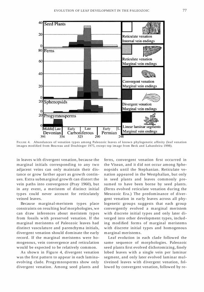
77EVOLUTION OF LEAF DEVELOPMENT IN THE PALEOZOIC
FIGURE 4. Abundances of venation types among Paleozoic leaves of known phylogenetic affinity (leaf venationimages modified from Boureau and Doubinger 1975, except top image from Beck and Labandeira 1998).
in leaves with divergent venation, because themarginal initials corresponding to any twoadjacent veins can only maintain their dis-tance or grow farther apart as growth contin-ues. Extra submarginal growth can distort thevein paths into convergence (Pray 1960), butin any event, a meristem of distinct initialtypes could never account for reticulatelyveined leaves.
Because marginal-meristem types placeconstraints on resulting leaf morphologies, wecan draw inferences about meristem typesfrom fossils with preserved venation. If themarginal meristems of Paleozoic leaves haddistinct vasculature and parenchyma initials,divergent venation should dominate the earlyrecord. If the marginal meristems were ho-mogenous, vein convergence and reticulationwould be expected to be relatively common.
As shown in Figure 4, divergent venationwas the first pattern to appear in each lamina-evolving clade. Progymnosperms show onlydivergent venation. Among seed plants and
ferns, convergent venation first occurred inthe Visean, and it did not occur among Sphe-nopsids until the Stephanian. Reticulate ve-nation appeared in the Westphalian, but onlyin seed plants and leaves commonly pre-sumed to have been borne by seed plants.(Ferns evolved reticulate venation during theMesozoic Era.) The predominance of diver-gent venation in early leaves across all phy-logenetic groups suggests that each groupconvergently evolved a marginal meristemwith discrete initial types and only later di-verged into other development types, includ-ing modified forms of marginal meristemswith discrete initial types and homogenousmarginal meristems.
Leaf evolution in each clade followed thesame sequence of morphologies. Paleozoicseed plants first evolved dichotomizing, finelylobed leaves with a single vein per laminarsegment, and only later evolved laminar mul-tiveined leaves with divergent venation, fol-lowed by convergent venation, followed by re-

78 C. KEVIN BOYCE AND ANDREW H. KNOLL
FIGURE 5. Abundances of venation types among all Paleozoic leaf species included in the analysis. Coding of ve-nation types as in Figure 4.
ticulation. The other clades followed the samesequence to a varying extent. It is proposedthat this repeated pattern reflects the limitednumber of ways that plants can form laminatephotosynthetic surfaces. The range of mor-phological possibilities is further constrainedby the common ancestry of the groups inquestion: evolution works by the modificationof preexisting structures and developmentalpathways, and the available underlying path-way common to the four clades and ultimatelyderived from a shared ancestor was stem de-velopment. Stems are indeterminate, cylindri-cal structures that grow from a discrete apicalmeristem, providing the source of an auxingradient involved in vascular patterning. Wehypothesize that stepwise modification of thegrowth and patterning employed in this an-cestral, axial system produced marginal mer-
istems and laminate leaves in each lineage (Ta-ble 1).
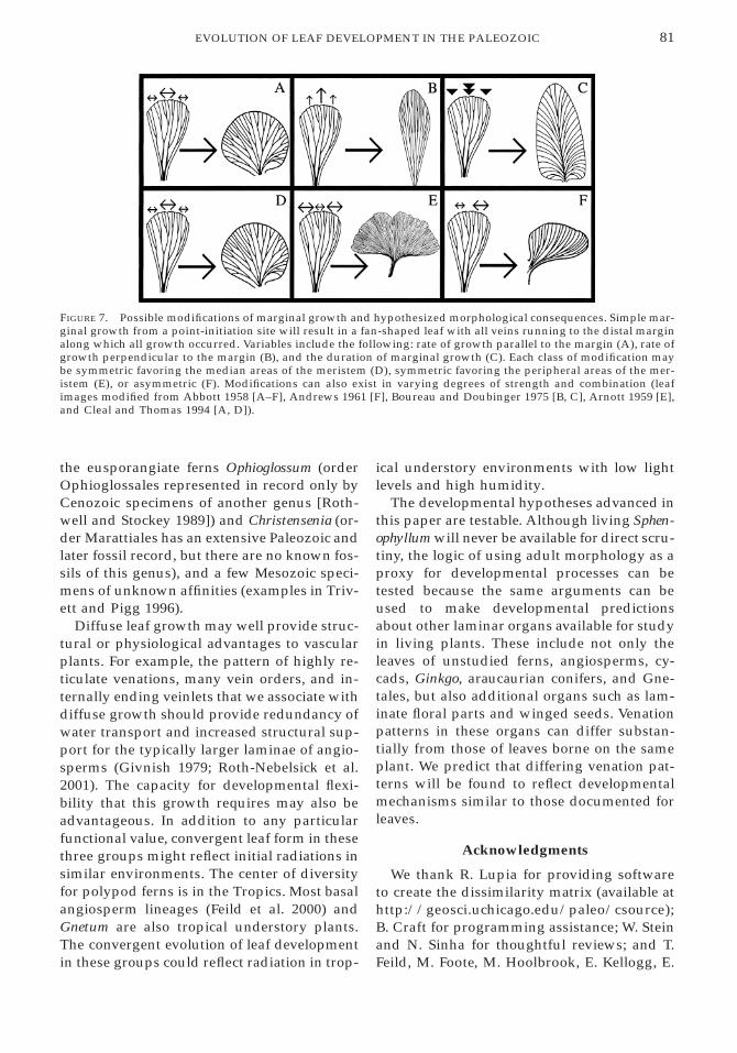
Just as the early steps of marginal meristemevolution were constrained by the scope of de-velopmental possibility, the ultimate range ofpossible leaf morphologies must have beenconstrained by available developmental mech-anisms. Early leaves consisting of linear, di-chotomizing segments had limited potentialfor morphological variability: they could bethree-dimensional or planar; dichotomiescould be equally distributed, concentrateddistally, or concentrated proximally; and therelative strengths of the two arms of a dichot-omy could be altered (Fig. 6). Each of thesemodifications had been explored by the LateDevonian.
The possibilities of marginal growth arealso limited. Simple marginal growth with

79EVOLUTION OF LEAF DEVELOPMENT IN THE PALEOZOIC
TABLE 1. Hypothesized sequence of developmental evolution and time of first occurrence of each innovation bylineage.
point initiation will result in a fan-shaped leafwith vein endings along the distal margin, theonly margin along which a marginal meri-stem would have been active. This system canbe modified by symmetric or asymmetric al-teration of the rate of growth parallel and/orperpendicular to the marginal meristem or byvariation in the duration of growing timealong the meristem (Fig. 7). Other possiblemodifications include the development of amidvein, and broad rather than point initia-tion of lamina growth. A rigorous documen-tation of possible leaf forms in terms of a the-oretical morphospace (McGhee 1999) basedon developmental mechanisms is needed, butit is fair to state that, even though the variouspossible combinations of these modificationscontinued to be shuffled within individuallineages, all specified variables had been ex-plored by the Namurian. The lack of furtherincrease in overall morphological disparity af-ter the Namurian, despite further increase intaxonomic diversity, likely reflects limitationson possible elaborations of marginal growth.
Discussion
The stratigraphic distributions of individu-al characters may provide crude tools for in-vestigating the evolution of leaf development,but the results are compelling. Internal veinendings are not present for the first 100 mil-lion years of leaf evolution; convergent vena-tion is absent for the first 50 million years. Themorphological constraints apparent in earlyleaves strongly suggest specific mechanismsof development. Although multiveined leavesevolved independently at least four times, theclose similarities of early leaves in all groupssuggest parallel evolution by modification ofcommon developmental mechanisms inherit-ed from ancestors whose photosynthetic or-gans consisted of apically growing, bifurcat-ing axial systems.
The evolution of laminated leaves may berelated to the evolution of vascular systemscompetent to support high levels of evapo-transpiration and other aspects of whole-plantfunction in increasingly stratified later Devo-

80 C. KEVIN BOYCE AND ANDREW H. KNOLL
FIGURE 6. Axially organized leaves with dichotomies.A, Evenly distributed. B, Proximally concentrated. C,Distally concentrated and with unequal strength be-tween the two arms of the dichotomies. (Leaf imagesmodified from Remy and Remy 1977 [A, C] and Baten-burg 1977 [B].)
nian and Early Carboniferous communities. Ithas also been proposed that appearance oflaminate leaves was causally linked to a dropin atmospheric CO2 concentration through the
Devonian (Beerling et al. 2001). Changing en-vironmental conditions may well have re-moved a physiological barrier to the evolutionof leaf lamination. Nonetheless, the staggeredtiming of lamina evolution among clades—inthe Devonian, multiveined, laminate leavesoccurred primarily in the Archaeopterid pro-gymnosperms—suggests that both intrinsicand extrinsic factors are necessary to explainthe observed patterns of leaf evolution.
After an initial period of parallel evolution,leaf morphologies within and among thegroups diverged; however, by Namuriantimes the limitations of marginal meristematicdevelopment had been reached and further in-creases in taxonomic diversity did not in-crease morphological disparity. Innovationsin frond architecture, leaf anatomy, and bio-chemistry continued to evolve, but the possi-bilities of marginal pinnule growth had beenexplored.
The use of vascular patterning as a proxyfor developmental mechanism can illuminateother patterns of developmental evolution andconvergence, including the evolution of dif-fuse leaf growth in post-Paleozoic time. An-giosperms radiated initially in the CretaceousPeriod. One of the hallmarks of angiospermmorphology, at least among dicots, is highlyreticulate leaf venation with multiple vein or-ders and freely ending internal veinlets in thevascular aureoles (Esau 1953; Gifford and Fos-ter 1989). On the basis of available experimen-tal evidence (Foster 1952; Pray 1955; Poethigand Sussex 1985a,b; Hagemann and Gleiss-berg 1996; Dolan and Poethig 1998), this pat-tern is interpreted as an indication of diffuseleaf growth. Venation patterns suggestive ofdiffuse leaf growth, however, are not limitedto the angiosperms. Species in two othergroups that radiated in the Cretaceous andearly Tertiary, the Gnetales and the fern cladethat includes the polypods and dryopterids,also possess leaves of this type. As in the Pa-leozoic, Mesozoic innovations in leaf devel-opment were convergent. The simultaneouslyradiating angiosperms, gnetaleans, and poly-pods were the only groups to evolve this typeof leaf, aside from a small leptosporangiatefern clade that includes Dipteris (fossil recorddating to the Late Triassic [Collinson 1996]),
81EVOLUTION OF LEAF DEVELOPMENT IN THE PALEOZOIC
FIGURE 7. Possible modifications of marginal growth and hypothesized morphological consequences. Simple mar-ginal growth from a point-initiation site will result in a fan-shaped leaf with all veins running to the distal marginalong which all growth occurred. Variables include the following: rate of growth parallel to the margin (A), rate ofgrowth perpendicular to the margin (B), and the duration of marginal growth (C). Each class of modification maybe symmetric favoring the median areas of the meristem (D), symmetric favoring the peripheral areas of the mer-istem (E), or asymmetric (F). Modifications can also exist in varying degrees of strength and combination (leafimages modified from Abbott 1958 [A–F], Andrews 1961 [F], Boureau and Doubinger 1975 [B, C], Arnott 1959 [E],and Cleal and Thomas 1994 [A, D]).
the eusporangiate ferns Ophioglossum (orderOphioglossales represented in record only byCenozoic specimens of another genus [Roth-well and Stockey 1989]) and Christensenia (or-der Marattiales has an extensive Paleozoic andlater fossil record, but there are no known fos-sils of this genus), and a few Mesozoic speci-mens of unknown affinities (examples in Triv-ett and Pigg 1996).
Diffuse leaf growth may well provide struc-tural or physiological advantages to vascularplants. For example, the pattern of highly re-ticulate venations, many vein orders, and in-ternally ending veinlets that we associate withdiffuse growth should provide redundancy ofwater transport and increased structural sup-port for the typically larger laminae of angio-sperms (Givnish 1979; Roth-Nebelsick et al.2001). The capacity for developmental flexi-bility that this growth requires may also beadvantageous. In addition to any particularfunctional value, convergent leaf form in thesethree groups might reflect initial radiations insimilar environments. The center of diversityfor polypod ferns is in the Tropics. Most basalangiosperm lineages (Feild et al. 2000) andGnetum are also tropical understory plants.The convergent evolution of leaf developmentin these groups could reflect radiation in trop-
ical understory environments with low lightlevels and high humidity.
The developmental hypotheses advanced inthis paper are testable. Although living Sphen-ophyllum will never be available for direct scru-tiny, the logic of using adult morphology as aproxy for developmental processes can betested because the same arguments can beused to make developmental predictionsabout other laminar organs available for studyin living plants. These include not only theleaves of unstudied ferns, angiosperms, cy-cads, Ginkgo, araucaurian conifers, and Gne-tales, but also additional organs such as lam-inate floral parts and winged seeds. Venationpatterns in these organs can differ substan-tially from those of leaves borne on the sameplant. We predict that differing venation pat-terns will be found to reflect developmentalmechanisms similar to those documented forleaves.
Acknowledgments
We thank R. Lupia for providing softwareto create the dissimilarity matrix (available athttp://geosci.uchicago.edu/paleo/csource);B. Craft for programming assistance; W. Steinand N. Sinha for thoughtful reviews; and T.Feild, M. Foote, M. Hoolbrook, E. Kellogg, E.
82 C. KEVIN BOYCE AND ANDREW H. KNOLL
Kramer, R. Lupia, C. Marshall, R. Moran, andM. Thompson for helpful discussions. Re-search leading to this paper was supported inpart by a National Science Foundation predoc-toral fellowship and by the NASA Astrobiol-ogy Institute.
Literature CitedAbbott, M. L. 1958. The American species of Asterophyllites, An-
nularia, and Sphenophyllum. Bulletin of American Paleontology38:289–390.
Andrews, H. N. 1961. Studies in Paleobotany. Wiley, New York.Arnott, H. J. 1959. Anastomoses in the venation of Ginkgo biloba.
American Journal of Botany 46:405–411.Barghoorn, E. S. 1940. The ontogenetic development and phy-
logenetic specialization of rays in the xylem of dicotyledons.I. The primitive ray structure. American Journal of Botany 27:918–928.
Bateman, R. M., P. R. Crane, W. A. DiMichele, P. R. Kenrick, N.P. Rowe, T. Speck, and W. E. Stein. 1998. Early evolution ofland plants: phylogeny, physiology, and ecology of the pri-mary terrestrial radiation. Annual Review of Ecology andSystematics 29:263–292.
Batenburg, L. H. 1977. The Sphenophyllum species in the Car-boniferous flora of Holz (Westphalian D, Saar Basin, Germa-ny). Review of Palaeobotany and Palynology 24:69–100.
Beck, A. L., and C. C. Labandeira. 1998. Early Permian insectfolivory on a gigantopterid-dominated riparian flora fromnorth-central Texas. Palaeogeography, Palaeoclimatology, Pa-laeoecology 142:139–173.
Beerling, D. J., C. P. Osborne, and W. G. Chaloner. 2001. Evolu-tion of leaf-form in land plants linked to atmospheric CO2 de-cline in the Late Palaeozoic era. Nature 410:352–354.
Berleth, T., J. Mattsson, and C. S. Hardtke. 2000. Vascular con-tinuity and auxin signals. Trends in Plant Science 5:387–393.
Bookstein, F. L. 1991. Morphometric tools for landmark data: ge-ometry and biology. Cambridge University Press, Cambridge.
Boureau, E., and J. Doubinger. 1975. Traite de paleobotanique,Tome IV, Fasc. 2. Pteridophylla (premiere partie). Masson,Paris.
Carland, F. M., and N. A. McHale. 1996. LOP1: a gene involvedin auxin transport and vascular patterning in Arabidopsis.Development 122:1811–1819.
Carland, F. M., B. L. Berg, J. N. FitzGerald, S. Jinamornphongs,T. Nelson, and B. Keith. 1999. Genetic regulation of vasculartissue patterning in Arabidopsis. The Plant Cell 11:2123–2137.
Caruso, J. L., V. C. Pence, and L. A. Leverone. 1995. Immuno-assay methods of plant hormone analysis. Pp. 433–447 in P. J.Davies, ed. Plant hormones: physiology, biochemistry andmolecular biology. Kluwer Academic, Netherlands.
Cleal, C. J., and B. A. Thomas. 1994. Plant fossils of the Britishcoal measures. The Palaeontological Association, London.
Collinson, M. E. 1996. ‘‘What use are fossil ferns?’’: 20 years on:with a review of the fossil history of extant pteridophyte fam-ilies and genera. Pp. 349–394 in J. M. Camus, M. Gibby, andR. J. Johns, eds. Pteridology in perspective. Royal Botanic Gar-dens, Kew, England.
Dolan, L. P., and R. S. Poethig. 1998. Clonal analysis of leaf de-velopment in cotton. American Journal of Botany 85:315–321.
Esau, K. 1953. Plant anatomy. Wiley, New York.Feild, T. S., M. A. Zweiniecki, T. Bodribb, T. Jaffre, M. J. Dono-
ghue, and N. M. Holbrook. 2000. Structure and function oftracheary elements in Amborella trichopoda. International Jour-nal of Plant Science 161:705–712.
Foote, M. 1995. Morphology of Carboniferous and Permian cri-
noids. Contributions from the Museum of Paleontology, Uni-versity of Michigan 29:135–184.
Foster, A. S. 1952. Foliar venation in angiosperms from an on-togenetic standpoint. American Journal of Botany 39:752–766.
Galweiler, L., C. Guan, A. Muller, E. Wisman, K. Mendgen, A.Yephremov, and K. Palme. 1998. Regulation of polar auxintransport by AtPIN1 in Arabidopsis vascular tissue. Science282:2226–2230.
Gifford, E. M., and A. S. Foster. 1989. Morphology and evolutionof vascular plants, 3d ed. W. H. Freeman, New York.
Givnish, T. 1979. On the adaptive significance of leaf form. Pp.375–407 in O. T. Solbrig, S. Jain, G. B. Johnson, and P. H. Raven,eds. Topics in plant population biology. Columbia UniversityPress, New York.
Gower, J. C. 1966. Some distance properties of latent root andvector methods used in multivariate analysis. Biometrika 53:325–338.
Hagemann, W., and S. Gleissberg. 1996. Organogenetic capacityof leaves: the significance of marginal blastozones in angio-sperms. Plant Systematics and Evolution 199:121–152.
Kenrick, P., and P. R. Crane. 1997. The origin and early diver-sification of land plants. Smithsonian Institution Press, Wash-ington, D.C.
Kerp, H. 2000. The modernization of landscapes during the LatePaleozoic–Early Mesozoic. In R. A. Gastaldo and W. A.DiMichele, eds. Phanerozoic terrestrial ecosystems. The Pa-leontological Society Papers 6:79–113.
Knauss, M. J., and W. H. Gillespie. 2001. Genselia compacta (Jong-mans et al.) Knaus et Gillespie comb. nov.: new insights intopossible developmental pathways of early photosyntheticunits. Palaeontographica, Abteilung B 256:69–94.
Knoll, A. H., and S. B. Carroll. 1999. Early animal evolution:emerging views from comparative biology and geology. Sci-ence 284:2129–2137.
Knoll, A. H., and K. J. Niklas. 1987. Adaptation, plant evolution,and the fossil record. Review of Palaeobotany and Palynology50:127–149.
Korn, R. W. 1998. Studies on vein formation in the leaf of thefern Thelypteris palustris Schott. International Journal of PlantScience 159:275–282.
Lupia, R. 1999. Discordant morphological disparity and taxo-nomic diversity during the Cretaceous angiosperm radiation:North American pollen record. Paleobiology 25:1–28.
Ma, Y., and T. A. Steeves. 1992. Auxin effects on vascular dif-ferentiation in Ostrich Fern. Annals of Botany 70:277–282.
MacLeod, N. 1999. Generalizing and extending the eigenshapemethod of shape space visualization and analysis. Paleobi-ology 25:107–138.
Mattsson, J., Z. R. Sung, and T. Berleth. 1999. Responses of plantvascular systems to auxin transport inhibition. Development126:2979–2991.
McElwain, J. C., D. J. Beerling, and F. I. Woodward. 1999. Fossilplants and global warming at the Triassic-Jurassic boundary.Science 285:1386–1390.
McGhee, G. R., Jr. 1999. Theoretical morphology: the conceptand its applications. Columbia University Press, New York.
Moctezuma, E., and L. J. Feldman. 1999. Auxin redistributes up-wards in graviresponding gynophores of the peanut plant.Planta 209:180–186.
Niklas, K. J. 1994. Morphological evolution through complex do-mains of fitness. Proceedings of the National Academy of Sci-ences USA 91:6772–6779.
———. 1997a. Adaptive walks through fitness landscapes forearly vascular plants. American Journal of Botany 84:16–25.
———. 1997b. The evolutionary biology of plants. University ofChicago Press, Chicago.
Poethig, R. S., and I. M. Sussex. 1985a. The developmental mor-
83EVOLUTION OF LEAF DEVELOPMENT IN THE PALEOZOIC
phology and growth dynamics of the tobacco leaf. Planta 165:158–169.
———. 1985b. The cellular parameters of leaf development intobacco: a clonal analysis. Planta 165:170–184.
Pray, T. R. 1955. Foliar venation of angiosperms. II. Histogenesisof the venation of Liriodendron. American Journal of Botany 42:18–27.
———. 1960. Ontogeny of the open dichotomous venation in thepinna of the fern Nephrolepis. American Journal of Botany 47:319–328.
———. 1962. Ontogeny of the closed dichotomous venation ofRegnellidium. American Journal of Botany 49:464–472.
Pryer, K. M., H. Schneider, A. R. Smith, R. Cranfill, P. G. Wolf,J. S. Hunt, and S. D. Sipes. 2001. Horsetails and ferns are amonophyletic group and the closest living relatives to theseed plants. Nature 409:618–622.
Raup, D. M. 1966. Geometric analysis of shell coiling: generalproblems. Journal of Paleontology 40:1178–1190.
Remy, R., and W. Remy. 1977. Die floren des Erdaltertums.Gluckauf, Essen.
Roth, A., and V. Mosbrugger. 1996. Numerical studies of waterconduction in land plants: evolution of early stele types. Pa-leobiology 22:411–421.
Roth-Nebelsick, A., D. Uhl, V. Mosbrugger, and H. Kerp. 2001.Evolution and function of leaf venation architecture: a review.Annals of Botany 87:553–566.
Rothwell, G. W., and R. A. Stockey. 1989. Fossil Ophioglossaceaein the Paleocene of western North America. American Journalof Botany 76:637–644.
Sachs, T. 1981. The control of the patterned differentiation ofvascular tissues. Advances in Botanical Research 9:151–262.
———. 1991. Pattern formation in plant tissues. CambridgeUniversity Press, Cambridge.
Scheckler, S. E. 1976. Ontogeny of progymnosperms. I. Shootsof Upper Devonian Aneurophytales. Canadian Journal of Bot-any 54:202–219.
———. 1978. Ontogeny of progymnosperms. II. Shoots of Up-per Devonian Archaeopteridales. Canadian Journal of Botany56:3136–3170.
Scott, D. H. 1909. Studies in fossil botany. Adam and CharlesBlack, London.
Shubin, N., C. Tabin, and S. Carroll. 1997. Fossils, genes and theevolution of animal limbs. Nature 388:639–648.
Sieburth, L. E. 1999. Auxin is required for leaf vein pattern inArabidopsis. Plant Physiology 121:1179–1190.
Smith, L. H., and P. M. Bunje. 1999. Morphological diversity ofinarticulate brachiopods through the Phanerozoic. Paleobi-ology 25:396–408.
Sneath, P. H. A., and R. R. Sokal. 1973. Numerical taxonomy. W.H. Freeman, San Francisco.
Stein, W. E. 1993. Modeling the evolution of stelar architecturein vascular plants. International Journal of Plant Science 154:229–263.
———. 1998. Developmental logic: establishing a relationshipbetween developmental process and phylogenetic pattern inprimitive vascular plants. Review of Palaeobotany and Paly-nology 102:15–42.
Steinmann, T., N. Geldner, M. Grebe, S. Mangold, C. L. Jackson,S. Paris, L. Galweiler, K. Palme, and G. Jurgens. 1999. Coor-dinated polar localization of auxin efflux carrier in PIN1 byGNOM ARF GEF. Science 286:316–318.
Taylor, T. N., and E. L. Taylor. 1993. The biology and evolutionof fossil plants. Prentice-Hall, Englewood Cliffs, N.J.
Trivett, M. L., and K. B. Pigg. 1996. A survey of reticulate ve-nation among fossil and living plants. Pp. in D. W. Taylor andL. J. Hickey, eds. Flowering plant origin, evolution and phy-logeny. Chapman and Hall, New York.
Valentine, J. W., D. H. Erwin, and D. Jablonski. 1999. Fossils, mol-
ecules and embryos: new perspectives on the Cambrian ex-plosion. Development 126:851–859.
Wagner, W. H. J. 1979. Reticulate veins in the systematics ofmodern ferns. Taxon 28:87–95.
White, R. A., and M. D. Turner. 1995. Anatomy and developmentof the fern sporophyte. Botanical Review 61:281–305.
Wight, D. C. 1987. Non-adaptive change in early land plant evo-lution. Paleobiology 13:208–214.
Wolpert, L. 1971. Positional information and pattern formation.Current Topics in Developmental Biology 6:183–192.
Zurakowski, K. A., and E. M. Gifford. 1988. Quantitative studiesof pinnule development in the ferns Adiantum raddianum andCheilanthes viridis. American Journal of Botany 75:1559–1570.
Appendix 1
Sources used to compile morphological characters of Paleo-zoic leaf species and to determine their stratigraphic ranges andsystematic affinities.
1. Abbott, M. L. 1958. The American species of Asterophyllites,Annularia, and Sphenophyllum. Bulletin of American Paleontol-ogy 38:298–389.
2. Alvarez Ramis, C., J. Doubinger, and R. Germer. 1978. Diesphenopteridischen Gewasche des Saarkarbons. 1. Sphenopterissensu stricto. Palaeontographica, Abteilung B 165:1–42.
3. ———. 1979. Die sphenopteridischen Gewasche des Saar-karbons. 2. Alloiopteris und Palmatopteris. Palaeontographica,Abteilung B 170:126–150.
4. Andrews, H. N., T. L. Phillips, and N. W. Radforth. 1965.Paleobotanical studies in Arctic Canada, part I: Archaeopterisfrom Ellesmere Island. Canadian Journal of Botany 43:545–556.
5. Arnold, C. A. 1939. Observations on fossil plants from theDevonian of eastern North America. IV. Plant remains from theCatskill Delta deposits of northern Pennsylvania and southernNew York. Contributions from the Museum of Paleontology,University of Michigan 5:271–314.
6. Batenburg, L. H. 1977. The Sphenophyllum species in theCarboniferous flora of Holz (Westphalian D, Saar Basin, Ger-many). Review of Palaeobotany and Palynology 24:69–99.
7. Beck, C. B. 1967. Eddya sullivanensis, gen. et sp. nov., a plantof gymnospermic morphology from the Upper Devonian ofNew York. Palaeontographica, Abteilung B 121:1–22.
8. Bell, W. A. 1938. Fossil flora of Sydney coalfield, Nova Sco-tia. Geological Survey of Canada Memoirs 215:1–334.
9. ———.1962. Flora of Pennsylvanian Pictou Group of NewBrunswick. Geological Survey of Canada Bulletin 87:1–71.
10. Bertrand, P. 1930. Neuropteridees: Bassin houiller de laSarre et de la Lorraine. Etudes des gıtes mineraux de la France.I. Flore fossile, Fasc. 1:1–58. Paris.
11. ———. 1932. Alethopteridees: Bassin houiller de la Sarreet de la Lorraine. Etudes des gıtes mineraux de la France I. Florefossile, Fasc. 2:1–40. Paris.
12. Boureau, E. 1964. Traite de paleobotanique, Tome III.Sphenophyta and noeggerathiophyta. Masson, Paris.
13. Boureau, E., and J. Doubinger. 1975. Traite de paleobota-nique, Tome IV, Fasc. 2. Pteridophylla (premiere partie). Mas-son, Paris.
14. Buisine, M. 1961. Contribution a l’etude de la flore do ter-rain houiller: les Alethopteridees du Nord de la France. Etudesgeologiques pour l’atlas de topographie souterraine. I. Flore fos-sile, Fasc. 4:1–317. Douriez-Bataille, Lille.
15. Carluccio, L. M., F. M. Hueber, and H. P. Banks. 1966. Ar-chaeopteris macilenta, anatomy and morphology of its frond.American Journal of Botany 53:719–730.
16. Cleal, C. J., and B. A. Thomas. 1994. Plant fossils of theBritish coal measures. Palaeontological Association FieldGuides to Fossils 6:1–222.
17. Corsin, P. 1932. Mariopteridees: Bassin houiller de la Sarre
84 C. KEVIN BOYCE AND ANDREW H. KNOLL
et de la Lorraine. Etudes des gıtes mineraux de la France. I. Florefossile, Fasc 3:111–173. Paris.
18. ———. 1951. Pecopteridees: bassin houiller de la Sarre etde la Lorraine. Etudes des gıtes mineraux de la France. I. Florefossile, Fasc. 4:174–370. Paris.
19. Crookall, R. 1955–1976. Fossil plants of the Carboniferousrocks of Great Britain. Memoirs of the Geological Survey ofGreat Britain, Palaeontology 4:1–1004.
20. Daber, R. 1955. Pflanzengeographische Besonderheitender Karbonflora des Zwickau-Lugauer Steinkohlenreviers: A,pteridophyllen (Farnlaubige Gewachse). Geologie 13:1–44.
21. ———. 1959. Die Mittel-Vise-Flora der Tiefbohrungen vonDoberlug-Kirchain. Geologie 26:1–83.
22. Dalinval, A. 1960. Contribution a letude des Pericopteri-dees: les Pecopteris de Bassin houiller de Nord de la France.Etudes geologiques pour l’atlas de topographie souterraine. I.Flore fossile, Fasc. 3. Service Geologique des H.B.N.P.C., Lille.
23. Danze, J. 1956. Les fougeres sphenopteridiennes du Bassinhouillier du Nord et du Pas-de-Calais. Etudes geologiques pourl’atlas de topographie souterrain. I. Flore fossile, Fasc.2. ServiceGeologique des H.B.N.P.C., Lille.
24. Danze-Corsin, P. 1953. Contribution a l’eetude des Mar-iopteridees: les Mariopteris du Nord de la France du Bassinhouillier du Nord et du Pas-le-Calais. etudes geologiques pourl’atlas de topographie souterrain, I. Flore fossile, Fasc. 1. ServiceGeologique des H.B.N.P.C., Lille.
25. Darrah, W. C. 1969. A critical review of Upper Pennsyl-vanian floras of eastern United States with notes on the MazonCreek flora of Illinois. W. C. Darrah, Gettysburg.
26. Dix, E. 1933. The succession of fossil plants in the mill-stone grit and the lower portion of the coal measures of theSouth Wales coalfield. Palaeontographica, Abteilung B 78:158–202.
27. Doubinger, J. 1956. Contribution a l’etude des Flores au-tuno-stephaniennes. Memoires de la Societe Geologique deFrance 75.
28. Doubinger, J., and R. Germer. 1971. Neue pecopteriden-funde im Saarkarbon. Palaeontographica, Abteilung B 133:72–88.
29. ———. 1971. Die Gattung Odontopteris im Saarkarbon. Pa-laeontographica, Abteilung B 136:129–147.
30. Doubinger, J., P. Vetter. J. Langiaux, J. Galtier, J. Broutin.1995. La flore fossile du bassin houiller de Saint-Etienne. Me-moires du Museum National d’Histoire Naturelle. 164.
31. Gastaldo, R. A., and L. C. Matten. 1978. Studies on NorthAmerican pecopterids. I. Pecopteris vera n. sp. from the MiddlePennsylvanian of southern Illinois. Palaeontographica, Abtei-lung B 165:43–52.
32. Gensel, P. G. 1988. On Neuropteris Brongniart and Cardiop-teridum Nathorst from the Early Carboniferous Price Formationin southwestern Virginia, U.S.A. Review of Palaeobotany andPalynology 54:105–119.
33. Gensel, P. G., and H. N. Andrews. 1984. Plant life in theDevonian. Praeger, New York.
34. Gillespie, W. M., and H. W. Pfefferkorn. 1986. Taeniopteridlamina on Phasmatocycas megasporophylls (Cycadales) from theLower Permian of Kansas, U.S.A. Review of Palaeobotany andPalynology 49:99–116.
35. Gillespie, W. M., J. A. Clendening, and H. W. Pfefferkorn.1978. Plant fossils of West Virginia. West Virginia Geologicaland Economic Survey Educational Series ED-3A.
36. Gothan, W., and W. Remy. 1957. Steinkohlenpflanzen.Gluckauf Verlag, Essen.
37. Guthorl, P. 1953. Querschnitt durch des Saar-LothringischeKarbon. Palaeontographica, Abteilung B 94:139–191.
38. Hill, S. A., S. E. Scheckler, and J. F. Basinger. 1997. Elles-meris sphenopteroides, Gen. et sp. nov., a new zygopterid fern
from the Upper Devonian (Frasnian) of Ellesmere, N.W.T. ArcticCanada. American Journal of Botany 84:85–103.
39. Høeg, O. A. 1931, 1935, 1945. Notes on the Devonian floraof western Norway. Kgl Norske Videnskabers Selskabs Skrifter6:1–18, 15:1–18, 25:183–192.
40. ———. 1942. The Downtownian and Devonian flora ofSpitzbergen. Norges Svalbad-og Ishaws-Unserskelser 83:1–229.
41. Kerp, J. H. F. 1988. Aspects of Permian of palaeobotanyand palynology. X. The west and central European species ofthe genus Autunia Krasser emend. Kerp (Peltaspermaceae) andthe form-genus Callipteris Brongniart 1849. Review of Palaeo-botany and Palynology 54:249–360.
42. Kidston, R. 1923–1925. Fossil plants of the Carboniferousrocks of Great Britain. Memoirs of the Geological Survey ofGreat Britain, Palaeontology 2:1–681.
43. Knauss, M. J., and W. M. Gillespie. 2001. Genselia compacta(Jongmans et al.) Knaus et Gillespie comb. nov.: new insightsinto possible developmental pathways of early photosyntheticunits. Palaeontographica, Abteilung B 256:69–94.
44. Krings, M., and H. Kerp. 1998. Epidermal anatomy of Bar-thelopteris germarii from the Upper Carboniferous and LowerPermian of France and Germany. American Journal of Botany85:553–562.
45. Laveine, J.-P. 1967. Les Neuropteridees du Nord de laFrance. Etudes geologiques pour l’atlas de topographie souter-rain. I. Flore fossile, Fasc. 5. Service Geologique des H.B.N.P.C.,Lille.
46. Laveine, J.-P., R. Coquel, and S. Loboziak. 1977. Phylogeniegenerale des Callipteridiacees (Pteridospermopsida). Geobios10:757–847.
47. Leary, R. L., and H. W. Pfefferkorn. 1977. An Early Penn-sylvanian flora with Megalopteris and Noeggerathiales fromwest-central Illinois. Illinois State Geological Survey Circular500:1–46.
48. Leclercq, S., and P. M. Bonamo. 1971. A study of the fruc-tification of Milleria (Protopteridium) Thomsonii Lang from theMiddle Devonian of Belgium. Palaeontographica, Abteilung B136:83–114.
49. Mamay, S. H. 1955. Acrangiophyllum, a new genus of Penn-sylvanian Pteropsida based on fertile foliage. American Journalof Botany 42:177–183.
50. ———. 1989. Evolsonia, a new genus of Gigantopterida-ceae from the Lower Permian Vale Formation, north-central Tex-as. American Journal of Botany 76:1299–1311.
51. Mapes, G., and J. T. Schabilion. 1979. A new species of Aci-theca (Marattiales) from the Middle Pennsylvanian ofOklahoma. Journal of Palaeontology 53:685–694.
52. Matten, L. C. 1981. Svalbardia banksii sp. nov. from the Up-per Devonian (Frasnian) of New York state. American Journal ofBotany 68:1383–1391.
53. Obrhel, J. V. 1961. Die flora der Srbsko-Schicten (Givet) desmittelbohmischen Devons. Sbornık Ustredniho Ustavu Geolo-gickeho 26:7–46.
54. Phillips, T. L., H. N. Andrews, and P. G. Gensel. 1972. Twoheterosporous species of Archaeopteris from the Upper Devoni-an of West Virginia. Palaeontographica, Abteilung B 139:47–71.
55. Read, C. B., and S. H. Mamay. 1964. Upper Paleozoic floralzones and floral provinces of the United States. U.S. GeologicalSurvey Professional Paper 454-K:1–35.
56. Remy, R., and W. Remy. 1959. Pflanzenfossilien. Akade-mie, Berlin.
57. ———. 1977. Die floren des Erdaltertums. Gluckauf, Es-sen.
58. ———. 1978. Die flora des Perms im Trompia-Tal und dieGrenze Saxon-Thuring in den Alpen. Argumenta Paleobotanica5:57–90.
59. Scheckler, S. E. 1976. Ontogeny of progymnosperms. I.

85EVOLUTION OF LEAF DEVELOPMENT IN THE PALEOZOIC
Shoots of Upper Devonian Aneurophytales. Canadian Journalof Botany 54:202–219.
60. Scheckler, S. E., and H. P. Banks. 1971. Proteokalon a newgenus of progymnosperms from the Devonian of New York stateand its bearing on phylogenetic trends in the group. AmericanJournal of Botany 58:874–884.
61. ———. 1986. Geology, floristics, and paleoecology of LateDevonian coal swamps from Appalachian North America. An-nales de la Societe Geologique de Belgique 109:209–222.
62. Schweitzer, H.-J. 1973. Die Mittledevon-flora von Lindlar(Rheinland), Part 3. Filicinae—Calamophyton primaevum Krauselund Weyland. Palaeontographica, Abteilung B 140:17–150.
63. ———. 1986. The land flora of the English and GermanZechstein sequences. In G. M. Harwood and D. B. Smith, eds.The English Zechstein and related topics. Geological Society ofLondon Special Publication 22:31–54.
64. Skog, J. E., and P. G. Gensel. 1980. A fertile species of Tri-phyllopteris from the early Carboniferous (Mississippian) ofsouthwestern Virginia. American Journal of Botany 67:440–451.
65. Stockmans, F. 1948. Vegetaux du Devonien superieur dela Belgique. Memoires du Musee Royal d’Histoire Naturelle deBelgique 110:1–85.
66. Stockmans, F., and Y. Williere. 1953. Vegetaux namuriensde la Belgique. Association pour l’Etude de la Paleontologie etStratigraphie Houilleres 13:1–382.
67. ———. 1955. Vegetaux namuriens de la Belgique. II. As-sise de Chokier, Zone de Bioul. Association pour l’Etude de laPaleontologie et Stratigraphie Houilleres 23:1–35.
68. ———. 1956. Vegetaux de la Zone d’Oupeye a Sarolay(Argenteau). Association pour l’Etude de la Paleontologie etStratigraphie Houilleres 25: Plates A, B.
69. ———. 1965. Documentos paleobotanicos para el estudiode la geologıa Hullera del Noroeste de Espana. Memoires duInstitut Royal des Sciences Naturelles de Belgique, 2e serie, Fasc.79.
70. Wagner, R. H. 1968. Upper Westphalian and Stephanianspecies of Alethopteris. Uitgevers-Maatschappij, Maastricht.
71. Walton, J. 1926. Contributions to the knowledge of LowerCarboniferous plants. Part 1, On the genus Rhacopteris. Part 2,On the morphology of Sphenopteris Teiliana, Kidston, and itsbearing on the position of the fructification on the frond of someLower Carboniferous plants. Philosophical Transactions of theRoyal Society of London B 215:201–224.
72. White, D. 1929. Flora of the Hermit Shale. Carnegie Insti-tution of Washington Publication 405:1–221.
73. Zodrow, E. L. 1989. Revision of Silesian sphenophyll bio-stratigraphy of Canada. Review of Palaeobotany and Palynolo-gy 58:301–332.
Appendix 2
Characters used for descriptions of leaf morphology. Inap-plicable or missing characters were coded as ‘‘?’’.
1. Lamina: 0, three-dimensional; 1, planar.2. Lamina: 0, lobed; 1, not lobed; 2, variable.3. Entire lamina: 0, simple; 1, sinuous margin; 2, variable.4. Margin: 0, smooth; 1, margin responds to each vein ending;
2, variable.5. Location of maximum width: 0, proximal; 1, central; 2, dis-
tal; 3, variable.6. Maximum width: 0, singular; 1, maintained; 2, variable (for
characters 5 and 6, linear or lobed 5? ).7. Attachment: 0, narrow; 1, broad; 2, variable.8. Narrow attachment type: 0, not stalked; 1, stalked; 2, vari-
able.9. Insertion: 0, perpendicular to somewhat angled (.608); 1,
severely angled (,608); 2, variable; 3, skew (Sphenophyllum; Cor-daites).
10. Attachment proximal: 0, straight; 1, constricted; 2, decur-rent; 3, variable.
11. Attachment distal: 0, straight; 1, constricted; 2, decurrent;3, variable.
12. Shape of pinnule body: 0, symmetric; 1, not symmetric; 2,variable.
13. Not-symmetric pinnules are: 0, falcate or otherwise reg-ular but asymmetric; 1, irregular; 2, variable.
14. Lamina (or lobe) tip: 0, regular; 1, irregular edge; 2, var-iable (linear, lobed leaves are 1).
15. Regular lamina tip: 0, acute; 1, rounded; 2, variably round-ed or acute; 3, wedge.
16. Wedge leaf distal margin: 0, curved; 1, straight; 2, acute;3, irregular; 4, variable.
17. Maximum number of veins in laminar segment: 0, one; 1,more than one; 2, variable.
18. Branching of veins within lamina: 0, absent; 1, present; 2,variable.
19. Number of distinct vein orders besides any midvein pre-sent: number.
20. Relations of vein orders: 0, strictly hierarchical; 1, not ; 2,variable (5? if no midvein).
21. Minimum number of branchings from base or midvein tomargin: number (from midvein 5 1).
22. Maximum number of branchings from base or midvein tomargin: number.
23. Branching of laterals: 0, just dichotomous; 1, also subdi-chotomous; 2, also pinnate.
24. Location of vein branching along proximal-distal axis: 0,only dispersed; 1, restricted at vein level; 2, restricted at wholelamina level; 3, restriction at both vein and whole lamina level(restrictions in linear leaves are considered at whole lamina lev-el 5 2).
25. Vein level restriction of branching favors: 0, distal; 1, cen-ter; 2, proximal; 3, variable.
26. Lamina level restriction of branching favors: 0, distal; 1,center; 2, proximal; 3, variable.
27. Location of vein branching from origin to edge: 0, onlydispersed; 1, restricted; 2, variable.
28. Location of vein branching from origin to edge favors: 0,origin; 1, edge; 2, variable.
29. Vein paths: 0, only divergent; 1, convergence present; 2,variable.
30. Vein convergence: 0, only passive; 1, strict; 2, variable.31. Location of convergence: 0, dispersed; 1, restricted; 2, var-
iable.32. Location of convergence restricted to: 0, vein origin; 1,
lamina edge; 2, variable.33. Vein reticulations: 0, no; 1, irregular; 2, regular.34. Minimum number of reticulations from base or midvein
to margin: number.35. Maximum number of reticulations from base or midvein
to margin: number.36. Location of reticulations: 0, dispersed; 1, restricted to or-
igin; 2, restricted to edge.37. Vein orders with reticulation: 0, only most distal; 1, more
than most distal.38. Enclosed space: 0, elongate perpendicular to margin; 1,
elongate parallel to margin; 2, isodiametric; 3, irregular; 4 var-iable.
39. Lamina innervated from rachis: 0, once; 1, more than once;2, variable.
40. Multiple lamina innervations: 0, all equivalent; 1, unequalstrength; 2, variable.
41. Lamina innervations from base: 0, evenly spaced; 1, un-evenly; 2, variable (centered single vein 5 0).
42. Lamina innervations from midvein: 0, evenly spaced; 1,unevenly; 2, variable.

86 C. KEVIN BOYCE AND ANDREW H. KNOLL
43. Innervation of lamina from base: 0, straight; 1, angled; 2,branches immediately; 3, irregular (with respect to main axis oflamina, a vein following the angled insertion of the laminawould be scored as straight).
44. Innervation of lamina from midvein: 0, straight; 1, angled;2, branches immediately; 3, irregular.
45. Midvein: 0, no; 1, yes; 2, variable.46. Midvein: 0, weak, not straight; 1, strong; 2, variable.47. Midvein: 0, included in lamina; 1, distinct from lamina; 2,
variable.48. Midvein length: 0, as long as lamina; 1, closer to distal
than other margins; 2, reaches all margins equally; 3, fartherfrom distal; 4 variable.
49. Midvein of uniform thickness: 0, no; 1, yes; 2, variable.50. Angle of midvein (or other innervation) insertion: 0, same
as pinnule; 1, different; 2, variable.51. ‘‘midvein’’ branches: 0, to both sides; 1, just 1, side; 2, var-
iable.52. Path of laterals to margin: 0, parallel; 1, not parallel; 2,
variable (if linear 5?).53. Lateral vein paths: 0, straight; 1, curved; 2, variable but
regular; 3, irregular paths.
54. Concavity of vein curvature: 0, concave up (distal); 1, con-cave down (proximal); 2, variable.
55. Location of vein endings: 0, all veins equally reach margin(or marginal vein); 1, some internal endings; 2, only internalendings; 3, no free endings.
56. Direction of vein paths: 0, only toward a margin; 1, inter-nally directly veins (perpendicular to or independent of mar-gin).
57. Marginal vein: 0, absent; 1, present.58. Vein endings: 0, just on distal edge; 1, all margins but ex-
panded base; 2, all margins;3, variable.59. Vein density: 0, uniform; 1, increases; or 2, decreases to-
ward margin; 3, irregular.60. Veins within a lamina lobe: 0, always include all of the con-
nected veins distal to the last shared dichotomy (i.e., alwaysforming a monophyletic group of veins); 1, not always the case(i.e., also paraphyletic vein groupings within lobes).
61. Lobing: 0, about each vein; 1, vein groups; 2, both typespresent.
62. Angle of marginal intersection of veins: 0, ;908; 1, angled;2, variable but consistent; 3, irregular.
63. Vein endings where present: 0, evenly spaced; 1, unevenbut predictable; 2, irregular; 3, variable type (if linear or lobedabout each vein, then 62 and 63 5? ).

87EVOLUTION OF LEAF DEVELOPMENT IN THE PALEOZOIC
Appendix 3Phylogenetic affinity, primary literature reference, and stratigraphic range for all taxa included in the analysis.

88 C. KEVIN BOYCE AND ANDREW H. KNOLL
Appendix 3. Continued.

89EVOLUTION OF LEAF DEVELOPMENT IN THE PALEOZOIC
Appendix 3. Continued.

90 C. KEVIN BOYCE AND ANDREW H. KNOLL
Appendix 3. Continued.

91EVOLUTION OF LEAF DEVELOPMENT IN THE PALEOZOIC
Appendix 3. Continued.

92 C. KEVIN BOYCE AND ANDREW H. KNOLL
Appendix 3. Continued.

93EVOLUTION OF LEAF DEVELOPMENT IN THE PALEOZOIC
Appendix 3. Continued.

94 C. KEVIN BOYCE AND ANDREW H. KNOLL
Appendix 3. Continued.

95EVOLUTION OF LEAF DEVELOPMENT IN THE PALEOZOIC
Appendix 3. Continued.

96 C. KEVIN BOYCE AND ANDREW H. KNOLL
Appendix 3. Continued.
97EVOLUTION OF LEAF DEVELOPMENT IN THE PALEOZOIC
Appendix 3. Continued.

98 C. KEVIN BOYCE AND ANDREW H. KNOLL
Appendix 3. Continued.

99EVOLUTION OF LEAF DEVELOPMENT IN THE PALEOZOIC
Appendix 3. Continued.
100 C. KEVIN BOYCE AND ANDREW H. KNOLL
Appendix 3. Continued.