evolutionary dynamics of the cellulose synthase gene superfamily
TRANSCRIPT

Evolutionary Dynamics of the Cellulose Synthase GeneSuperfamily in Grasses1[OPEN]
Julian G. Schwerdt, Katrin MacKenzie, Frank Wright, Daniel Oehme, John M. Wagner, Andrew J. Harvey,Neil J. Shirley, Rachel A. Burton, Miriam Schreiber, Claire Halpin, Jochen Zimmer, David F. Marshall,Robbie Waugh, and Geoffrey B. Fincher*
Australian Research Council Centre of Excellence in Plant Cell Walls, University of Adelaide, Waite Campus,Glen Osmond, South Australia 5064, Australia (J.G.S., N.J.S., R.A.B., G.B.F.); Biomathematics and StatisticsScotland, Invergowrie, Dundee DD2 5DA United Kingdom (K.M., F.W); IBM Research Collaboratory for LifeSciences, University of Melbourne, Parkville, Victoria 3053, Australia (D.O., J.M.W.); Department of Geneticsand Bioengineering, Yeditepe University, Kayisdagi, Istanbul 34755, Turkey (A.J.H.); Division of PlantSciences, University of Dundee at the James Hutton Institute, Invergowrie, Dundee DD2 5DA, UnitedKingdom (M.S., C.H., R.W.); University of Virginia, School of Medicine, Charlottesville, Virginia 22908(J.Z.); and James Hutton Institute, Invergowrie, Dundee DD2 5DA, United Kingdom (D.F.M., R.W.)
ORCID IDs: 0000-0002-9567-1856 (K.M.); 0000-0002-3488-0531 (A.J.H.); 0000-0002-1808-8130 (C.H.).
Phylogenetic analyses of cellulose synthase (CesA) and cellulose synthase-like (Csl) families from the cellulose synthase genesuperfamily were used to reconstruct their evolutionary origins and selection histories. Counterintuitively, genes encodingprimary cell wall CesAs have undergone extensive expansion and diversification following an ancestral duplication from asecondary cell wall-associated CesA. Selection pressure across entire CesA and Csl clades appears to be low, but this concealsconsiderable variation within individual clades. Genes in the CslF clade are of particular interest because some mediate thesynthesis of (1,3;1,4)-b-glucan, a polysaccharide characteristic of the evolutionarily successful grasses that is not widelydistributed elsewhere in the plant kingdom. The phylogeny suggests that duplication of either CslF6 and/or CslF7 produced theancestor of a highly conserved cluster of CslF genes that remain located in syntenic regions of all the grass genomes examined.A CslF6-specific insert encoding approximately 55 amino acid residues has subsequently been incorporated into the gene, or possiblylost from other CslFs, and the CslF7 clade has undergone a significant long-term shift in selection pressure. Homology modeling andmolecular dynamics of the CslF6 protein were used to define the three-dimensional dispositions of individual amino acids that aresubject to strong ongoing selection, together with the position of the conserved 55-amino acid insert that is known to influence theamounts and fine structures of (1,3;1,4)-b-glucans synthesized. These wall polysaccharides are attracting renewed interest because oftheir central roles as sources of dietary fiber in human health and for the generation of renewable liquid biofuels.
Recent attempts to better understand the chemistryand biology of plant cell walls have been driven by theimportance of these walls as biomass sources for biofuelproduction systems, as sources of dietary fiber that isincreasingly recognized as being highly beneficial forhuman health, and as key components of livestock forageand fodder. Plant cell walls consist predominantly ofpolysaccharides and lignin. In addition to cellulose, wallscontain a wide array of complex noncellulosic polysac-charides that vary across the plant kingdom (Carpita,1996; Popper and Fry, 2003; Niklas, 2004; Popper andTuohy, 2010). In the dicotyledons, pectic polysaccharidesand xyloglucans predominate, although smaller amountsof heteroxylans and heteromannans are also found. Inevolutionary terms, a major change in noncellulosic wallcomposition is observed with the emergence of thePoaceae family, which contains the grasses and impor-tant cereal species. In contrast to dicots, walls of thePoaceae have relatively low levels of pectic polysac-charides and xyloglucans and correspondingly higherlevels of heteroxylans, which appear to constitute thecore noncellulosic wall polysaccharides in this family. In
1 This work was supported by the Australian Research Council,the Grains Research and Development Corporation, the Scottish Gov-ernment Research Program WP5.2, the Biotechnology and BiologicalSciences Research Council of the United Kingdom, the Victorian LifeSciences Computation Initiative of the Victorian Government, Aus-tralia (grant no. VR0278), a Rural and Environment Science and An-alytical Services studentship (to M.S.), and a Royal Society WolfsonResearch Merit Award (to C.H.).
* Address correspondence to [email protected] author responsible for distribution of materials integral to the
findings presented in this article in accordance with the policy de-scribed in the Instructions for Authors (www.plantphysiol.org) is:Geoffrey B. Fincher ([email protected]).
J.G.S. performed the phylogenetic and selection analyses, with as-sistance from K.M., F.W., and D.F.M.; A.J.H. identified the conservedinsert in the CslF6 proteins; N.J.S., R.A.B., M.S., and C.H. defined thechromosomal locations of the genes; J.G.S. performed the homologymodeling; D.O. and J.M.W. performed the molecular dynamics sim-ulations; J.Z. provided key coordinates and assisted with the struc-tural biology; D.F.M., R.W., J.G.S., and G.B.F. conceived the projectand designed the experiments.
[OPEN] Articles can be viewed without a subscription.www.plantphysiol.org/cgi/doi/10.1104/pp.15.00140
968 Plant Physiology�, July 2015, Vol. 168, pp. 968–983, www.plantphysiol.org � 2015 American Society of Plant Biologists. All Rights Reserved. www.plantphysiol.orgon April 7, 2018 - Published by Downloaded from
Copyright © 2015 American Society of Plant Biologists. All rights reserved.
addition, walls of the Poaceae often contain (1,3;1,4)-b-glucans, which are not widely distributed in dicot-yledons or other monocotyledons (Carpita, 1996; Fincher,2009).Following the identification of the genes that encode
cellulose synthases, which were designated CesA genes(Pear et al., 1996; Arioli et al., 1998), analyses of ESTdatabases quickly revealed that the CesA group ofcellulose synthase genes was in fact just one clade of amuch larger superfamily that contained up to about 50genes in most land plants (Richmond and Somerville,2000; Hazen et al., 2002). The other members of thelarge gene family were designated cellulose synthase-like genes (Csl), which represent several clades in theoverall phylogeny of the superfamily (SupplementalFig. S1). The plant CesA genes were shown to haveboth conserved and hypervariable regions (Delmer,1999; Doblin et al., 2002) and, together with the relatedCsl genes, were predicted to be integral membraneproteins and to have conserved, active-site D,D,D,QxxRW amino acid sequences. The CesA and Csl genesare members of the GT2 family of glycosyltransferases(Cantarel et al., 2009; http://www.cazy.org/).Several of the Csl genes have now been implicated in
the biosynthesis of noncellulosic wall polysaccharides.Certain CslA genes mediate mannan and glucomannansynthesis (Dhugga et al., 2004; Liepman et al., 2005).Genes in the CslC clade are believed to be involved inxyloglucan biosynthesis (Cocuron et al., 2007), whilegenes from both the CslF and CslH clades mediate(1,3;1,4)-b-glucan synthesis in the Poaceae (Burtonet al., 2006; Doblin et al., 2009). The CslJ group of en-zymes is also believed to be involved in (1,3;1,4)-b-glucan synthesis (Farrokhi et al., 2006; Fincher,2009), but the phylogeny of this group of genes re-mains unresolved (Yin et al., 2009). The fact that theCslF group does not form a clade with the CslH andCslJ groups on the phylogenetic tree (SupplementalFig. S1) led to the suggestion that the genes mediating(1,3;1,4)-b-glucan synthesis have evolved indepen-dently on more than one occasion (Doblin et al., 2009;Fincher, 2009).Against this background and considering the se-
quence similarities between genes in the cellulosesynthase gene superfamily, we have used Bayesianphylogenetic analyses of these genes from seven fullysequenced taxa to reconstruct the evolutionary originsof the CesA and Csl families in the grasses and, inparticular, to investigate the evolution of the CslF,CslH, and CslJ genes. The distributions of the genesacross genomes were compared, CslF gene clusterswere analyzed, and the rates of synonymous andnonsynonymous nucleotide substitution were esti-mated to assess and compare selection histories ofindividual members of clades within the gene su-perfamily. Finally, we have constructed a refinedmodel of the barley CslF6 enzyme to observe howselection on specific residues and regions of theenzyme has operated in a structural and functionalcontext.
RESULTS
Distribution of CesA/Csl Genes across the Genome
Distribution mapping using four well-annotatedgenome sequences, rice (Oryza sativa; Ouyang et al.,2007), sorghum (Sorghum bicolor; Paterson et al., 2009),Brachypodium distachyon (Vogel et al., 2010), and barley(Hordeum vulgare; Mayer et al., 2012), showed that theCesA and Csl genes are distributed across the genomes.The syntenic positions for barley, B. distachyon, andrice CesA and Csl genes are compared in Figure 1A.Gene clusters were not commonly detected, with theexception of a large cluster of CslF genes, some ofwhich have been shown to direct (1,3;1,4)-b-glucansynthesis in grasses (Burton et al., 2006). The barleyHvCslF gene family was originally thought to consistof seven members, which had been mapped to loci thatoften corresponded to quantitative trait loci for the(1,3;1,4)-b-glucan content of barley grain and includeda cluster of genes on chromosome 2H (Burton et al.,2008). The publication of the barley genome scaffoldsequence (Mayer et al., 2012) revealed the presence ofthree additional HvCslF genes, two of which appear tobe functional (Schreiber et al., 2014) and are includedin Figure 1A. The conserved synteny of the CslF geneclusters in different grass species is demonstrated inFigure 1B.
Thus, five of the nine barley HvCslF genes map to asingle locus on the long arm of chromosome 2H(Burton et al., 2008; Schreiber et al., 2014). A similarcluster of tandemly arranged CslF genes can bedetected in conserved syntenic positions in all theother grass genomes, although different numbers ofCslF genes are found in the clusters of different species(Fig. 2). Hence, six of the eight OsCslF genes clusteredon rice chromosome 7 within an interval of approxi-mately 100 kb, while five of eight HvCslF genes ofbarley, seven of the 10 SbCslF genes from sorghum,and five of the seven BdCslF genes from B. distachyonmap to a single locus in syntenic regions of these otherspecies. Smaller clusters of two to three CslA, CslE, andCslH genes were detected in conserved syntenic posi-tions of barley chromosomes 7H, 5H, and 2H, respec-tively (Fig. 1A).
Reconstructing Phylogeny and Substitution Rates
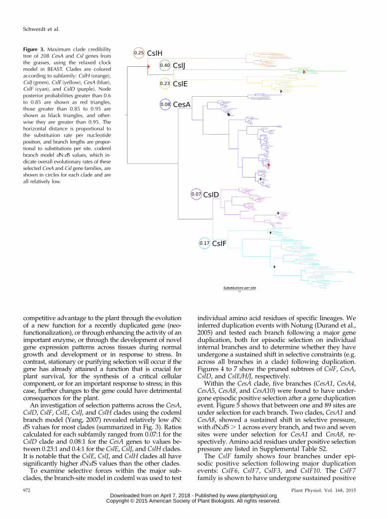
Figure 3 shows the Bayesian (BEAST; Drummondet al., 2012) phylogeny reconstructed using 208 PF03552cellulose synthase PFAM domains from fully se-quenced grasses. PFAM is a database of protein func-tional regions storing profile hidden Markov modelsbuilt from a large sequence database. As such, it pre-sents a comprehensive source of homologous, func-tionally informative amino acids for analysis. PF03552encompasses CesA, CslB, CslD, CslE, CslF, CslG, CslH,and CslJ. The CslB and CslG groups are not includedin this analysis because they are not represented inthe grasses. The CslA and CslC families do not contain a
Plant Physiol. Vol. 168, 2015 969
Evolution of CesA and Csl Gene Families
www.plantphysiol.orgon April 7, 2018 - Published by Downloaded from Copyright © 2015 American Society of Plant Biologists. All rights reserved.
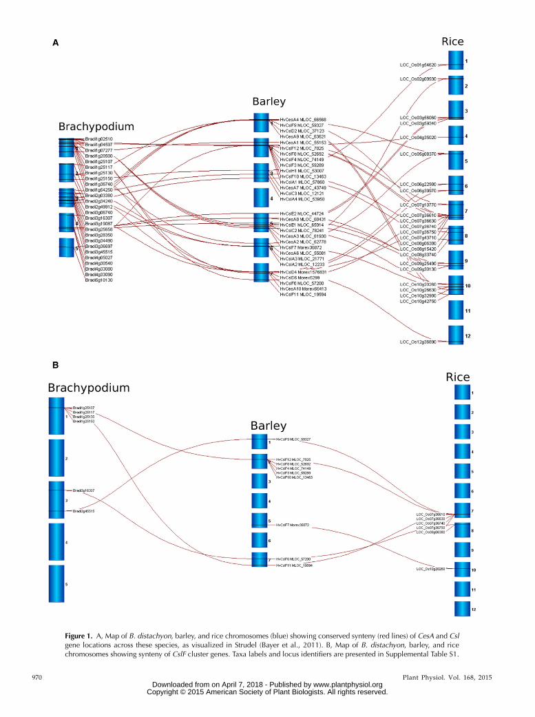
Figure 1. A, Map of B. distachyon, barley, and rice chromosomes (blue) showing conserved synteny (red lines) of CesA and Cslgene locations across these species, as visualized in Strudel (Bayer et al., 2011). B, Map of B. distachyon, barley, and ricechromosomes showing synteny of CslF cluster genes. Taxa labels and locus identifiers are presented in Supplemental Table S1.
970 Plant Physiol. Vol. 168, 2015 www.plantphysiol.orgon April 7, 2018 - Published by Downloaded from
Copyright © 2015 American Society of Plant Biologists. All rights reserved.

PF03552 domain, which is consistent with earlier sug-gestions that the CslA and CslC families evolved from aseparate cyanobacterial endosymbiotic event (Yin et al.,2009), and have also been excluded. Two alternativemolecular clock models were tested: a strict clock thatassumes that nucleotide substitutions accumulate at anapproximately constant rate across all branches ina phylogeny, and a relaxed uncorrelated log-normalclock, which allows for differences in substitutionrates among and along branches (Drummond et al.,2012). In biological terms, the strict clock assumes thatthe substitution rate in a particular phylogeny is con-stant with time. In contrast, the relaxed molecularclock model allows the inclusion in the analysis ofdifferences in substitution rates that might arise due tovariation in selection pressures, gene duplications, etc.Here, the models were compared by running the anal-yses for 2 3 108 generations and partitioning the align-ment by the three codon positions.Clock model comparison using Bayes factors in Tracer
version 1.6 (Rambaut and Drummond, 2007) showedthe relaxed clock to provide a better fit to the data thanthe strict clock (21,048 Log likelihood). The relaxed clockmodel yielded a tree with highly unequal branch lengthsamong terminal lineages. To assess the overall rate het-erogeneity, we inspected the coefficient of variation us-ing Tracer version 1.6. A coefficient of variation closer to0 indicates that the data are clock like, whereas a valuecloser to 1 indicates extremely high rate heterogeneity.Our tree had a coefficient of 0.542 (95% highest posteriordensity, 0.483–0.604), which suggests a moderately highlevel of heterogeneity in molecular branch rates.The maximum clade credibility tree topology is con-
sistent with previous studies (Richmond and Somerville,2000; Burton et al., 2006), supporting two major mono-phyletic groups. One group includes CesA, CslD, andCslF, while the other includes CslE, CslH, and CslJ. Withinthe CesA clade, the previously undocumented CesA10is sister to all other CesAs. The second branching CesAgroup includes HvCesA4, HvCesA8, OsCesA7, and
OsCesA9. A clade of HvCesA5 and OsCesA4 split next,followed by two monophyletic groups: (1) HvCesA3,OsCesA2, HvCesA6/9, and OsCesA1; and (2) HvCesA2,OsCesA3, OsCesA5, and OsCesA6. The CslD group hasthe HvCslD4 and OsCslD4 clade branching first and amonophyletic group including HvCslD1, HvCslD6,OsCslD3, and OsCslD5 sister to HvCslD2, HvCslD3,OsCslD1, and OsCslD2. The CslFs are sister to the CslDs.The CslF6 gene branches first, followed by CslF7 andthen CslF4, and finally two monophyletic clades, CslF9and CslF10. Two CslH clades branch before a single CslJclade that is sister to two CslE clades (Fig. 3).
The maximum likelihood tree reconstructed in RAxML(Stamatakis, 2006) showed two minor topological dif-ferences from the BEAST tree, such that CslF4 is resolvedas sister to CslF8/CslF9 and CesA4 as sister to CesA5/CesA1/CesA2/CesA3/CesA6. However, these differenceswere poorly supported in the maximum likelihood tree,with bootstrap proportions of less than 60%. They areindicated in Supplemental Figure S1.
Selection Pressure
Nucleotide substitutions are divided into non-synonymous substitutions (N), which result in changes ofamino acid residues in the encoded proteins, and syn-onymous substitutions (S), which do not cause changes inamino acid residues. Thus, positive selection can beconsidered to have occurred if the dN:dS is greater than 1,where dN is the rate of nonsynonymous nucleotide sub-stitution and dS is the rate of synonymous nucleotidesubstitution, while stationary (stabilizing) or purifying(negative) selection is indicated by dN:dS values thatapproach 0 (Yang and Bielawski, 2000). By way of ex-ample, positive selection might occur if a gene is involvedin such functions as the plant-pathogen competition,where it is important that the plant genes evolve newforms to counter and keep up with new forms of fungalgenes. Similarly, positive selection may occur if there is a
Figure 2. Structure of the conserved CslF gene cluster in the grasses. These clusters are conserved in syntenic regions of grassgenomes (Fig. 1B) but include variable numbers of genes in different orientations. Chr indicates the chromosome number for theparticular species. In some cases, relatively recent duplications are evident, through much higher values for sequence identity.The orientations of the genes are indicated by the arrows, and recently duplicated genes (sequence identity of about 90% ormore) are shown in the same color in a particular species. No pseudogenes were detected in the clusters.
Plant Physiol. Vol. 168, 2015 971
Evolution of CesA and Csl Gene Families
www.plantphysiol.orgon April 7, 2018 - Published by Downloaded from Copyright © 2015 American Society of Plant Biologists. All rights reserved.
competitive advantage to the plant through the evolutionof a new function for a recently duplicated gene (neo-functionalization), or through enhancing the activity of animportant enzyme, or through the development of novelgene expression patterns across tissues during normalgrowth and development or in response to stress. Incontrast, stationary or purifying selection will occur if thegene has already attained a function that is crucial forplant survival, for the synthesis of a critical cellularcomponent, or for an important response to stress; in thiscase, further changes to the gene could have detrimentalconsequences for the plant.
An investigation of selection patterns across the CesA,CslD, CslF, CslE, CslJ, and CslH clades using the codemlbranch model (Yang, 2007) revealed relatively low dN:dS values for most clades (summarized in Fig. 3). Ratioscalculated for each subfamily ranged from 0.07:1 for theCslD clade and 0.08:1 for the CesA genes to values be-tween 0.23:1 and 0.4:1 for the CslE, CslJ, and CslH clades.It is notable that the CslE, CslJ, and CslH clades all havesignificantly higher dN:dS values than the other clades.
To examine selective forces within the major sub-clades, the branch-site model in codeml was used to test
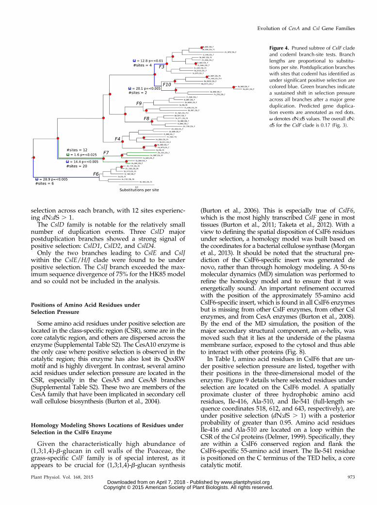
individual amino acid residues of specific lineages. Weinferred duplication events with Notung (Durand et al.,2005) and tested each branch following a major geneduplication, both for episodic selection on individualinternal branches and to determine whether they haveundergone a sustained shift in selective constraints (e.g.across all branches in a clade) following duplication.Figures 4 to 7 show the pruned subtrees of CslF, CesA,CslD, and CslE/H/J, respectively.
Within the CesA clade, five branches (CesA1, CesA4,CesA5, CesA8, and CesA10) were found to have under-gone episodic positive selection after a gene duplicationevent. Figure 5 shows that between one and 89 sites areunder selection for each branch. Two clades, CesA1 andCesA8, showed a sustained shift in selective pressure,with dN:dS. 1 across every branch, and two and sevensites were under selection for CesA1 and CesA8, re-spectively. Amino acid residues under positive selectionpressure are listed in Supplemental Table S2.
The CslF family shows four branches under epi-sodic positive selection following major duplicationevents: CslF6, CslF7, CslF3, and CslF10. The CslF7family is shown to have undergone sustained positive
Figure 3. Maximum clade credibilitytree of 208 CesA and Csl genes fromthe grasses, using the relaxed clockmodel in BEAST. Clades are coloredaccording to subfamily: CslH (orange),CslJ (green), CslE (yellow), CesA (blue),CslF (cyan), and CslD (purple). Nodeposterior probabilities greater than 0.6to 0.85 are shown as red triangles,those greater than 0.85 to 0.95 areshown as black triangles, and other-wise they are greater than 0.95. Thehorizontal distance is proportional tothe substitution rate per nucleotideposition, and branch lengths are propor-tional to substitutions per site. codemlbranch model dN:dS values, which in-dicate overall evolutionary rates of theseselected CesA and Csl gene families, areshown in circles for each clade and areall relatively low.
972 Plant Physiol. Vol. 168, 2015
Schwerdt et al.
www.plantphysiol.orgon April 7, 2018 - Published by Downloaded from Copyright © 2015 American Society of Plant Biologists. All rights reserved.
selection across each branch, with 12 sites experienc-ing dN:dS . 1.The CslD family is notable for the relatively small
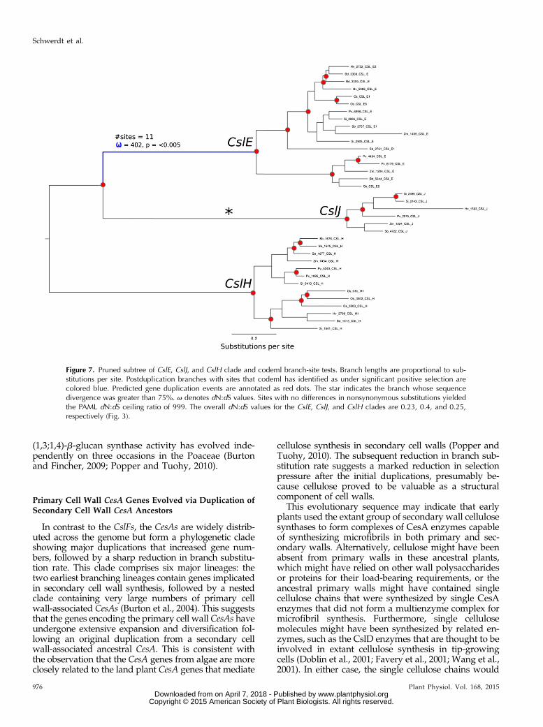
number of duplication events. Three CslD majorpostduplication branches showed a strong signal ofpositive selection: CslD1, CslD2, and CslD4.Only the two branches leading to CslE and CslJ
within the CslE/H/J clade were found to be underpositive selection. The CslJ branch exceeded the max-imum sequence divergence of 75% for the HK85 modeland so could not be included in the analysis.
Positions of Amino Acid Residues underSelection Pressure
Some amino acid residues under positive selection arelocated in the class-specific region (CSR), some are in thecore catalytic region, and others are dispersed across theenzyme (Supplemental Table S2). The CesA10 enzyme isthe only case where positive selection is observed in thecatalytic region; this enzyme has also lost its QxxRWmotif and is highly divergent. In contrast, several aminoacid residues under selection pressure are located in theCSR, especially in the CesA5 and CesA8 branches(Supplemental Table S2). These two are members of theCesA family that have been implicated in secondary cellwall cellulose biosynthesis (Burton et al., 2004).
Homology Modeling Shows Locations of Residues underSelection in the CslF6 Enzyme
Given the characteristically high abundance of(1,3;1,4)-b-glucan in cell walls of the Poaceae, thegrass-specific CslF family is of special interest, as itappears to be crucial for (1,3;1,4)-b-glucan synthesis
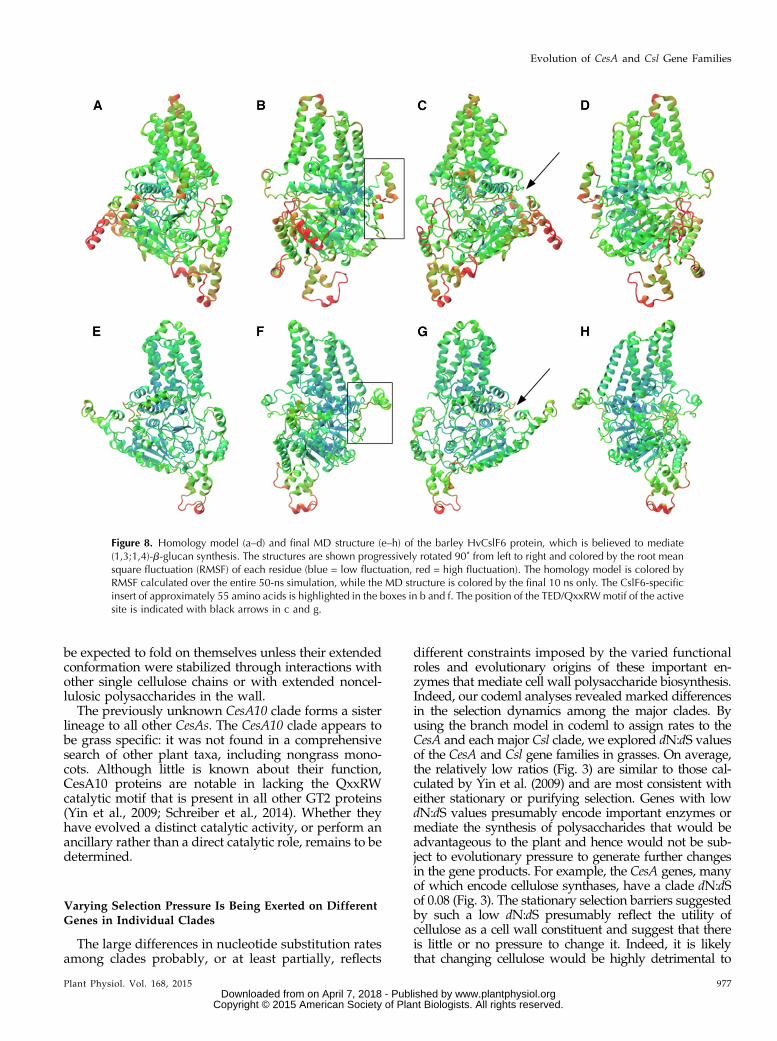
(Burton et al., 2006). This is especially true of CslF6,which is the most highly transcribed CslF gene in mosttissues (Burton et al., 2011; Taketa et al., 2012). With aview to defining the spatial disposition of CslF6 residuesunder selection, a homology model was built based onthe coordinates for a bacterial cellulose synthase (Morganet al., 2013). It should be noted that the structural pre-diction of the CslF6-specific insert was generated denovo, rather than through homology modeling. A 50-nsmolecular dynamics (MD) simulation was performed torefine the homology model and to ensure that it wasenergetically sound. An important refinement occurredwith the position of the approximately 55-amino acidCslF6-specific insert, which is found in all CslF6 enzymesbut is missing from other CslF enzymes, from other Cslenzymes, and from CesA enzymes (Burton et al., 2008).By the end of the MD simulation, the position of themajor secondary structural component, an a-helix, wasmoved such that it lies at the underside of the plasmamembrane surface, exposed to the cytosol and thus ableto interact with other proteins (Fig. 8).
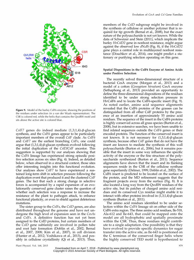
In Table I, amino acid residues in CslF6 that are un-der positive selection pressure are listed, together withtheir positions in the three-dimensional model of theenzyme. Figure 9 details where selected residues underselection are located on the CslF6 model. A spatiallyproximate cluster of three hydrophobic amino acidresidues, Ile-416, Ala-510, and Ile-541 (full-length se-quence coordinates 518, 612, and 643, respectively), areunder positive selection (dN:dS . 1) with a posteriorprobability of greater than 0.95. Amino acid residuesIle-416 and Ala-510 are located on a loop within theCSR of the Csl proteins (Delmer, 1999). Specifically, theyare within a CslF6 conserved region and flank theCslF6-specific 55-amino acid insert. The Ile-541 residueis positioned on the C terminus of the TED helix, a corecatalytic motif.
Figure 4. Pruned subtree of CslF cladeand codeml branch-site tests. Branchlengths are proportional to substitu-tions per site. Postduplication brancheswith sites that codeml has identified asunder significant positive selection arecolored blue. Green branches indicatea sustained shift in selection pressureacross all branches after a major geneduplication. Predicted gene duplica-tion events are annotated as red dots.v denotes dN:dS values. The overall dN:dS for the CslF clade is 0.17 (Fig. 3).
Plant Physiol. Vol. 168, 2015 973
Evolution of CesA and Csl Gene Families
www.plantphysiol.orgon April 7, 2018 - Published by Downloaded from Copyright © 2015 American Society of Plant Biologists. All rights reserved.

DISCUSSION
A Cluster of CslF Genes Is Conserved in the Grasses
In Figure 1, the distribution of the approximately50 CesA and Csl genes across the genomes of barley,B. distachyon, and rice is compared. Generally, the genesare scattered across all chromosomes, but in one case,there is evidence for clustering of genes. Specifically, asingle cluster of CslF genes is observed in conservedsyntenic regions (Moore et al., 1995) of all the grassesexamined so far (Fig. 2). Within the cluster, individualCslF genes exhibit 65% to 75% sequence identity at thenucleotide level and form a well-supported clade on thetree, which suggests that the cluster itself has beenconserved over a considerable time period while geneswithin it have been free to evolve. Indeed, because thiscluster has been observed in all fully sequenced grasses,it possibly originated when the Poaceae split from theother Poales. Phylogenetic reconstruction of genes in thecluster (Fig. 2) showed that the clusters occasionallycontain highly similar (90% sequence similarity) paral-ogous pairs, such as the rice OsCslF1 and OsCslF2 genesand the sorghum SbCslF3 and SbCslF13 genes. Given thelikely recent origins of these paralogs, they are probablyexplained by unequal crossing over during meiosisrather than by recombination. Comparison of the syn-teny map and phylogeny for genes such as HvCslF9 and
BdCslF9 shows that, although they form a monophyleticclade with other CslFs, they are located elsewhere on thegrass genomes, indicating that these may have escapedfrom the cluster through recognizable recombinationand genome duplication events (Moore et al., 1995).
The CslF6 gene is strongly recovered as the earliestbranching lineage within the CslFs, followed by CslF7and the cluster CslFs. Allowing for local reorganizationsin synteny within the CslF cluster, the phylogeny sug-gests that a duplication event involving their commonancestor with CslF6 and/or CslF7 led to the CslF cluster.The fact that the branch leading to the cluster of CslFs hasthe highest amount of substitution of all branches in thephylogeny indicates a rapid and dynamic period of di-versification following duplication. Thus, the CslF geneclusters appear to be taxonomically conserved within thegrasses, but they are nevertheless dynamic insofar asrelatively recent expansions and contractions of clustersize can be detected. Therefore, one could conclude thatthere is some selection pressure on maintaining the CslFgenes in clusters, but genes in the cluster are transcribedat relatively low levels in most tissues during normalgrowth and development (Burton et al., 2008), and pos-sible functional advantages of keeping the CslF genes inclusters are not yet demonstrated. One possibility is thatthe conservation of the cluster might be attributable toselection pressure on the grasses to rapidly synthesize
Figure 5. Pruned subtree of CesA clade and codeml branch-site tests. Branch lengths are proportional to substitutions per site.Postduplication branches with sites that codeml has identified as under significant positive selection are colored blue. Greenbranches indicate a sustained shift in selection pressure across all branches after a major gene duplication. Predicted geneduplication events are annotated as red dots. v denotes dN:dS values. Sites with no differences in nonsynonymous substitutionsyielded the PAML dN:dS ceiling ratio of 999. The overall dN:dS for the CesA clade is 0.08 (Fig. 3).
974 Plant Physiol. Vol. 168, 2015
Schwerdt et al.
www.plantphysiol.orgon April 7, 2018 - Published by Downloaded from Copyright © 2015 American Society of Plant Biologists. All rights reserved.
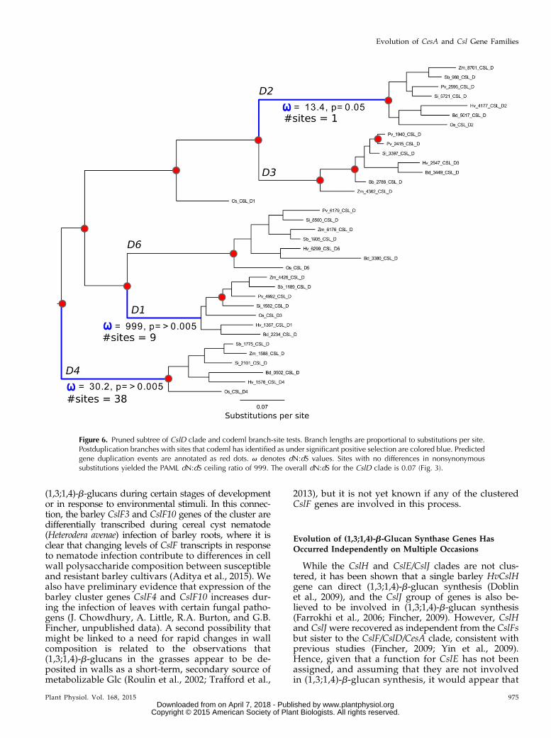
(1,3;1,4)-b-glucans during certain stages of developmentor in response to environmental stimuli. In this connec-tion, the barley CslF3 and CslF10 genes of the cluster aredifferentially transcribed during cereal cyst nematode(Heterodera avenae) infection of barley roots, where it isclear that changing levels of CslF transcripts in responseto nematode infection contribute to differences in cellwall polysaccharide composition between susceptibleand resistant barley cultivars (Aditya et al., 2015). Wealso have preliminary evidence that expression of thebarley cluster genes CslF4 and CslF10 increases dur-ing the infection of leaves with certain fungal patho-gens (J. Chowdhury, A. Little, R.A. Burton, and G.B.Fincher, unpublished data). A second possibility thatmight be linked to a need for rapid changes in wallcomposition is related to the observations that(1,3;1,4)-b-glucans in the grasses appear to be de-posited in walls as a short-term, secondary source ofmetabolizable Glc (Roulin et al., 2002; Trafford et al.,
2013), but it is not yet known if any of the clusteredCslF genes are involved in this process.
Evolution of (1,3;1,4)-b-Glucan Synthase Genes HasOccurred Independently on Multiple Occasions
While the CslH and CslE/CslJ clades are not clus-tered, it has been shown that a single barley HvCslHgene can direct (1,3;1,4)-b-glucan synthesis (Doblinet al., 2009), and the CslJ group of genes is also be-lieved to be involved in (1,3;1,4)-b-glucan synthesis(Farrokhi et al., 2006; Fincher, 2009). However, CslHand CslJ were recovered as independent from the CslFsbut sister to the CslF/CslD/CesA clade, consistent withprevious studies (Fincher, 2009; Yin et al., 2009).Hence, given that a function for CslE has not beenassigned, and assuming that they are not involvedin (1,3;1,4)-b-glucan synthesis, it would appear that
Figure 6. Pruned subtree of CslD clade and codeml branch-site tests. Branch lengths are proportional to substitutions per site.Postduplication branches with sites that codeml has identified as under significant positive selection are colored blue. Predictedgene duplication events are annotated as red dots. v denotes dN:dS values. Sites with no differences in nonsynonymoussubstitutions yielded the PAML dN:dS ceiling ratio of 999. The overall dN:dS for the CslD clade is 0.07 (Fig. 3).
Plant Physiol. Vol. 168, 2015 975
Evolution of CesA and Csl Gene Families
www.plantphysiol.orgon April 7, 2018 - Published by Downloaded from Copyright © 2015 American Society of Plant Biologists. All rights reserved.
(1,3;1,4)-b-glucan synthase activity has evolved inde-pendently on three occasions in the Poaceae (Burtonand Fincher, 2009; Popper and Tuohy, 2010).
Primary Cell Wall CesA Genes Evolved via Duplication ofSecondary Cell Wall CesA Ancestors
In contrast to the CslFs, the CesAs are widely distrib-uted across the genome but form a phylogenetic cladeshowing major duplications that increased gene num-bers, followed by a sharp reduction in branch substitu-tion rate. This clade comprises six major lineages: thetwo earliest branching lineages contain genes implicatedin secondary cell wall synthesis, followed by a nestedclade containing very large numbers of primary cellwall-associated CesAs (Burton et al., 2004). This suggeststhat the genes encoding the primary cell wall CesAs haveundergone extensive expansion and diversification fol-lowing an original duplication from a secondary cellwall-associated ancestral CesA. This is consistent withthe observation that the CesA genes from algae are moreclosely related to the land plant CesA genes that mediate
cellulose synthesis in secondary cell walls (Popper andTuohy, 2010). The subsequent reduction in branch sub-stitution rate suggests a marked reduction in selectionpressure after the initial duplications, presumably be-cause cellulose proved to be valuable as a structuralcomponent of cell walls.
This evolutionary sequence may indicate that earlyplants used the extant group of secondary wall cellulosesynthases to form complexes of CesA enzymes capableof synthesizing microfibrils in both primary and sec-ondary walls. Alternatively, cellulose might have beenabsent from primary walls in these ancestral plants,which might have relied on other wall polysaccharidesor proteins for their load-bearing requirements, or theancestral primary walls might have contained singlecellulose chains that were synthesized by single CesAenzymes that did not form a multienzyme complex formicrofibril synthesis. Furthermore, single cellulosemolecules might have been synthesized by related en-zymes, such as the CslD enzymes that are thought to beinvolved in extant cellulose synthesis in tip-growingcells (Doblin et al., 2001; Favery et al., 2001; Wang et al.,2001). In either case, the single cellulose chains would
Figure 7. Pruned subtree of CslE, CslJ, and CslH clade and codeml branch-site tests. Branch lengths are proportional to sub-stitutions per site. Postduplication branches with sites that codeml has identified as under significant positive selection arecolored blue. Predicted gene duplication events are annotated as red dots. The star indicates the branch whose sequencedivergence was greater than 75%. v denotes dN:dS values. Sites with no differences in nonsynonymous substitutions yieldedthe PAML dN:dS ceiling ratio of 999. The overall dN:dS values for the CslE, CslJ, and CslH clades are 0.23, 0.4, and 0.25,respectively (Fig. 3).
976 Plant Physiol. Vol. 168, 2015
Schwerdt et al.
www.plantphysiol.orgon April 7, 2018 - Published by Downloaded from Copyright © 2015 American Society of Plant Biologists. All rights reserved.
be expected to fold on themselves unless their extendedconformation were stabilized through interactions withother single cellulose chains or with extended noncel-lulosic polysaccharides in the wall.The previously unknown CesA10 clade forms a sister
lineage to all other CesAs. The CesA10 clade appears tobe grass specific: it was not found in a comprehensivesearch of other plant taxa, including nongrass mono-cots. Although little is known about their function,CesA10 proteins are notable in lacking the QxxRWcatalytic motif that is present in all other GT2 proteins(Yin et al., 2009; Schreiber et al., 2014). Whether theyhave evolved a distinct catalytic activity, or perform anancillary rather than a direct catalytic role, remains to bedetermined.
Varying Selection Pressure Is Being Exerted on DifferentGenes in Individual Clades
The large differences in nucleotide substitution ratesamong clades probably, or at least partially, reflects
different constraints imposed by the varied functionalroles and evolutionary origins of these important en-zymes that mediate cell wall polysaccharide biosynthesis.Indeed, our codeml analyses revealed marked differencesin the selection dynamics among the major clades. Byusing the branch model in codeml to assign rates to theCesA and each major Csl clade, we explored dN:dS valuesof the CesA and Csl gene families in grasses. On average,the relatively low ratios (Fig. 3) are similar to those cal-culated by Yin et al. (2009) and are most consistent witheither stationary or purifying selection. Genes with lowdN:dS values presumably encode important enzymes ormediate the synthesis of polysaccharides that would beadvantageous to the plant and hence would not be sub-ject to evolutionary pressure to generate further changesin the gene products. For example, the CesA genes, manyof which encode cellulose synthases, have a clade dN:dSof 0.08 (Fig. 3). The stationary selection barriers suggestedby such a low dN:dS presumably reflect the utility ofcellulose as a cell wall constituent and suggest that thereis little or no pressure to change it. Indeed, it is likelythat changing cellulose would be highly detrimental to
Figure 8. Homology model (a–d) and final MD structure (e–h) of the barley HvCslF6 protein, which is believed to mediate(1,3;1,4)-b-glucan synthesis. The structures are shown progressively rotated 90˚ from left to right and colored by the root meansquare fluctuation (RMSF) of each residue (blue = low fluctuation, red = high fluctuation). The homology model is colored byRMSF calculated over the entire 50-ns simulation, while the MD structure is colored by the final 10 ns only. The CslF6-specificinsert of approximately 55 amino acids is highlighted in the boxes in b and f. The position of the TED/QxxRWmotif of the activesite is indicated with black arrows in c and g.
Plant Physiol. Vol. 168, 2015 977
Evolution of CesA and Csl Gene Families
www.plantphysiol.orgon April 7, 2018 - Published by Downloaded from Copyright © 2015 American Society of Plant Biologists. All rights reserved.
survival and that there would be pressure to conserve itin its present form.
However, the low overall dN:dS values for the variousclades conceal considerable variability for individual lin-eages within those clades. Figure 5 highlights the selectiveforces experienced by the CesA family throughout itsevolution, where there is evidence for rapid CesA geneduplication and diversification after the ancestral geneacquired its current enzymatic function. The branch-sitemodel implemented in codeml was used to explore theevolutionary dynamics of lineages, specifically testing foran episodic burst of selection or sustained but moregradual shifts in selective pressure. As indicated in Figure5, five CesA lineages have undergone strong episodicpositive selection, with one to 89 sites having a posteriorprobability of greater than 0.95, following major geneduplication events. In such cases, it is not known whetherselection was driving the evolution of a novel function orfixing a polymorphism or another adaptive mechanism(Innan and Kondrashov, 2010). In land plants, CesA en-zymes have been observed to form a six-subunit rosettestructure spanning the plasma membrane called the ter-minal complex, which may be associated with microtu-bules (Doblin et al., 2002; Paredez et al., 2006). So nameddue to their position at the end of the cellulose microfibril,terminal complexes are thought to play a critical role inthe pattern of cell expansion (Green, 1962). One possi-bility, given the rapid CesA diversification, is that theadaptive advantage of the CesA terminal complex was solarge that positive selection has been driven by the
structural maintenance of its constituent members. In-deed, our finding that the primary cell wall-associatedCesA1 and the secondary cell wall-associated CesA8clades have experienced a sustained shift in selectionperhaps indicates similar evolutionary pressures on dif-ferent complexes.
The CslE, CslJ, and CslH clade comprises a muchsmaller radiation of genes compared with the CesAs.They also have numerous postspeciation duplications(Yin et al., 2009), the highest nucleotide substitutionrates, and the highest subfamily dN:dS values. Strongselection leading to the CslE clade could indicate suchevolutionary mechanisms as modified duplication orneofunctionalization, and perhaps their relatively highsubstitution rates in comparison with the CesAs indicatethat less of the protein is under selection, with more ofthe gene allowed to accumulate neutral mutations. Sucha hypothesis might suggest that the proteins encoded byCslH, CslE, and CslJ are not structurally constrained likethe CesAs. That the CslJ genes have such a long branchfollowing their split from CslE is curious, given the evi-dence for its involvement in (1,3;1,4)-b-glucan synthesis(Farrokhi et al., 2006; Fincher, 2009). Thus, a more de-tailed study of the evolutionary dynamics in this specificgroup might become a priority for future study.
Although it is widely assumed that the CslF genes inthe grasses are involved in (1,3;1,4)-b-glucan synthesis,not all the genes in the CslF clade have yet been shownto direct (1,3;1,4)-b-glucan synthesis in heterologous ortransgenic systems (Burton et al., 2006). The CslF6 and
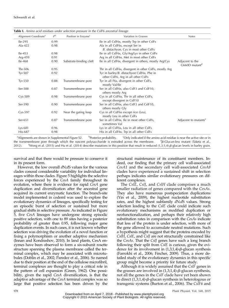
Table I. Amino acid residues under selection pressure in the CslF6 ancestral lineage
Alignment Coordinatea P b Position in Enzymec Variation in Grasses Notes
Ile-295 0.99 Ile in all CslF6s, mostly Trp in other CslFsAla-422 0.98 Ala in all CslF6s, except Ser in
B. distachyon, Cys in most other CslFsIle-453 0.98 Ile in all CslF6s, Gly/Arg/Lys in other CslFsArg-459 0.91 Arg in all CslF6s, Met in most other CslFsIle-468 0.90 Substrate-binding cleft Ile in all CslF6s, divergent in others, mostly Arg/Cys Adjacent to the
G660D mutantd
Thr-506 0.95 Thr in all CslF6s, divergent in other CslFs, mostly ArgTyr-507 0.92 Tyr in barley/B. distachyon CslF6s, Phe in
other CslF6, Arg in all other CslFsTyr-550 0.88 Transmembrane pore Tyr in all F6s, divergent in other CslFs,
mostly Val/IleSer-588 0.87 Transmembrane pore Ser in all CslF6s, also CslF3 and CslF10,
others mostly ArgCys-589 0.98 Transmembrane pore Cys in all CslF6s, Thr in all other CslFs,
except divergent in CslF10Ser-590 0.90 Transmembrane pore Ser in all CslF6s, also CslF3 and CslF10,
others mostly GlyCys-597 0.92 Near the gating loop Cys in all CslF6s except rice (Leu),
mostly Leu in other CslFsSer-651 0.87 Transmembrane pore Ser in all CslF6s, Ile in most other CslFs,
sometimes ValAdjacent to mutantse
Lys-681 0.96 Lys in all CslF6s, Leu in all other CslFsHis-687 0.98 His in all CslF6s, Trp in all other CslFs
aAlignments are shown in Supplemental Figure S2. bPosterior probability. cOnly indicated if the amino acid residue is near the active site or inthe transmembrane pore through which the nascent polysaccharide is extruded across the membrane. db-Glucan-less mutant (Taketa et al.,2012). eWong et al. (2015) and Hu et al. (2014) describe mutations in this position that result in reduced (1,3;1,4)-b-glucan levels in barley grain.
978 Plant Physiol. Vol. 168, 2015
Schwerdt et al.
www.plantphysiol.orgon April 7, 2018 - Published by Downloaded from Copyright © 2015 American Society of Plant Biologists. All rights reserved.
CslF7 genes do indeed mediate (1,3;1,4)-b-glucansynthesis, and the CslF6 genes appear to be particularlyimportant members of the overall CslF clade. As CslF6and CslF7 are the earliest branching CslFs, one couldargue that (1,3;1,4)-b-glucan synthesis evolved followingthe initial duplication of the CslD/CslF ancestor. Thissuggestion is supported by our analyses showing thatthe CslF6 lineage has experienced strong episodic posi-tive selection across six sites (Fig. 4). Indeed, as detailedbelow, when observed in a structural context, those sitesoffer interesting insights into this functional evolution.Our analyses show CslF7 to have experienced a sus-tained long-term shift in selection pressure following theduplication event that produced it and the clustered CslFgenes. The fact that such a strong change in selectiveforces is accompanied by a rapid expansion of an evo-lutionarily conserved gene cluster raises the question ofwhether such selection was a response to increase theresistance of the plant to pathogen attack, to enhancefunctional plasticity, or even to shield against deleteriousmutations.The sister group to the CslFs, the CslD genes, are also
distributed throughout the genome and have not un-dergone the high level of expansion seen in the CesAsand CslFs. A definitive function has not yet beenassigned to the CslD proteins, but certain members ofthe CslD gene subfamily are involved in pollen tubeand root hair formation (Doblin et al., 2002; Bernalet al., 2007, 2008; Kim et al., 2007), in cell division(Hunter et al., 2012; Yoshikawa et al., 2013), and pos-sibly in cellulose crystallinity (Qi et al., 2013). Thus,
members of the CslD subgroup might be involved inthe synthesis of cellulose or another polymer that is re-quired for tip growth (Bernal et al., 2008), but the exactnature of the polysaccharide is not yet known. While thedata of Schweizer and Stein (2011), which implicate thebarley HvCslD2 gene in nonhost resistance, might argueagainst the observed low dN:dS (Fig. 8), if the HvCslD2gene plays a central role in multilayered nonhost resis-tance (Douchkov et al., 2014), one might predict a sta-tionary or purifying selection operating on this gene.
Spatial Dispositions in the CslF6 Enzyme of Amino Acidsunder Positive Selection
The recently solved three-dimensional structure of abacterial CesA enzyme (Morgan et al., 2013) and amodel of a cotton (Gossypium hirsutum) CesA enzyme(Sethaphong et al., 2013) provided an opportunity todefine the three-dimensional dispositions of the residuesidentified to be under strong selection pressure inHvCslF6 and to locate the CslF6-specific insert (Fig. 9).As noted earlier, amino acid sequence alignmentsrevealed that the CslF6 proteins of the grasses can bedistinguished from all other CslF proteins by the pres-ence of an insertion of approximately 55 amino acidresidues. The sequence of the insert in the CslF6 proteinsis highly conserved across all grass species (Burton et al.,2008), but in extensive searches, we have been unable tofind related sequences outside the CslF6 genes or theirencoded proteins. The function of the conserved insert isnot known. It is not necessary for (1,3;1,4)-b-glucansynthesis, because other CslF genes that do not have theinsert are known to mediate the synthesis of this wallpolysaccharide (Burton et al., 2006), but it remains pos-sible that the insert in some way influences the specificactivity of the enzyme or the fine structure of the poly-saccharide synthesized (Burton et al., 2011). Sequencealignments have shown that the insert and its flankingsequences reside in the CSR of the cellulose synthasegene superfamily (Delmer, 1999; Doblin et al., 2002). TheCslF6 insert is predicted to be located on the surface ofthe protein, and the MD refinement suggests that thefragment projects away from the surface (Fig. 8). It isalso located a long way from the QxxRW residues of theactive site, but its patches of charged amino acid resi-dues and its conserved Cys residue might enable it tointeract with other proteins involved in (1,3;1,4)-b-glucansynthesis (Burton et al., 2011).
The amino acid residues identified to be under se-lection within the CslF6 lineage sit on either side of theCslF6 extra region. The three amino acid residues, Ile-518,Ala-612 and Ile-643, that could be mapped onto themodel are all hydrophobic and spatially proximatewithin the CSR. Thus, these residues might contrib-ute to a single adaptation. One possibility is that theyhave evolved to provide specific dynamics for sugartransfer into the active site, as Ile-643 is positioned onthe C terminus of the conserved finger helix wherethe highly conserved TED motif is hypothesized to
Figure 9. Model of the barley CslF6 enzyme, showing the positions ofthe residues under selection (in a van der Waals representation). TheCSR is colored red, while the helix that contains the QxxRW motif andsits above the active site is colored blue.
Plant Physiol. Vol. 168, 2015 979
Evolution of CesA and Csl Gene Families
www.plantphysiol.orgon April 7, 2018 - Published by Downloaded from Copyright © 2015 American Society of Plant Biologists. All rights reserved.
contact the acceptor glycosyl residue of the polysac-charide chain (Morgan et al., 2014).
Several of the residues under positive selection in theCslF6 enzyme are located near regions involved in ca-talysis and in the extrusion of the nascent polysaccha-ride chain across the membrane, through the enzyme’stransmembrane pore. Thus, Ile-468 and Cys-597 arewithin the substrate-binding cleft and near the gating loop,respectively (Table I), while Tyr-550, Ser-588, Cys-589,Ser-590, and Ser-651 are located immediately adjacentto or actually within the transmembrane pore of theenzyme (Table I). The Ile-468 residue is immediately adja-cent to a mutated Gly residue that results in a b-glucan-less mutant (Taketa et al., 2012). Similarly, mutation ofthe Ser-651 residue results in barley grain with greatlyreduced levels of (1,3;1,4)-b-glucan (Hu et al., 2014;Wong et al., 2015; Table I). These examples go part of theway toward the validation of the selection pressureanalyses, but it will now be possible to investigate therelative importance of the residues under positive selec-tion pressure with respect to their influence on enzymeactivity and on the fine structure of the polysaccharideproduct of enzymic action.
Evolutionary Data Raise Questions of Extant Functions
In any case, the analyses described here have raised anumber of central biological questions. First, did cellu-lose microfibrils of the type found in land plants firstappear in secondary cell walls and only later in primarywalls? Second, what adaptive advantage might be re-sponsible for the conserved clustering of CslF genesacross grass genomes? Currently, we are investigatingindications that clustering might be advantageous forthe rapid expression of multiple genes in response topathogen attack or for the short-term storage of excessphotosynthate. The third question is related to the clearimportance of the CslF6 enzyme in (1,3;1,4)-b-glucansynthesis. Did an ancient duplication event involvingthe immediate ancestor of the CslF6/F7 gene add acrucial protein fragment that greatly improved thefunction of the CslF6 enzyme? Also embodied in thisquestion is the role and origin of the CslF6-specificinsert of about 55 amino acids that appears to be animportant determinant of the fine structure of the(1,3;1,4)-b-glucan synthesized by the CslF6 enzyme. Thephylogenetic hypotheses generated here, in combina-tion with the three-dimensional model of the CslF6enzyme, will now be used to guide domain and singleamino acid swaps within the CslF enzymes, with a viewto defining the evolution and mechanisms of enzymicaction and substrate specificity at the molecular andthree-dimensional structural levels. Finally, the dataraise the question of why some CslF genes are underpositive selection pressure while other members of thesame clade are apparently constrained by selectionbarriers. Here, the effects of abiotic and biotic stress onthe transcription of CslF genes that show high rates ofnucleotide substitution and appear to be under positive
selection pressure will be defined, and we will investi-gate possible links between the selective conservation ofthe CslF gene cluster and selection pressure on indi-vidual CslF genes within the cluster.
MATERIALS AND METHODS
Multiple Sequence Alignment
Cellulose synthase superfamily sequences from rice (Oryza sativa), sorghum(Sorghum bicolor), Brachypodium distachyon, Setaria italica, maize (Zea mays), andPannicum virgatum were retrieved from Phytozome (Goodstein et al., 2012)using BioMart with queries limited to the PF03552 PFAM and verified usingBLAST (Altschul et al., 1990). Barley (Hordeum vulgare) sequence data weresourced from the Morex assembly (Mayer et al., 2012). Candidate sequencegene models were assessed for accuracy with an FGENESH+ (Solovyev, 2002)perl pipeline using a local database of well-characterized PF03552 sequencesas templates. The hmmalign program within the HMMER (Finn et al., 2011)package was used to assign the residues to the PF03552 PFAM hidden Markovmodel profile. Codon sequences were mapped to the protein alignment usingPAL2NAL (Suyama et al., 2006), and sites with a posterior probability of less than0.6 were manually removed from the alignment using Jalview (Waterhouse et al.,2009).
Phylogenetic Analyses
Phylogenies for the CesA and Csl superfamily were reconstructed using theBayesian MCMC package BEAST version 1.8.0 (Drummond et al., 2012). Inputalignments were partitioned into the three separate codon positions withunlinked substitution models that included rate heterogeneity parameters,stationary base frequencies, and transitions/transversion frequencies. Eachanalysis was run with a relaxed clock (log-normal distribution with uncorre-lated branch rate variation) and repeated with a strict clock prior. Bayes fac-tors calculated in TRACER version 1.5 (Rambaut and Drummond, 2007) wereused to test whether the relaxed clock provided a better fit to the data than thestrict clock. The GTR+I+G substitution model, as selected by jModelTest(Posada, 2008), and a Yule tree prior were used for all analyses. Convergencewas monitored in TRACER version 1.5 by assessing the effective sample sizevalues, trace plots, and posterior probabilities of the estimated parameters.Each analysis was run for at least 200,000,000 generations, or until effectivesample size values were over 200, sampling every 1,000 states.
Maximum likelihood phylogenies were also reconstructed. RAxML(Stamatakis, 2006) was run using the GTRGAMMA substitution model on acodon position partitioned data set with 1,000 bootstraps. Putative duplicationevents were identified by reconciling a grass species tree to the CesA/Csl gene treeusing Notung (Durand et al., 2005).
Taxa labels of phylogenetic trees and their associated locus identifiers areshown in Supplemental Table S1.
dN:dS Estimation
The codeml program of the PAML 4.7 (Yang, 2007) package and a spe-cifically optimized version of codeml, slimcodeml (Schabauer et al., 2012),were used to estimate dN:dS. codeml and slimcodeml were used with thebranch and branch-site models, respectively.
To explore how selection has operated on each of the majorCesA/Csl clades,we used a branch model (model = 2, NSsites = 0) to estimate dN:dS values forCesA, CslD, CslE, CslF, CslH, and CslJ. The branches of each major clade wereset to foreground, with the remaining branches of the full gene tree assigned tothe background and dN:dS values estimated using fix_omega = 0, omega = 1 (re-peating with omega = 2 and omega = 4).
Additionally, a branch model was used to estimate rates for the CslE/CslH/CslJ and CesA/CslD/CslF clades separately to compare levels of selectionpressure in these groups.
To test whether the major ancestral gene duplications (red nodes in Figs. 4—7)were followed by a strong shift in selective constraints, we set these nodes as theforeground branches in a branch model analysis (model = 2, NSsites = 0), with allremaining branches in the full gene tree set as background.
To determine which amino acid sites have had a shift in selective pressuresthroughout the evolution of the family, we conducted x branch-site tests.
980 Plant Physiol. Vol. 168, 2015
Schwerdt et al.
www.plantphysiol.orgon April 7, 2018 - Published by Downloaded from Copyright © 2015 American Society of Plant Biologists. All rights reserved.
Subtrees of the major CesA/Csl divisions were extracted using nw_utils (Junierand Zdobnov, 2010). Subsequent branch-site model analyses were performedwith the branches following ancestral gene duplications (and leading to themajor clades within the CesA/Csl subtrees) set as foreground, with theremaining subtree branches as background. The branch-site model (model = 2,NSsites = 2), with a dN:dS value allowed to vary (fix_omega = 0, omega = 1),was used to calculate the likelihood of positive selection at each site along thebranch. To explore the likelihood landscape, the initial dN:dS value was variedand all analyses were repeated (dN:dS = 2, 4, and 6).
Furthermore, to test whether postduplication selection represented a sus-tained shift in selective pressure or a burst of functional differentiation, werepeated the branch-site (model = 2, NSsites = 2) analyses including all branchesfollowing the duplication event within the foreground.
CslJ was problematic in that the genetic distance between the CslJ branchesand CslE and CslH is greater than 75%. This exceeded the limits of the NG86model used by codeml.
Amino Acid Site Mapping
The amino acid sites identified in the slimcodeml branch-site analyses weremapped onto the CslF6 homology model using Pymol (version 1.5.0.4,Schrödinger).
Homology Modeling and MD Simulations
The GT2 PFAMdomain (PF00535) and two transmembrane helices on eitherside of it were taken from the BcsA crystal structure (Morgan et al., 2013),manually aligned to homologous regions of the HvCSLF6 amino acid se-quence, and assessed using hydrophobic cluster analysis. The structures ofgap regions of greater than 11 amino acids were solved de novo using theI-TASSER server (Zhang, 2008; Roy et al., 2010) and RaptorX (CNFsearch,CNFalign; Källberg et al., 2012) and along with the BcsA structure used astemplates for modeler (Sali and Blundell, 1993). Candidate models wereassessed using internal DOPE functions of modeler, ProSA, and Procheck.Top-scoring models were taken through a loop-refining script for modeler,with a final model selected based on modeler DOPE, modeler GA32, ProSA,and Procheck outputs.
As noted above, the model was first generated including transmembranehelices TMH1 through TMH4 of CslF6, which were modeled corresponding toTMH3 to TMH6 of the bacterial RsBcsA (Morgan et al., 2013). Based onstructural and biochemical data from the bacterial enzyme, TMH5 of CslF6,which is predicted to form a transmembrane helix, more likely forms anamphipathic interface helix that runs along the cytoplasmic side of themembrane in juxtaposition with a conserved gating loop that is implicated insubstrate binding (Morgan et al., 2014). Thus, the CslF6 TMH5 was modeledas a cytosolic interface helix, corresponding to interface helix 3 of the bacterialRsBcsA structure (Morgan et al., 2014). TMH6 and TMH7 of CslF6 were againmodeled based on RsBcsA’s TMH7 and TMH8. The last C-terminal CslF6 helixis not constantly predicted to be a transmembrane helix by different predictionalgorithms and therefore was omitted from the final model.
The homology model of CslF6 was embedded in a preequilibrated POPEbilayer using the membrane plugin of VMD 1.9.1 (http://www.ks.uiuc.edu/research/vmd; Humphrey et al., 1996). Lipid and water molecules thatoverlapped the protein were removed, and the system was fully solvated in a116- 3 116- 3 140-Å box of transferable intermolecular potential with threepoints water. Na+ and Cl2 ions were added to neutralize the system and gavea final ionic concentration of 0.15 M, consistent with standard MD simulations(Hille, 2001).
MD simulations were performed using NAMD 2.9 (http://www.ks.uiuc.edu/Research/namd; Phillips et al., 2005) on 32 nodes of the IBM Blue Gene/Qsupercomputer (Avoca) at the Victorian Life Sciences Computation Initiative.A nonbonded cutoff of 12 Å was used with a smoothing function applied at 10Å. Pair lists were updated every 20 steps for van der Waals interactions andevery 40 steps for electrostatic interactions, at distances of 14 Å. These inter-actions were calculated every one and two steps, respectively, with the elec-trostatics calculated using the particle-mesh Ewald method (Darden et al.,1993). Charmm27 parameters with CMAP corrections (Mackerell et al., 2004)were used for all protein parameters, while lipid parameters were defined bythe lipid c36 parameters (Klauda et al., 2010). Periodic boundary conditionswere utilized with all atoms wrapped when exiting the periodic boundary. TheSHAKE algorithm was applied to restrain hydrogen-heavy atom-bonded dis-tances for water molecules in optimization simulations and to all hydrogen-
heavy atom bonds in the equilibration and production phase simulationsallowing for a 2-fs time step.
A four-step MD protocol was followed with temperature and pressuremaintained at 300 K and 1 atm, respectively, using Langevin dynamics andpiston (Martyna et al., 1994; Feller et al., 1995). The positions of lipid tails wereinitially optimized by performing a 0.1-ns constant pressure, constant tem-perature (NPT) simulation where all atoms except those in the lipid tails werefixed. A further 0.1-ns NPT simulation was performed to optimize the positionof all lipid and water atoms by placing 5 kcal mol21 Å22 restraints on proteinatoms. The entire system was equilibrated with a 10-ns NPT simulation afterall restraints had been removed. Finally, a 50-ns simulation was performedwith the x-y area of the system held at constant pressure, constant area, andconstant temperature.
RMSF values were calculated on a per residue basis, averaged over all atomsof each residue. All images from the MD simulations were created with VMD.
Supplemental Data
The following supplemental materials are available.
Supplemental Figure S1. Phylogeny of CesA and Csl genes in grasses.
Supplemental Figure S2. Sequence alignments.
Supplemental Table S1. Locus identifiers.
Supplemental Table S2. Amino acids under positive selection.
ACKNOWLEDGMENTS
We thank Natalie Kibble and Karen Chance for assistance in preparing thearticle.
Received January 29, 2015; accepted May 19, 2015; published May 21, 2015.
LITERATURE CITED
Aditya J, Lewis J, Shirley NJ, Tan H, Henderson M, Fincher GB, BurtonRA, Mather DE, Tucker MR (2015) Temporal differences during cerealcyst nematode infection in barley lead to specific changes in cell wallcomposition and transcript abundance. New Phytol 207: 135–147
Altschul SF, Gish W, Miller W, Myers EW, Lipman DJ (1990) Basic localalignment search tool. J Mol Biol 215: 403–410
Arioli T, Peng L, Betzner AS, Burn J, Wittke W, Herth W, Camilleri C,Höfte H, Plazinski J, Birch R, et al (1998) Molecular analysis of cellulosebiosynthesis in Arabidopsis. Science 279: 717–720
Bailey TL, Boden M, Buske FA, Frith M, Grant CE, Clementi L, Ren J, LiWW, Noble WS (2009) MEME SUITE: tools for motif discovery andsearching. Nucleic Acids Res 37: W202–W208
Bayer M, Milne I, Stephen G, Shaw P, Cardle L, Wright F, Marshall D(2011) Comparative visualization of genetic and physical maps withStrudel. Bioinformatics 27: 1307–1308
Bernal AJ, Jensen JK, Harholt J, Sørensen S, Moller I, Blaukopf C,Johansen B, de Lotto R, Pauly M, Scheller HV, et al (2007) Disruption ofATCSLD5 results in reduced growth, reduced xylan and homogalacturonansynthase activity and altered xylan occurrence in Arabidopsis. Plant J 52:791–802
Bernal AJ, Yoo CM, Mutwil M, Jensen JK, Hou G, Blaukopf C, SørensenI, Blancaflor EB, Scheller HV, Willats WG (2008) Functional analysis ofthe cellulose synthase-like genes CSLD1, CSLD2, and CSLD4 in tip-growing Arabidopsis cells. Plant Physiol 148: 1238–1253
Burton RA, Collins HM, Kibble NAJ, Smith JA, Shirley NJ, Jobling SA,Henderson M, Singh RR, Pettolino F, Wilson SM, et al (2011) Over-expression of specific HvCslF cellulose synthase-like genes in transgenicbarley increases the levels of cell wall (1,3;1,4)-b-D-glucans and alterstheir fine structure. Plant Biotechnol J 9: 117–135
Burton RA, Fincher GB (2009) (1,3;1,4)-b-D-Glucans in cell walls of thePoaceae, lower plants, and fungi: a tale of two linkages. Mol Plant 2:873–882
Burton RA, Jobling SA, Harvey AJ, Shirley NJ, Mather DE, Bacic A,Fincher GB (2008) The genetics and transcriptional profiles of the cel-lulose synthase-like HvCslF gene family in barley. Plant Physiol 146:1821–1833
Plant Physiol. Vol. 168, 2015 981
Evolution of CesA and Csl Gene Families
www.plantphysiol.orgon April 7, 2018 - Published by Downloaded from Copyright © 2015 American Society of Plant Biologists. All rights reserved.
Burton RA, Shirley NJ, King BJ, Harvey AJ, Fincher GB (2004) The CesAgene family of barley: quantitative analysis of transcripts reveals twogroups of co-expressed genes. Plant Physiol 134: 224–236
Burton RA, Wilson SM, Hrmova M, Harvey AJ, Shirley NJ, Medhurst A,Stone BA, Newbigin EJ, Bacic A, Fincher GB (2006) Cellulose synthase-like CslF genes mediate the synthesis of cell wall (1,3;1,4)-b-D-glucans.Science 311: 1940–1942
Cantarel BL, Coutinho PM, Rancurel C, Bernard T, Lombard V, HenrissatB (2009) The Carbohydrate-Active EnZymes database (CAZy): an expertresource for glycogenomics. Nucleic Acids Res 37: D233–D238
Carpita NC (1996) Structure and biogenesis of the cell walls of grasses:review. Annu Rev Plant Physiol Plant Mol Biol 47: 445–476
Cocuron JC, Lerouxel O, Drakakaki G, Alonso AP, Liepman AH, Keegstra K,Raikhel N, Wilkerson CG (2007) A gene from the cellulose synthase-likeC family encodes a beta-1,4 glucan synthase. Proc Natl Acad Sci USA 104:8550–8555
Darden T, York D, Pedersen L (1993) Particle mesh Ewald: an N.log(N)method for Ewald sums in large systems. J Chem Phys 98: 10089–10092
Delmer DP (1999) Cellulose biosynthesis: exciting times for a difficult fieldof study. Annu Rev Plant Physiol Plant Mol Biol 50: 245–276
Dhugga KS, Barreiro R, Whitten B, Stecca K, Hazebroek J, Randhawa GS,Dolan M, Kinney AJ, Tomes D, Nichols S, et al (2004) Guar seed beta-mannan synthase is a member of the cellulose synthase super genefamily. Science 303: 363–366
Doblin MS, De Melis L, Newbigin E, Bacic A, Read SM (2001) Pollentubes of Nicotiana alata express two genes from different beta-glucansynthase families. Plant Physiol 125: 2040–2052
Doblin MS, Kurek I, Jacob-Wilk D, Delmer DP (2002) Cellulose biosyn-thesis in plants: from genes to rosettes. Plant Cell Physiol 43: 1407–1420
Doblin MS, Pettolino FA, Wilson SM, Campbell R, Burton RA, FincherGB, Newbigin E, Bacic A (2009) A barley cellulose synthase-like CSLHgene mediates (1,3;1,4)-beta-D-glucan synthesis in transgenic Arabi-dopsis. Proc Natl Acad Sci USA 106: 5996–6001
Douchkov D, Lück S, Johrde A, Nowara D, Himmelbach A, Rajaraman J,Stein N, Sharma R, Kilian B, Schweizer P (2014) Discovery of genesaffecting resistance of barley to adapted and non-adapted powderymildew fungi. Genome Biol 15: 518
Drummond AJ, Suchard MA, Xie D, Rambaut A (2012) Bayesian phylo-genetics with BEAUti and the BEAST 1.7. Mol Biol Evol 29: 1969–1973
Durand D, Halldorsson BV, Vernot B (2005) A hybrid micro-macroevolutionaryapproach to gene tree reconstruction. J Comput Biol 13: 320–335
Farrokhi N, Burton RA, Brownfield L, Hrmova M, Wilson SM, Bacic A,Fincher GB (2006) Plant cell wall biosynthesis: genetic, biochemical andfunctional genomics approaches to the identification of key genes. PlantBiotechnol J 4: 145–167
Favery B, Ryan E, Foreman J, Linstead P, Boudonck K, Steer M, Shaw P,Dolan L (2001) KOJAK encodes a cellulose synthase-like protein re-quired for root hair cell morphogenesis in Arabidopsis. Genes Dev 15:79–89
Feller SE, Zhang Y, Pastor RW, Brooks BR (1995) Constant pressure mo-lecular dynamics simulation: the Langevin piston method. J Chem Phys103: 4613–4621
Fincher GB (2009) Revolutionary times in our understanding of cell wallbiosynthesis and remodeling in the grasses. Plant Physiol 149: 27–37
Finn RD, Clements J, Eddy SR (2011) HMMER web server: interactivesequence similarity searching. Nucleic Acids Res 39: W29–W37
Goodstein DM, Shu S, Howson R, Neupane R, Hayes RD, Fazo J, MitrosT, Dirks W, Hellsten U, Putnam N, et al (2012) Phytozome: a com-parative platform for green plant genomics. Nucleic Acids Res 40:D1178–D1186
Green PB (1962) Mechanism for plant cellular morphogenesis. Science 138:1404–1405
Hazen SP, Scott-Craig JS, Walton JD (2002) Cellulose synthase-like genesof rice. Plant Physiol 128: 336–340
Hille B (2001) Ion Channels of Excitable Membranes. Sinauer Associates,Sunderland, MA
Hu G, Burton C, Hong Z, Jackson E (2014) A mutation of the cellulose-synthase-like (CslF6) gene in barley (Hordeum vulgare L.) partially af-fects the b-glucan content in grains. J Cer Sci 59: 189–195
Humphrey M, Dalke A, Schulten K (1996) VMD: Visual Molecular Dy-namics. J Mol Graph 14: 33–38
Hunter S, Jones P, Mitchell A, Apweiler R, Attwood TK, Bateman A,Bernard T, Binns D, Bork P, Burge S, et al (2012) InterPro in 2011: new
developments in the family and domain prediction database. NucleicAcids Res 40: D306–D312
Innan H, Kondrashov F (2010) The evolution of gene duplications: classi-fying and distinguishing between models. Nat Rev Genet 11: 97–108
Junier T, Zdobnov EM (2010) The Newick Utilities: high-throughputphylogenetic tree processing in the UNIX shell. Bioinformatics 26: 1669–1670
Källberg M, Wang H, Wang S, Peng J, Wang Z, Lu H, Xu J (2012) Template-based protein structure modeling using the RaptorX web server. Nat Protoc7: 1511–1522
Kim CM, Park SH, Je BI, Park SH, Park SJ, Piao HL, Eun MY, Dolan L,Han CD (2007) OsCSLD1, a cellulose synthase-like D1 gene, is requiredfor root hair morphogenesis in rice. Plant Physiol 143: 1220–1230
Klauda JB, Venable RM, Freites JA, O’Connor JW, Tobias DJ,Mondragon-Ramirez C, Vorobyov I, MacKerell AD Jr, Pastor RW(2010) Update of the CHARMM all-atom additive force field for lipids:validation on six lipid types. J Phys Chem B 114: 7830–7843
Liepman AH, Wilkerson CG, Keegstra K (2005) Expression of cellulosesynthase-like (Csl) genes in insect cells reveals that CslA family mem-bers encode mannan synthases. Proc Natl Acad Sci USA 102: 2221–2226
Mackerell AD Jr, Feig M, Brooks CL III (2004) Extending the treatment ofbackbone energetics in protein force fields: limitations of gas-phasequantum mechanics in reproducing protein conformational distribu-tions in molecular dynamics simulations. J Comput Chem 25: 1400–1415
Martyna GJ, Tobias DJ, Klein ML (1994) Constant pressure moleculardynamics algorithms. J Chem Phys 101: 4177–4189
Mayer KF, Waugh R, Brown JW, Schulman A, Langridge P, Platzer M,Fincher GB, Muehlbauer GJ, Sato K, Close TJ, et al (2012) A physical,genetic and functional sequence assembly of the barley genome. Nature491: 711–716
Moore G, Devos KM, Wang Z, Gale MD (1995) Cereal genome evolution:grasses, line up and form a circle. Curr Biol 5: 737–739
Morgan JL, McNamara JT, Zimmer J (2014) Mechanism of activation ofbacterial cellulose synthase by cyclic di-GMP. Nat Struct Mol Biol 21:489–496
Morgan JL, Strumillo J, Zimmer J (2013) Crystallographic snapshot ofcellulose synthesis and membrane translocation. Nature 493: 181–186
Niklas KJ (2004) The cell walls that bind the tree of life. Bioscience 54: 831–841
Ouyang S, Zhu W, Hamilton J, Lin H, Campbell M, Childs K, Thibaud-Nissen F, Malek RL, Lee Y, Zheng L, et al (2007) The TIGR Rice Ge-nome Annotation Resource: improvements and new features. NucleicAcids Res 35: D883–D887
Paredez AR, Somerville CR, Ehrhardt DW (2006) Visualization of cellulosesynthase demonstrates functional association with microtubules. Sci-ence 312: 1491–1495
Paterson AH, Bowers JE, Bruggmann R, Dubchak I, Grimwood J,Gundlach H, Haberer G, Hellsten U, Mitros T, Poliakov A, et al (2009)The Sorghum bicolor genome and the diversification of grasses. Nature457: 551–556
Pear JR, Kawagoe Y, Schreckengost WE, Delmer DP, Stalker DM (1996)Higher plants contain homologs of the bacterial celA genes encoding thecatalytic subunit of cellulose synthase. Proc Natl Acad Sci USA 93:12637–12642
Phillips JC, Braun R, Wang W, Gumbart J, Tajkhorshid E, Villa E, ChipotC, Skeel RD, Kalé L, Schulten K (2005) Scalable molecular dynamicswith NAMD. J Comput Chem 26: 1781–1802
Popper ZA, Fry SC (2003) Primary cell wall composition of bryophytes andcharophytes. Ann Bot (Lond) 91: 1–12
Popper ZA, Tuohy MG (2010) Beyond the green: understanding the evo-lutionary puzzle of plant and algal cell walls. Plant Physiol 153: 373–383
Posada D (2008) jModelTest: phylogenetic model averaging. Mol Biol Evol25: 1253–1256
Qi G, Hu R, Yu L, Chai G, Cao Y, Zuo R, Kong Y, Zhou G (2013) Two poplarcellulose synthase-like D genes, PdCSLD5 and PdCSLD6, are functionallyconserved with Arabidopsis CSLD3. J Plant Physiol 170: 1267–1276
Rambaut A, Drummond A (2007) Tracer version 1.4. http://beast.bio.ed.ac.uk/Tracer (December 2010)
Richmond TA, Somerville CR (2000) The cellulose synthase superfamily.Plant Physiol 124: 495–498
Roulin S, Buchala AJ, Fincher GB (2002) Induction of (1→3,1→4)-beta-D-glucan hydrolases in leaves of dark-incubated barley seedlings. Planta215: 51–59
982 Plant Physiol. Vol. 168, 2015
Schwerdt et al.
www.plantphysiol.orgon April 7, 2018 - Published by Downloaded from Copyright © 2015 American Society of Plant Biologists. All rights reserved.
Roy A, Kucukural A, Zhang Y (2010) I-TASSER: a unified platform forautomated protein structure and function prediction. Nat Protoc 5: 725–738
Sali A, Blundell TL (1993) Comparative protein modelling by satisfactionof spatial restraints. J Mol Biol 234: 779–815
Schabauer H, Valle M, Pacher C, Stockinger H, Stamatakis A, Robinson-Rechavi M, Yang Z, Salamin N (2012) SlimCodeML: an optimized version ofCodeML for the branch-site model. HiCOMB (IEEE International Workshopon High Performance Computational Biology) 11: 706–714
Schreiber M, Wright F, MacKenzie K, Hedley PE, Schwerdt JG, Little A,Burton RA, Fincher GB, Marshall D, Waugh R, et al (2014) The barleygenome sequence assembly reveals three additional members of the CslF(1,3;1,4)-b-glucan synthase gene family. PLoS ONE 9: 3
Schweizer P, Stein N (2011) Large-scale data integration reveals colocalization ofgene functional groups with meta-QTL for multiple disease resistance inbarley. Mol Plant Microbe Interact 24: 1492–1501
Sethaphong L, Haigler CH, Kubicki JD, Zimmer J, Bonetta D, DeBolt S,Yingling YG (2013) Tertiary model of a plant cellulose synthase. ProcNatl Acad Sci USA 110: 7512–7517
Solovyev V (2002) Finding genes by computer: probabilistic and discrim-inative approaches. In T Jiang, T Smith, Y Xu, M Zhang, eds, CurrentTopics in Computational Biology. MIT Press, Cambridge, MA, pp 365–401
Stamatakis A (2006) RAxML-VI-HPC: Maximum likelihood-based phylo-genetic analyses with thousands of taxa and mixed models. Bio-informatics 22: 2688–2690
Suyama M, Torrents D, Bork P (2006) PAL2NAL: robust conversion ofprotein sequence alignments into the corresponding codon alignments.Nucleic Acids Res 34: W609–W612
Taketa S, Yuo T, Tonooka T, Tsumuraya Y, Inagaki Y, Haruyama N,Larroque O, Jobling SA (2012) Functional characterization of barley
betaglucanless mutants demonstrates a unique role for CslF6 in (1,3;1,4)-b-D-glucan biosynthesis. J Exp Bot 63: 381–392
Trafford K, Haleux P, Henderson M, Parker M, Shirley NJ, Tucker MR,Fincher GB, Burton RA (2013) Grain development in Brachypodiumand other grasses: possible interactions between cell expansion, starchdeposition, and cell-wall synthesis. J Exp Bot 64: 5033–5047
Vogel JP, Garvin DF, Mockler TC, Schmutz J, Rokhsar D, Bevan MW,Barry K, Lucas S, Harmon-Smith M, Lail K, et al (2010) Genome se-quencing and analysis of the model grass Brachypodium distachyon. Na-ture 463: 763–768
Wang X, Cnops G, Vanderhaeghen R, De Block S, Van Montagu M, VanLijsebettens M (2001) AtCSLD3, a cellulose synthase-like gene impor-tant for root hair growth in Arabidopsis. Plant Physiol 126: 575–586
Waterhouse AM, Procter JB, Martin DM, Clamp M, Barton GJ (2009)Jalview Version 2: a multiple sequence alignment editor and analysisworkbench. Bioinformatics 25: 1189–1191
Wong SC, Burton RA, Shirley NJ, Fincher GB, Mather DE (2015) Differ-ential expression of the HvCslF6 gene late in grain development mayexplain quantitative differences in (1,3;1,4)-b-glucan concentration inbarley. Mol Breed 35: 20
Yang Z (2007) PAML 4: phylogenetic analysis by maximum likelihood. MolBiol Evol 24: 1586–1591
Yang Z, Bielawski JP (2000) Statistical methods for detecting molecularadaptation. Trends Ecol Evol 15: 496–503
Yin Y, Huang J, Xu Y (2009) The cellulose synthase superfamily in fullysequenced plants and algae. BMC Plant Biol 9: 99
Yoshikawa T, Eiguchi M, Hibara K, Ito J, Nagato Y (2013) Rice slender leaf1 gene encodes cellulose synthase-like D4 and is specifically expressed inM-phase cells to regulate cell proliferation. J Exp Bot 64: 2049–2061
Zhang Y (2008) I-TASSER server for protein 3D structure prediction. BMCBioinformatics 9: 40
Plant Physiol. Vol. 168, 2015 983
Evolution of CesA and Csl Gene Families
www.plantphysiol.orgon April 7, 2018 - Published by Downloaded from Copyright © 2015 American Society of Plant Biologists. All rights reserved.