expanding an expanded genome: long-read sequencing of ... · although the genome of trypanosoma...
TRANSCRIPT

Downloaded from www.microbiologyresearch.org by
IP: 190.64.49.90
On: Wed, 02 May 2018 11:43:20
Expanding an expanded genome: long-read sequencing ofTrypanosoma cruzi
Luisa Bern�a,1 Matias Rodriguez,2 María Laura Chiribao,1,3 Adriana Parodi-Talice,1,4 Sebasti�an Pita,1,4 Gastón Rijo,1
Fernando Alvarez-Valin2,* and Carlos Robello1,3,*
Abstract
Although the genome of Trypanosoma cruzi, the causative agent of Chagas disease, was first made available in 2005, with
additional strains reported later, the intrinsic genome complexity of this parasite (the abundance of repetitive sequences and
genes organized in tandem) has traditionally hindered high-quality genome assembly and annotation. This also limits diverse
types of analyses that require high degrees of precision. Long reads generated by third-generation sequencing technologies
are particularly suitable to address the challenges associated with T. cruzi’s genome since they permit direct determination
of the full sequence of large clusters of repetitive sequences without collapsing them. This, in turn, not only allows accurate
estimation of gene copy numbers but also circumvents assembly fragmentation. Here, we present the analysis of the
genome sequences of two T. cruzi clones: the hybrid TCC (TcVI) and the non-hybrid Dm28c (TcI), determined by PacBio Single
Molecular Real-Time (SMRT) technology. The improved assemblies herein obtained permitted us to accurately estimate gene
copy numbers, abundance and distribution of repetitive sequences (including satellites and retroelements). We found that the
genome of T. cruzi is composed of a ‘core compartment’ and a ‘disruptive compartment’ which exhibit opposite GC content
and gene composition. Novel tandem and dispersed repetitive sequences were identified, including some located inside
coding sequences. Additionally, homologous chromosomes were separately assembled, allowing us to retrieve haplotypes as
separate contigs instead of a unique mosaic sequence. Finally, manual annotation of surface multigene families, mucins and
trans-sialidases allows now a better overview of these complex groups of genes.
DATA SUMMARY
The genome assemblies and annotation of TCC and Dm28chave been deposited in GenBank with the accession num-bers: PRJNA432753 and PRJNA433042, respectively. Rawsequences of TCC and Dm28c have been deposited inGenBank with the accession numbers SRP134374 andSRP134013, respectively. Supporting Data tables are avail-able on Figshare data: https://figshare.com/s/38da1c-de5667299b3a1e. We confirm that all supporting data, codeand protocols have been provided within the article orthrough supplementary data files.
INTRODUCTION
The year 2005 represents a landmark in the study of trypa-nosomatid biology with the simultaneous publication of theLeishmania major, Trypanosoma brucei and Trypanosoma
cruzi genomes; three species considered to be representativeof the parasitic diversity of the group [1–3]. The new infor-mation obtained from these genomes opened a new era thatallowed researchers to conduct studies with unprecedentedcomprehensiveness. Several new types of analyses, such aslarge-scale comparative genomics, aimed at understandingthe common evolutionary basis of parasitism and pathogen-esis, or searching for new vaccine candidates and drugtargets [4, 5] were made possible. These three genomes,however, were published with very different degrees of fin-ishing. The genome of L. major was precisely assembled andannotated. The T. brucei genome was also of very goodquality but was mostly focused on megabase chromosomes,whereas minichromosomes were not included. Conversely,the assembly of the T. cruzi genome (CL Brener strain) wasextremely fragmented (4098 contigs) with very few contigsexceeding 100 kb in size. In fact, only 12 contigs met this
Received 19 February 2018; Accepted 3 April 2018Author affiliations: 1Laboratory of Host Pathogen Interactions-UBM, Institut Pasteur de Montevideo, Montevideo, Uruguay; 2Sección Biomatem�atica -Unidad de Genómica Evolutiva, Facultad de Ciencias-UDELAR, Montevideo, Uruguay; 3Departamento de Bioquímica, Facultad de Medicina-UDELAR,Montevideo, Uruguay; 4Sección Gen�etica, Facultad de Ciencias-UDELAR, Montevideo, Uruguay.*Correspondence: Fernando Alvarez-Valin, [email protected]; Carlos Robello, [email protected]: Trypanosoma cruzi; PacBio; whole genome sequencing; Chagas disease.Abbreviations: HSP, high-scoring pairs; SMRT, Single Molecular Real-Time.Data statement: All supporting data, code and protocols have been provided within the article or through supplementary data files. Fivesupplementary figures and five supplementary tables are available with the online version of this article.
RESEARCH ARTICLE
Bern�a et al., Microbial Genomics 2018;4
DOI 10.1099/mgen.0.000177
000177 ã 2018 The AuthorsThis is an open access article under the terms of the http://creativecommons.org/licenses/by/4.0/, which permits unrestricted use, distribution and reproduction in any medium, provided the originalauthor and source are credited.
1
Downloaded from www.microbiologyresearch.org by
IP: 190.64.49.90
On: Wed, 02 May 2018 11:43:20
threshold and none was larger than 150 kb [3]. Despite thisdegree of fragmentation, this draft genome was highly valu-able because it led to the identification of several novel spe-cies-specific multigene families and gave, for the first time, adraft overview of the genome architecture of T. cruzi. Thisdraft, also made it possible to identify the vast majority ofconserved trypanosomatid genes, as indicated by the factthat very few genes conserved between Leishmania andT. brucei were not found in the draft T. cruzi genome [4].Most likely this can be attributed to the fact that genes areshort in trypanosomatids (since they lack introns), henceeven moderately small contigs can contain complete codingsequences (CDS). The genome sequences of additional T.cruzi strains Dm28c [6], Sylvio X10/1 [7], and T. c. marin-kellei [8] have been reported since this initial publicationusing new sequencing technologies, such as Roche 454 aloneor in combinations with other methodologies (Illumina andSanger). A common feature of all the assemblies reportedfor these strains is again high fragmentation, which waseven higher than that originally reported for CL Brener.
To tackle the problem of assembly fragmentation, Weath-erly et al. [9] used complementary sources of data to scaffoldT. cruzi (CL Brener strain) contigs, aiming to recover full-length chromosome sequences. Their approach used a com-bined strategy based on sequencing bacterial artificial chro-mosome (BAC) ends, as well as their co-location onchromosomes and synteny conservation with T. brucei andLeishmania chromosomes. This strategy enabled theseauthors not only to obtain an assembly that represented asubstantial improvement in comparison to previous ver-sions of the genome but also to reconstruct chromosomes(with a few gaps), with only a relatively minor portion of thegenome remaining unassigned to chromosomes. Nonethe-less, the issue of assembly fragmentation is a limitation thatposes a number of complications for diverse types of analy-ses that require high precision.
Third-generation sequencing technologies are particularlysuitable for addressing the challenges associated with thepeculiarities of the T. cruzi’s genome since they allowsequencing reads of more than 15 kb in average length, andmany much longer can be obtained. This opens up the pos-sibility of directly determining the full sequence of largeclusters of repetitive sequences (without collapsing them), aswell as determining the single-copy sequences that flankboth sides of these clusters. The collapse of tandemlyarrayed gene copies erases any variability that these copiesmight eventually exhibit, thus precluding any assessment inwhich this variability may be relevant (for instance intra-cluster differential gene expression). As a consequence,assembly fragmentation is largely overcome. In fact, usingthis technology has made it possible to obtain much betterquality genome assemblies (compared with pre-existingones) in other parasitic protozoa that also have highly repet-itive genomes, such as Plasmodium coatneyi [10]. Morerecently the full length chromosome sequences (withoutgaps and collapsed segments) were obtained for Plasmodium
malariae [11]. Furthermore, this technology allows obtain-
ing separated assemblies of homologous chromosomes, such
that haplotypes can be retrieved as separated contigs/scaf-
folds instead of a unique mosaic sequence. This feature is
particularly desirable in genomes with a high level of hetero-
zygosity, as in the case of some T. cruzi hybrid strains that
have arisen from relatively divergent ancestors, in which the
divergence between haplotypes is higher than 5% [3]. Need-
less to say, a single mosaic assembly, representing a mixture
of two parental sequences, can produce distortions in several
types of analyses, such as detection of recombination or
aneuploidies.
In order to contribute to the genomic information on thisparasite, we report here the genome sequence of two T. cruzistrains, TCC and Dm28c, obtained using long-readsequencing. Our results significantly improve the quality ofthe genome assembly and annotation available for this para-site, implying more precise estimations of genome sizes,gene copy numbers and repetitive sequence distribution.
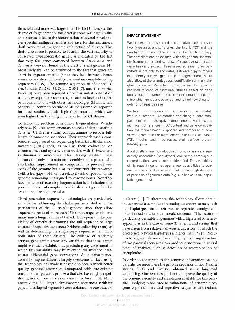
IMPACT STATEMENT
We present the assembled and annotated genomes of
two Trypanosoma cruzi clones, the hybrid TCC and the
non-hybrid Dm28c, obtained using PacBio technology.
The complications associated with this genome (assem-
bly fragmentation and collapse of repetitive sequences)
were basically solved. These improved assemblies per-
mitted us not only to accurately estimate copy numbers
of tandemly arrayed genes and multigene families but
also allowed the unambiguous identification of many sin-
gle-copy genes. Reliable information on the latter is
required to conduct functional studies based on gene
knock-out, a fundamental source of information to deter-
mine which genes are essential and to find new drug tar-
gets for Chagas disease.
We found that the genome of T. cruzi is compartmental-
ized in a isochore-like manner, containing a ‘core com-
partment’ and a ‘disruptive compartment’, which exhibit
significant differences in GC content and gene composi-
tion, the former being GC-poorer and composed of con-
served genes and the latter enriched in trans-sialidases
(TS), mucins and mucin-associated surface protein
(MASP) genes.
Additionally, many homologous chromosomes were sep-
arately assembled (haplotypes), and some homologous
recombination events could be identified. The availability
of high-quality genomes opens new possibilities to con-
duct analysis on this parasite that require high degrees
of precision of genomic data (e.g. allelic exclusion, popu-
lation genomics).
Bern�a et al., Microbial Genomics 2018;4
2
Downloaded from www.microbiologyresearch.org by
IP: 190.64.49.90
On: Wed, 02 May 2018 11:43:20
These results also reveal multiple unique aspects of genomearchitecture previously overlooked, such as whole-genomecompartmentalization into regions of different base compo-sition and biased distribution of genes.
METHODS
Parasites and DNA isolation
T. cruzi strains Dm28c [12] and TCC [13], were usedthroughout this work. Epimastigotes were grown in liverinfusion tryptose (LIT) medium supplemented with 10%fetal calf serum at 28
�
C; total DNA was extracted using theQuick DNA Universal kit (Zymo Research).
Sequencing
PacBio library preparation and sequencing were done bythe University of Washington PacBio Sequencing Services(Washington, USA). Briefly, DNA was mechanically frag-mented using a Covaris g-TUBE device, and concentratedwith AMPure PB magnetic beads. The final long-insert Pac-Bio libraries were size-selected for fragments larger than10 kb using the BluePippin device.
A total of eight Single Molecular Real-Time (SMRT) cellswere used, five for TCC and three for Dm28c, yielding751 460 and 601 168 raw reads, respectively. Subreads wereobtained using the SMRT Analysis RS.Subreads.1 pipeline(minimum polymerase read quality=0.85; minimum poly-merase read length and minimum subread length=500 bp).An Illumina MiSeq platform was used with a paired-endlibrary (2�150 cycles). Briefly, a Nextera XT (Illumina)library preparation kit was used with 1 ng total DNAaccording to the manufacturer’s instructions. Index primerswere added to each library to allow sequence multiplexing.After 12 PCR cycles, the final library was purified withAMPure XP (Benchman) and quantified with the QubitdsDNA HS assay kit (Invitrogen). Quality and length of thelibrary were assessed with the Agilent high-sensitivity DNAkit (Agilent) using the 2100 Bioanalyzer (Agilent). The rawreads obtained from the two genomes were deposited atSequence Read Archive (SRA) NCBI repositories (AccessionIDs: SRP134374 and SRP134013).
Genome assembly
The assembly was performed using the SMRT Analysis toolsimplemented in the Hierarchical Genome Assembly Process(HGAP) pipeline V3 [14]. It was run with the default parame-ters modifying only the expected genome size, which was setto 110 Mb, generating 1978 and 1143 contigs with a shortestsequence length at 50% of the genome (N50) of 73 kb and129 kb for TCC and Dm28c, respectively. Subsequently thescaffolding pipeline IPA was applied [15], specifically thosescripts aimed to merge overlapping contigs using the assem-bled output and Illumina paired-end reads. This pipeline startsby self-mapping the contigs using megablast, with the parame-ter of word-size set at 40 and an e-value of 1e�80. Self-con-tained contigs and those with an identity below 99% andshorter than 500 bp were discarded for further analysis. ThenIllumina reads were mapped over the remaining sequences
using the alignment software SMALT. Finally, if there areenough pairs of Illumina reads where each member of the pairmaps to two different contigs, it can be argued that the paircomes from the same contiguous sequence. This informationwas used to merge contigs by their ends. The final assemblieswere of 86.7 and 53.2Mb with a N50 of 265 and 318 kb forTCC and Dm28c respectively. Both genomes and their anno-tations were deposited at the NCBI repository (BioProjectsPRJNA432753 and PRJNA433042).
Genome annotation
For the annotation of the coding sequences we extractedfrom each assembled genome the open reading frames of atleast 150 amino acids in length, between a start and a stopcodon, using the getorf tool from the EMBOSS suite [16].These sequences were mapped using BLASTP [17] against adatabase of kinetoplastid proteins from TriTrypDB using ascut-off an e-value of 1e�10. The database of kinetoplastidproteins consists of a curated collection of CDS from variousspecies of the genera Trypanosoma (excluding Trypanosomagrayi and Trypanosoma rangeli), Leishmania, Leptomonas,Crithidia and Endotrypanum.
For practical purposes the annotation was divided into sev-eral steps. First the surface multigene families i.e. mucin-associated surface proteins (MASPs), mucins, retrotranspo-son hot spot proteins (RHS), zinc metalloprotease gp63(GP63), trans-sialidases (TS), dispersed gene family protein1 (DGF-1) and the recently identified Trypomastigote, ala-nine, serine, valine proteins (TASV) [18] were annotatedusing a stricter cut-off, an e-value of 1e�30. Then we anno-tated the genes conserved among kinetoplastids. In this stepwe chose the best hit in T. cruzi CL-Brener Esmeraldo andnon-Esmeraldo strains, and the best hit outside T. cruzi. Ifthere were hits only outside T. cruzi these ORFs were alsoannotated with the best two hits. The selection of the besthigh-scoring pairs (HSP) was performed using a personal-ized script that scanned for the lowest possible e-value witha meaningful description. If the only description availablewas ‘hypothetical protein’ or a similar non-descriptive tag,then the lowest e-value was selected. For annotating shortproteins, those smaller than 150 amino acids, we extractedthe open reading frames of between 50 and 150 amino acidsgenerating a massive amount of sequences that were subse-quently mapped only against a T. cruzi database by BLASTP
searches (e-value 1e�03, identity >80% and query coverage>80%). Initital methionines were verified using RNA-seqdata. Specifically, T. cruzi RNA-seq reads were downloadedfrom the SRA repository, bioproject PRJNA251583 [19],and the reads containing a spliced leader sequence wereidentified. The spliced leader segment was trimmed fromthese reads and the remaining segment was mapped to theT. cruzi genome to identify splice-acceptor sites (furtherdetails in [20]). Once the splice-acceptor sites were deter-mined, we annotated as the initiation codon, the in-frameATG triplet closest to (and downstream of) the splice-accep-tor site. Additional information was included in the annota-tion by predicting signal peptide (SP), using SignalP v4.1
Bern�a et al., Microbial Genomics 2018;4
3

Downloaded from www.microbiologyresearch.org by
IP: 190.64.49.90
On: Wed, 02 May 2018 11:43:20
[21], glycosylphosphatidylinositol (GPI) anchor, predictedby predGPI [22], and trans-membrane domains, usingTMHMM v2.0 [23].
A curated non-coding RNAs and interspersed repeats data-base (from TriTrypDB and NCBI) was used for the annota-tion of these elements. The sequences in these databaseswere mapped using BLASTN against the assembled contigs,then filtering the results and keeping only those hits whichmapped more than an 80% of the length of the queriedsequence with at least an 80% identity. An overview of theworkflow is presented in Fig. S1 (available in the online ver-sion of this article).
Customized scripts were written in Python [24], Perl [25]and R [26]. Alignments of short reads (Illumina and 454reads) were performed with Bowtie2 [27] and Rsubreads(from within R [28]). Samtools utilities [29] were used tomanipulate alignments and perform coverage analyses
Contigs comparison
Dotplot view
A self-comparative mapping of each contig, showing theresults in a dotplot, was performed with BLASTN. We usedthe YASS web server [30] with the default options to createthese plots.
Circos view
To assess the similarities between contigs of each assembly,we run a BLASTN of all-against-all the contigs longer than50 kb. For practical purposes we generated two tables, onewith HSPs longer than 10 kb and other with HSPs between5 and 10 kb. To graphically represent these data and avoidan entangled plot with an over-representation of hits frommultigene families, the hits from these sequences wereremoved from the final results. The mappings between con-tigs are shown in a circular layout using the Circos software[31]. In these plots (Fig. 2d and http://bioinformatica.fcien.edu.uy) the target sequence is shown at 12 o’clock andextends clockwise proportional to its length. The ten contigswith the largest overall mappings were selected, and are rep-resented according to their length in the plot. The genomicsimilarities of the target sequence and the contigs are repre-sented as wide lines that show the length of the mappingsand their coordinates and share the same color with thecontig of origin.
The web interface
An online genome browser was developed (http://bioinfor-matica.fcien.edu.uy/cruzi) where the whole annotation isdisplayed for contigs longer than 50 kb. Each contig isshown as a long rectangular block where genes and sequen-ces of interest are represented as rectangles of different col-ors according to their classification. The width and positionof each rectangle is proportional to their length and coordi-nates in the contig. Each represented contig has at the bot-tom a smaller rectangle that indicates the direction 5¢�3¢:sense (red) or antisense (green). In the visualizationinterface the annotation is divided into different categories.
The CDSs are divided according to the main multigene fam-ily they belong to, conserved genes are divided betweenthose with a meaningful description and those annotated ashypothetical proteins. The repeated elements are also classi-fied as tandem, interspersed repeats or satellite. Each cate-gory is indicated by a different color and can be displayedor hidden as a whole using the check boxes in the upperpart of the page. When the cursor is over an annotatedsequence it displays a tooltip with a description of the best
BLAST hits and hyperlinks to the nucleic and amino acidic
sequence if possible. Under each contig there is a white rect-angular block with more information. There are hyperlinksto the contig sequence in fasta format. At the bottom-rightof the page there are five buttons, two with the magnifyingglass allow zooming in and out in the sequences, the onewith the stripes allows changing of views between the stan-dard view and a six frames view, and the text box and binoc-ulars allows searching for sequence descriptions within thepage. It is also possible to extract a customized nucleotidesequence from the contig by clicking at the start and end ofthe desired area. After doing this an emergent windowappears at the bottom of the page, allowing correction of thecoordinates and retrieval of the genomic sequence.
Chromosome and contig visualization
Besides the developed web interface, we used ACT [32] andARTEMIS [33] to visualize and compare contigs from ourassembly and previous assemblies. Both software packagesuse the output of the BLASTN search and the contigs in fastaformat. BLASTN searches for these comparisons were per-formed with a stringent e-value <1e�200. IGV [34] soft-ware was used to visualize the alignments of reads over theassemblies (including Illumina, 454 or PacBio reads of dif-ferent sources).
Manual curation of surface multigene familiesannotation
The annotation of surface multigene families was based onthe coding sequences identified by the annotation pipeline.Furthermore, we manually inspected the sequences, analyz-ing specific characteristics relative to their function.
For mucins we use the length, the number of repetitions ofthreonine tandems, the presence of conserved sequenceswithin the SP, the region surrounding the GPI and the S, Tand P amino acid composition [35]. The identification ofconserved N- and C-terminal domains as well as character-istic sequences and tandem repeats present in mucins wereperformed with custom scripts. Genes without a clear signalpeptide and GPI anchor signal, but containing other mucinproperties were designated as pseudogenes (most of themcorresponded to genes that had lost the N- or C- terminalextremes). Finally, analysis of RNA seq data [19, 36] andcorrection of the initial methionine were performed whereapplicable.
For trans-sialidase sequences we considered the length, thepresence of VTV and ASP box motifs, the GPI anchor signalprobability and the presence of a SP sequence (using the same
Bern�a et al., Microbial Genomics 2018;4
4
Downloaded from www.microbiologyresearch.org by
IP: 190.64.49.90
On: Wed, 02 May 2018 11:43:20
software as above). SXDXGXXTW and VTVxNVxLYNRsequences [37] were searched using PatMatch [38] and theoutput parsed with customized scripts.
For MASP genes, we consider the presence of highly con-served domains. N-terminal domain MAMMMTGRVLLV-CALCVLWCG and C-terminal domain GDSDGSTAVSHTTSPLLLLLVVACAAAAAVVAA [3, 39] were searchedusing HHsearch [40]. Genes having only one domain wereannotated as pseudogenes.
Fragmented DGF-1 pseudogenes were reconstructed into asingle sequence. The partial pseudogenic ORFs were mergedand the “full length” pseudogene thus obtained was com-pared with a functional copy used as template.
Gene cluster analyses
Protein-coding genes were clustered into gene families usingMarkov Cluster Algorithm (MCL) [41] with BLASTP �log e-values (e�20). A fairly stringent inflation value which deter-mines the granularity (or size of the output clusters) of 4was used. A customized script was used to parse the clusteri-zation output and generate the final results. Clusterizationwas performed to identify gene families, novel gene familiesand single copy genes.
Characterization of transposable elements
Canonical complete elements for each transposable element(TE) were used to perform queries against the genomesusing BLASTN (e-value 1e�10, identity >80%, query coverage>90%). Non autonomous elements (SIRE and NARTc)were filtered when mapped into the parental elements(VIPER and L1Tc). L1Tc was also identified in the CLBrener assembly (from TriTrypDB v28 T. cruzi CL Brener,T. cruzi CL Brener Esmeraldo-like and T. cruzi CL Brenernon-Esmeraldo-like) and the Sylvio X10/1 assembly (fromNCBI BioProject PRJNA40815) with same parameters. Atotal of seven L1Tc elements were identified in CL Brenerand 156 in Sylvio X10/1. Multiple alignments were per-formed with MAFFT [42] using the e-ins-i Iterative refine-ment method. Phylogenetic trees were computed by PhyML3.1 [43] through the SeView platform [44]. A maximumlikelihood tree based on the GTR+imodel was recon-structed with 1000 bootstrap replications. Final representa-tion was performed with iTOL [45].
Characterization of tandem repeats
To identify and annotate tandem repeats we used the Tan-dem Repeats Finder software [46] with the parameters ofminimum alignment score of 20, maximum period size of2000 and an alignment score of 2 for a matching base and�7 for mismatches and insertions and deletions (indels).All entries with at least ten repeated periods of at least threenucleotides in length were considered for analysis. Theresults that share an overlap over 80% in both sequenceswere merged and reported as one. The grouping of thesesequences was done in several steps. First, we used personal-ized scripts to generate all the variants of a period and scan
the results to group the identical ones. This strategy is bettersuited to grouping short periods without internal variability.
The second step of clusterization consisted of creating amultifasta file with a sequence from each period and group-ing them using blastclust with the parameters -S80 -L0.8 -bT that restricts the results to those that share at least 80%of the sequence length with and 80% of identity for bothsequences. This grouping step is mainly aimed at clusteringlonger sequences with few indels or mismatches.
For further grouping, a third step was used, where eachsequence was extended by repeating the period up to at least300 bp. Then a self-comparative BLAST was performed andresults with an identity lower than 80% were discarded.Using a personalized script, for each sequence all the HSPwere summed and if this sum was at least an 80% of the tar-get sequence, they were grouped. This step is particularlyuseful for grouping sequences where the shortest reportedperiod is in fact a multiple of a shorter one of another clus-ter. The final output is a table with the coordinates of eachtandem repeat and the group they belong. An overview ofthe workflow is presented in Fig. S2
RESULTS AND DISCUSSION
Genome assembly
To obtain a more complete assembly of the complex T. cruzigenome using the PacBio technology, library preparationincluded fragmentation and size selection with a cut-off of10 kb. This size threshold also prevents the inclusion ofminicircles, whose presence leads to a substantial reductionin sequencing depth of nuclear genomic DNA (they repre-sent about 20% of total DNA). To include both clinicallyand evolutionarily relevant strains, we chose to sequence thehybrid TCC strain (TcVI), derived from Tulahuen 2 andclosely related to CL Brener [13], and the non-hybrid strainDm28c (TcI) [12]. It should be mentioned that TcI is themost abundant DTU and has a wide distribution in theAmericas, evolving separately from a common ancestor [47]while TcVI, present in several countries of South America,is a hybrid of the two distinct lineages TcII and TcIII [48].
After filtering by read quality, we obtained 5.2 and 4.0Gb ofsequences comprising 343 384 and 261 392 reads for TCC andDm28c, respectively (Table 1). The average read length was16 kb for both strains, and the longest reads were larger than60 kb (Fig. S3). The availability of such long reads is essentialto disentangle the sequence repetitions; a hallmark of T. cruzi.HGAPv.3 assembler [49] was used to correct and assemblethe initial reads. IPA [15] was used to collapse overlappingcontigs and cleanup the assembly incorporating Illuminareads. The length of the reads enabled us to directly obtain thefull sequences of large clusters of repetitive sequences withoutcollapsing them, as well as to determine the non-repetitivesequences flanking those repeats. Illustrative examples of howPacBio reads allowed us to overcome some of the commoncomplications associated with T. cruzi assemblies, fragmenta-tion and collapse of repeats, are presented in Fig. 1. The figure
Bern�a et al., Microbial Genomics 2018;4
5

Downloaded from www.microbiologyresearch.org by
IP: 190.64.49.90
On: Wed, 02 May 2018 11:43:20
shows the comparison between chromosome 30P from CLBrener (assembly reported in [9]) with TCC and Dm28c con-tigs reported here using long reads. In order to build the vir-tual chromosome 30P (from CL Brenner) Weatherly et al. [9]connected many contigs by segments of unknown sequence(filled with N letters, shadowed in green in Fig. 1a). These ‘Nregions’ were fully resolved in our assemblies, both in TCCand Dm28c (Fig. 1a). It is worth noting that the lengths of ‘Nregions’ were precisely estimated byWeatherly et al. (Fig. 1).
A second noticeable improvement is related to the repetitivesequences clusters, which were collapsed in the previousassembly, and now are disaggregated into the actual copynumber (Fig. 1). This is more clearly shown in Fig 1b thatshows a magnification of a fragment from the same CLBrener chromosome, containing several clusters of tandemrepeats. The figure also shows that some of these collapsedclusters contributed to assembly fragmentation, since theyare located at contig boundaries.
A comparison between the same chromosome of TCC andDm28c (Fig. 1c) revealed additional aspects concerningcluster repeats and structural variation between strains.
Specifically, the figure shows copy number variationsbetween the two strains in four groups (indicated by num-bers 1 to 6 in Fig. 1c) as well as strain-specific insertions/deletions. Among the latter, it is worth mentioning a largesegment located on the (left) telomere present in TCC andCL Brener, but absent in Dm28c, containing tandem repeatsof some species-specific genes (such as DGF1).
Overall, in terms of integrity (i.e. low fragmentation levels)the assemblies obtained for both strains, represent notableimprovements (Table 1) compared with previous ones.Their N50 values were 265 and 317 kb for TCC and Dm28crespectively, representing more than a ten-fold increase inthis index. Furthermore, several contigs correspond toentire chromosomes (for instance contigs TCC_3, TCC_4,TCC_10, Dm28c_6, Dm28c_22 and Dm28c_58, being thelargest ones, 1.65Mb for Dm28c and 1.35Mb for TCC).Other contigs are considerably smaller (50 kb) and are com-posed uniquely of a well-known satellite sequence of 195 bpthat encompasses more than 5% of the genome (see below).This extremely abundant repeat is one of the factors thatcontributes the most to assembly fragmentation. Thisoccurred not only in previous assemblies with short reads,but its incidence is even significant in assemblies based onlong reads, such as that presented here. Thus, this indicatesthat the size of some of its clusters exceeds that of the reads.
Besides fragmentation, another important element to con-sider when evaluating assembly quality is its size. In the caseof Dm28c, the assembly size was 53.2Mb, consistent withits haploid size as previously estimated using a fluorescentnucleic acid dye [50]. Previous efforts to sequence Dm28cand Sylvio X10/1 strains (two closely related TcI strains)using other technologies [6, 7] resulted not only in assemblyfragmentation but also in genome sizes of roughly one halfof the actual haploid size (27Mb for Dm28c and 23Mb forSylvio X10/1). This size underestimation is in all likelihooddue to the limitations of short-read technologies for assem-bling complex genomes. Keeping this limitation in mind,Franz�en et al. [8] recalibrated their genome size estimationfor Sylvio X10/1 to 44Mb, extrapolating on the basis ofnon-assembled reads.
The TCC strain is closely related to CL Brener, hence, weexpected their genomes to be very similar in terms of size andsequence composition. Indeed, sequence identity is higherthan 99.7% over aligned segments longer than 10 kb(Table S1). The CL Brener genome has been estimated to bebetween 106 [3] and 122Mb [50]. The assembly of TCCreported here consists of a ‘diploid’ genome of 86.7Mb. How-ever, the total assembly length is virtually the same if only con-tigs longer than 10 kb are considered (85.5Mb). This value isbelow the expected genome (diploid) size, if one assumes thatit should be similar to CL Brener, yet it represents a very sig-nificant improvement compared with the best previous assem-bly obtained by Weatherly et al. [9], in which both CL Brenerhaplotypes combined (Esmeraldo like and Non-Esmeraldo)have a total added size of only 54Mb [9]. The smaller size ofour assembly can be attributed to two factors. First, like CL
Table 1. Summary of the assembly and annotation of T. cruzi TCC and
T. cruzi Dm28c genomes
Assembly properties TCC Dm28c
Total reads 751 460 601 161
Filtered reads 343 384 261 392
Read N50 20 939 21 011
Total base pairs 5 200 759 160 4 037 444 749
Coverage ~60� ~76�
Genome properties
Size* (bp) 86 772 227 53 163 602
N50 265 169 317 638
Number of contigs 1142 599
DNA G+C content (%) 51.7 51.6
Percentage coding 49.6 49.8
Protein-coding genes
Number of gene models 27 522 17 371
Mean CDS length 1388 1484
DNA G+C content (%) 54.3 53.6
Gene density (genes per Mb) 320 326
Intergenic regions
Mean length (bp) 1690 1660
DNA G+C content (%) 46.0 46.8
RNA genes
tRNA 115 94
rRNA locus** 8 14
rRNA 5S 193 77
SL-RNA 206 622
snRNA 16 13
snoRNA 1561 1024
*Includes all contigs >5Kb.
**Includes SSU+5.8S+LSU.
Bern�a et al., Microbial Genomics 2018;4
6

Downloaded from www.microbiologyresearch.org by
IP: 190.64.49.90
On: Wed, 02 May 2018 11:43:20
Brener, TCC is a hybrid clone composed of two relatively
divergent parental lineages similarly to Esmeraldo and non-
Esmeraldo. This would imply that the distinction and ‘segre-
gation’ of parental haplotypes was partial (about 40% appears
to have remained un-separated). It is likely that some genomic
regions exhibit interallelic (inter-haplotype) divergence below
the identity threshold that the assembler requires to discrimi-
nate between them. Secondly, it is possible that some clusters
of repetitive sequences, especially the largest ones, were not
completely uncollapsed, thus contributing to assembly
shortening. It is difficult to estimate the effect of this second
source of uncertainty on the assembly, since in the case of
Dm28c, in spite of repetitions, the assembly was not smaller
than the expected genome size. Importantly, however, for
TCC we were able to distinguish and assemble separately (to a
substantial degree) the parental haplotypes (Fig. 2).
Fig. 1. Chromosomal assembly improvements. (a) ACT alignment of homologous chromosomes from three strains: TCC (contig
TCC_10), Dm28c (contig Dm28c_6) and CL Brener (chromosome TcChr30-P). Previously undetermined sequences filled by Ns in CL
Brener are marked in green. (b) Magnification of a fragment of a (boxed and shadowed in grey). The six frames and the DNA G+C con-
tent of each chromosome are plotted. Previously collapsed repetitive sequences (boxed in orange) are disaggregated in the new
assembly. c) Visualization of the alignment of the same homologous chromosome showing additional details in TCC and Dm28c. The
color patterns in the annotation bars (bottom and top-most horizontal stripped bars) correspond to the annotation as they appear in
the web interface (DGF1 in red, GP63 in orange, RHS in brown, conserved genes in green). The six reading frames are also shown. (1)
Terminal DGF-1 gene cluster present only in TCC. (2) Non-homologous region present only in Dm28c. (3) Repetitive region present in
both strains. (4) Expansion of a GP63 cluster in TCC (four copies versus two copies in Dm28c). (5) Strain-specific amplifications of
two different genes. There are seven GP63 copies (orange strips on the top annotation bar) in TCC but only one in Dm28c; moreover
Dm28c contains four RHS copies in the same region. (6) Repetitive element present in both genomes having fewer copies in TCC (20
copies in TCC and 44 copies in Dm28c). The segment is followed by another strain-specific amplification consisting of a cluster of 14
GP63 genes in TCC and only one copy in Dm28c.
Bern�a et al., Microbial Genomics 2018;4
7
Downloaded from www.microbiologyresearch.org by
IP: 190.64.49.90
On: Wed, 02 May 2018 11:43:20
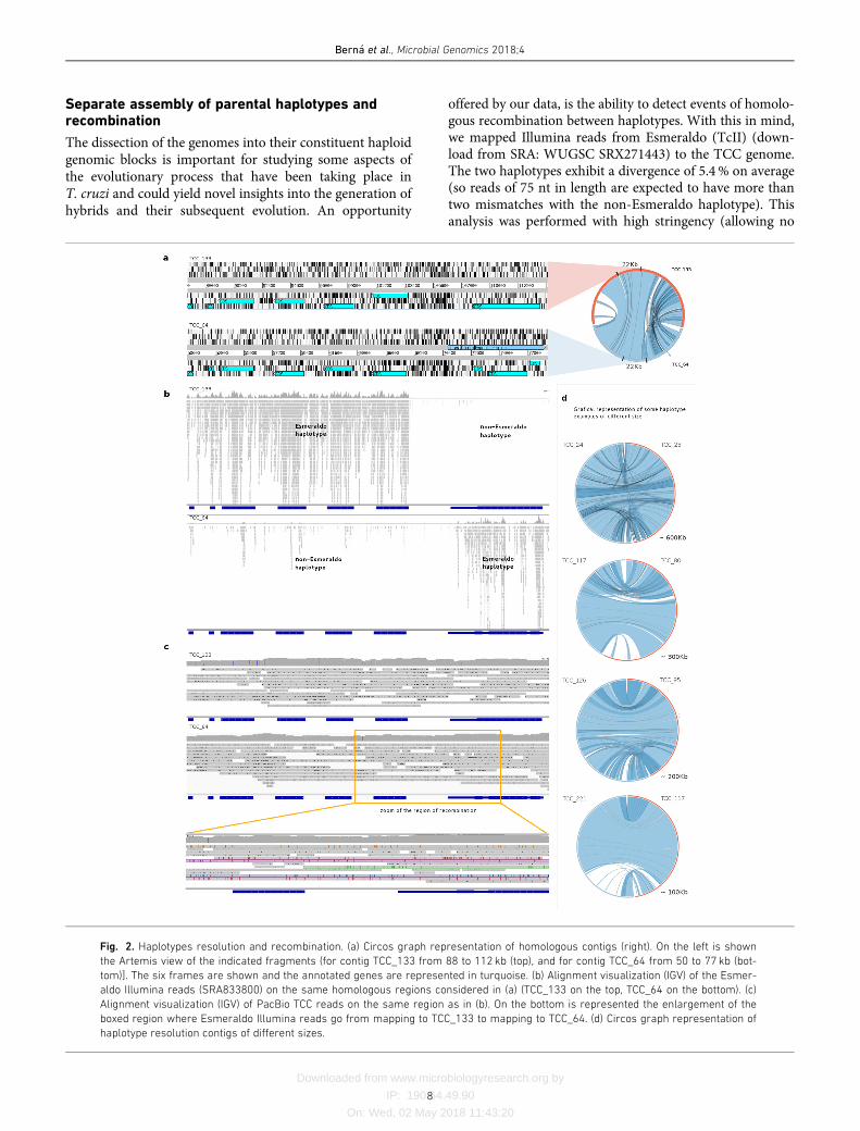
Separate assembly of parental haplotypes andrecombination
The dissection of the genomes into their constituent haploidgenomic blocks is important for studying some aspects ofthe evolutionary process that have been taking place inT. cruzi and could yield novel insights into the generation ofhybrids and their subsequent evolution. An opportunity
offered by our data, is the ability to detect events of homolo-gous recombination between haplotypes. With this in mind,we mapped Illumina reads from Esmeraldo (TcII) (down-load from SRA: WUGSC SRX271443) to the TCC genome.The two haplotypes exhibit a divergence of 5.4% on average(so reads of 75 nt in length are expected to have more thantwo mismatches with the non-Esmeraldo haplotype). Thisanalysis was performed with high stringency (allowing no
Fig. 2. Haplotypes resolution and recombination. (a) Circos graph representation of homologous contigs (right). On the left is shown
the Artemis view of the indicated fragments (for contig TCC_133 from 88 to 112 kb (top), and for contig TCC_64 from 50 to 77 kb (bot-
tom)]. The six frames are shown and the annotated genes are represented in turquoise. (b) Alignment visualization (IGV) of the Esmer-
aldo Illumina reads (SRA833800) on the same homologous regions considered in (a) (TCC_133 on the top, TCC_64 on the bottom). (c)
Alignment visualization (IGV) of PacBio TCC reads on the same region as in (b). On the bottom is represented the enlargement of the
boxed region where Esmeraldo Illumina reads go from mapping to TCC_133 to mapping to TCC_64. (d) Circos graph representation of
haplotype resolution contigs of different sizes.
Bern�a et al., Microbial Genomics 2018;4
8
Downloaded from www.microbiologyresearch.org by
IP: 190.64.49.90
On: Wed, 02 May 2018 11:43:20
mismatches) so that reads would basically map only to theEsmeraldo-like haplotype. The separation was evidenced bythe read mapping distribution: almost all of them mappedonly to one of the homologous contigs (Fig. 2b). However,in sites where recombination presumably took place, thereis a switch in the mapping pattern and reads start to map onthe homolog contig, as shown in Fig. 2(b). To confirm thatthe switch in the mapping preference was not an assemblyartifact; namely a chimera generated by mixing the two hap-lotypes, we mapped PacBio reads that span the region ofrecombination to these sites (Fig. 2c). As shown in Fig. 2(c),mismatches between reads and the contigs are evenly andrandomly distributed along the genome. In contrast, whenmapped into chimerical assembled contigs (Fig. S4), PacBioreads have low or high abundance of mismatches beforeand after the artificial ‘recombination’ point. The mappingpattern of Illumina reads onto chimerical contigs is, never-theless, indistinguishable from that expected in actualrecombination zones.
Another noteworthy observation from Fig. 2(b) is anextreme scarcity of Illumina reads mapping on this putativerecombination hotspot. To investigate this we tested severalpossible causes. First, we wondered whether this could bethe result of our stringent mapping conditions. However,relaxing mapping restrictions to allow more mismatchesonly caused a marginal increase in mapping. The same istrue for 454 reads (from NCBI PRJNA50493); very few ofthem map here, even when a large percentage of mis-matches is tolerated. To us, this indicated that this region iseither refractory to sequencing or an artifact of our assem-bly. To test the latter, we searched for the presence ofthe region in other assemblies and strains. It was foundin Dm28c (our assembly), Sylvio X10/1 (from NCBIPRJNA40815, PacBio) and CL Brener (Sanger). This led usto conclude that this region is poorly sequenced using otherNGS technologies. Further analysis of the region, revealed apoor GC content, averaging 30%, (with segments longerthan 100 nt with GC content lower than 20%). This is inkeeping with previous reports showing that genomic seg-ments with very low GC content are under-represented inIllumina sequencing [51]. Using GC content and Illuminasequencing depth as criteria for identifying similar regionsin the genome of TCC, we found more than 500 regionssimultaneously matching these two features: very low GCcontent and very few or no mapping reads (with segmentswhere sequencing depth is as low as zero). A similar searchin Dm28c (PacBio assembly), yielded 160 such regions.Comparing with other assemblies we found that most GC-low regions from TCC are also present in CL Brener assem-bly, but less than half of them can be found in our assemblyof Dm28c. The comparison of two Dm28c assembliesreveals that only 90 of the 160 found in the PacBio assem-bly, reported here, were present in the Dm28c assemblyobtained using Roche 454 reads. Similar figures are obtainedwhen the comparison is performed with Sylvio X10/1 (Illu-mina and Roche 454). Furthermore, most of these regions,or fragments of them, are located in contigs ends (for
Illumina and Roche 454) indicating that assembly washalted at these positions due to insufficient coverage. Takentogether, these results indicate that these low-GC regionsare another important factor in assembly fragmentation inT. cruzi genomes for Illumina and 454 technologies, butthey do not appear to affect Sanger- or PacBio-basedassemblies.
Genome annotation
Genome annotation is an error-prone task, which is particu-larly intricate in T. cruzi due to its intrinsic genomic com-plexities, including large multigene families, pseudogenesand the absence of fully annotated genomes from phyloge-netically closely related species. To address these problems,we followed an annotation strategy designed to handle thepeculiarities of this species. Since the vast majority of try-panosome genes lack introns, we used a ‘prokaryotic-like’approach to identify them. Specifically, as gene models to beused downstream in the annotation pipeline we used agreedy criterion: all open reading frames (ORFs) longerthan 450 nt and starting with Met. Shorter CDSs were dealtwith separately (see below). Since many long ORFs are notprotein-coding, this initial group of potential protein-codinggenes very probably contains a large number of false posi-tives. However, only those ORFs encoding proteins willyield BLASTP hits with relatively distant species, hence, thesecan be readily identified and excluded by subsequent filter-ing with BLASTP, using appropriate protein reference datasets. Therefore these data sets are crucial to get accurateannotations. Indeed a common drawback in genome anno-tation is inheriting (by homology transfer of information)spurious and erroneous annotations from other genomesused as references. To work around this potential source ofinaccuracy the following datasets were built/used: (i) A care-fully curated databases of T. cruzi multigene families (TS,mucin, MASP, GP63, TASV, DGF-1, RHS). Each of thesefamilies was thoroughly scrutinized manually to determinethe complete and probable functional copies, their sub-groups and pseudogenes. (ii) Trypanosomatid proteomedatabases from which T. cruzi strains as well as other speciesnot very distant from T. cruzi (T. rangeli, T. grayi) wereexcluded. The rationale for this criterion is that whereasevolutionary conservation is an indication of functional rel-evance (i.e. if the amino acid sequence encoded by the ORFhas been maintained over time this indicates a real protein-coding gene), conservation among not distant taxa does notguarantee functional relevance; instead, it could merelyreflect phylogenetic inertia (i.e. there not being enough timeto diverge). (iii) A curated protein database from non-trypa-nosomatids (iv) Additionally, to cross-check our annota-tions, we used protein annotations from T. cruzi, exclusivelyfrom CL Brener, since this is the strain most accuratelyannotated. Whenever inconsistencies arose, problematicORFs were further analyzed and manually curated. An over-view of the workflow is presented in Fig. S1.
In order to include, and annotate, protein-coding genesshorter than 450 nt, the search was conducted backwards;
Bern�a et al., Microbial Genomics 2018;4
9

Downloaded from www.microbiologyresearch.org by
IP: 190.64.49.90
On: Wed, 02 May 2018 11:43:20
namely a dataset of short proteins was built and used tosearch for them (using TBLASTN) in the assemblies. Diversetools (see methods) were used to incorporate additionalinformation in the annotation. Non-coding RNAs, trans-posable elements and repetitive elements, were annotatedusing dedicated software run on specific databases that werebuilt for these purposes.
Although this strategy is intended to minimize misannota-tions, further manual curation was needed, especially withgenes belonging to multigene families and their associatedpseudogenes. In particular, several of these genes had beenfrequently excluded from previous assemblies, as they wereimpossible to accurately position [9]. The genomes of thetwo strains presented here contain 27 522 and 17 371 pro-tein coding genes (TCC and Dm28c respectively) and thenumber of hypothetical genes has been reduced from~50 to ~39% by means of the removal of spurious annota-tion due to the inclusion of new gene functions that couldbe classified due to the improvements of the TriTrypDB, orbecause they were coding sequences that were not assem-bled before.
To visualize and comfortably handle the information on theT. cruzi genomes we generated a web platform (http://bioin-formatica.fcien.edu.uy). A genome browser can be used tonavigate the annotation of contigs longer than 50 kb. It alsooffers built in tools to select groups of genes and other geno-mic features that are distinctive for T. cruzi, such as surfacemultigene families, repetitive elements and directional geneclusters. Among other functionalities of the interface, it ispossible to visualize gene annotations and retrieve theirnucleotide and amino acid sequences. It is also possible toconduct different searches (by annotation or by keywords)and to visualize graphical representation of repetitions andhaplotypes (haplotype information is available). The inter-face is intuitive and user friendly. See Methods for furtherdetails.
The genome of T. cruzi is compartmentalized
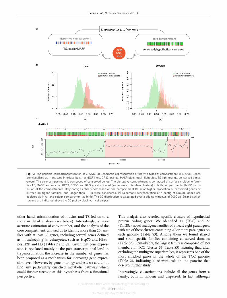
Two groups of protein-coding genes were very well describedin T. cruzi: the multigene families with hundreds of copies:DGF-1, GP63, MASP, mucins, RHS and TS, and those generi-cally defined as ‘conserved’, which can be further divided intotwo groups: genes encoding proteins with a known functioncalled ‘conserved genes’, and genes without an assigned func-tion but present in more than one trypanosomatid speciescalled ‘hypothetical conserved genes’. We found that thegenome of T. cruzi is compartmentalized in two clearlydefined regions: a ‘core compartment’ composed of conservedand hypothetical conserved genes, and a ‘non-syntenic’ ‘dis-ruptive compartment’ composed of the multigene families TS,MASP and mucins (Fig. 3a). On the other hand, GP63, DGF-1 and RHS multigene families have a dispersed distribution inthe genome, being present in both compartments, which mayin turn be organized as unique or in tandem array distribu-tion. Since the ‘core compartment’ corresponds to the previ-ously described syntenic blocks in T. brucei and L. major [4],and the ‘disruptive compartment’ is mainly composed of
species- or genus-specific genes, the latter can be consideredas a recent region of the genome. In turn, all of the ncRNAsare located in the core compartment (see web interface, e.g.contigs TCC:1,TCC_9, TCC_57 and Dm28c_2Dm28c_30). Itis noteworthy that the members of the disruptive compart-ment have been previously referred to as sub-telomeric; how-ever, as is evident from our results, these genes can be locatedin any position in the chromosome with the only conditionthat they cover wide ranges of distances, and the location canbe anything from internal chromosomal regions to extremesof chromosomes, and even comprise whole chromosomes.Therefore the term sub-telomeric, probably inherited from thegenome organization of VSG genes in T. brucei, is inappropri-ate and can lead to confusion. We suggest it should bereplaced by the more encompassing concept of‘compartments’.
In order to identify general features distinguishing the com-partments, we performed compositional analysis of thegenome, and found that the disruptive compartment consis-tently exhibits higher GC content than the core compart-ment (Fig. 3b). Interestingly, strand-switch regions showGC peaks that indicate particular compositions as shown inFig. 3(c). In this figure is shown the GC distribution overthe length of a contig, and the bias in GC content becomesclear (Fig. 3c). This genome organization resembles iso-chore-like structures. In fact, although originally onlydescribed in vertebrates [52, 53], the ‘isochoric’ level ofgenome organization was recently proposed to be a featureof all eukaryotes including unicellular ones [54, 55]. Ourobservations are consistent with this concept. This distribu-tion deserves further study to examine how compartmental-ization emerged and evolved.
Multigene families
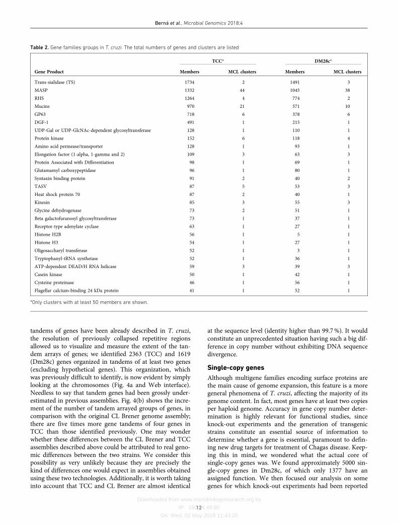
One of the known characteristics of the T. cruzi genome isits widely expanded content, mainly due to the large num-ber of multigene families [35, 56, 57]. In fact, when the firstgenome of T. cruzi was sequenced, authors pointed out therisk of underestimating the number of units of genes orga-nized in tandem [3]. Later, Arner and collaborators exposedexamples of copy number underestimation and misassem-bly and proposed that the number of protein-coding genesand pseudogenes may be twice the previous estimates [58].Our genome assembly makes it possible to determine thereal extent of the T. cruzi gene expansion. Defining a genefamily as that which presents eight or more genes, we iden-tified 90 and 74 families in TCC and Dm28c, respectively. Amore detailed analysis of families by MCL clustering indi-cated that they can be subdivided; TCC presents 190 paralo-gous gene clusters and Dm28c presents 139 (Table S2). InTable 2 we show only those gene families with more than 50members in at least one sequenced genome. As previouslydescribed, the most enriched ones are TS, MASP, RHS,mucins, DGF-1 and GP63 [3]. Remarkably, whereas RHS,DGF-1, GP63 and TS fall into six or fewer clusters in bothstrains, mucins, and particularly MASPs, harbor highernumber of subfamilies (clusters) (Tables 2 and S2). On the
Bern�a et al., Microbial Genomics 2018;4
10
Downloaded from www.microbiologyresearch.org by
IP: 190.64.49.90
On: Wed, 02 May 2018 11:43:20
other hand, misannotation of mucins and TS led us to amore in detail analysis (see below). Interestingly, a more
accurate estimation of copy number, and the analysis of the
core compartment, allowed us to identify more than 20 fam-ilies with at least 50 genes, including several genes defined
as ‘housekeeping’ in eukaryotes, such as Hsp70 and Histo-
nes H2B and H3 (Tables 2 and S2). Given that gene expres-sion is regulated mainly at the post-transcriptional level in
trypanosomatids, the increase in the number of genes hasbeen proposed as a mechanism for increasing gene expres-
sion level. However, by gene ontology analysis we could not
find any particularly enriched metabolic pathway whichcould further strengthen this hypothesis from a functional
perspective.
This analysis also revealed specific clusters of hypotheticalprotein coding genes. We identified 47 (TCC) and 27(Dm28c) novel multigene families of at least eight paralogues,with ten of these clusters containing 20 or more paralogues oneach genome (Table S3). Among them we found sharedand strain-specific families containing conserved domains(Table S3). Remarkably, the largest family is composed of 158members in TCC (cluster 35, Table S3) meaning that, afterexcluding the multigene superfamilies, it represents one of themost enriched genes in the whole of the TCC genome(Table 2), indicating a relevant role in the parasite thatdeserves further study.
Interestingly, clusterizations include all the genes from afamily, both in tandem and dispersed. In fact, although
Fig. 3. The genome compartmentalization of T. cruzi. (a) Schematic representation of the two types of compartment in T. cruzi. Genes
are visualized as in the web interface by strips (DGF1 red, GP63 orange, MASP blue, mucin light blue, TS light orange, conserved genes
green). The core compartment is composed of conserved genes. The disruptive compartment is composed of surface multigene fami-
lies TS, MASP and mucins. GP63, DGF-1 and RHS are distributed (sometimes in tandem clusters) in both compartments. (b) GC distri-
bution of the compartments. Only contigs entirely composed of one compartment (80% or higher proportion of conserved genes or
surface multigene families) and longer than 10 kb were considered. (c) Schematic representation of a contig of Dm28c; genes are
depicted as in (a) and colour compartment as in (b). The GC distribution is calculated over a sliding windows of 7000 bp. Strand-switch
regions are indicated above the GC plot by black vertical stripes.
Bern�a et al., Microbial Genomics 2018;4
11
Downloaded from www.microbiologyresearch.org by
IP: 190.64.49.90
On: Wed, 02 May 2018 11:43:20
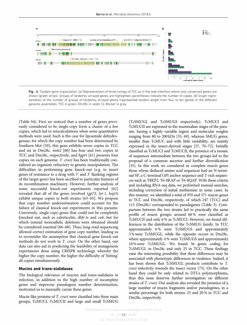
tandems of genes have been already described in T. cruzi,the resolution of previously collapsed repetitive regionsallowed us to visualize and measure the extent of the tan-dem arrays of genes; we identified 2363 (TCC) and 1619(Dm28c) genes organized in tandems of at least two genes(excluding hypothetical genes). This organization, whichwas previously difficult to identify, is now evident by simplylooking at the chromosomes (Fig. 4a and Web interface).Needless to say that tandem genes had been grossly under-estimated in previous assemblies. Fig. 4(b) shows the incre-ment of the number of tandem arrayed groups of genes, incomparison with the original CL Brener genome assembly;there are five times more gene tandems of four genes inTCC than those identified previously. One may wonderwhether these differences between the CL Brener and TCCassemblies described above could be attributed to real geno-mic differences between the two strains. We consider thispossibility as very unlikely because they are precisely thekind of differences one would expect in assemblies obtainedusing these two technologies. Additionally, it is worth takinginto account that TCC and CL Brener are almost identical
at the sequence level (identity higher than 99.7%). It wouldconstitute an unprecedented situation having such a big dif-ference in copy number without exhibiting DNA sequencedivergence.
Single-copy genes
Although multigene families encoding surface proteins arethe main cause of genome expansion, this feature is a moregeneral phenomena of T. cruzi, affecting the majority of itsgenome content. In fact, most genes have at least two copiesper haploid genome. Accuracy in gene copy number deter-mination is highly relevant for functional studies, sinceknock-out experiments and the generation of transgenicstrains constitute an essential source of information todetermine whether a gene is essential, paramount to defin-ing new drug targets for treatment of Chagas disease. Keep-ing this in mind, we wondered what the actual core ofsingle-copy genes was. We found approximately 5000 sin-gle-copy genes in Dm28c, of which only 1377 have anassigned function. We then focused our analysis on somegenes for which knock-out experiments had been reported
Table 2. Gene families groups in T. cruzi. The total numbers of genes and clusters are listed
TCC* DM28c*
Gene Product Members MCL clusters Members MCL clusters
Trans-sialidase (TS) 1734 2 1491 3
MASP 1332 44 1045 38
RHS 1264 4 774 2
Mucins 970 21 571 10
GP63 718 6 378 6
DGF-1 491 1 215 1
UDP-Gal or UDP-GlcNAc-dependent glycosyltransferase 128 1 110 1
Protein kinase 152 6 118 4
Amino acid permease/transporter 128 1 93 1
Elongation factor (1-alpha, 1-gamma and 2) 109 3 63 3
Protein Associated with Differentiation 98 1 69 1
Glutamamyl carboxypeptidase 96 1 80 1
Syntaxin binding protein 91 2 40 2
TASV 87 5 53 3
Heat shock protein 70 87 2 40 1
Kinesin 85 3 55 3
Glycine dehydrogenase 73 2 51 1
Beta galactofuranosyl glycosyltransferase 73 1 37 1
Receptor-type adenylate cyclase 63 1 27 1
Histone H2B 56 1 5 1
Histone H3 54 1 27 1
Oligosaccharyl transferase 52 1 3 1
Tryptophanyl-tRNA synthetase 52 1 36 1
ATP-dependent DEAD/H RNA helicase 59 3 39 3
Casein kinase 50 1 42 1
Cysteine proteinase 46 1 56 1
Flagellar calcium-binding 24 kDa protein 41 1 52 1
*Only clusters with at least 50 members are shown.
Bern�a et al., Microbial Genomics 2018;4
12
Downloaded from www.microbiologyresearch.org by
IP: 190.64.49.90
On: Wed, 02 May 2018 11:43:20
(Table S4). First we noticed that a number of genes previ-ously considered to be single-copy form a cluster of a fewcopies, which led to miscalculations when semi-quantitativemethods were used. Such is the case for lipoamide dehydro-genase, for which the copy number had been determined bySouthern blot [59]; this gene exhibits seven copies in TCCand six in Dm28c. msh2 [60] has four and two copies inTCC and Dm28c, respectively, and hgprt [61] presents fourcopies on each genome. T. cruzi has been traditionally con-sidered an organism refractory to genetic manipulation. Thedifficulties in performing gene knock-out (e.g. to insertgenes of resistance to a drug with 5¢ and 3¢ flanking regionsof the target gene) has been ascribed to particular features ofits recombination machinery. However, further analysis ofsome successful knock-out experiments reported [62]revealed that all of the genes involved (gp72, lyt-1, kap3)exhibit unique copies in both strains [63–65]. We proposethat copy number underestimation could account for thefailure of classical knock-out experiments in this parasite.Conversely, single-copy genes that could not be completelyknocked out, such as calreticulin, dhfr-ts and cub, but forwhich instead monoallelic deletions were obtained shouldbe considered essential [66–68]. Thus, long-read sequencingallowed correct estimation of gene copy number, leading usto reconsider the assumption that classical gene knock-outmethods do not work in T. cruzi. On the other hand, ourdata can also aid in predicting the feasibility of mutagenesisexperiments done using CRISPR technology whereby thehigher the copy number, the higher the difficulty of ‘hitting’all copies simultaneously.
Mucins and trans-sialidases
The biological relevance of mucins and trans-sialidases ininfection, in addition to the high number of incompletegenes and imprecise pseudogene number determination,motivated us to manually curate these genes.
Mucin-like proteins of T. cruzi were classified into three maingroups, TcMUCI, TcMUCII and large and small TcSMUG
(TcSMUGL and TcSMUGS respectively). TcMUCI andTcMUCII are expressed in the mammalian stages of the para-site, having a highly variable region and molecular weightsranging from 80 to 200 kDa [35, 69], whereas SMUG genes,smaller than TcMUC and with little variability, are mainlyexpressed in the insect-derived stages [57, 70–72]. Initiallyclassified as TcMUCI and TcMUCII, the presence of a mosaicof sequences intermediate between the two groups led to theproposal of a common ancestor and further diversification[35]. In this work we considered as complete mucin genesthose whose deduced amino acid sequences had an N-termi-nal SP, a C-terminal GPI anchor sequence and T-rich sequen-ces such as T8KP2, T6-8KAP or T6-8QAP.With these criteriaand including RNA-seq data, we performed manual searches,including correction of initial methionine in some cases. Inthis manner, we identified a total of 970 and 571 mucin genesin TCC and Dm28c, respectively, of which 247 (TCC) and113 (Dm28c) corresponded to pseudogenes (Table 3). Com-parison between the two strains led to practically the sameprofile of mucin groups; around 60% were classified asTcMUCII and only 6% as TcMUCI. However, we found dif-ferences in the distribution of the TcSMUG family. In TCCapproximately 6% were TcSMUGS and approximately3%were TcSMUGL, while the opposite occurs in Dm28c,where approximately 4% were TcSMUGS and approximately10%were TcSMUGL. We found 56 genes coding forTcSMUGL in Dm28c and only 25 in TCC. These findingsraise the interesting possibility that these differences may beassociated with phenotypic differences in virulence. Indeed, ithas been shown that TcSMUGL products contribute to T.cruzi infectivity towards the insect vector [73]. On the otherhand they could be only related to DTUs polymorphisms,thus this issue deserves further investigation on differentstrains of T. cruzi. Our analysis also revealed the presence of alarge number of mucin fragments and/or pseudogenes, in asimilar percentage for both strains: 25 and 20% in TCC andDm28c, respectively.
Fig. 4. Tandem gene organization. (a) Representation of three contigs of TCC as in the web interface where only conserved genes are
shown (green strips). Groups of tandemly arrayed genes are highlighted; parentheses indicate the number of copies. (b) Graph repre-
sentation of the number of groups of tandemly arrayed genes (represented tandem length from four to ten genes) in the different
genome assemblies. TCC in green, Dm28c in violet, CL Brener in gray.
Bern�a et al., Microbial Genomics 2018;4
13

Downloaded from www.microbiologyresearch.org by
IP: 190.64.49.90
On: Wed, 02 May 2018 11:43:20
Trans-sialidase/trans-sialidase-like proteins constitute alarge and polymorphic superfamily of around 1400 mem-bers. This heterogeneous family is currently classified ineight different groups [37], and although most of them donot exhibit trans-sialidase activity they play relevant roles ininfectivity and virulence [37]. TS contain an N-terminal SPand a C-terminal GPI anchor signal. Functional members ofthe TS family possess the characteristic VTVxNVLLYNRmotif. Additionally, some groups have N-terminal ASP boxsequences. Members of Groups I and IV also contain SAPArepeats, an extremely antigenic region whose role is toincrease the half-life of TS in blood [37, 74]. The initialannotated TS sequences were manually filtered using thefollowing criteria: presence of VTV and ASP motifs, GPIanchor signal probability and presence of a SP sequence.Using these criteria we found 1734 TS genes in TCC and1491 genes in Dm28c. However, using the same methodol-ogy as Freitas et al. [37], we could not identify the eightgroups described for TS, indicating that this classification isnot robust enough or might be species-specific. Our analysisalso revealed the presence of a large number of pseudogenes
or fragments; 721 and 567 for TCC and Dm28c, respectively(Table 3). These results highlight the importance of manualcuration for the most expanded and complex multigenefamilies. Finally, the fact that TS genes, present also in T.brucei, were found in the disruptive compartment deservesfuture analysis, in order to evaluate how the more ancestralgenes are distributed and to obtain clues about the evolutionof this family.
Transposable elements
Transposable elements (TEs) are dynamic drivers of evolution-ary processes that contribute to genomic plasticity. Usuallypresent as repetitive sequences in the T. cruzi genome, they areabundant and commonlymisannotated.T. cruzi presents threefamilies of autonomous genomic elements, as well as theirnon-autonomous pairs. Here, we were able to identify theentire sequences of all TEs families present in both genomesand their flanking sequences (Table 4). Namely, VIPER, a tyro-sine recombinase (YR) element which belongs to the DIRSorder; L1Tc, a non-LTR element of the INGI clade; and CZAR,also a non-LTR element from the CRE clade which is site-spe-cific inserting only in the SL gene [3, 75–77]. On the other
Table 3. Manually annotated surface multigene families
TCC Dm28c
Total Genes Pseudogenes Total Genes Pseudogenes
TS 1734 689 1045 1491 709 782
MASP 1332 941 391 1045 736 309
GP63 718 237 481 378 96 282
DGF-1 491 191 300 215 75 140
Mucin 970 723 247 571 458 113
Mucin Total TcMUCII TcMUCI TcSMUGS TcSMUGL Pseudogenes
TCC 970 (100%) 585 (60.3%) 57 (5.9%) 56 (5.8%) 25 (2.6%) 247 (25.5%)
Dm28c 571 (100%) 351 (61.5%) 29 (5.1%) 22 (3.9%) 56 (9.8%) 113 (19.8%)
Table 4. Complete retrotransposon copy numbers in T. cruzi
RetrotransposonsTCC Dm28c
Complete
copies
Length
(kb)
Identity
(%)
GC content
(%)
Complete
copies
Length
(kb)
Identity
(%)
GC content
(%)
Non-LTR
retrotransposons
CZAR 43 6.497 93.2 55.7 57 6.442 91.9 56.3
L1Tc 43(13) 4.874 90.7 53.0 54(18) 4.749 96.5 53.3
NARTc* 110 0.257 92.6 51.7 55 0.258 90.5 51.7
TcTREZO 978 1.459 92.1 50.9 297 1.423 95.2 50.3
YR retrotransposons
VIPER 244 3.454 85.0 55.2 194 3.423 87.3 54.8
SIRE* 851 0.440 87.4 44.0 669 0.441 88.6 44.2
*Non autonomous.
Parentheses indicate the number of putative active copies.
Bern�a et al., Microbial Genomics 2018;4
14

Downloaded from www.microbiologyresearch.org by
IP: 190.64.49.90
On: Wed, 02 May 2018 11:43:20
hand, non-autonomous elements have also been identified.Namely SIDER, which has sequence similarity to VIPER´s 5¢and 3¢ ends; NARTc, the non-autonomous pair of L1Tc ele-ments; and TcTREZO [78]. Putative active and defective copiescould be detected for all families. None of the VIPER, CZARand TcTREZO elements had complete domains, indicatingthat all copies are defective, whereas L1Tc was the only one toshow putative active copies in both genomes.
To shed some light on the evolutionary dynamics of L1Tctransposon, we first built a maximum likelihood phyloge-netic tree using only complete, and hence putatively func-tional sequences of this element. The resulting tree (Fig. 5),exhibits three main clades, one containing L1Tc copies fromDm28c, and two clades containing TCC copies. To furtherexplore this configuration, we analyzed (full length) L1Tcelements from CL Brener, discriminating Esmeraldo andnon-Esmeraldo haplotypes. It becomes immediately appar-ent that while one TCC cluster is associated with the Esmer-aldo haplotype, the second TCC cluster is related to thenon-Esmeraldo haplotype (Fig. 5). This provides evidencethat the functional copies of L1Tc had significant activityand underwent substantial evolutionary divergence after thetwo ancestral T. cruzi lineages comprising TCC (and CLBrener) split apart. The fact that branches in these two clus-ters are considerably longer, denotes a low transposition/divergence ratio. It is surprising, however, that there arehardly any intermingled copies, meaning that any inter-
copy variability predating the separation of these two ances-
tral lineages (i.e. already present in the ancestral genome)
has been erased. This is consistent with the fact that Dm28c
copies are also isolated, forming a single cluster. Neverthe-
less, in this case most copies are very similar to each other
(short branches) indicating that in this strain, L1Tc exhibits
a much higher transposition : divergence ratio. One can
attribute this to the fact that the new copies did not have
enough time to diverge after their emergence owing to a
high transposition rate. Alternatively, this might be due to a
much slower nucleotide evolutionary (divergence) rate.
Incorporating in the analysis the set of L1Tc sequences from
the strain Sylvio X10/1 (from NCBI PRJNA40815) gives
some clues as to the cause of the different dynamics. In
effect, these sequences cluster together with those of
Dm28c, yet a sharp division between the two strains is
observed (Fig. 5). Since the two strains are very close rela-
tives (with virtually identical nucleotide sequences genome-
wide), short branches in this part of the tree cannot be
attributed to a slow divergence rate, favoring the hypothesis
of very high transposition rate in this DTU. In any case, it
denotes a markedly different dynamic from that observed in
the hybrid TcVI. Whether the different dynamics are related
to the hybrid/non-hybrid nature of the strains being consid-
ered is a subject that deserves further assessment.
Fig. 5. L1Tc phylogeny. Maximum-likelihood phylogeny of complete sequences of L1Tc. Elements from TCC in green, Dm28c in violet,
SylvioX10/1 in light violet, CL Brener Esmeraldo-like in light violet grey, CL Brener non-Esmeraldo-like in b.
Bern�a et al., Microbial Genomics 2018;4
15

Downloaded from www.microbiologyresearch.org by
IP: 190.64.49.90
On: Wed, 02 May 2018 11:43:20
Tandem repeats
Tandem repeats (TR) are considered ‘neglected’ sequencesin genome analyses, since short reads cause often unsolv-able problems for de novo assemblies in TR regions. As aconsequence of long reads we could resolve TR-enrichedregions previously fragmented. As expected, thousands ofcopies of the already well characterized 195nt satellite [79,80] were found: 41 061 (8.3Mb) and 12 244 (2.5Mb) copiesfor TCC and Dm28c, respectively (Table 5). These find-ings are in keeping with previous estimates made by celland molecular biology methods [78, 81]. The difference incopy number between the genomes analyzed are not aconsequence of the different genome sizes only. In fact, ithas been observed that satellite amounts are variableamong strains, reaching differences of up to sixfold, butorganized similarly throughout the genome [81]. Theresults of our analysis indicate that the satellite is twotimes more abundant in TCC than in Dm28c, accountingfor 9.5 and 4.7% of the total size of the genomes, respec-tively (Table 5). This coincides with the differences esti-mated between TcVI and TcI groups by Souza et al. [78],the repeats in the former being two to four times moreabundant than in the latter. As mentioned, the abundanceof the satellite led to a high level of assembly fragmenta-tion. In this work we recovered satellites reaching lengthsof up to 45 and 42 kb in TCC and Dm28c, respectively,although the majority were of smaller size, with an averageof more than 10 kb, as previously reported [81]. We couldassemble several contigs containing satellite sequences sur-rounded by single-copy DNA, meaning that in these casesit was possible to pass over these repetitive clusters (Fig.S5). However, the contribution of this satellite to fragmen-tation is notorious, since some satellite sequences arelocated at the end of many small and medium-sized con-tigs (see for example TCC_301, 303, 312, and Dm28c_122,126, 146).
We then aimed to characterize the remaining TR sequenceswith a minimal length of three nucleotides and containing
at least ten monomers. We identified 261 and 205 groups ofdifferent sizes the longest period in both genomes beingapproximately 1960 bp (Table 5).
The identified TR (related or not to coding sequences)described here are shared between the strains analyzed.Around 85% of TR are present in both genomes, althoughonly 107 groups of TR are shared. This means that theremainder, even when they are present in the other genome,do not meet the selection criteria; they do not have at leastten copies or enough identity between copies (i.e. they aremore degenerate in one genome than in the other).
It is noteworthy that several TR are composed of protein-coding genes organized in tandems where the intergenic
regions are highly conserved. Such is the case of FlagellarAttachment Zone protein 1, R27-2 protein, Kinesin-likeprotein and histone H4 among others (Table S5). This couldrepresent recent events of consecutive gene duplication that,
in contrast with the tandem arrays of protein-coding genesdescribed above, still retain high levels of identity amongtheir intergenic regions. In fact, around 90% of the TR withperiods from 500 to 2000 bp correspond to these arrays, andthe tandem intra-identity is above 97.5% (Table 5). Con-
versely, we detected TR of shorter periods present withincoding sequences (Table 5). These internal tandem repeatsare multiples of three, which indicates that they will betranslated as a tandem of amino acids. This feature was first
identified on the shed antigen SAPA [82], and subsequently,by immunoscreening of expression libraries [83, 84]. Addi-tionally, TRs have been observed in TS, mucins and MASP,and in several other hypothetical proteins exhibiting signalsequences or transmembrane domains, some of which have
been determined to be antigenic [85]. Antigenicity of thesetandems, and their wide distribution along CDS indicatethat they constitute a general mechanism for includingunspecific polyclonal immune responses. These could act as
‘smoke screens’, which could allow the parasite to evade thehost immune response.
Table 5. Tandem repeats in TCC and Dm28c
A Satellite Number of tandems Total length (bp) Percentage of genome Mean identity (%)
TCC 195 bp 41 062 8 303 881 9.5 95.4
Dm28c 195 bp 12 244 2 483 120 4.7 95.1
B Tandem length Number of groups Total length (bp) Mean identity (%) Belong to or include genes (%)
TCC 3 to 10 74 154 516 85.5 39.2
11 to 100 135 833 791 84.2 74.1
101 to 500 26 343 243 94.7 80.8
501 to 2000 26 1 528 929 97.6 92.3
Dm28c 3 to 10 59 117 028 85.0 37.3
11 to 100 104 185 816 84.1 62.5
101 to 500 18 301 938 96.0 66.7
501 to 2000 24 1 091 688 97.9 91.7
Bern�a et al., Microbial Genomics 2018;4
16
Downloaded from www.microbiologyresearch.org by
IP: 190.64.49.90
On: Wed, 02 May 2018 11:43:20
CONCLUSIONS
In this work we present the assembled and annotated genomesof two T. cruzi clones: the hybrid TCC and the non-hybridDm28c obtained using PacBio long-read technology. Thisallowed us to overcome, to a great extent, the common com-plications associated with this genome: assembly fragmenta-tion and collapse of repetitive sequences. The improvedassemblies obtained herein permitted us not only to accuratelyestimate copy numbers of tandemly arrayed genes and multi-gene families, but also to unambiguously identify many single-copy genes. We also found that the genome of T. cruzi is com-posed of a ‘core compartment’ and a ‘disruptive compartment’which exhibit significant differences in DNA GC content andgene composition, the former being GC-poorer and composedof conserved genes, and the latter enriched in TS, mucins andMASP genes. In addition, many homologous chromosomes(haplotypes) were separately assembled, and some homolo-gous recombination events could be identified. Finally, man-ual annotation of surface multigene families, mucins andtrans-sialidases allows now a better overview of these complexgroups of genes. These genomes and their annotation can bevisualized and navigated using a customized web interface cre-ated in this work (http://bioinformatica.fcien.edu.uy/) and arealso available in public repositories (NCBI and TriTrypDB).
Funding information
This work was funded by Agencia Nacional de Investigación e Innova-ción (UY) DCI-ALA/2011/023–502, ‘Contrato de apoyo a las políticas deinnovación y cohesión territorial’ and Fondo para la ConvergenciaEstructural del Mercado Común del Sur (FOCEM) 03/11; and byResearch Council United Kingdom Grand Challenges Research Funder‘A Global Network for Neglected Tropical Diseases’ grant number MR/P027989/1. L. B., A. P. T., F. A. V. and C. R. are members of the SistemaNacional de Investigadores (SNI-ANII, UY)
Acknowledgements
We thanks Paola Zago (Universidad Nacional de Salta, Argentina) andSamuel Goldenberg (Instituto Carlos Chagas-FIOCRUZ Paran�a, Brazil)for providing clones TCC and Dm28c respectively; María Eugenia Fran-cia and Lucía Spangenberg (Institut Pasteur de Montevideo) for criticalreading of the manuscript and Gonzalo Greif, Natalia Rego and HugoNaya (Institut Pasteur de Montevideo) for technical assistance in bioin-formatics and molecular biology.
Conflicts of interest
The authors declare that there are no conflicts of interest.
References
1. Ivens AC, Peacock CS, Worthey EA, Murphy L, Aggarwal G et al.
The genome of the kinetoplastid parasite, Leishmania major.Science 2005;309:436–442.
2. Berriman M, Ghedin E, Hertz-Fowler C, Blandin G, Renauld H
et al. The genome of the African trypanosome Trypanosoma bru-
cei. Science 2005;309:416–422.
3. El-Sayed NM, Myler PJ, Bartholomeu DC, Nilsson D, Aggarwal G
et al. The genome sequence of Trypanosoma cruzi, etiologic agentof Chagas disease. Science 2005;309:409–415.
4. El-Sayed NM, Myler PJ, Blandin G, Berriman M, Crabtree J et al.
Comparative genomics of trypanosomatid parasitic protozoa.Science 2005;309:404–409.
5. Kissinger JC. A tale of three genomes: the kinetoplastids havearrived. Trends Parasitol 2006;22:240–243.
6. Grisard EC, Teixeira SM, de Almeida LG, Stoco PH, Gerber AL
et al. Trypanosoma cruzi clone Dm28c draft genome sequence.Genome Announc 2014;2:e01114-13.
7. Franz�en O, Ochaya S, Sherwood E, Lewis MD, Llewellyn MS et al.
Shotgun sequencing analysis of Trypanosoma cruzi I Sylvio X10/1and comparison with T. cruzi VI CL brener. PLoS Negl Trop Dis
2011;5:e984.
8. Franz�en O, Talavera-López C, Ochaya S, Butler CE, Messenger
LA et al. Comparative genomic analysis of human infective Trypa-
nosoma cruzi lineages with the bat-restricted subspecies T. cruzi
marinkellei. BMC Genomics 2012;13:531.
9. Weatherly DB, Boehlke C, Tarleton RL. Chromosome level assem-bly of the hybrid Trypanosoma cruzi genome. BMC Genomics 2009;10:255.
10. Chien JT, Pakala SB, Geraldo JA, Lapp SA, Humphrey JC et al.
High-quality genome assembly and annotation for Plasmodium
coatneyi, generated using single-molecule real-time PacBio tech-nology. Genome Announc 20164:e00883-16.
11. Rutledge GG, Marr I, Huang GKL, Auburn S, Marfurt J et al. Geno-mic characterization of recrudescent Plasmodium malariae aftertreatment with artemether/lumefantrine. Emerg Infect Dis 2017;23:1300–1307.
12. Contreras VT, Araujo-Jorge TC, Bonaldo MC, Thomaz N, Barbosa
HS et al. Biological aspects of the Dm 28c clone of Trypanosoma
cruzi after metacyclogenesis in chemically defined media. MemInst Oswaldo Cruz 1988;83:123–133.
13. Basombrío MA, Besuschio S, Cossio PM. Side effects of immuni-zation with liver attenuated Trypanosoma cruzi in mice and rab-bits. Infect Immun 1982;36:342–350.
14. Chin CS, Alexander DH, Marks P, Klammer AA, Drake J et al.
Nonhybrid, finished microbial genome assemblies from long-readSMRT sequencing data. Nat Methods 2013;10:563–569.
15. ThomasDOtto. IPA: Script to improve long read (pacbio) assem-blies [Internet]. 2017. Available from: https://github.com/Thomas-DOtto/IPA.
16. Rice P, Longden I, Bleasby A. EMBOSS: the European molecularbiology open software suite. Trends Genet 2000;16:276–277.
17. Camacho C, Coulouris G, Avagyan V, Ma N, Papadopoulos J et al.
BLAST+: architecture and applications. BMC Bioinformatics 2009;10:421.
18. García EA, Ziliani M, Agüero F, Bernabó G, S�anchez DO et al.
TcTASV: a novel protein family in Trypanosoma cruzi identifiedfrom a subtractive trypomastigote cDNA library. PLoS Negl Trop
Dis 2010;4:e841.
19. Li Y, Shah-Simpson S, Okrah K, Belew AT, Choi J et al. Transcrip-tome remodeling in Trypanosoma cruzi and human cells duringintracellular infection. PLoS Pathog 2016;12:e1005511.
20. Greif G, Ponce de Leon M, Lamolle G, Rodriguez M, Piñeyro D
et al. Transcriptome analysis of the bloodstream stage from theparasite Trypanosoma vivax. BMC Genomics 2013;14:149.
21. Nielsen H. Predicting secretory proteins with SignalP. MethodsMol Biol 2017;1611:59–73.
22. Pierleoni A, Martelli PL, Casadio R. PredGPI: a GPI-anchor predic-tor. BMC Bioinformatics 2008;9:392.
23. Krogh A, Larsson B, von Heijne G, Sonnhammer EL. Predictingtransmembrane protein topology with a hidden Markov model:application to complete genomes. J Mol Biol 2001;305:567–580.
24. Python.org. Welcome to Python.org [Internet]. Available from:www.python.org/.
25. The Perl Programming Language - www.perl.org [Internet]. Avail-able from: https://www.perl.org/.
26. R Development Core Team. R: a Language and Environment for
Statistical Computing Version 2.0.1. Vienna, Austria: R Foundationfor Statistical Computing; 2004.
27. Langmead B, Trapnell C, Pop M, Salzberg SL. Ultrafast and mem-ory-efficient alignment of short DNA sequences to the humangenome. Genome Biol 2009;10:R25.
Bern�a et al., Microbial Genomics 2018;4
17

Downloaded from www.microbiologyresearch.org by
IP: 190.64.49.90
On: Wed, 02 May 2018 11:43:20
28. Liao Y, Smyth GK, Shi W. The Subread aligner: fast, accurate and
scalable read mapping by seed-and-vote. Nucleic Acids Res 2013;41:e108.
29. Li H, Handsaker B, Wysoker A, Fennell T, Ruan J . 1000 Genome
Project Data Processing Subgroup et al. The Sequence Align-ment/Map format and SAMtools. Bioinformatics 2009;25:2078–2079.
30. No�e L, Kucherov G. YASS: enhancing the sensitivity of DNA simi-
larity search. Nucleic Acids Res 200533:W540–W543.
31. Krzywinski M, Schein J, Birol I, Connors J, Gascoyne R et al. Cir-cos: an information aesthetic for comparative genomics. Genome
Res 2009;19:1639–1645.
32. Carver TJ, Rutherford KM, Berriman M, Rajandream M-A, Barrell
BG et al. ACT: the Artemis comparison tool. Bioinformatics 2005;
21:3422–3423.
33. Rutherford K, Parkhill J, Crook J, Horsnell T, Rice P et al. Arte-
mis: sequence visualization and annotation. Bioinformatics 2000;16:944–945.
34. Thorvaldsdóttir H, Robinson JT, Mesirov JP. Integrative Genomics
Viewer (IGV): high-performance genomics data visualization andexploration. Brief Bioinform 2013;14:178–192.
35. Buscaglia CA, Campo VA, Frasch AC, di Noia JM. Trypanosoma
cruzi surface mucins: host-dependent coat diversity. Nat Rev
Microbiol 2006;4:229–236.
36. Bern�a L, Chiribao ML, Greif G, Rodriguez M, Alvarez-Valin F et al.
Transcriptomic analysis reveals metabolic switches and surfaceremodeling as key processes for stage transition in Trypanosoma
cruzi. PeerJ 2017;5:e3017.
37. Freitas LM, dos Santos SL, Rodrigues-Luiz GF, Mendes TA,
Rodrigues TS et al. Genomic analyses, gene expression and anti-
genic profile of the trans-sialidase superfamily of Trypanosoma
cruzi reveal an undetected level of complexity. PLoS One 2011;6:e25914.
38. Yan T, Yoo D, Berardini TZ, Mueller LA, Weems DC et al. Pat-Match: a program for finding patterns in peptide and nucleotidesequences. Nucleic Acids Res 200533:W262–W266.
39. Bartholomeu DC, Cerqueira GC, Le~ao AC, Darocha WD, Pais FS
et al. Genomic organization and expression profile of the mucin-
associated surface protein (masp) family of the human pathogenTrypanosoma cruzi. Nucleic Acids Res 2009;37:3407–3417.
40. Söding J. Protein homology detection by HMM–HMM comparison.
Bioinformatics 2005;21:951–960.
41. Enright AJ, van Dongen S, Ouzounis CA. An efficient algorithm for
large-scale detection of protein families. Nucleic Acids Res 2002;30:1575–1584.
42. Katoh K, Standley DM. MAFFT multiple sequence alignment soft-ware version 7: improvements in performance and usability. MolBiol Evol 2013;30:772–780.
43. Guindon S, Dufayard JF, Lefort V, Anisimova M, Hordijk W et al.
New algorithms and methods to estimate maximum-likelihoodphylogenies: assessing the performance of PhyML 3.0. Syst Biol2010;59:307–321.
44. Gouy M, Guindon S, Gascuel O. SeaView version 4: A multiplat-form graphical user interface for sequence alignment and phylo-genetic tree building. Mol Biol Evol 2010;27:221–224.
45. Letunic I, Bork P. Interactive tree of life (iTOL) v3: an online tool
for the display and annotation of phylogenetic and other trees.Nucleic Acids Res 201644:W242–W245.
46. Benson G. Tandem repeats finder: a program to analyze DNAsequences. Nucleic Acids Res 1999;27:573–580.
47. Zingales B, Miles MA, Campbell DA, Tibayrenc M, Macedo AM
et al. The revised Trypanosoma cruzi subspecific nomenclature:rationale, epidemiological relevance and research applications.Infect Genet Evol 2012;12:240–253.
48. Zingales B, Andrade SG, Briones MR, Campbell DA, Chiari E .
Second Satellite Meeting et al. A new consensus for Trypanosoma
cruzi intraspecific nomenclature: second revision meeting recom-
mends TcI to TcVI. Mem Inst Oswaldo Cruz 2009;104:1051–1054.
49. Chin CS, Peluso P, Sedlazeck FJ, Nattestad M, Concepcion GT
et al. Phased diploid genome assembly with single-molecule real-
time sequencing. Nat Methods 2016;13:1050–1054.
50. Souza RT, Lima FM, Barros RM, Cortez DR, Santos MF et al.
Genome size, karyotype polymorphism and chromosomal evolu-
tion in Trypanosoma cruzi. PLoS One 2011;6:e23042.
51. Chen YC, Liu T, Yu CH, Chiang TY, Hwang CC. Effects of GC bias in
next-generation-sequencing data on de novo genome assembly.
PLoS One 2013;8:e62856.
52. Bernardi G, Olofsson B, Filipski J, Zerial M, Salinas J et al. The
mosaic genome of warm-blooded vertebrates. Science 1985;228:
953–958.
53. Costantini M, Cammarano R, Bernardi G. The evolution of isochore
patterns in vertebrate genomes. BMC Genomics 2009;10:146.
54. Costantini M, Alvarez-Valin F, Costantini S, Cammarano R,
Bernardi G. Compositional patterns in the genomes of unicellular
eukaryotes. BMC Genomics 2013;14:755.
55. Costantini M, Musto H. The isochores as a fundamental level of
genome structure and organization: a general overview. J Mol
Evol 2017;84:93–103.
56. Frasch AC. Functional diversity in the trans-sialidase and mucin
families in Trypanosoma cruzi. Parasitol Today 2000;16:282–286.
57. Acosta-Serrano A, Almeida IC, Freitas-Junior LH, Yoshida N,
Schenkman S. The mucin-like glycoprotein super-family of Trypa-
nosoma cruzi: structure and biological roles. Mol Biochem Parasitol
2001;114:143–150.
58. Arner E, Kindlund E, Nilsson D, Farzana F, Ferella M et al. Data-
base of Trypanosoma cruzi repeated genes: 20,000 additional gene
variants. BMC Genomics 2007;8:391.
59. Dos Santos PF, Moreira DS, Baba EH, Volpe CMO, Ruiz JC et al.
Molecular characterization of lipoamide dehydrogenase gene in
Trypanosoma cruzi populations susceptible and resistant to benz-
nidazole. Exp Parasitol 2016;170:1–9.
60. Augusto-Pinto L, Bartholomeu DC, Teixeira SM, Pena SD,
Machado CR. Molecular cloning and characterization of the DNA
mismatch repair gene class 2 from the Trypanosoma cruzi. Gene
2001;272:323–333.
61. Allen TE, Ullman B. Molecular characterization and overexpres-
sion of the hypoxanthine-guanine phosphoribosyltransferase gene
from Trypanosoma cruzi. Mol Biochem Parasitol 1994;65:233–245.
62. Xu D, Brand�an CP, Basombrío MA, Tarleton RL. Evaluation of high
efficiency gene knockout strategies for Trypanosoma cruzi. BMC
Microbiol 2009;9:90.
63. Cooper R, de Jesus AR, Cross GA. Deletion of an immunodomi-
nant Trypanosoma cruzi surface glycoprotein disrupts flagellum-
cell adhesion. J Cell Biol 1993;122:149–156.
64. Manning-Cela R, Cort�es A, Gonz�alez-Rey E, van Voorhis WC,
Swindle J et al. LYT1 protein is required for efficient in vitro infec-
tion by Trypanosoma cruzi. Infect Immun 2001;69:3916–3923.
65. de Souza FS, Rampazzo RC, Manhaes L, Soares MJ, Cavalcanti
DP et al. Knockout of the gene encoding the kinetoplast-associ-
ated protein 3 (KAP3) in Trypanosoma cruzi: effect on kinetoplast
organization, cell proliferation and differentiation. Mol Biochem
Parasitol 2010;172:90–98.
66. S�anchez Vald�ez FJ, P�erez Brand�an C, Zago MP, Labriola C,
Ferreira A et al. Trypanosoma cruzi carrying a monoallelic deletion
of the calreticulin (TcCRT) gene are susceptible to complement
mediated killing and defective in their metacyclogenesis. Mol
Immunol 2013;53:198–205.
67. Perez Brandan C, Padilla AM, Xu D, Tarleton RL, Basombrio MA.
Knockout of the dhfr-ts gene in Trypanosoma cruzi generates
attenuated parasites able to confer protection against a virulent
challenge. PLoS Negl Trop Dis 2011;5:e1418.
Bern�a et al., Microbial Genomics 2018;4
18
Downloaded from www.microbiologyresearch.org by
IP: 190.64.49.90
On: Wed, 02 May 2018 11:43:20
68. Ajioka J, Swindle J. The calmodulin-ubiquitin (CUB) genes of Try-panosoma cruzi are essential for parasite viability. Mol BiochemParasitol 1996;78:217–225.
69. Buscaglia CA, Campo VA, di Noia JM, Torrecilhas AC, de Marchi
CR et al. The surface coat of the mammal-dwelling infective trypo-mastigote stage of Trypanosoma cruzi is formed by highly diverseimmunogenic mucins. J Biol Chem 2004;279:15860–15869.
70. Urban I, Santurio LB, Chidichimo A, Yu H, Chen X et al. Moleculardiversity of the Trypanosoma cruzi TcSMUG family of mucin genesand proteins. Biochem J 2011;438:303–313.
71. Di Noia JM, Pollevick GD, Xavier MT, Previato JO, Mendoça-
Previato L et al. High diversity in mucin genes and mucin moleculesin Trypanosoma cruzi. J Biol Chem 1996;271:32078–32083.
72. Nakayasu ES, Yashunsky DV, Nohara LL, Torrecilhas AC,
Nikolaev AV et al. GPIomics: global analysis of glycosylphosphati-dylinositol-anchored molecules of Trypanosoma cruzi. Mol Syst
Biol 2009;5:261.
73. Gonzalez MS, Souza MS, Garcia ES, Nogueira NF, Mello CB et al.
Trypanosoma cruzi TcSMUG L-surface mucins promote develop-ment and infectivity in the triatomine vector Rhodnius prolixus.PLoS Negl Trop Dis 2013;7:e2552.
74. Buscaglia CA, Campetella O, Leguizamón MS, Frasch AC. Therepetitive domain of Trypanosoma cruzi trans-sialidase enhancesthe immune response against the catalytic domain. J Infect Dis
1998;177:431–436.
75. Bringaud F, Ghedin E, El-Sayed NM, Papadopoulou B. Role oftransposable elements in trypanosomatids. Microbes Infect 2008;10:575–581.
76. Thomas MC, Macias F, Alonso C, López MC. The biology and evo-lution of transposable elements in parasites. Trends Parasitol
2010;26:350–362.
77. Wicker T, Sabot F, Hua-van A, Bennetzen JL, Capy P et al. A uni-
fied classification system for eukaryotic transposable elements.Nat Rev Genet 2007;8:973–982.
78. Souza RT, Santos MR, Lima FM, El-Sayed NM, Myler PJ et al. New
Trypanosoma cruzi repeated element that shows site specificityfor insertion. Eukaryot Cell 2007;6:1228–1238.
79. Gonzalez A, Prediger E, Huecas ME, Nogueira N, Lizardi PM. Mini-
chromosomal repetitive DNA in Trypanosoma cruzi: its use in ahigh-sensitivity parasite detection assay. Proc Natl Acad Sci USA
1984;81:3356–3360.
80. Sloof P, Bos JL, Konings AF, Menke HH, Borst P et al. Characteri-
zation of satellite DNA in Trypanosoma brucei and Trypanosoma
cruzi. J Mol Biol 1983;167:1–21.
81. Elias MC, Vargas NS, Zingales B, Schenkman S. Organization of
satellite DNA in the genome of Trypanosoma cruzi. Mol BiochemParasitol 2003;129:1–9.
82. Affranchino JL, Ibañez CF, Luquetti AO, Rassi A, Reyes MB et al.
Identification of a Trypanosoma cruzi antigen that is shed duringthe acute phase of Chagas’ disease. Mol Biochem Parasitol 1989;34:221–228.
83. Ibañez CF, Affranchino JL, Macina RA, Reyes MB, Leguizamon S
et al. Multiple Trypanosoma cruzi antigens containing tandemly
repeated amino acid sequence motifs. Mol Biochem Parasitol
1988;30:27–33.
84. Hoft DF, Kim KS, Otsu K, Moser DR, Yost WJ et al. Trypanosoma
cruzi expresses diverse repetitive protein antigens. Infect Immun
1989;57:1959–1967.
85. Goto Y, Carter D, Reed SG. Immunological dominance of Trypano-
soma cruzi tandem repeat proteins. Infect Immun 2008;76:3967–
3974.
Bern�a et al., Microbial Genomics 2018;4
19
Five reasons to publish your next article with a Microbiology Society journal
1. The Microbiology Society is a not-for-profit organization.
2. We offer fast and rigorous peer review – average time to first decision is 4–6 weeks.
3. Our journals have a global readership with subscriptions held in research institutions aroundthe world.
4. 80% of our authors rate our submission process as ‘excellent’ or ‘very good’.
5. Your article will be published on an interactive journal platform with advanced metrics.
Find out more and submit your article at microbiologyresearch.org.