experimental brain research - freewexler.free.fr/library/files/hietanen (1993) motion sensitive...
TRANSCRIPT

Exp Brain Res (1993) 93:117-128
Experimental Brain Research �9 Springer-Verlag 1993
Motion sensitive cells in the macaque superior temporal polysensory area
I. Lack of response to the sight of the animal's own limb movement
J.K. Hietanen*, D.I. Perrett
Department of Psychology, University of St. Andrews, St. Andrews, Fife KY16 9JU, UK
Received: 26 March 1992 / Accepted: 16 September 1992
Abstract. An animal's own behaviour can give rise to sensory stimulation that is very similar to stimulation ,of completely external origin. Much of this self-induced stimulation has little informative value to the animal and may even interfere with the processing of externally in- duced stimulation. We have measured responses of visual movement sensitive neurons in the anterior part of the dorsal superior temporal sulcus of monkeys to stimula- tion caused by the animal's own active movements. These cells responded to any stimuli moved by the experiment- er, but gave no response to the sight of animal's own limb movements. The cells remained responsive to external stimulation, however, while the monkey's own hand was moving in view. Responses to self-induced movements were recovered if the monkey introduced a novel object in its hand into view. Various possible neural mecha- nisms for explaining the results are discussed, and it is suggested that the studied neurons belong to a system that detects unexpected and hence behaviourally relevant sensory events.
Key words: Self-induced stimulation - Expectat ion - Visual motion - Superior temporal polysensory area - Monkey
Introduction
Active behaviour in natural surroundings causes con- tinuous stimulation of sensory systems as an inevitable consequence of mere action. An animal is stimulated not only by sources in the environment but also by itself. In fact, an animal's own behaviour can give rise to sensory stimulation that is very similar to stimulation of com- pletely external origin. In some cases, this self-induced
* Present address: Dept. of Physiology, University of Helsinki, Siltavuorenpenger 20 J, SF~)0170 Helsinki, Finland
Correspondence to: D. Perrett
stimulation is used to provide information about an animal's own activity in relation to environment and hence can monitor the ongoing motor activity, but there are instances where self-induced stimulation has little informative value to the animal and may even interfere with the processing of externally induced stimulation.
Examples of sensory systems where stimulation result- ing from animal's own actions is discriminated from equivalent externally induced stimulation can be found in a diversity of species in the animal kingdom. The most familiar and most studied example is the perception of stable visual world during voluntary eye movements. Even though the retinal image moves across the retina, we do not experience movement of the visual environ- ment. This phenomenon is a necessary prerequisite for the stabilization of the visuo-spatial environment. The nervous system must, therefore, process visual informa- tion resulting from self-induced eye movements different- ly from that arising when the eyes are still and the en- vironment moves. Descriptions of the phenomenon and theories of the underlying neural basis have a long his- tory extending back to Mach, James, von Helmholtz and Descartes (for a historical review see Griisser 1986). In modern theories the core idea has been that, in addition to sending messages to oculomotor centres for moving eyes, the motor command centres send a corollary dis- charge (Sperry 1950) or an efference copy (von Holst and Mittelstaedt 1950) to the visual centres to compensate for, or cancel, the retinal displacement resulting from the eye movement. A computationally less demanding role for corollary discharges was suggested by MacKay (1973). He proposed that perceivers build up an internal representation of their environment with the expectation that it is unchanging. The function of corollary dis- charges is to provide information to the central mecha- nisms when the incoming afferent sensory signals do not require an adjustment to be made to the internal repre- sentation of environment (i.e. in the case of self-induced stimulation).
Since the early theories, neurophysiological investiga- tions have found single cell activity related not to the

118
movement across the receptive field on the retina per se but to the "real movement" of objects in the visual field, independently of the eye movements. Image motion caused by an animal 's own eye movements has been observed to elicit reduced neuronal responses compared to real mot ion in the superior colliculus and pulvinar (Straschill and Hoffmann 1970; Robinson and Wurtz 1976; Richmond and Wurtz 1980; Robinson and Peter- sen 1992) and the cortical visual areas V1, V2, V3a and MSTd (Fischer et al. 1981; Galletti et al. 1984, 1988, 1990; Toyama et al. 1984; Erickson and Thier 1991) of monkeys and cats.
The examples of cases where self-produced stimula- tion is treated differently f rom the equivalent external stimulation are by no means restricted to the visual system of mammals . Differential responses to "self- vocalized" versus "playback" vocalizations have been recorded within the auditory system of bats and mon- keys. It has been found that the responses of neurons in thenucleus of the lateral lemniscus of bats differentiate between self-emitted sounds and the same sounds played back f rom an audio tape, even when the auditory nerve response is the same for both sound stimuli (Suga and Schlegel 1972; Suga and Shimozawa 1974). A similar response differentiation between self-produced vocaliza- tions and externally produced playback vocalizations has been found in neuron responses in monkey thalamus and auditory cortex (Mfiller-Preuss 1983, 1986). The biologi- cal purpose of this discriminative capacity seems to be common amongst these diverse examples; to ensure max- imally effective extraction and processing of behavioural- ly relevant stimulation f rom the environment and to be able to ignore self-produced reafferent stimulation.
Recent studies have shown that cells at a high level of somatosensory system of macaque monkeys (in the superior temporal polysensory area, STP) do not re- spond to tactile stimulation arising f rom the monkey 's active exploration of familiar surfaces, but do respond to passive stimulation - for example f rom the touch of the experimenter (Mistlin and Perrett 1990). Furthermore, the responses of these cells have been shown to be depen- dent on "expectation" of the stimulus and hence the cells have been suggested to be a part of a general system for detecting (unexpected) stimulation arising f rom other animals. These findings prompted us to study whether similar response selectivity is present within the visual modali ty as well.
Cells in the anterior portions of the superior temporal sulcus are well known for their extremely selective visual responses, for example to hands, human and monkey faces, and body movements (Gross et al. 1972; Perrett et al. 1982, 1984, 1985a, b 1991; Desimone et al. 1984; Rolls 1984; Baylis et al. 1985; Rolls and Baylis 1986; Hasselmo et al. 1989; Hietanen et al. 1992). Surprisingly, this area also contains cells which appear to lack any kind of selectivity for visual form. These cells are often, however, sensitive to simple mot ion (including translation in the fronto-parallel plane or in depth) over very large recep- tive fields which often cover the whole visual field (Bruce et al. 1981; Perrett et al. 1985a). We decided to study whether this particular group of cells might discriminate
between self-induced and externally induced mot ion stimulation.
In this paper, we describe a novel situation showing that one populat ion of neurones in the visual system discriminate between self- and non-self-produced image movement. In our situation, the animal 's actions do not, however, result in the movement of the entire retinal surface and hence in the movement of the whole visual receptive field, as is the case with eye or whole body movements (Straschill and Hoffmann 1970; Robinson and Wurtz 1976; Richmond and Wurtz 1980; Fischer et al. 1981 ; Galletti et al. 1984, 1988, 1990; Roy and Wurtz 1990; Erickson and Thier 1991). Instead, the functional connection between the motor commands and conse- quent sensory events is much more complex, as the dis- criminated self-produced motion is restricted to a limited par t of the receptive field.
Materials and methods
Visual discrimination task and eye movement recording
Before beginning recordings, the subjects were trained to sit in a primate chair with head restraint. The monkeys were taught to direct their attention to small LED lights on a large white screen at a distance of 4 m in front of them. There were five LEDs on the screen, the central one located directly in front of the monkey approximately at eye level. Two lateral LEDs were located at the same level, 15 deg of visual angle to left and right from the central fixation point. Another pair of vertically aligned LEDs were located 10 deg of visual angle above and below the central fixation point. The monkeys were trained to discriminate between the red or green colour of any one of the LED lights. The sequence of events during a trial was as follows: (a) a trial started with a delivery of a 500 ms warning tone signal; (b) this was followed by a presentation of either a green or red LED light for 1.0 s (the colour of the LED lights was changed in random order across trials, controlled by a computer programme); and (c) behavioural response by the mon- key. The correct behavioural response on trials with a green LED was a lick of a tube for fruit juice reward and the latency for this response was measured. Lick responses to the red LED were dis- couraged with the delivery of a weak saline solution; therefore, a correct behavioural response on these trials was to withhold the lick. The monkeys performed the LED colour discrimination task at a high level of accuracy (>90%, reaction time 300-500 ms).
Horizontal and vertical eye movements were monitored (and recorded during the electrophysiological experiments) by using an infra-red corneal reflection system (ACS) adapted to allow record- ing of both signals from one eye. The eye position signals were digitized every 5 ms and stored together with the single unit activity. At the beginning of each recording session the eye-movement re- cording system was calibrated by requiring the monkey to perform the red/green colour discrimination task with each of the LED locations. Over the central field of view (:k 15 deg), this simple calibration procedure achieved an accuracy of :k 3.0 deg from such trials, which was adequate for the purposes of this study.
Testing procedure
After isolating a cell by spike waveform and amplitude its respon- sivity to visual stimuli was initially tested using a 20-cm-square liquid crystal shutter (Screen Print Technology Ltd., rise time < 15 ms) placed 15 cm in front of the monkey's eyes. On each trial, 3D stimuli were presented from behind the shutter, which became transparent for 1.0 s after a 0.5 s signal tone. Otherwise the shutter
119
remained opaque white. The central fixation LED was also visible during the period the shutter was open.
First, it was established whether the cell response showed any selectivity for stimulus movement over responses to static stimuli. For this purpose, the cell was tested for responses to the sight of hand-held objects within and outside peri-personal space (0.2-1.0 m) moving in different directions (left/right, up/down, away/towards) and the experimenter walking in different directions at a range of distances from the monkey (1.0-3.5 m). If stimulus motion was observed to affect the responses, selectivity for the direction of movement was tested systematically. Second, the cell responses were tested for form selectivity. Various 3D laboratory objects of different shape, size, colour and texture (human faces and bodies, fruit, tools, boxes, fur etc.) were presented to the monkey in the shutter. Each stimulus was moved in the cell's preferred direction and at least in one other direction (usually 180 deg from the preferred direction).
Cells were selected for further testing on the basis of whether or not they fulfilled two criteria: (a) the cell should respond when a stimulus entered the visual field from below, at a distance of 10-20 cm from the monkey, and (b) the cell should not show selectivity for stimulus form, colour or velocity. Further testing included comparing the cell responsiveness to the sight of the mon- key's own arm with that to various control objects entering the view from below.
While sitting in the primate chair, the monkeys were naturally interested in exploring the surroundings with their hands, and when a slit in the front panel of the primate chair was opened, the monkeys usually pushed their arm through it. They would spon- taneously raise the hand into view, inspect the hand and occasion- ally manipulate the lick-tubes just in front of their mouth, or, if given a piece of food, feed themselves. The monkeys did not need much encouragement to get them to move their own hands into view. Because of the head restraint and the edges of the primate chair walls, it was possible to determine quite accurately the borders of the monkey's field of view when looking out from the primate chair into the testing laboratory. This visual space was restricted because of the occlusion by the primate chair walls and was thus independent of the eye movements. Objects located behind these walls could not be seen, but as soon as a moving object crossed the border of this visual field, it became visible to the monkey.
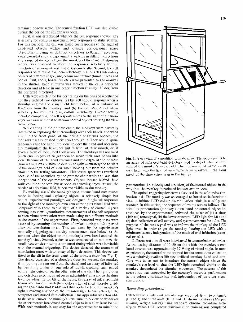
By making use of the monkey's spontaneous hand movements made in feeding and exploring objects, a relatively simple but natural experimental paradigm was designed. Single cell responses to the sight of the monkey's own arm entering its visual field were compared with those to the sight of a variety of control objects coming into view. Quantitative measurements of the cell responses to such visual stimulation were made using two different methods in the course of the experiments. First, neuronal responses were assessed by counting the number of spikes during a 1.0-s period after the stimulation onset. This was done by the experimenter manually triggering cell activity measurement (see below) at the moment when the object or the monkey's own hand entered the monkey's view. Second, a device was constructed to minimize the small inaccuracies in stimulation onset timing which were inevitable with the manual triggering. The device detected the moment of stimulation onset with an array of light detectors. This device was fitted to the slit in the front panel of the primate chair (see Fig. 1). The device consisted of a closeable door (to prevent the monkey from putting its arm out from the chair) and an array of infra-red light-emitting diodes on one side of the slit opening, each paired with a light detector on the other side of the slit. The light diodes and detectors were mounted on an adjustable frame above the door hole. By adjusting the tilt of the frame, the array of infra-red light beams were lined up with the monkey's line of sight, thereby divid- ing the space into that visible and that occluded from the monkey's sight. Breaking any one of the infra-red light beams activated the computer and started data collection. This apparatus was thus able to detect whenever the monkey's arm came into view or whenever the experimenter introduced control objects into view from below. With both methods, it was easy for the experimenter to mimic the
\ ,
Fig. 1. A drawing of a modified primate chair. The arrow points to an array of infra-red light detectors used to detect when stimuli entered the monkey's visual field. The monkey could introduce its own hand into the field of view through an aperture in the front panel of the chair (dark area in the figure)
presentation (i.e. velocity and direction) of the control objects in the way that the monkey introduced its own arm in view.
The optical triggering device was also used in the colonr discrim- ination task. The monkey was encouraged to introduce its hand into view to initiate LED colour discrimination trials in a self-paced manner. In this setting, the sequence of events was as follows. The stimulus presentation (monkey's own hand or control object in- troduced by the experimenter) activated the onset of (a) a short (100-ms) tone signal, (b) the lower or central LED light for 1.0 s and (c) data collection of cell activity and eye movements for 1.0 s. The purpose of the tone signal was to inform the monkey of the LED light onset in order to get the monkey fixating the LED with a minimum latency independent of the mode of trial initiation (exter- nal or self).
Different test stimuli were interleaved in counterbalanced order. At the testing distance of 10-20 cm the width the monkey's own hand covered was approximately 17-9 deg of visual angle. In most experiments, the control stimulus used for the actual data collection was a relatively realistic life-size artificial monkey hand and arm. Care was taken not to introduce the control object above the monkey's eye level so that the LED light remained visible to the monkey throughout the stimulus movement. The success of this precaution was supported by the monkey's accurate performance in the colour discrimination task independent of the other visual stimulation.
Recordin9 procedures
Extracellular single unit activity was recorded from two female (F and J) and three male (B, D and H) rhesus monkeys (Macaca mulatta, weight 4-8 kg) using standard chronic recording tech- niques. When LED colour discrimination training was completed

120
each monkey was sedated with a weight-dependent dose of intra- muscular ketamine (10 mg/kg i.m.) and anaesthetized with intrave- nous pentobarbitone sodium (Sagatal 25 mg/kg i.v.). Full sterile precautions were then employed while two stainless steel recording wells (16 mm internal diameter, ID) were implanted 10 mm anterior to the interaural plane and 12 mm to the left and right of midline. Plastic tubes (5 mm ID) were fixed horizontally with dental acrylic in front of and behind the wells. Metal rods could be passed through these tubes to restrain the monkey's head during recording sessions.
Two weeks after implantation, the subjects were retrained to perform the discrimination task for 1 4 h in the primate chair with head restraint. For each recording session, topical anaesthetic [lig- nocaine hydrochloride (Xylocaine, 40 mg/ml)] was applied to the dura and a David Kopf micro-positioner fixed to the recording well. On each recording track a guide tube (outer diameter, OD, 1.0 mm) was inserted 3-5 mm through the dura and a tungsten in glass microelectrode (OD 0.5mm, Merrill and Ainsworth 1972) ad- vanced with a hydraulic micro-drive to the temporal cortex. These procedures allowed recordings to be made repeatedly (over periods of up to 2 years) without intracranial infection. The target area for recording was the STP in the anterior parts of the dorsal superior temporal sulcus (Bruce et al. 1981).
Data collection and analysis
The cell activity was amplified, filtered (bandpass 800-20 000 Hz), monitored with an oscilloscope and an audiomonitor, converted to TTL pulses by a spike processor (Digitimer D130), and sampled with a AT-compatible PC microcomputer every 5 ms (Hyundai 286 or Dell 386). The horizontal and vertical eye position signals were filtered, digitized every 5 ms, and stored together with the single unit activity on the computer hard disc. Quantitative measurements of cell responses to different type of visual stimuli and spontaneous activity were analysed with one-way ANOVA and post-hoc tests (protected least significant difference, PLSD; Snedecor and Coch- ran 1980).
In some experiments, the filtered cell activity, together with the eye movement signal and stimulus onset signal, were additionally recorded on a four-channel FM tape recorder (RACAL) for off-line analysis. This method also provided the most convenient way for inspecting of pre-stimulus cell activity for self-initiated trials. In some experiments, a close-up of the upper part of the primate chair from side view was filmed with a video camera and recorded on a 0.75-in. U-matic videotape. Afterwards, the film was played back, frame by frame, and the number of frames (25 frames/s) taken for the monkey's hand or control object to move a measured distance was recorded. Given the distance from the monkey's eyes to the stimuli, it was possible to calculate a reasonably accurate estimation of the retinal velocity for the movements of the hand-held control objects and the monkey's own arm.
Cell localization
After each recording track, frontal and lateral X-radiographs were taken to allow the position of the metal microelectrode to be recon- structed from subsequent histology. Reconstruction of electrode position was achieved by reference to the positions of micro-lesions (10 gA DC for 30 s) made at the end of some electrode tracks which were subsequently identified using standard histological techniques. In one monkey (D), additional markers used in calibration of electrode position were provided by micro-injection of anatomical tracers (horseradish peroxidase and the fluorescent dyes true blue and diamadino yellow) at the site of cell recording on three record- ing tracks. For these markers, the position of injection, recorded in X-radiographs, could be compared to the anatomical location of injection revealed through normal or fluorescence microscopy.
Following the last recording session, a sedating dose of keta- mine was administered followed by a lethal dose of barbiturate
anaesthetic. The monkey was then perfused transcardially with phosphate buffered saline and 4% glutaraldehyde/paraformal- dehyde fixative. The brain was removed and sunk in successively higher concentrations (10, 20 and 30%) of sucrose solution or 2% dimethylsulphoxide and 20 % glycerol (Rosene et al. 1986). Coronal sections (50 gm thick) were collected every 0.25 mm and subjected to routine histological procedures.
Results
General response properties
Movement-sensi t ive cells showing no selectivity for fo rm const i tuted between 5 % and 7 % o f all cells tested in the anterior por t ions o f the superior tempora l sulcus. Within this area, 47 neurons o f this type were isolated which fulfilled the requirements o f (a) lacking fo rm selectivity and (b) responding to the entry o f objects into the visual field f rom below. These were tested for possible differ- ences in responses to self-produced and externally p roduced moving visual stimuli.
Eighteen o f these cells were selective for stimulus mo t ion in view and eight cells were selective to entry into view. In the latter case there was no response to the cont inuous movemen t in view, but only a transient burs t o f activity to the stimulus entry into view. The remaining 21 cells responded weakly to static stimuli, with stimulus mot ion further increasing the activity. Typically, the cells responded over a wide range o f stimulus velocities (20-400 deg/s). Transient response type was more typical than sustained responses. W h e n stimuli were presented f rom behind a liquid crystal shutter, the cell responses were observed to occur with latencies o f 90-150 ms. Re- sponse habi tua t ion for the effective stimulus presenta t ion was no t observed, and the responses mainta ined their s trength for at least 10 consecutive identical stimulus presentations.
Figure 2 shows an example o f an STP cell sensitive to visual stimulus mot ion. The cell gave a transient response to stimulus mo t ion with a slight directional selectivity for movemen t up, whereas a static control object did no t increase the cell activity above spontaneous level.
Response selectivity for motion direction
T h o r o u g h tests for the directional selectivity o f the neu- rons examined here were no t per formed systematically across the cell popula t ion . O f the 26 cells tested for directionality, 14 were observed to be responsive to all directions o f mo t ion in the fronto-paral lel plane. Nine cells exhibited a preference for certain directions with three cells responding to only upwards movements . Cells with preferences for other directions o f movemen t were c o m m o n in the STP but were not included in the present experiments (Oram Perrett and Hietanen, in prepara- tion). This directional selectivity limited the number o f neurons to be studied, because the testing pa rad igm necessitated responses to upward movements .

3 0 0
I1) v
Q .
" " 150 e.. o CL U~ (U re
0
3001
~150 0~
t -
O
| re
A
C
" , " , " ' l l r | J - - , I,
0 0 .5
0 0.5
3 0 0
150
300]
150-
B
H
eL.. ' ' ' .- ' . 0 0.5
O-
D
t H
0 0.5 time (s)
12l
Fig. 2A-D. Peristimulus-time histo- grams of responses of an STP cell sen- sitive to stimulus motion. The re- sponses were collected by presenting the stimuli behind a shutter for 1 s. Sight of a static control object (B) did not increase the cell activity above spontaneous level (A), whereas the cell gave a strong response to the sight of the same control object moving up- wards. The cell exhibited an additional slight selectivity for direction. A con- trol object moving upwards (D) elicited a stronger response than the sight of the same control object moving down- wards (C) (protected least significant difference, PLSD, P<0.02). (overall effect of condition, one-way ANOVA: F3,x6 = 15.4, P<0.0005). The histo- grams show data collected from five trials. Bin width 20 ms in each histo- gram
Feature selectivity
As explained in Materials and methods, particular atten- tion was paid to the possibility that the observed differ- ences in cell responses might have been caused by visual selectivity for form or simple features. Forty-three of the cells fulfilled the criterion of lacking form selectivity completely. These 43 cells were found to exhibit indistin- guishable responses to a variety of laboratory objects as long as the object movement occurred in the cell's pre- ferred direction. A further 4 cells were discovered to exhibit some degree of feature selectivity. Two of these showed a selectivity for stimulus size at the testing dis- tance preferring large objects (e.g. a book) over smaller ones (e.g. a pen), but as the control objects presented to the monkey were matched in size to the monkey's arm, there was no reason to exclude these two cells from the data analysis. Two other cells showed a selectivity for form in that they responded equally well to many objects of differing visual characters, but not at all to faces. These two cells were also included in the data analysis again because the form selectivity present in the cells could not account for any response difference between the sight of the monkey's hand and control objects used for testing.
Differential responses to the sight of object motion and motion of own hand
Thirty-nine (83%) of the 47 cells tested exhibited differ- ences between responses to the sight of a control object moving and to the sight of the monkey's own arm moving into view in the same direction. Thirty-eight of these 39 cells failed completely to respond to the self- induced motion stimulation (i.e. cell responses were not significantly different from spontaneous activity). One cell did respond to the sight of the monkey's own hand, but still gave significantly stronger responses to external- ly moved objects. No cells were found responding selec- tively to the sight of the monkey's own arm movements. The remaining 17 % of the cells tested gave equally strong responses to both stimulus types.
Figure 3 shows histogram presentation of responses (spikes/s) of one cell (H40 27.82) to the sight of a control object and monkey's own hand moving in the same direction (upwards) and the cell's spontaneous activity. Quantitative measurements of the responsivity of this cell was collected by using both the manually triggered spike counting method and the light-detector device. Results collected with both methods showed the same pattern, with a significantly larger response to the sight of a control object moving than to the sight of the monkey's own arm moving or spontaneous activity. A two-way ANOVA performed on the data (with stimulation type and method of data collection as main factors) showed

122
50.
40.
~030. o
o .
�9
220
CD
10.
I manual tr iggering
optical tr iggering
own hand s.a.
\ \ \ \ \ \ \ \ \ \ \ \ \ \ \ \ \ \ \ \ \ \ \ \ \ \ \ \ \ \ \ ' - , \ \ \ \ \ \ \ \
% \ \ \ N . \ \ \
control object
Histogram Fig. 3. presentation of mean responses (spikes/s + 1 SEM) of one cell to the sight of a control object and the monkey's own hand moving upwards and the cell's spontaneous activity. The spike counting was triggered both manually and with a light-detector device. Results collected with both methods showed the same pattern with significantly larger responses to the sight of a control object moving than to the sight of the monkey's own arm moving or spontaneous activity (P<0.0005, each comparison). One-way ANOVA (manual triggering): F2,1a=12.8, P<0.003, number of trials per condition n = 7; one-way ANOVA (light detec- tor): F2,16=31.6, P<0.0005, n=7, 7, 5
a significant effect of stimulation type (F1,23=40.2, P<0.0005), but no effect of method (F1,23=0.37, P=0.551) and no interaction between stimulation type and method of data collection (F1,23=1.0, P=0.328) . Thus, testing the same cell with these two methods showed that the manually triggered spike counting method was accurate enough for catching the stimulus onset and neuronal response.
Responsiveness to external stimuli durin9 self-induced stimulation
It was essential to study whether the cells continued exhibiting visual responses to external stimuli while mon- key's own hand was in view. The reasons for this inves- tigation were twofold. First, it was possible that the mechanisms producing a lack of responsiveness to self- produced visual stimulation caused some kind of general cessation of processing all visual information to the re- corded cell. Second, the lack of responsiveness to the sight of the monkey ' s own arm might have been caused simply by an unstable recording during the monkey 's movements. Therefore, the monkey was encouraged to lift its a rm in view and at the same time the experimenter
40,
"30
, m
~20
~10'
--I- +
N o w n h a n d
exp's own hand hand &
exp's hand
s . a ,
Fig. 4. Mean response (-4- 1 SEM) of an STP cell to different visual stimuli. The experimenter's hand (and various control objects) entering the monkey's visual field from below elicited a response above the cell's spontaneous activity (s.a.), whereas the sight of the monkey's own arm entering into view in the same direction did not activate the cell. The presence of the monkey's own arm in view, however, did not affect the visual responses to the entry of control objects. In any case, the sight of the control object elicited responses significantly stronger than those elicited by the monkey's own hand moving in view or the cell's spontaneous activity (P < 0.0005, each comparison). One-way ANOVA: F3,19 = 51.4, P < 0.0005, n = 10, 8, 5,7
introduced stimuli into the monkey ' s visual field as de- scribed. All the seven neurons tested in this way con- tinued responding to external visual stimulation while the monkey 's own arm was moving and visible. Further, in all seven cases the cells exhibited no decrease in their responsiveness compared to the condition where only control objects presented by the experimenter were vis- ible.
Figure 4 presents the results of one such experiment. The cell (D201 30.02) responded briskly to the sight of the experimenter 's hand or control objects moving in the view in all directions. The presence or absence of the monkey 's own hand in sight at the same time did not make any difference to the responses to the experiment- er's hand moving in view. In both conditions, the sight of experimenter 's hand elicited significantly stronger re- sponses than monkey 's own hand moving in view or the cell's spontaneous activity.
Unexpected self-produced stimulation
In these experiments, the two lines of study described before were combined. Again, an object was introduced to the monkey 's field of vision, but this time the object

123
30.
20' ~.~
~. tO.
r r
+
-1
v+-q o b j e c t own o b j e c t s.a.
h a n d in own hand
Fig. 5. An STP cell responded to various objects (fur, glove, feather, black bar, model monkey arm and the experimenter's hand) enter- ing into view, but gave no response to the monkey's own hand entering the visual field. The cell gave a response when the monkey brought objects (e.g. a piece of apple) into view with its own hand. These responses were stronger than those to the sight of the mon- key's arm moving alone or spontaneous activity (P< 0.0005, each comparison), but weaker than responses to objects moved by the experimenter (P<0.004). The monkey did not know the visual appearance of the objects, which were placed in its hand out of sight and changed after each presentation. One-way ANOVA: F3,26= 32.0, P<0.001, n=10, 8, 5, 7
mot ion was caused by the monkey itself. An object, e.g. a small piece of food was put into the monkey 's hand out of sight and then the monkey was allowed to bring the object in its hand into view. On each trial, a new object was placed into the monkey 's hand. Seven cells were studied with this procedure, all o f which had proved unresponsive to the sight of the monkey 's hand alone in preliminary testing. Five of these cells did give responses when the monkey brought an object into view in its hand. Figure 5 shows results for one cell tested in this way. The cell (H5 26.18) responded to control objects (fur, glove, feather, black bar etc.) and the experimenter 's hand alone entering into view, but gave no response to the monkey ' s own hand entering the visual field. The cell, however, responded to the monkey ' s hand bringing an object into view more strongly than to the monkey ' s a rm moving alone or spontaneous activity.
Eye movements during self-produced and external visual stimulation
Monitoring of eye movements was essential to ensure that differences in cell responses to the sight of stimuli moved by the experimenter or by the monkey were not caused by differences in fixation or tracking. For exam- ple, the monkey ' s eyes might follow movements caused
control object monkey's own arm
J
i
i l H I I I I I I I I I l i l l l l l l l I I I I I I I I I l l l l l I I I I I I I I I I I I I I I I I I I I I I I I I I I I I I I I I I I I I I I I I II I I I
I I I I I I I I I I I I I I I I I I I I I I I I I I I I I I I I I I I I I I ~ I~ l l l I I l l , . , I I I I I I I I I I I I I I I I I I I I I I I I I I I I I I I I
I i r I [ I
100
0 0 500
- - f L _ _
" - ' , - - _ F
I I I I I I
I I I I I I i I I [ ]
ii I I I I II I il I I I I I I I I I I I I
I I I I I I I I I I I I t l I i I
'~176 1
0 500 Time (ms)
Fig. 6. Vertical eye position tracks and activity of an STP cell which responded to the sight of an object entering the monkey's visual field (left column), but not to the sight of the monkey's own arm coming into view (right column). The vertical eye position is illustrated for five (ran- domly interleaved) trials in both conditions, and the sixth row shows the summed eye move- ments during these trials. The post-stimulus time rasterograms show spike activity (short vertical dashes) during these five trials retaining the same order. At the bottom the cell activity is depicted in post-stimulus time histograms (PSTHs). The ordinate axis in the eye position recordings gives a scale of =L 20 deg and the ordinate axis of the PSTHs shows cell respon- sivity in spikes/s

124
B
~a
C D
leee
E
Fig. 7. A lateral view of the right hemisphere of a rhesus macaque brain showing the major sulci. STS, superior temporal sulcus; IOS, inferior occipital sulcus; CS, central sulcus; IPS, intraparietal sul- cus; LS, lunate sulcus; AS, arcuate sulcus; PS, principal sulcus. The STS is opened to reveal the bottom and both banks of the sulcus. The two pairs of arrows show the interaural plane and a plane 20 mm anterior to it. B A coronal section of the right hemisphere showing the subareas within the STS according to Seltzer and Pandya (1978). C-E Three enlarged coronal sections of the STS
12 ram, 15 mm and 18 mm anterior to the interaural plane. The recorded cells were located between + 10 mm and + 20 mm along the rostro-caudal extent of the STS. For illustration, the cells stud- ied from both hemispheres which were located 10-14mm, 14-17 mm and 17-20 mm along this plane are shown in C, D and E, respectively. Thefilled circles mark the locations of cells respond- ing selectively to externally induced movement, and the open trian- gles mark the locations of cells failing to show this discrimination
by the experimenter but not the movements of its own hand.
Figure 6 presents vertical eye movements and spike activity of a cell to externally induced and self-induced visual stimulation. The testing was performed by using the light-detector device, and the cell was tested with the upward movement of the control artificial monkey arm or the monkey 's own arm. During this particular testing, the LED fixation light which was usually located at the level of monkey ' s sight (see Materials and methods) was switched to a bo t tom position (10 deg below central position). In this way the monkey was biased to direct its gaze in the direction f rom which the stimulus would appear.
Figure 6 illustrates that the monkey made a variety of different fixations and saccades during the presentation of the control stimulus by the experimenter. Despite this
range of eye movements, the cell responded on every trial. When the monkey initiated the trial by bringing its own arm into view, eye movements again showed the same variation in pat tern of fixation, saccades and track- ing, yet in this condition the cell always remained unre- sponsive. It is also evident f rom the recording that the cell's response was not modulated in any obvious way by the presence of saccades. It might be anticipated that the monkey would be more interested in the movement of control stimuli than its own hand but the eye movement records indicate a comparable interest (or disinterest) in both stimuli.
Thus eye movements did not cause difference to the responses to self-induced movement and object motion. Cell response or lack of response was dependent on the stimulus type and independent of relatively large varia- tions in the eye movement patterns.

125
Location of cells
Histological reconstruction in monkeys B, F and D in- dicated that 34 of the 38 tested cells in these monkeys were located in area STP (areas TPO and PGa of Seltzer and Pandya 1978). X-ray measurements of recording positions in two other monkeys (two in subject H and seven in subject J) indicated that the tested cells from these subjects were also located within the same area. Thus, from the histological evidence and reconstructions based on X-ray measurements, a total of 43 cells (out of the 47 tested) were recorded from areas TPO and PGa. Of these, 37 (86%) exhibited selective responses for ex- ternally induced motion. The cells described here were within the same area as those responsive to the static views of the head that have been described in earlier studies (Perrett et al. 1982, 1984, 1991).
Even though the recordings were aimed at the dorsal bank of the superior temporal sulcus, histological recon- struction indicated that four of the studied cells were in the ventral bank of the superior temporal sulcus. In monkey F, the two cells showed selective responses to the externally-induced motion, but in monkey D both cells responded equally well to the sight of the monkey's own hand and moving control objects. Figure 7 shows the results of the histological reconstruction in monkey (D) from which the majority of the cells were recorded (27/47).
Discussion
Neurophysiological studies of single cell responses in the anterior parts of the dorsal superior temporal sulcus have almost exclusively concentrated on selective responses to complex visual stimuli. The most frequently studied cell type has been the one responsive to the face and other views of the head (Bruce et al. 1981; Perrett et al. 1982, 1984, 1985b, 1991; Desimone et al. 1984; Rolls 1984; Baylis et al. 1985; Rolls and Baylis 1986; Hasselmo et al. 1989; Hietanen et al. 1992). Cells with highly selective responses to specific body movements (e.g. walking in one direction with one body view) have also been studied extensively (Perrett et al. 1985a, 1989, 1990a, b). How- ever, the earliest single-cell studies of this area, in anaes- thetized monkeys, reported the existence of visually re- sponsive cells sensitive to stimulus motion but lacking any selectivity for form (Desimone and Gross 1979; Bruce et al. 1981).
The existence of cells lacking form selectivity within STP seems rather surprising, for two reasons. First, from the point of view of object recognition it is very difficult to think what the functional value of units which respond to all moving objects would be. Second, from the point of view of motion processing it is difficult to think of a functional role of motion-sensitive cells lacking direc- tional tuning or pronounced velocity sensitivity at a stage of analysis after very detailed processing of motion in- formation has been performed earlier in the "motion pathway" within posterior parts of the same sulcus (i.e. areas MT and MST).
It appears, however, that when studied in awake, behaving monkeys, STP neurons non-selective for form but sensitive to movement have very complex selectivity discriminating between externally induced stimulus mo- tion and visual movement which results from the ani- mal's own action. A very high percentage of the cells studied in the STP (86%) responded to the sight of any object moved into view by the experimenter, but failed to respond above spontaneous activity to the sight of the monkey's own hand and arm movements.
It could be argued that the differences in responsivity to these two classes of stimuli reflects the effects of arous- al rather than discriminative sensory processing between externally induced and self-induced stimulation. This possibility, however, seems very unlikely. First, this kind of explanation has been considered for STP cells respon- sive to faces and no evidence for relation to arousal has been found (Perrett et al. 1982, 1989). Second, if the responses to a moving control object were only due to its arousing nature, one would expect a sight of a static face or food to evoke a comparable "arousal response". This was not the case, as cells studied here did not respond to such static stimuli. Third, the neuronal responses were time-locked to the visual stimulation, occurred at short latencies and were transient in most cases. Fourth, the responses to motion exhibited different types of direc- tional selectivity. All these response characteristics are unlikely if the cells merely reflected arousal.
The observed response discrimination might reflect differences in interest or the attention paid to the exter- nally-moved control objects and monkey's own hand. Eye movements can be used as an indicator of interest in the moving stimuli. Records of the monkey's eye move- ments indicated that the monkey's fixation of its own hand and control objects was equivalently variable. Re- cordings therefore do not indicate one stimulus class as more interesting. Eye position recordings also showed that the responses were not related to the eye movements (Fig. 6). The observed lack of habituation in responses to control objects moving into the field of view also speaks against the responses being related to the level of interest.
In order to get insight into the functions of the STP cells studied here, the results are discussed in the context of neurophysiological studies of cells in other brain areas which resemble the present experiments.
Visual guidance of hand actions
The posterior parietal cortex has been shown to be heavi- ly involved in combining visuo-spatial and motor in- formation and in visual guidance of hand projections (Hyvfirinen and Poranen 1974; Mountcastle et al. 1975). In a recent study, Taira et al. (1990) found that a majority (69%) of their "hand-movement related" neurons showed activity changes in response to hand manipula- tion, with the activity of the cells being greater when the hand movement took place in light. This was taken as an evidence that these cells received a motor input as well

126
as a visual input related to the object and/or the moving hand. Furthermore, a role in monitoring (rather than in commanding, cf. Mountcastle et al. 1975) the ongoing motor activity was assigned to the parietal neurons by Taira et al. (1990).
In contrast, the present study showed that the cells in the anterior dorsal bank of the superior temporal sulcus selectively failed to respond to the sight of the monkey's own arm movements. This was also the case when the monkey projected its arm and hand into view in order to reach for a piece of food, i.e. during goal-directed move- ments under visual guidance. It is noteworthy that cells selective for the sight of manipulative hand actions found from the ventral bank of the superior temporal sulcus (area TEa) do respond to the sight of the monkey's own hands performing the appropriate hand actions (Perrett et al. 1990c).
Some of the properties of the STP cells described here may well depend on interconnections with the parietal cortex (Seltzer and Pandya 1978, 1984; Pandya and Selt- zer 1982; Morel and Bullier 1990; Baizer et al. 1991). These connections may provide the inputs required for the STP cells to "ignore" the monkey's own limb move- ments. It is interesting to consider what kind of informa- tion the STP cells require for the observed response selectivity. In the parietal processing the "hand- movement-related" neurons combine the visual and mo- tor/kinaesthetic information to produce maximal re- sponses during visually guided hand movements. By con- trast, the processing performed within STP suggest that visual input and motor/kinaesthetic signals work antago- nistically, the motor/kinaesthetic input inhibiting the vi- sual responses to the sight of own arm movements. This inhibition, however, must be very selective. In the experi- ments where a control object was introduced to the visual field while the monkey's own arm was simultaneously moving in view, the neurons continued responding to the sight of control object motion. The inhibition does not prevent all visual processing in the STP cells, but acts selectively (perhaps presynaptically) on the visual motion signal resulting from the own hand movements. Therefore, the inhibition must contain information about the position, trajectory and velocity of the limb motion in three-dimensional space. Kinaesthetic in- formation may well be used additionally to give an accu- rate description about the ongoing motor activity. To match the visual input the motor/kinaesthetic signals about hand movements in three-dimensional space must be converted to a retinal coordinate system and this necessitates that the dynamic head and eye position must also be taken into account. As posterior parietal cortex is known to be heavily involved in these functions (see Andersen 1989), it seems highly possible that this in- formation is used as an inhibitory input and fed either directly or indirectly to the STP cells. In summary the discrimination against predictable stimuli that is ex- hibited by STP cells requires very complex and con- tinuous mapping of expectations about the form, posi- tion and direction of moving objects within the world to the appropriate coordinates within the continuously moving visual receptive fields.
Processin9 of visual motion which results from eye movements
As described in the Introduction, neurons in several visual areas have been observed to respond to object motion but not to retinal motion stimulation which is caused by the animal's own eye movements. It is interest- ing that the discrimination against self-produced stimula- tion is increasingly pronounced at higher levels of motion processing in areas MT and MST (Erickson and Thier 1991). From this trend, one might expect to see the more complex effects of stimulus predictability (of the type we have studied here) only in the anterior areas of the superior temporal sulcus. It should be noted, however, that throughout the visual system high level areas send back projections down to particular lower areas (Felle- man and Van Essen 1991). These selective back connec- tions from MSTd to MT, V3a and V2 (but not V4) might well mediate the influences of eye motion observed in V 1, V2 and V3a by Galletti et al. (1984, 1988, 1990).
In most cases, the spontaneous activity of the real- motion neurons in V3A is not affected by tracking move- ments alone which has been interpreted as suggesting that the eye-motion input selectively inhibits the visual input reaching the real-motion cells (GaUetti et al. 1984, 1988, 1990). In this respect, the results of the present study are comparable. The movements of the monkey's own hand were not observed to have any effects on the cell's spontaneous activity, and, more importantly, the presence of the monkey's own hand in view did not affect the responsivity to simultaneous externally induced mo- tion. Thus, the inhibition must have acted selectively on the visual input carrying information about the appear- ance and spatial movements of the monkey's own hand. Indeed, it would be a very maladaptive neural mecha- nism which shuts down the processing of all external information during self-induced stimulation.
In respect to perceptual experience, the responses of real-motion cells offer a physiological basis for the stabil- ity of the visual world despite self-induced eye move- ments. There is not, however, such a clear difference in perception of our own limb movements and the motion of external objects. What, then, could be the functions of the STP cells we have recorded from?
Expectation
Recordings from the parietal cortex, superior temporal sulcus and frontal cortex of monkeys revealed bimodal cells which gave a visual response whenever the body part corresponding to the cell's tactile receptive field was approached by the investigator as though contact would be made (Hyviirinen and Poranen 1974; Sakata 1975; Leinonen et al. 1979; Leinonen 1980; Rizzolatti et al. 1981; MacKay and Crammond 1987; Gentilucci et al. 1988 ; Mistlin and Perrett 1990). As the visual and tactile receptive fields coincide, it has been suggested that the function of these neurons is essentially predictive, i.e.

127
they provide information about the impending tactile collision and prepare the animal for an adequate behav- ioural reaction.
The response properties of the apparent ly unimodal somatosensory neurons in the STP were found to be very complex (Mistlin and Perrett 1990). Even though visual stimuli alone did not have any effects on the cell re- sponses, if the monkey was allowed to see the approach- ing object before skin contact the responses were re- duced. Moreover, not only visual information but also previous experience with the tactile surroundings was capable of inhibiting the effects on the tactile responses. Tactile stimulation resulting f rom active exploration of a familiar pr imate chair failed to drive these cells, but as soon as the monkey contacted a novel surface the cells responded vigorously. It was proposed that the respon- sivity of the tactile STP neurons reflected the "expecta- tion" of the stimulation (Mistlin and Perrett 1990).
The results of the present study can be interpreted in the context of the effects of expectation as well. When the monkey raised its a rm and empty hand into view, the visual appearance of the hand and arm was predictable and hence the cells did not respond, but when an object was placed in the monkey ' s hand out of view and the monkey did not know the visual characteristics of ob- jects, the visual appearance of the compound stimulus (hand + object) was unpredictable and the cells respond- ed accordingly when the compound stimulus came in sight.
I f the observed response selectivity were based on this kind of expectation, it would mean that some type of matching process must be performed. The mechanisms performing this matching would have to be supplied with information on the visual appearance of the monkey ' s a rm and hand (the STP is known to contain cells selective to hands: Bruce et al. 1981; Perrett et al. 1989) and this "expected" image would be compared to the input carry- ing information about the actual visual stimulation. I f they coincided, they would cancel each other. As the cells we recorded f rom did not modify firing to the sight of monkey ' s own a rm this means that the actual "com- parison" was performed on the inputs to the recorded cell or at an earlier stage "upstream".
We have also investigated responses to predictable visual mot ion of objects other than the monkey ' s own arm (Hietanen and Perrett, in preparation). One monkey has been taught to turn a handle (out of sight) which connects to a cylinder covered with a visible black/white striped grating pattern. Eleven cells (of 18 tested) were found to give stronger responses to the grating rotat ion when the movement was controlled by the experimenter than when the same grating movement was produced by the monkey turning the handle. These results imply that (at least in some cases) it is the predictability of visual movement rather than the specific visual characteristics of the stimulus which controls the responses of STP cells to motion.
Conceptually, the results show parallels with the effect of expectation on somatosensory processing in the STP (Mistlin and Perrett 1990). Unexpected sensory events usually derive f rom other animals and are therefore be-
haviourally important . By contrast, predictable sensory consequences of an animal 's own actions do not gener- ally require reaction. Cells selectively responsive to the sight of faces and body movements have also been found in the STP. The STP therefore appears to be well suited as a filter for behaviourally and socially relevant sensory events.
Acknowledgements. We acknowledge the help of M.W. Oram, M.H. Harries and P.J. Benson with the physiological recordings. This research was funded by grants from the SERC (GR/F 96723) and ONR (United States) and NEDO (Japan). J.H. was supported by the Pirkanmaan Kulttuurirahasto, Kordelinin S/i/iti6, Aaltosen S/i~iti6, and Tampereen Kaupungin Tiederahasto (Finland), and D.P. by a Royal Society University research fellowship.
References
Andersen RA (1989) Visual and eye movement functions of the posterior parietal cortex. Ann Rev Neurosci 12:377-403
Baizer JS, Ungerleider LG, Desimone R (1991) Organization of visual inputs to the inferior temporal and posterior parietal cortex in macaques. J Neurosci 11:168-190
Baylis GC, Rolls ET, Leonard CM (1985) Selectivity between faces in responses of a population of neurons in the cortex of the superior temporal sutcus of the macaque monkey. Brain Res 342:91-102
Bruce C, Desimone R, Gross CG (1981) Visual properties of neu- rons in a polysensory area in superior temporal sulcus of the macaque. J Neurophysiol 46:369-484
Desimone R, Gross CG (1979) Visual areas in the temporal cortex of the macaque. Brain Res 178 : 363-380
Desimone R, Albright TD, Gross CG, Bruce C (1984) Stimulus- selective properties of inferior temporal neurons in the macaque. J Neurosci 8 : 2051-2062
Erickson RG, Thier P (1991) A neuronal correlate of spatial during periods of self-induced visual motion. Exp Brain Res 86:608-616
Felleman DJ, Van Essen DC (1991) Distributed hierarchical processing in the primate cerebral cortex. Cerebral Cortex 1:147
Fischer B, Boch R, Bach M (1981) Stimulus versus eye movements: comparison of neural activity in the striate and prelunate visual cortex (A17 and A19) of trained monkey. Exp Brain Res 43 : 69-77
Galletti C, Squatrito S, Battaglini PP, Maioli MG (1984) 'Real- motion' cells in the primary visual cortex of macaque monkeys. Brain Res 301:95-110
Galletti C, Battaglini PP, Aicardi G (1988) 'Real-motion' cells in visual area V2 of behaving macaque monkeys. Exp Brains Res 69: 279-288
Galletti C, Battaglini PP, Fattori P (1990) 'Real-motion' cells in area V3A of macaque visual cortex. Exp Brain Res 82:67-76
Gentilucci M, Fogassi L, Luppino G, Matelli M, Camarda R, Rizzolatti G (1988) Functional organization of inferior area 6 in the macaque monkey. I. Somatotopy and the control of proximal movements. Exp Brain Res 71:475490
Gross CG, Rocha-Miranda CE, Bender DB (1972) Visual proper- ties of neurons in the inferotemporal cortex of the macaque. J Neurophysiol 35:96-111
Grtisser O-J (1986) Interaction of efferent and afferent signals in visual perception: a history of ideas and experimental par- adigms. Acta Psychol 63:3-21
Hasselmo ME, Rolls ET, Baylis GC, Nalwa V (1989) Object- centred encoding by face-selective neurons in the cortex in the superior temporal sulcus of the monkey. Exp Brain Res 75:417-429
Hietanen JK, Perrett DI, Oram MW, Benson PJ, Dittrich WH (1992) The effects of lighting conditions on responses of cells

128
selective for face views in the macaque temporal cortex. Exp Brain Res 89:157-171
Holst E yon, Mittelstaedt H (1950) Das Reafferenzprinzip (Wech- selwirkungen zwischen Zentralnervensystem und Peripherie). Naturwissenschaften 37:464-476
Hyv/irinen J, Poranen A (1974) Function of the parietal associative area 7 as revealed from cellular discharges in alert monkeys. Brain 97: 673-692
Leinonen L (1980) Functional properties of neurones in the pos- terior part of area 7 in awake monkeys. Acta Physiol Scand 108:301-308
Leinonen L, Hyv/irinen J, Nyman G, Linnankoski I (1979) Func- tional properties of neurons in lateral part of associative area 7 in awake monkey. Exp Brain Res 34:299-320
MacKay DM (1973) Visual stability and voluntary eye movements. In: Jung R (ed) Handbook of sensory physiology, vol VII/3A. Springer, Heidelberg, Berlin New York, pp 307-332
MacKay WA, Crammond DJ (1987) Neuronal correlates in pos- terior parietal lobe of the expectation of events. Behav Brain Res 24:167-179
Merrill EG, Ainsworth A (1972) Glass-coated platinum-plated tungsten microelectrodes. Med Biol Eng 10:662-672
Mistlin AJ, Perrett DI (1990) Visual and somatosensory processing in the macaque temporal cortex: the role of expectation. Exp Brain Res 82:437-450
Morel A, Bullier J (1990) Anatomical segregation of two cortical visual pathways in the macaque monkey. Vis Neurosci 4:555-578
Mountcastle VB, Lynch JC, Georgopoulos A, Sakata H, Acuna C (1975) Posterior parietal association cortex of the monkey: command functions for operation within extrapersonal space. J Neurophysiol 38:871-908
Miiller-Preuss P (1983) Inhibition of auditory neurons during phonation: evidence of feed-forward mechanisms in brain processes controlling audio-vocal behavior. In: JP Ewert, RR Capranica, JP Ingle (eds) Advances in vertebrate neuroethol- ogy. NATO ASI Series, pp 919-923
Miiller-Preuss P (1986) On the mechanisms of call coding through auditory neurons in the squirrel monkey. Eur Arch Psychiat Neurol Sci 236 : 50-55
Pandya DN, Seltzer BJ (1982) Intrinsic connections and architec- tonics of posterior parietal cortex in the rhesus monkey. J Comp Neurol 204:196-210
Perrett DI, Rolls ET, Caan W (1982) Visual neurons responsive to faces in the monkey temporal cortex. Exp Brain Res 47: 329-342
Perrett DI, Smith PAJ, Potter DD, Mistlin A J, Head AS, Milner AD, Jeeves MA (1984) Neurons responsive to faces in the temporal cortex: studies of functional organization, sensitivity to identity and relation to perception. Hum Neurobiol 3 : 197-208
Perrett DI, Smith PAJ, Mistlin A J, Chitty A J, Head AS, Potter DD, Broennimann R, Milner AD, Jeeves MA (1985a) Visual analysis of body movements by neurons in the temporal cortex of the macaque monkey: a preliminary report. Behav Brain Res 16:153-170
Perrett DI, Smith PAJ, Potter DD, Mistlin AJ, Head AS, Milner AD, Jeeves MA (1985b) Visual cells in the temporal cortex sensitive to face view and gaze direction. Proc R Soc (Lond) B 3:293-317
Perrett DI, Harries MH, Bevan R, Thomas S, Benson PJ, Mistlin AJ, Chitty AJ, Hietanen JK, Ortega JE (1989) Frameworks of analysis for the neural representation of animate objects and actions. J Exp Biol 146:87-114
Perrett DI, Harries MH, Benson PJ, Chitty AJ, Mistlin AJ (1990a) Retrieval of structure from rigid and biological motion: an analysis of the visual responses of neurons in the macaque temporal cortex. In: Troscianko T, Blake A (eds) AI and the eye. Wiley, Chichester, pp 181-201
Perrett DI, Harries MH, Chitty A J, Mistlin AJ (1990b) Three stages in the classification of body movements by visual neurons. In : HB Barlow, C Blakemore, M Weston-Smith (eds) Images and understanding. Cambridge Univ Press, Cambridge, pp 94-108
Perrett DI, Mistlin AJ, Harries MH, Chitty AJ (1990c) Understand- ing the visual appearance and consequences of hand actions. In: Goodale MA (ed) Vision and action: the control of grasping. Ablex Publishing Corp, Worwood, NJ, pp 163-180
Perrett DI, Oram MW, Harries MH, Bevan R, Hietanen JK, Ben- son PJ, Thomas S (1991) Viewer-centred and object centred encoding of heads by cells in the superior temporal sulcus of the rhesus monkey. Exp Brain Res 86:159-173
Richmond BJ, Wurtz RH (1980) Vision during saccadic eye move- ments. II. A corollary discharge to monkey superior colliculus. J Neurophysiol 43 : 1156-1167
Rizzolatti G, Scandolara C, Matelli M, Gentilucci M (1981) Affer- ent properties of periarcuate neurons in macaque monkeys. II. Visual responses. Behav Brain Res 2:147-163
Robinson DL, Petersen SE (1992) The pulvinar and visual salience. Trends Neurosci 15 : 127-132
Robinson DL, Wurtz RH (1976) Use of an extraretinal signal by monkey superior colliculus neurons to distinguish real from self-induced stimulus movement. J Neurophysiol 39:852-870
Rolls ET (1984) Neurons in the cortex of the temporal lobe and in the amygdala of the monkey with responses selective for faces. Hum Neurobiol 3 : 209-222
Rolls ET, Baylis CG (1986) Size and contrast have only small effects on the responses to faces of neurons in the cortex of the superior temporal sulcus of the macaque monkey. Exp Brain Res 65:38-48
Rosene DL, Roy N J, Davis BJ (1986) A cryoprotection method that facilitates cutting frozen sections of whole monkey brains for histological and histochemical processing without freezing arti- fact. J Histochem Cytochem 34:1301-1315
Roy J-P, Wurtz RH (1990) The role of disparity-sensitive cortical neurons in signalling the direction of self-motion. Nature 348:160-162
Sakata H (1975) Somatic sensory responses of neurons in the pari- etal association area (area 5) of monkeys. In: Kornhuber HH (ed) The somatosensory system. Thieme, Stuttgart, pp 250-261
Seltzer B, Pandya DN (1978) Afferent cortical connections and architectonics of the superior temporal sulcus and surrounding cortex in the rhesus monkey. Brain Res 149:1-24
Seltzer B, Pandya DN (1984) Further observations on parieto- temporal connections in the rhesus monkey. Exp Brain Res 55:301-312
Snedecor GW, Cochran WG (1980) Statistical methods, 7th edn. Iowa State University Press, Ames, Iowa
Sperry RW (1950) Neural basis of the spontaneous optokinetic response produced by visual neural inversion. J Comp Physiol Psychol 43 : 482-489
Straschill M, Hoffmann KP (1970) Activity of movement sensitive neurons of the cat's tectum opticum during spontaneous eye movements. Exp Brain Res 11 : 318-326
Suga N, Schlegel P (1972) Neural attenuation of responses to emit- ted sounds in echolocating bats. Science 177:82-84
Suga N, Shimozawa T (1974) Site of neural attenuation of re- sponses to self-vocalized sound in echolocating bats. Science 183:1211-1213
Taira M, Mine S, Georgopoulos AP, Murata A, Sakata H (1990) Parietal cortex neurons of the monkey related to the visual guidance of hand movement. Exp Brain Res 83 : 29-36
Toyama K, Komatsu Y, Shibuki K (1984) Integration of retinal and motor signals of eye movements in striate cortex cells of the alert cat. J Neurophysiol 51:649-665