exploring molecular mechanisms of itch and touch in the...
TRANSCRIPT

Exploring molecular mechanisms of itch and touch in the mammalian somatosensory system.
By Kristin Anne Thompson Gerhold
A dissertation submitted in partial satisfaction of the requirements for the degree of
Doctor of Philosophy in
Molecular and Cell Biology in the
Graduate Division of the
University of California, Berkeley
Committee in charge:
Professor Diana M. Bautista, Chair Professor Kristin Scott Professor Marla Feller
Professor Song Li
Fall 2013


1
Abstract by
Kristin Anne Thompson Gerhold Doctor of Philosophy Molecular and Cell Biology
University of California, Berkeley Professor Diana Bautista, Chair
Unlike other sensory systems, the somatosensory system is required for encoding information about multiple sensory modalities. Somatosensory neurons can respond to a variety of different stimuli including temperature, pressure and chemical irritants. These neurons are a heterogeneous population and utilize a variety of molecular pathways to transduce somatosensory stimuli. Because of this diversity of pathways and the diversity of sensory neuron subtypes, our understanding of transduction machinery in the somatosensory system lags behind our understanding of other sensory systems. We have worked to establish new tools for identifying new candidate molecules involved in transducing somatosensory stimuli and to test the role of these molecules in vitro and in vivo. Our work has focused on the transduction of two types of stimuli: mechanical forces that do not cause pain and chemical irritants that elicit sensations of itch. We have shown that TRPA1 is a convergence point for multiple itch causing compounds. We have also generated a new set of putative mechanotransducers by exploiting the extreme specialization of the hypertrophied epidermis of the star organ in the star-nosed mole. We have tested the role of one of these candidates, Cnga2, in somatosensation in mice. Our data suggests that Cnga2 plays a role in texture discrimination though altering the mechanical responses of somatosensory neurons.
i
ACKNOWLEDGEMENTS For contributing to Chapter I, I would like to thank Dr. Marion Kollarik for advice on single cell PCR, Dr. Rachel Brem for use of equipment, Takeshi Morita for technical support and Drs. Brem, Ngai, Sack, for helpful discussions and critical reading of the manuscript. For contributing to Chapter II, I would like to thank Dr. Allan Basbaum for generously providing the substance P antibody and Dr. John Ngai and members of my labaratory for helpful discussions. For contributing to Chapter III, I would like to thank Dr. Diego Restrepo for providing us with the Cnga2 knockout mouse line, Dr. Weihong Lin for performing preliminary staining of Cnga2-GFP mouse tissue and Dr. John Ngai and members of his laboratory for advice on care and breeding of Cnga2 mice. I would also like to thank my committee Drs. Feller, Scott and Li for critical reading of the manuscript and Blair Citron for editing and formatting.
ii
TABLE OF CONTENTS
• Introduction iii-xvi o The Somatosensory System iii o Fiber Types iii-x o Mechanotransduction x-xv o Directions of Study xv-xvi
• Chapter I: TRPA1 is required for histamine-independent, Mas-related G
protein-coupled receptor-mediated itch. 1-19 o Summary and Introduction 1-3 o Results 3-7 o Discussion 7-9 o Experimental Procedures 9-11 o Figures 1-6 12-17 o Supporting Figure 1 18
• Chapter II: The star-nosed mole reveals cues to the molecular basis of
mammalian touch. 19-36 o Summary and Introduction 19-20 o Results 20-23 o Discussion 23-25 o Experimental Procedures 25-28 o Figures 1-5 29-33 o Supporting Figures 1-2 34-35 o Supporting Table 1 36
• Chapter III: The Role of CNGA2 in mammalian mechanotransduction. 37-49
o Summary and Introduction 37-38 o Results 38-40 o Discussion 41 o Experimental Procedures 42-44 o Figures 1-5 45-48
• Summary 49
• References 50-63

iii
THE SOMATOSENSORY SYSTEM In this introduction I will summarize the current understanding of the molecular mechanisms of sensory transduction in the mammalian somatosensory system, focusing on mechanotransduction. My work expands on previous work—both in vitro and in vivo—that has attempted to characterize the function of specific transduction proteins from the cellular level to the behavioral. Our understanding of these mechanisms lags behind our understanding of other sensory systems for two major reasons: the diversity of subtypes of sensory neurons and the anatomy of the system. Sensory systems are typically specialized to encode information about a specific sensory modality. For example, the visual system is adapted to respond to light, the auditory system to mechanical forces, the taste system to soluble chemical ligands, and the olfactory system to volatile chemicals. Each of these sensory systems utilizes a specialized cell type and set of molecular pathways to transduce these physical stimuli into signals that can be interrupted by the brain. The somatosensory system responds to not one, but three different types of stimuli: mechanical forces, temperature and chemical ligands. The primary detectors in this system are a set of specialized primary sensory neurons. These neurons, however, are not a single homogeneous population and show differing receptor expression profiles, innervation patterns, and second order connections depending on the types of stimuli they respond to. This means that there are often multiple non-overlapping molecular mechanisms within different cell types that contribute to the same sensation. The cell bodies of primary somatosensory neurons reside in the dorsal root ganglia (DRG) and trigeminal ganglia (TG). These neurons are pseudo-bipolar and send long afferent projections to the skin and viscera where detection of stimuli occurs. They also send efferent projections to either the dorsal horn of the spinal cord (in the case of DRG neurons, where they form synapses on spinal cord interneuron that project to the brain stem and thalamus) or directly to the brain stem (in the case of TG neurons). In the skin the primary afferent fibers terminate either as free nerve endings or in complex with epidermal lineage structures such as Merkel cells, lamellated corpuscles, or hair follicles. It has thus far been impossible to record receptor potentials in vivo in mammals because of the anatomy of this system, specifically the distance between the point of detection and cell bodies and the diffuse localization of the terminals in their target organs. This means that the characteristics of transduction channels must be extrapolated from what can be observed in in vitro experiments. FIBER TYPES Integrating what is known about sensation in vivo and what can be discovered about transduction in vitro is not always a straightforward task. For example, estimates of the number of types of primary somatosensory neurons vary greatly and depend both on the criteria used to define populations and whether the responses are measured in vitro or in vivo. To overcome this difficultly, somatosensation research has utilized a combination of in vivo (microneurography) and ex vivo (skin-nerve preparation) techniques that allow the measurement of sensory neuron action potential firing in

iv
response to stimulation of the skin. These techniques have grouped sensory neurons into three basic subtypes based on conduction velocity: unmyelinated slow conducting C-fibers, lightly myelinated intermediately conducting Aδ-fibers, and heavily myelinated fast conducting Aβ-fibers. These classes relate to the function of the fiber. C-fibers are generally polymodal and responsible for the sensation of throbbing pain. Aδ-fibers can be either polymodal or mechanosensitive and the majority contributes to the sensation of sharp pain. Aβ-fibers are mechanosensitive and contribute to the sensation of gentle touch and to proprioception. These fibers can be further subdivided based on their responses to the three types of somatosensory stimuli, temperature, mechanical force, and chemical ligands. In this section I will describe what is known about each of these subtypes at a molecular level and how these fibers are thought to contribute to somatosensation C-Fibers C-fibers are the most homogeneous of the three fiber subtypes. Most C-fibers that innervate the skin share characteristic innervation patterns: their afferent projections terminate in the epidermis as free nerve endings, they project from the DRG into the spinal cord, and they make connections with interneurons in the outer laminae (I-II) of the dorsal horn (1). C-fibers are predominantly polymodal, responding to a variety of temperatures, mechanical stimuli, and inflammatory chemicals. Within this population, however, there appear to be some fibers that play differing roles in pain sensation and itch. Inflammatory Fibers C-fibers that contribute to neurogenic inflammation and play a role in altering pain thresholds in injury and disease make up one of the best characterized C-fiber subpopulations. These neurons are peptidergic, meaning that they contain large dense core vesicles that release inflammatory peptides (including substance P and Calcitonin gene related peptide (CGRP)) at central and peripheral terminals. Peptidergic fibers share a common anatomy, terminating in the outermost layers of the epidermis and the most dorsal lamina of the spinal cord, lamina I. Activation of these fibers leads to release of the dense core vesicles which in turn leads to increased permeability of the vasculature, recruitment of immune cells in the skin, and sensitization at the central terminals. At a molecular level, these fibers express channels that respond to chemical nociofensive and inflammatory cues. For example, these fibers express the transient receptor potential (TRP) channel TRPA1 (2), acid sensing cation channels such as ASICs and HCN, and prurinergic receptors like P2X. TRPA1 is the best characterized of these channels. It is known to respond to a variety of exogenous and endogenous pro-inflammatory compounds (3, 4). Its activation can lead to release of peptides (5) and is required for the development of both inflammatory pain responses (6-8) and mechanical hyperalgesia in response to some inflammatory agents (6, 9). This suggests that the peptidergic population of C-fibers not only initiates inflammation, but also responds to it in a positive feedback loop that may play a role in some of the persistent alterations in pain threshold that occur with tissue damage and disease.

v
Heat Sensitive Fibers Heat sensitive C-fibers are important for the burning sensation caused by noxious heat. The majority of C-Fibers can be activated by temperatures above 43°C and the majority of heat pain is thought to be transduced by the ion channel TRPV1. TRPV1 was initially discovered in a screen for proteins that are activated by capsaicin, a compound produced by chili peppers that mimics the sensation of burning heat (10). This channel is highly heat sensitive, with a threshold of activation around 43°C, near the calculated threshold for burning pain in humans. When knocked out in mouse models, the neuronal and behavioral responses to capsaicin are eliminated and the responses to noxious thermal stimuli are decreased (11), indicating that this channel is essential for the transduction of the sensation of painful heat TRPV1 is not the only channel involved in the sensation of hot temperatures. Loss of TRPV1 does not affect the ability of the mouse to detect ambient warm temperatures and does not eliminate behavioral responses to painful heat (12). It has been hypothesized that other TRPV channels, including TRPV3 (13), TRPV4 (14), and TRPV2 (15) (which are sensitive to heat when heterologously expressed), are also involved in the sensation of warmth. However, loss of these channels does not appear to alter cutaneous heat perception (16, 17). Recently, another TRP channel, TRPM3, has been implicated in the detection of noxious heat; however, the fiber and cell type specificity of this protein has yet to be elucidated (18). Cold Sensitive Fibers: Cold sensitive C-fibers are thought to be important for the sensation of cold pain. In vitro, 10-15% of DRG neurons respond to rapid cooling but whether these cells are C-fibers is unclear. In mice the majority of fibers that respond to cold in the skin-nerve preparation are C-fibers; however, there are also a smaller number of Aδ fibers that respond to cold. It is unknown whether the two fiber types code for different sensations or their roles overlap; however, both of these fiber types seem to share conserved transduction machinery. The ion channel TRPM8 plays a key role in sensing noxious cold. Similar to the discovery of TRPV1, menthol, a natural product that mimics the sensation of cooling, was utilized to identify TRPM8 as a cold transducer in sensory neurons (19). Both C-fibers and Aδ fibers require TRPM8 for robust responses to cooling and TRPM8 knockout animals are incapable of distinguishing cool temperatures above 20°C (20). In addition, pharmacological blockade of TRPM8 reduces cold-evoked behavioral responses and inflammation-evoked cold hypersensitivity (21), highlighting the importance of this channel for cold detection in both normal and injury states. Like TRPV1, however, loss of TRPM8 does not fully eliminate cold sensitivity. Animals lacking TRPM8 retain both behavioral responses to noxious cold and a small percentage of sensory neurons that respond to temperatures below 12°C, suggesting that other channels may be involved in cold transduction. TRPC5 has also been proposed to play a role in cold sensitivity. Yet while knockout of TRPC5 reduces cold

vi
induced firing in peripheral nerves, it causes these changes in the TRPM8 dependent fiber population and does not affect cold evoked behaviors (22). This suggests that TRPC5 may modulate the response of TRPM8 positive cells to cold, rather than acting as a detector itself. What channels or proteins are involved in the response to extremely low temperatures remains unknown. Itch Sensitive Fibers The heat, cold, and inflammatory chemicals detected by C-fibers all elicit sensations of pain. C-fibers, however, respond not only to pain causing stimuli, but also to itch causing compounds or puritogens. Unlike heat or cold, which are coded by a specific subpopulation of C-fibers, the sensation of itch was originally proposed to be coded by the limited activation of a fraction of the C-fibers within a receptive field. This hypothesis is supported by the observation that some compounds can act as puritogens at low concentrations but can cause pain behaviors at higher concentrations or at different depths in the epidermis (23, 24). Indeed, the neurons that respond to puritogens, such as histamine, also express TRPV1, indicating that they can respond to alogens as well as puritogens. Yet not all TRPV1 positive cells respond to puritogens and different puritogens activate different overlapping populations, suggesting that there is a specific population of itch encoding C-fibers. At the level of the spinal cord, ablation of specific ascending spinal cord neurons (those that express GRPR), attenuates itch behavior, leaving pain behavior intact and suggesting a “labeled line” may be involved in itch processing (25). We hypothesize that a specific population of C-fibers, defined by the expression of puritogen receptors, encode for sensation of itch. In our work, we have attempted characterize these populations of itch neurons and describe the pathways by which these fibers are activated. Itch also differs from other sensations encoded by C-fibers in that puritogens bind GPCRs rather than directly acting on channels like heat and cold do. Several families of GPCRs have been implicated in detection of puritogens in sensory neurons. Among these are the histamine receptors (26), the protease-activated receptors PAR-1 and PAR-2 (27), the endothelin receptors ET-A and ET-B (28-30), and the Mas-related G-protein coupled receptors (Mrgpr) (31). The best studied of these puritogen receptors is the histamine receptor. Evidence suggests that the H1, H3, and H4 subtypes of the histamine receptor are expressed in sensory neurons and are involved in somatosensory signaling (26). In order for a signal from a GPCR to stimulate afferent signals in the spinal cord, it must couple to a depolarizing ion channel in the plasma membrane that induces action potential firing in the sensory fiber. The coupling of GPCRs to ion channels typically occurs though a G-protein mediated intracellular signaling pathway. For the histamine receptors, this pathway is Gq coupled, requiring PLCβ3 for the majority of histamine-evoked signaling (32). Downstream of PLCβ3, histamine activates TRPV1 (33), indicating that this channel has dual functions in itch and pain transduction. Since other TRP channels can couple to other inflammatory GPCRs such as bradykinin (34), we hypothesize that TRP channels may act as puritogen-coupled channels, playing an important role not only in pain signaling but also in itch. We have worked to describe the

vii
intracellular signaling pathways that transduce itch signals and characterize the role of TRP channels in itch. C-LTMR One of the more elusive subtypes of C-fibers is the C-fiber low threshold mechanoreceptor (C-LMTR). This population can be observed using the skin-nerve preparation as a group of slow conducting fibers that respond to low magnitude mechanical force. It has been purposed to code for pleasurable light touch based on testing in humans who lack other light touch fibers; however, this has not been explored experimentally. Several attempts have been made to characterize C-LMTRs molecularly. MrgprB4 was proposed to be a molecular maker of C-LMTRs because it is expressed in a small percentage of small-diameter, non-peptidergic DRG neurons; however, the response characteristics and functional relevance of subpopulation have not been characterized (35). More recently Vglut3 (36) and tyrosine hydroxylase (TH) (37) GFP knock-in mice have been used to characterize overlapping populations of small diameter low mechanical threshold neurons. Somewhat surprisingly, TH neurons innervate hair follicles in a manner similar to D-Hair fibers and project more ventral regions of the dorsal horn (lamina II) compared to other C-fibers (37). In mice the Vglut3 population seems to play a role in mechanical hypersensitivity in mouse models of injury, suggesting that these fibers may play different roles in injury and normal states (36). Aδ-Fibers Unlike C-fibers, Aδ-Fibers do not share a single characteristic anatomy. There are instead two anatomically distinct subpopulations, the Aδ-nocioceptors and the D-hair receptors. These two subpopulations code for two distinct sensations, sharp pain and some form of light touch. Aδ-nocioceptors: Most Aδ-fibers respond to painful stimuli, specifically heat, cold, and high intensity mechanical forces. As Aδ-fibers are lightly myelinated and thus transduce signals faster then C-fibers, they are thought to be responsible for the sensation of sharp pain. Like C-fibers, Aδ-nocioceptors terminate in the skin as free nerve endings. These fibers, however, tend to terminate in the dermis, further form the surface of the skin than their C-fiber counterparts (38). Like C-fibers, these nocicpetors project to laminae I in the spinal cord. Yet they also project to the wide dynamic range neurons in more ventral regions of the spinal cord (laminae IV), which integrate signals from both noxious and innocuous detectors (39). A fraction of these fibers appear to be heat sensitive or cold sensitive in the skin nerve preparation, suggesting this subtype may consist of multiple functionally distinct subpopulations. TRPM8 is required for the response of these fibers to cold, though whether TRPV1 is required for heat sensitivity is unclear (11, 40). D-Hairs D-hairs do not respond to noxious stimuli, but rather to low intensity mechanical stimulation, similar to C-LMTRs. The sensation mechanical activation of these fibers

viii
evokes is unclear, but chemical activation with hydroxy-α-sanshool appears to lead to tingling parathesia (41). These fibers are anatomically distinct from Aδ nocioceptors in that they project to more ventral areas of the dorsal horn (laminae III) (39). These fibers also terminate in the skin, wrapping around a subset of hair follicles. Recently, these some of these fibers were shown to innervate a population of hair follicles also innervated by C-LMTRs and D-hairs. The endings from different fiber types interdigitate (37), suggesting that all of these low threshold mehcanoreceptors may have overlapping functions. Like all purely mechanically sensitive fibers, nothing is known about the transduction machinery required for the activity of D-hairs. This specific cell population, however, requires specialized growth factor signaling for their development and mechanical sensitivity. D-Hair survival during development is dependent on the growth factor receptor NT4 (42) and their mechanical responsiveness requires the growth factor receptor p75 (43). These developmental makers may help to better characterize this population in vitro in future experiments. Aβ-fibers Heavily myelinated Aβ-fibers are the most anatomically diverse of the fiber types and innervate diverse non-neuronal structures in the skin and muscle. Almost all Aβ-fiber types are mechanosensitive, meaning that very little is known about the transduction machinery they express. From skin-nerve recordings, subpopulations of Aβ-fibers have been defined by adaptation proprieties, thresholds, and response profiles to different types of mechanical perturbation. For example, Aβ-fibers are classified as rapidly adapting, firing at the onset or at the onset and offset of a sustained mechanical force, or slowly adapting, firing during the entire duration of a sustained force. The specialized response properties of different subtypes of Aβ-fibers appear to depend on the end organ they innervate, but whether the sensory neurons share a common set of transduction machinery is unclear. There are four defined subtypes of Aβ-fibers, those that innervate Merkel cells, those that innervate hair follicles, those that innervate corpuscles, and those that innervate muscle tissue. Merkel Cells Due to their response properties and density in the skin of the hand, Merkel cell-neurite complexes have long been thought to be important for the detection of texture and discrimination of edges. The Merkel cell-neurite complex consists of an epidermial lineage (44) Merkel cell and a slowly adapting Aβ-fiber, which form a synapse-like junction at the dermis/epidermis border (45). The relative importance of these two components in the mechanical sensitivity of the complex and their relative importance for texture discrimination are currently unknown. Like somatosensory neurons, Merkel cells are mechanically sensitive and respond with a rise in intracellular calcium to multiple types of mechanical stimuli in vitro (46). These cells contain large dense core vesicles and all of the synaptic release machinery required to release glutamate onto the innervating Aβ-fibers (47-49). However, loss of Merkel cells (50) and block of glutamate signaling in the periphery (51) does not affect

ix
the mechanical threshold or number of mechanically sensitive Aβ-fibers in the skin, indicating that Merkel cells do not confer mechanosensitivity on the innervating Aβ-fibers. In the absence of Merkel cells, the characteristic slowly-adapting response of Merkel cell innervating Aβ-fibers is lost and more Aβ-fibers display a rapidly-adapting response profile (50). This suggests that Merkel cells are required for prolonged firing in the presence of a sustained mechanical force, and that the innervating Aβ-fibers themselves are also mechanically sensitive. (52). Recent evidence from mouse models with genetically ablated Merkel cells suggests that both components of the Merkel cell-neurite complex are required for texture sensitivity (53), but how the glutamatergic signaling from the Merkel cell integrates with the innate mechanosensitivity of the innervating Aβ-fiber is unclear. Guard hairs Hair follicles are innervated by a wide variety of different sensory neurons, including both myelinated and unmyelinated fibers (54). The myelinated fibers primarily form lanceolate endings, bulbous protrusions from a central fiber that wraps around the hair follicle (55). In fact, the majority of rapidly adapting Aβ-fibers that innervate hairy skin are thought to form the lanceolate endings around guard hairs, a subtype of hair that is longer than the surrounding hairs and has Merkel cells adjacent to the mouth of the follicle (56). Recent work has demonstrated that a subset of myelinated somatosensory neurons which respond to prolonged mechanical stimuli with a rapidly adapting response terminate in the skin as lanceolate endings; however, some of these fibers project to a subset of shorter hairs which are also innervated by CLMTRs and D-Hairs (37). This suggests that there may be multiple different types of hair follicle innervating Aβ-fibers that are indistinguishable from one another based on mechanical response properties. Corpuscles Different types of corpuscles appear to play different roles in mechanosensation. There are three major types of corpuscles present in mammalian skin: Meissner corpuscles, Pacinian corpuscles, and Ruffini endings. All of these structures are innervated by Aβ-fibers, although Meissner corpuscles are also innervated by C-fibers (57). The Aβ-fibers that innervate Meissner corpuscles are rapidly adapting, respond to low frequency vibrations (58), and are thought to be essential for feedback of grip control in primates (59). The Aβ-fibers that innervate Pacinian corpuscles are also rapidly adapting, although they respond preferentially to high frequency vibration (60). Like Merkel cells, the corpuscle surrounding the Aβ-fiber in Pacinian corpuscles modulates the fibers adaptation to mechanical forces; however, in this case the corpuscle seems to increase the amount of adaptation to prolonged force rather then decreasing it (61). Unlike both Meissner and Pacinian corpuscles, Ruffini endings exhibit slowly adapting responses to mechanical force (62). These endings respond preferentially to skin stretch (63) and may be partially responsible for the sensation of skin strain as joints flex (64). Golgi tendons and muscle spindles Proprioceptive neurons innervate Golgi tendon organs or muscle spindles and are required for awareness of relative limb position. They are heavily myelinated and among
x
the fastest conducting Aβ-fibers. All of these subtype Aβ-fibers show a biphasic adaptation with both fast and slow components (65). Golgi tendon organ fibers tend to be higher threshold and slower conducting than muscle spindle fibers. All proprioceptive neurons express the growth factor receptor TRKC and tend to be of very large diameter in culture (66). There are no known specific markers for this fiber type that do not also mark some other types of Aβ-fibers. MECHANOTRANSDUCTION Despite the fact that the vast majority of fibers innervating the skin are mechanically sensitive, less is known about the molecular mechanisms of mechanotransduction in sensory neurons than the mechanisms of transduction for other types of somatosensory stimuli. For example, is unclear whether the different response properties of different fiber types are due to different molecular machinery or whether there is a common mechanosensory apparatus. Since it has proven difficult to isolate distinct populations of mechanically sensitive neurons in vitro and correlate their activity with the previously described fiber types, this question has not been addressed directly. Instead, many researchers have utilized other organisms with simpler somatosensory systems to identify candidate mechanotransducers. While this approach has told us a great deal about the behavior of mechanosensitive channels and the types of mechanosensitive complexes that can be formed, the majority of the proteins identified as mechanotransducers do not play a conserved role in mammals. For this reason, our work has focused on identifying candidate mechanotransducers in a mammalian sensory system specialized to detect light touch, the star organ of the star-nosed mole. This section addresses what is known about sensory mechanotransduction in non-mammalian systems and how this work does or does not relate to mechanotransduction in mammals. Bacterial MscS and MscL The most comprehensive biophysical models of mechanical gating of ion channels have been derived from study of the bacterial channels MscS and MscL. These channels appear to act as emergency ion efflux valves, preventing hypotonic busting and promoting the survival of bacteria exposed to low tonicity solutions (67). These channels have been particularly easy to study, as they are homomeric and require no additional interacting proteins for mechanosensitivity (67, 68). MscS has a smaller conductance (~1nS), a lower mechanical threshold of activation, a sustained open state, and a slight preference for anions (69). In contrast, MscL has a larger conductance (~3nS), a higher mechanical threshold, displays a flickering open state, and is nonselective (69). MscS and MscL both form pentameric channels, however, their structures differ significantly. MscL subunits contain two transmembrane domains (70). The first transmembrane domain forms the iris-like gate and lines the pore (71), while the second interacts with membrane lipids. Opening the channel leads to a large (~30Å) pore (72) and an overall decrease in channel thickness (73). In contrast, MscS subunits contain three transmembrane domains, the third of which lines the pore and the first and second of which are thought to sense changes in membrane environment (74). Both MscS and MscL appear to be gated by a change in rotation and tilt of the pore lining
xi
transmembrane domain; however, how this change is brought about by the application of force is unclear. The question of what exactly MscS and MscL sense is still somewhat open for debate, though the majority of evidence suggests that these channels are gated directly by membrane tension. These channels clearly respond directly to forces applied to the membrane and not forces translated to the channel through the cytoskeleton or extracellular matrix; however, several changes occur when force is applied to a lipid bilayer, all of which could potentially lead to gating. The most fundamental distinction is whether channels are gated by pressure differential across the membrane or by deformation of the membrane in response to that pressure. Both modeling of channel behavior (75) and gating of the channel by asymmetric intercalation of amphipaths into the membrane (76) strongly suggest that these channels sense changes in the membrane. Tension within the membrane could be sensed, however, as could changes in membrane curvature or membrane thickness that occur in response to membrane tension. The role of membrane thickness has been tested directly by reconstituting MscL in artificial membranes formed from lipids with different chain lengths. The shorter chain lengths decreased the mechanical threshold for channel opening, suggesting membrane thickness is indeed relevant to gating. Spontaneous activity was not changed, however, suggesting that a reduction in thickness is not sufficient to gate the channel but rather promotes change to a more mechanosensitive closed state (77). Intercalation of amphipathic molecules asymmetrically into the membrane changes both membrane curvature and tension. Careful measurement of the curvature in patch of membrane and channel activity within that patch suggests that these two phenomena are not directly correlated, suggesting that MscS is most likely directly gated by membrane tension (75). While study of these channels has yielded a great deal of information about how channels can be mechanically gated, these models may not be relevant to mechanotransduction in mammals. No homologs of the Msc family channels have been identified in animals and no other families of channels have been shown to directly sense membrane tension. It is possible that the Msc’s are specifically adapted to function in bacteria and plants, and that the role they play in these organisms is not required for survival in animals. It is also plausible that osmotically gated channels in mammals have taken over this role, but primarily act as sensors of ion concentrations and not sensors of pressure. Therefore, it is unclear as to whether a mammalian mechanosensor would display any structural homology to the Msc family and it is possible that mammalian mechanosensors detect force on the membrane by an entirely different mechanism. DEG/ENaC channels The MEC channel complex in Caenorhabditis elegans (C. elegans) is the best characterized animal mechanosensory complex. The components of this complex were identified by screening for genes required for gentle touch responses in C. elegans (78, 79). This screen identified two ion channel subunits, MEC-4 (80) and MEC-10 (81),

xii
which form the pore of the mechanosensitive channel (82). These channel subunits belong to the DEG/ENaC family of non-voltage gated sodium channels. Unlike MscS and MscL, MEC-4/10 channels are not mechanosensitive when expressed by themselves (83), suggesting that multiple mec genes may form a complex that confers mechanosensitivity on the MEC4/10 channel. Two additional mec genes, mec-2 and mec-6 have been shown to directly interact with the MEC-4/10 channel (84, 85), suggesting that these components are likely part of the MEC complex. Neither of these genes have functions that obviously relate to mechanosensitivity. The mec-2 gene codes for an inner leaflet protein that binds cholesterol (86), while mec-6 codes for a single transmembrane domain with some similarity to paraoxonases (84). How or whether these genes are required for the function of the MEC complex is still unclear. Some of the other genes isolated from the screen code for components of the extracellular matrix (mec-5 and mec-9 (87)) and 15-protofilament microtubules (mec-7(88), mec-12(89)). This led to the hypothesis that the mechanotransduction complex may be tethered to the extracellular matrix, the cytoskeleton, or both. In this model force may be translated to the MEC-4/10 channel by one or both of these matrices. Careful analysis of protein localization, however, shows that MEC-4/10 channels do not co-localize with mec-5 collagen or 15-protofilament microtubules (90). Thus, how this channel senses force and what components of the complex confer mechanical sensitivity remain unknown. Whether homologues of MEC-4/10 channels play a role in mechanotransduction in organisms other then C. elegans remains a thorny issue. In Drosophila melanogaster (Drosophila), the DEG/ENaC channel pickpocket is required for mechanical pain responses in larvae; however it has not been shown to directly affect mechanically evoked currents and whether this channel plays a role in adult systems is unknown (91). The role of DEG/ENaC channels in mammalian systems is even less clear. Initial reports in mice suggested that two members of the acid-sensing cation channel family (a mammalian family of DEG/ENaC channel), ASIC3 (92) and ASIC2 (93), play a role in mechanotransduction in rapidly adapting Aβ fibers. Yet while knockout of these channels affected the rate of firing of Aβ fibers in response to mechanical stimuli, they do not seem to affect the mechanical threshold, suggesting that they may act as modulators of mechanosensitivity and not as transducers. These channels also seem to reduce mechanical responses of some fibers innervating the viscera; however, other fibers showed enhanced mechanical responsiveness (94). Problematically, an additional study found no difference in mechanically evoked currents in vitro from ASIC2, ASIC3, or ASIC2/3 double knockouts (95). Mice also show no change in behavioral mechanical thresholds in ASIC1, 2, or 3 knockout animals (96), and a triple knockout of the ASIC channels showed enhanced mechanical sensitivity (97), suggesting that the role that these channels play in mechanotransduction in mammals is limited. Two other DEG/ENaC channel family members, βENaC and γENaC, have also been investigated, but no mechanosensory phenotypes were observed (98).

xiii
Somewhat surprisingly, the MEC-2 homologues stomatin and stomatin-like 3 (Stoml3) play a more prominent role in mammalian mechanotransduction. Stoml3 knockout animals show a decrease in the number of mechanically sensitive Aβ and Aδ fibers and a behavioral defect in texture sensitivity (99), while stomatin knockouts showed a decreased mechanically-evoked firing in D-hairs (100). Whether these phenotypes are due to effects on DEG/ENaC channel function or whether these proteins interact with other mechanosensitive channel subtypes has not been determined. TRP Channels TRP channels play a well-characterized role in thermal and chemosensation in the mammalian somatosensory system. However, several TRP channel family members have been implicated in mechanotransduction in non-mammalian systems. While many of these family members clearly do not play a conserved role in the mammalian somatosensory system, TRPV4 and TRPA1 may act as modulators of mechanical sensitivity in some fiber types. TRPV channels While the TRPV channel family is better known for its role in thermal sensation in mice, the first identified member of this family, OSM-9, was discovered in a screen of genes required for nose touch responses in C. elegans. Unlike the gentle touch neurons, which rely on MEC4/10 for mechanical responses, the nose touch neuron is polymodal (101). OSM-9 is required for behavioral responses to nose touch, osmotic gradients, and chemical agonists that act on the nose touch neuron (102). Further characterization showed that while OSM-9 is required for calcium responses in nose touch neurons (103), it is not required for the generation of mechanically evoked currents (104). This suggests that TRPV channels may function downstream of a mechanotransducer; in the nose touch neuron this transducer appears to be a DEG/ENaC channel formed for UNC-8 and DEG-1 (104). In mammals, there are 6 members of the TRPV family, two of which, TRPV4 and TRPV2, are sensitive to osmotic stimuli. Touch and osmotic sensitivity in the C. elegans nose touch neuron can be regained by expression of TRPV4, suggesting that this channel may share conserved function to OSM-9 (105). In mice, loss of TRPV4 has a mild affect on responses to painful pressure, but does not affect mechanical threshold (106), suggesting that TRPV4 may modulate mechanical sensitivity. TRPV2 knockout animals show normal mechanical sensitivity (107), indicating that this channel does not play a role in mechanotransduction. TRPN channels The TRPN channel family member NOMPC was identified in a screen of mechanosensitive mutants in Drosophila and is required for mechanically-evoked currents in the neurons that innervate sensory bristles (108). This channel also appears to play a role in modulating mechanical responses in Drosophila auditory neurons (109, 110). More strikingly, NOMPC appears to play a conserved role in mechanotransduction among multiple organisms as homologs have been implicated in auditory hair cell transduction in zebrafish (111) and proprioception in C. elegans (112). The best

xiv
evidence that TRPN channels function as transduction channels comes from work in C. elegans, where TRP-4 is not only required for mechanical responses in cephalic neurons, but also where mutations in the putative pore of TRP-4 alter the permeability of the mechano-evoked current (113). Unlike TRPV channels, TRPN channels have a large intracellular N-terminal made up of 29 ankyrin repeats. This domain is hypothesized to act as a tethered spring, translating force from the cytoskeleton to the channel gate (114). The function of TRPN channels, however, has not been recapitulated in vitro and the TRPN family does not appear to exist in mammals. TRPA channels Though TRPN channels and TRPA channels show significant sequence divergence, TRPA channels are the most structurally similar TRP channel family to the TRPN family that is found in mammals. Both TRPA and TRPN channels have a large intracellular domain made up of ankyrin repeats, which is thought to be important for mechanosensitivity in the TRPN channel. Mammalian TRPA1 also has a number of biophysical properties in common with the hair cell transduction channel, making this channel a particularly appealing candidate mechanotransducer (115). In Drosophila there are three members of the TRPA family, painless, trpa1, and pyrexia. All three channels act as heat sensors and control thermal responses (116-118). Painless is also required for responses to painful mechanical stimuli, and both painless and pyrexia are required for gravity sensing (118, 119). The ankyrin repeat domain of painless is required for thermal sensation but is dispensable for mechanotransduction, suggesting that the idea of an ankyrin repeat spring is incorrect (120). In C. elegans TRPA-1 is required for behavioral responses to head touch but, like OSM-9, it does not appear to act as a transducer but rather as a modulator that affects responses to repetitive stimuli (121). This channel may also play a role in cold transduction (122). In mammals TPRA1, the sole member of the TRPA family, appears to predominantly act as a sensor of inflammatory agents. It may also play a role in amplifying mechanical responses, perhaps similar to the activity of OSM-9 in C. elegans (123-125). K2P channels Several members of the two-pore potassium channel family, the TWIK-related K+ channels or TREKs, have been shown to be mechanically sensitive in vitro. Some of these channels also appear to be gated by unequal intercalation of amphipathic molecules (126, 127), much like the bacterial MscS and MscL channels. Yet the opening of these channels leads to efflux of potassium and hyperpolarization, therefore they are likely to inhibit neuronal responses to mechanical stimuli and not act a primary mechanotransducers. Indeed, knockout of one of the mechanosensitive K2P channels, TREK-1, increases sensitivity to painful mechanical stimuli in mice (128). This suggests that these channels may play a key role in setting mechanical thresholds at a cellular level, even though they are not required for mechanosensitivity. Fam38a and b (Peizo1 and 2) Recently a new putative mechanotransducer, Fam38a, was shown to be required for cellular mechanotransduction in a neuronal cell line (129). Both Fam38a and the related

xv
protein Fam38b can induce mechanically gated currents in mechanically insensitive cell types, suggesting that these proteins may code for mechanically gated channels (129). Indeed, reconstituted Fam38a appears to form a constitutively active channel in liposomes (130). However, Fam38a and b code for large multi membrane pass proteins that show no homology to any known channels and no structural characteristics consistent with channel activity. Fam38a was initially characterized as an endoplasmic reticulum protein that interacts with the small GTPase R-Ras to promote calcium release from stores, increase integrin activation, and promote cell adhesion (131). Thus, these proteins could affect mechanosensitivity by altering cellular adhesion. While the mechanism is unclear, Fam38 proteins do affect mechanosensation. Fam38b is highly expressed in somatosensory neurons in mice and knockdown of this protein alters cellular mechanotransduction. The Drosophila homologue of the Fam38 proteins, Dmpiezo, reduces responses painful mechanical stimuli in larvae (130), indicating that this protein also plays a role in mechanosensation at an organismal level. How Fam38 proteins affect mechanotransduction in mammalian sensory fibers has yet to be determined. TMC1 and 2 Like the somatosensory system, the auditory system transduces mechanical stimuli to cellular responses. In mammals the auditory system relies on a specialized group of sensory cells called hair cells. These cells have a bundle of stereocilia that protrude from the apical surface of the cell. Hair cells respond to mechanical forces through a large complex called the tip link, which connects adjacent stereocilia. One side of the tip link is anchored to the side of a stereocilia through myosin and additional interacting proteins. The other side is tethered to a transduction channel on the tip of an adjacent stereocilia. When the hair cell is perturbed by mechanical forces, adjacent stereocilia slide against one another, applying force to the transduction channel through the tip link (132). Until recently, the molecular identity of the transduction channel was unknown. TMC1 was initially identified as a gene that underlies nonsyndromic hearing loss in humans and mice (133). This gene codes for a six transmembrane domain pass protein that is part of a larger family of seven proteins in mice and humans (134). Initially, characterization of mice with mutations in TMC1 suggested that this protein is required for maturation and survival of hair cells (135). Additionally, while TMC-1 from C. elegans forms a sodium selective channel in heterologous systems, expression of mammalian TMCs does not produce functional channels (136). Recent work, however, has shown that TMC1 is required for both auditory and vestibular behaviors and that the loss of either TMC1 or TMC2 alters mechanically gated currents in hair cells (137). Mutations in TMC1 also alter the conductance of the mechanically gated channel in hair cells (138). This strongly implies that TMC1 is a pore-forming subunit of the mechanotransduction channel in hair cells. It also suggests that members of the TMC family could play roles in other mechanosensitive systems, such as the somatosensory system. DIRECTIONS OF STUDY As mentioned briefly above, our work has focused on two areas: defining the molecular machinery associated with transduction of itch signals and identifying and testing the

xvi
role of new candidate mechanotransduction molecules. Our work on itch was built off of previous studies implicating both the Mrgpr family of GPCR and the TRP channel TRPV1 in itch transduction. We were interested in characterizing how the population of sensory neurons respond to different puritogens in vitro and in attempting to determine what channel or channels were responsible for depolarizing those neurons downstream of Mrgpr’s. Identifying the molecular mechanisms underlying itch should not only lead the way towards better tools for clinical intervention in cases of chronic itch, but should also allow us to design specific mouse models in which we can specifically activate or suppress itch signaling and determine if itch-receptor-expressing neurons contribute to pain sensation. Our work on mechanotransduction has mainly focused on candidate generation. Despite much interest and investigation in the last few years, very little progress has been made in identifying the molecular components that control mechanosensitivity in sensory neurons. We chose to utilize a mammalian system to generate our candidate genes because of the apparent lack of conservation of molecular machinery between mammalian and non-mammalian systems. To this end, we characterized genes that were highly expressed in the ganglia that innervate the ultrasensitive touch organ of the star-nosed mole. We then investigated the function of one of these candidate mechanotransducers both in vitro and in vivo in mice. We believe that several of our candidate genes will contribute to mechanical sensitivity in one or more of the mechanically sensitive fiber types in mice and humans.

1
CHAPTER I: TRPA1 is required for histamine-independent, Mas-related G protein-coupled receptor-mediated itch. This chapter is a reproduction of the paper by the same name published in Nature Neuroscience May 2011. My contributions were to Figure 1c, 2d, 3d, 5d and S1. For this paper I performed cellular imaging, electrophysiology and PCR experiments and made figures and wrote the manuscript with S.R.W. and D.M.B. SUMMARY Itch, the unpleasant sensation that evokes a desire to scratch, accompanies numerous skin and nervous system disorders. In many cases, pathological itch is insensitive to antihistamine treatment. Recent studies have identified members of the Mas-related GPCR (Mrgpr) family that are activated by mast cell mediators and promote histamine-independent itch. MrgprA3 and MrgprC11 act as receptors for the pruritogens chloroquine and BAM8–22, respectively. However, the signaling pathways and transduction channels activated downstream of these pruritogens are largely unknown. We found that TRPA1 is the downstream target of both MrgprA3 and MrgprC11, in cultured sensory neurons and heterologous cells. TRPA1 is required for Mrgpr-mediated signaling, as sensory neurons from TRPA1-deficient mice exhibited profoundly diminished responses to chloroquine and BAM8–22. Likewise, TRPA1-deficient mice displayed little to no scratching in response to these pruritogens. Our findings demonstrate that TRPA1 is an essential component of the signaling pathways that promote histamine-independent itch. INTRODUCTION Acute pruritis, or itch, serves an important protective function by warning against harmful agents in the environment such as insects, toxic plants or other irritants. Itch also promotes scratching, which aids in clearing pruritogens and attenuates itch sensations. In contrast, pruritus can also be a debilitating condition that accompanies numerous skin, systemic, and nervous system disorders(139). While many forms of itch are mediated by histamine signaling, there are clearly other key neural pathways. For example, a side effect of the antimalarial drug chloroquine (CQ) is antihistamine-resistant, intolerable itch(140). Likewise, spicules from the plant Mucuna pruriens produce intense itch via a histamine-independent pathway(141-143). Moreover, immune cells release a variety of pruritogens that mediate allergy-evoked itch, psoriasis and eczema, and anti-histamines are not effective in treating the full spectrum of allergic disorders(144, 145). Finally, most pathophysiological itch conditions are insensitive to antihistamine treatment and therapeutic targets have yet to be identified(146-149). While the molecular and cellular mechanisms of itch have yet to be fully elucidated, recent studies have begun to delineate the basic characteristics of the itch circuitry. There is now evidence implicating dedicated neuronal pathways for itch, separate from pain(150, 151). Mice lacking gastrin-releasing peptide receptor (GRPR)-positive cells in dorsal horn of the spinal cord display reduced itch behaviors, but normal pain

2
behaviors(152). Distinct subsets of primary afferent neurons mediating itch have also been identified. Approximately 5–20% of primary afferent C-fibers are activated by endogenous itch-producing compounds released by non-neuronal cells in the skin (e.g., mast cells), as well as by exogenous pruritogens, such as chloroquine(31, 33, 139). Itch-sensitive C-fibers can be divided into multiple subgroups based on pruritogen-sensitivity. A subset of primary afferent C-fibers that express the capsaicin receptor, TRPV1, can be divided into three groups based on receptor expression and pruritogen sensitivity. The first group expresses the 5-hydroxytryptamine receptor 3 and the H1 histamine receptor, and mediates itch-evoked responses to serotonin and histamine(33). A second group expresses Mas-related GPCR A3 (MrgprA3) that mediates itch-evoked responses to CQ. The third group expresses both MrgprA3 and MrgprC11, the receptor for the endogenous pruritogen, BAM8–22 (BAM)(31). MrgprA3 and MrgprC11 are members of the newly identified, sensory neuron-specific Mas-related G protein-coupled receptor family. While the function of most Mrgprs remain unknown, MrgprA3 and MrgprC11 have been shown to play key roles in histamine-independent pruritus. MrgprC11 is targeted by mast cell pruritogens released during allergic inflammation(153). MrgprA3 is activated by the antimalarial drug CQ, which causes acute itch in rodents and intolerable itch in some patients. The signaling mechanisms by which pruritogen-evoked activation of MrgprA3 and MrgprC11 leads to neuronal excitation remain unknown. MrgprA3 and MrgprC11 are expressed in a subset of TRPV1 positive afferents. In addition, MrgprA3-evoked excitation is inhibited by ruthenium red, a blocker of TRPA1 and TRPV1 channels(31). While TRPV1-expressing afferents mediate responses to a variety of pruritogens, mice lacking functional TRPV1 channels display reduced responses to histamine, but normal responses to serotonin and endothelin-1(33). These data imply that other ion channels are also activated by pruritogens in TRPV1-expressing afferents. These findings suggest that both TRPV1 and TRPA1 are candidate transduction channels in the Mrgpr-pruritic pathways. The irritant receptor TRPA1 is highly expressed in a subset of TRPV1-positive neurons. TRPA1 is activated by a number of pain producing compounds such as isothiocyanates, the pungent compounds present in mustard oil and other Brassica plants, cinnamon oil, and cannabinoids. Additionally, TRPA1 is activated downstream of G protein-coupled receptors, including the pro-algesic bradykinin receptor(3, 154). Histamine, serotonin, chloroquine and BAM8–22 all evoke itch by acting on G protein-coupled receptors(31, 155, 156). Thus, TRPA1 is a key candidate transduction channel for itch. Here we show that TRPA1 is an essential player in the transduction of Mrgpr-mediated itch. Cultured sensory neurons from TRPA1-deficient mice exhibit profoundly diminished responses to both chloroquine and BAM8–22. The functional coupling between MrgprA3 and TRPA1 is attenuated by disruption of Gβγ signaling, while coupling between MrgprC11 and TRPA1 requires PLC signaling. TRPA1 is required for Mrgpr-

3
evoked itch in vivo, as mice lacking TRPA1 do not display the chloroquine- or BAM8–22-evoked itch behaviors typical of wild type animals. Our findings support an emerging role for TRP channels in the transduction of pruritic stimuli. RESULTS BAM8–22 and CQ activate TRPA1 and TRPV1-expressing neurons
The endogenous pruritogen BAM8–22 and the pruritic antimalarial drug chloroquine activate a subset of TRPV1-positive neurons(31). To determine whether these pruritogens activate the subset of TRPV1-positive neurons that also express TRPA1, we used ratiometric calcium (Ca2+) imaging to examine overlap between BAM- and CQ-sensitivity, and sensitivity to the TRPA1 agonist, allyl isothiocyanate (mustard oil; Fig. 1). We found that 9.8±1.2% of dorsal root ganglia (DRG) neurons and 16.1±2.3% of trigeminal (TG) neurons (Fig. 1a–c; n≥1050 neurons) showed robust increases in intracellular Ca2+ following CQ (1 mM) application, while only 6.2±1.2% of DRG and 5.4±0.9% of TG neurons were responsive to both CQ and BAM (100 µM; Fig. 1a–c; n≥390 neurons). Subsequent exposure to mustard oil (MO; 200 µM) or capsaicin (Cap; 1 µM) produced further increases in Ca2+ levels in all CQ- and BAM-positive cells (Fig. 1b–c). These results suggest that BAM and CQ activate a subset of TRPV1-positive sensory neurons that also express the ion channel TRPA1. To further test this, we used PCR to correlate TRPA1 gene expression with CQ and BAM sensitivity in individual sensory neurons, as determined by calcium imaging. Cells were subjected to RT-PCR using MrgprA3, MrgprC11 and TRPA1-specific primers. As previously reported, BAM- and CQ-sensitive neurons showed amplification of MrgprA3 and MrgprC11, respectively (Fig. 1d; Supplementary Figure 1). Likewise, all BAM-sensitive neurons also expressed MrgprA3 (7 of 7) consistent with previous studies(157), and our imaging data (Fig. 1b). In addition, the TRPA1 was amplified from all CQ-sensitive neurons (CQ+; n=7) and BAM-responsive (BAM+; n=7) neurons (Fig. 1d; Supplementary Figure 1). In contrast, BAM-, CQ-, and MO-insensitive cells did not display MrgprA3, MrgprC11 or TRPA1 expression (BAM– and CQ–; Fig. 1d; Supplementary Figure 1). These results clearly show that CQ activates a subset of sensory neurons that express TRPA1 and TRPV1; BAM, in turn, activates a subset of CQ-sensitive cells. Histamine and other phospholipase C (PLC)-coupled receptor agonists promote the release of Ca2+ from intracellular stores and subsequent activation of TRP channels. Consistent with a previous study showing that the BAM receptor, MrgprC11, couples to PLC(158), BAM application evokes Ca2+ release from intracellular stores in the absence of extracellular Ca2+ (Ca2+
EXT; Fig. 1e). Subsequent addition of Ca2+EXT triggers a rise
intracellular Ca2+due to influx (Fig. 1e). Unlike BAM, CQ application in the absence of Ca2+
EXT fails to mobilize Ca2+ release from stores. However, CQ application in extracellular Ca2+ triggers influx across the plasma membrane (Fig. 1f). This demonstrates that both BAM and CQ trigger the influx of Ca2+ through transduction channels in the plasma membrane. TRPV1 and TRPA1 are likely candidate transducers because they are expressed in CQ- and BAM-sensitive cells (Fig. 1) and are inhibited by ruthenium red, which abolishes CQ-evoked signaling(31). We thus asked whether

4
BAM- and CQ-evoked excitation is attenuated by pharmacological or genetic knockdown of TRPV1 or TRPA1 channels. TRPV1 is not required for BAM or CQ signaling
We first compared BAM- and CQ-evoked Ca2+ signals in neurons isolated from TRPV1-deficient mice to those isolated from wild type littermates (Fig. 2a–c). Cultured neurons isolated from TRPV1-deficient mice showed a decrease in the proportion of BAM-sensitive neurons (Fig. 2a,c) but no change in the magnitude of the Ca2+ signal in the responsive cells, as compared to wild type (WT peak=1.38±0.11; V1–/– peak=1.52±0.16; p=0.59). Similar results were observed in wild type neurons treated with the TRPV1 antagonist, capsazepine (Fig. 2c). In contrast, no significant differences in the amplitude (WT peak=1.57±0.18; V1–/– peak= 1.62±0.21) or prevalence (Fig. 2a,c) of CQ-evoked signals were observed. Wild type neurons treated with capsazepine displayed normal CQ-evoked signals (Fig. 2c). To further probe the role of TRPV1 in CQ and BAM signaling, we next performed current-clamp recording of CQ-and BAM-evoked action potential firing in wild type and TRPV1-deficient neurons (Fig. 2d). No significant differences in action potential firing were observed between wild type and TRPV1-deficient neurons following application of BAM (WT: 39.1±10.5; Trpv1–/–: 46.0 ±15.0; p=0.73; Fig. 2d) or CQ (WT: 8.0±1.8; Trpv1–
/–: 7.2±1.6; p=0.74; Fig. 2d). Taken together, these results demonstrate that functional TRPV1 channels are not required for BAM- or CQ-evoked excitation. TRPA1 is required for BAM and CQ-evoked neuronal excitation We next asked whether deficiencies in TRPA1 would alter neuronal CQ and BAM sensitivity. Unlike TRPV1-deficient neurons that display a partial attenuation of BAM responses (Fig. 2), BAM-evoked Ca2+ signaling is ablated in TRPA1-deficient neurons (Fig. 3a,c). Similarly, pharmacological inhibition of TRPA1 with the selective antagonist HC-030031 (HC; 100 µM)(8, 9, 159) significantly decreased neuronal sensitivity to BAM (Fig. 3c; Trpa1+/+: 6.18±1.49; Trpa1–/–: 0.57±0.36; HC-treated: 1.12±0.71). We also examined the role of TRPA1 in CQ-evoked neuronal activation. CQ-evoked Ca2+ signals were significantly attenuated in TRPA1-deficient neurons (Fig. 3b,c) as compared to wild type neurons (Fig. 3b). Consistent with previous studies, MO-evoked responses were also attenuated in TRPA1-deficient neurons (Fig. 3a,b). Likewise, pharmacological inhibition of TRPA1 with HC-030031 (HC; 100 µM) significantly decreased neuronal sensitivity to CQ (Fig. 3c). Importantly, the prevalence of capsaicin-responsive cells was similar in wild type, mutant, and HC-treated neurons (Trpa1+/+: 52.1±5.17%; Trpa1–/–: 56.7±6.9%; HC-treated: 55.5±6.1%).
Finally, we used current-clamp recording to probe the role of TRPA1 in CQ- and BAM-evoked neuronal excitation. CQ- and BAM-evoked action potential firing was compared in DRG neurons treated with vehicle versus HC-030031 (Fig. 3d). TRPA1 inhibition

5
significantly attenuated CQ-evoked action potential firing (CQ+ vehicle=6.2±1.2; 100 µM HC-030031=0.25±0.1; p=0.003; Fig. 3d) and BAM-evoked firing (BAM+ vehicle=21.3±4.2; 100 µM HC-030031=2.40±0.98; p=0.002; Fig. 3d). Together, our results clearly show that functional TRPA1 channels are required for CQ and BAM-evoked neuronal excitability. While TRPA1 is required for CQ and BAM signaling, it does not mediate all forms of itch. Neurons isolated from TRPA1-deficient animals (Fig. 3c) or treated with HC-030031 (100 µM; Fig. 3c) display normal histamine-evoked responses. These findings are consistent with previous studies showing that TRPV1, but not TRPA1, is required for histamine signaling in sensory neurons(33, 160, 161). MrgprA3 and MrgprC11 functionally couple to TRPA1 The GPCRs MrgprA3 and MrgprC11 are required for CQ and BAM signaling in sensory neurons, respectively(31). In addition to being activated directly by endogenous and exogenous irritants, TRPA1 is a receptor-operated channel that can be activated by bradykinin, or other GPCR-coupled inflammatory mediators(6, 162). We therefore asked whether CQ or BAM could activate heterologous TRPA1 channels expressed in the CQ- and BAM-insensitive neuroblastoma cell line, NG108. CQ and BAM fail to trigger Ca2+ influx into TRPA1-transfected cells (Fig. 4a). However, these cells responded robustly to application of MO (200 µM; Fig. 4a), confirming the presence of functional TRPA1 channels. CQ-evoked Ca2+ signals were not observed in NG108 cells transfected with Mrgpra3 alone (Fig. 4ab). Consistent with our findings in TRPV1-deficient neurons (Fig. 2), CQ failed to trigger Ca2+ signals in cells expressing Mrgpra3 and TRPV1 (Fig. 4b). In contrast, NG108 cells transfected with both TRPA1 and Mrgpra3 (A1/A3) displayed robust increases in intracellular Ca2+ following CQ application (Fig. 4a–b); these responses were attenuated by HC-030031 (100 µM; not shown). Thus, both MrgprA3 and TRPA1 receptors are required to confer CQ-sensitivity to NG108 cells. BAM-evoked Ca2+ signals were observed in NG108 cells transfected with Mrgprc11 alone, but not cells transfected with TRPA1, TRPV1 or vector alone (Fig. 4c). This is consistent with our data showing that MrgprC11 activation leads to Ca2+-release from stores (Fig. 1e), and previous studies linking MrgprC11 and PLC(32). Co-transfection of TRPV1 with Mrgprc11 caused an increase in the amplitude of the BAM response (30.1% increase; p=.005 Fig. 4c, middle). However, co-expression of TRPA1 with Mrgprc11 led to an even more robust increase in intracellular Ca2+, 81% higher than with Mrgprc11 alone (p=.0001; Fig. 4c). These data suggest that both TRPV1 and TRPA1 couple to MrgprC11, consistent with our findings that both channels contribute to BAM-evoked Ca2+ responses in neurons (Figs. 2c and 3c). MrgprA3 and MrgprC11 couple to TRPA1 via distinct mechanisms We next examined the mechanisms by which MrgprA3 preferentially activates TRPA1, but not TRPV1. Because many TRP channels are activated or modulated by PLC-

6
coupled receptors(163), and many pruritogens and members of the Mrgpr family activate PLC signaling(158), we first tested the role of PLC in CQ-evoked signaling. The PLC inhibitor, U73122, had no effect on the amplitude (Fig. 5a) or prevalence (Fig. 5b) of CQ-evoked Ca2+ signals in cultured neurons or A1/A3 NG108 cells (not shown). BAM activation of MrgprC11 has been previously demonstrated to act through PLC(158). Consistent with these findings, U73122 significantly reduced both the amplitude of BAM-evoked Ca2+ signals in cultured neurons (Fig. 5b) and the prevalence of BAM-sensitive neurons (Fig. 5c). Likewise, U73122 significantly attenuated histamine signaling in neurons (Fig. 5c). These data show that while PLC signaling is required for BAM- and histamine-evoked signaling, it is not required for MrgprA3 mediated activation of TRPA1. GPCR signaling leads to the dissociation of both Gα and Gβγ subunits. In addition, Gbg signaling has been shown to directly open ion channels(164). We thus asked whether Gbg signaling is required for MrgprA3-evoked activation of TRPA1. Pre-treatment of neurons with gallein, a small molecule inhibitor of Gβγ, dramatically reduced both the amplitude of CQ-evoked Ca2+ signals (Fig. 5a), and the number of CQ-sensitive cells (vehicle: 17.6±1.1%; gallein: 4.6±1.1%; Fig. 5c). Gallein does not act directly on TRPA1, as mustard oil-evoked activation of TRPA1 is not altered by this inhibitor (not shown). Likewise, gallein has no effect on histamine-evoked signaling in neurons (Fig. 5c). We also probed the role of Gβγβ in CQ-evoked neuronal excitation using current-clamp recording. Gallein significantly attenuated membrane depolarization and action potential firing caused by CQ application (vehicle: 17.00±13.24; 100 µM gallein: 1.33±1.53; Fig. 5d). Finally, we explored the role of Gβγ in the coupling between MrgprA3 and TRPA1 in heterologous cells. Co-expression of phosducin (Pdc), a Gβγ chelating peptide(165), or treatment with gallein, significantly attenuates CQ responses in NG108 cells (Fig. 5e; control=82.34±9.61; phosducin=58.21±11.61; p=0.003; and data not shown). These experiments suggest that Gβγββ signaling is required for MrgprA3 coupling to TRPA1. Gβγ signaling has also been shown to open channels via PLC(166, 167). Thus, we asked whether Gbg signaling is also required for the PLC-dependent coupling between MrgprC11 and TRPA1. Pre-treatment of neurons with gallein had no significant effect on the amplitude of BAM-evoked Ca2+ signals in neurons (Fig. 5b), or the fraction of BAM-sensitive cells (Fig. 5c; vehicle (VEH): 7.06±1.94%; U73122 (U7): 0.98±0.95%; gallein (GAL): 6.54±3.46%). Similarly, overexpression of Pdc in TRPA1/Mrgprc11 NG108 cells fails to attenuate BAM-evoked responses (Fig. 5e; control=82.37±6.55; phosducin=81.95±6.11; p=0.887). These experiments provide evidence that PLC signaling through Gαq is required for MrgprC11 evoked neuronal activation, and may explain why MrgprC11 can couple to both TRPA1 and TRPV1, similar to the bradykinin receptor(3, 6). TRPA1 is required for CQ- and BAM-evoked itch Given the requirement for TRPA1 in the cellular actions of CQ and BAM, we asked whether TRPA1-deficient mice also exhibit behavioral deficits in CQ- and BAM-evoked itch. We examined scratching following injection of these pruritogens into the nape of

7
the neck. CQ and BAM evoked robust scratching behaviors in wild type mice. (Fig. 6a). The time spent scratching was significantly attenuated in TRPA1-deficient littermates (Fig. 6a) to levels similar to vehicle injection (Fig. 6a). In contrast, no differences between wild type mice and TRPV1-deficient littermates were observed for either CQ or BAM injection (Fig. 6a). These results suggest that while TRPV1 partially contributes to the cellular responses to BAM in culture, the residual BAM-sensitivity in the TRPV1-deficient neurons drives BAM-evoked itch behaviors and requires functional TRPA1 channels. To distinguish between CQ- and BAM-evoked itch and pain behavior, we used the “cheek” model of itch, where an irritant is injected into the cheek, rather than the neck(168). Injection of CQ or BAM evokes robust scratching of the cheek with the hindlimb (Fig. 6b–c). In contrast, injection of an irritant, such as mustard oil (1 mM), evokes wiping of the cheek with one of the forelimbs (Fig. 6b). Standard grooming behaviors always involve rubbing the head or face with both forelimbs (not shown). Wiping was never observed following injection of CQ or BAM. Thus we used this model to better examine the in vivo role of TRPA1 in CQ- and BAM-evoked itch. Using the cheek assay, CQ and BAM evoked prolonged periods of scratching in wild type mice. No significant differences were observed between Trpa1+/+ mice and Trpv1+/+ mice (BAM: A1-WT=49.2 s; V1-WT=50.5 s; p=0.90; one-way ANOVA; CQ: A1-WT=111 s; V1-WT=104.8 s; p=0.94; one-way ANOVA), thus data from these animals were combined (Fig. 6c). Similarly, no significant differences in BAM- or CQ-evoked scratching were observed between wild type and TRPV1-deficient mice (Fig. 6c). In contrast, this scratching behavior was never observed in TRPA1-deficient mice (Fig. 6c). TRPA1-deficient mice were not generally incapable of scratching, or insensitive to all pruritogens, as cheek injection of alpha-methyl-serotonin (2 µM) evoked robust scratching (WT=48.3±10.8 s; Trpa1–/– =51.0±10.4 s; p=0.87; n=11/genotype). These experiments demonstrate that TRPA1 is required for both CQ and BAM-evoked itch. DISCUSSION Itch is mediated by both histamine-dependent and independent pathways. Chronic itch associated with skin and systemic diseases is insensitive to antihistamine treatment, and even allergic itch is only marginally inhibited by histamine receptor antagonists(169). However, little is known about the mechanisms underlying histamine-independent itch. The GPCRs MrgprA3 and MrgprC11 are receptors for CQ and BAM8–22, respectively, two pruritogens that elicit robust antihistamine-insensitive itch(31, 170). Our results clearly demonstrate that TRPA1 is activated downstream of both MrgprA3 and MrgprC11, and is the primary transduction channel mediating CQ- and BAM-evoked signaling and itch behaviors. Most Mrgprs are orphan GPCRs and their underlying mechanisms of signal transduction are largely unknown. However, MrgprC11 has been shown to couple to the Gαq/11 pathway and activate PLC in heterologous cells(158). Consistent with these findings, we show that MrgprC11-evoked excitation requires functional PLC signaling in neurons. Most TRP channels are activated or modulated by PLC, making them likely

8
downstream targets of MrgprC11. Indeed, MrgprC11-positive BAM-sensitive neurons express both TRPA1 and TRPV1. Thus, it is not surprising that BAM activates both TRPA1 and TRPV1 in heterologous cells or that both channels contribute to BAM-evoked calcium signals in neurons. It is surprising, however, that TRPA1, but not TRPV1, is required for BAM-evoked itch behaviors. This finding is similar to bradykinin-evoked signaling whereby PLC activation robustly activates TRPA1, and weakly activates TPRV1 to promote calcium influx; because calcium also activates TRPA1(171, 172), calcium permeation through TRPV1 opens additional TRPA1 channels, leading ultimately to mechanical and thermal hypersensitivity. Similar to BAM, loss of TRPV1 or TRPA1 leads to diminished bradykinin-evoked calcium signaling in vitro, but only the loss of TRPA1 leads to attenuation of inflammatory behavioral responses. Thus TRPA1 plays a dominant role in both bradykinin and BAM signaling in vivo. Unlike BAM, pharmacological inhibition of PLC does not alter CQ-evoked activation of TRPA1 in sensory neurons or transfected cell lines. These findings are consistent with a previous study showing that CQ-evoked itch is normal in mice lacking PLCb(33). In addition, CQ-evoked signaling does not require functional TRPV1 channels in neurons, and MrgprA3 fails to couple to TRPV1 in heterologous cells. What signaling pathway mediates the functional coupling of MrgprA3 to TRPA1, but not TRPV1? In somatosensation, Gbg is required for morphine-evoked analgesia and directly activates N- and P/Q-type calcium channels in cultured dorsal root ganglia neurons(173, 174). Here we show that Gbg may be yet another signaling molecule capable of modulating the activity of a TRP channel. Gallein, a small molecule inhibitor of Gbg and the Gbg chelating peptide of phosducin specifically attenuate CQ-evoked signaling, with no effects on histamine or BAM signaling. Taken together, these data indicate that Gbg is a likely candidate for mediating the specific coupling of MrgprA3 and TRPA1. Gbg modulates several ion channels via direct binding, including members of the G protein-coupled inwardly-rectifying potassium channel and voltage-gated calcium channel families(164). Future studies will elucidate whether Gβγ opens TRPA1 channels directly, or via another signaling intermediate. Our findings support the hypothesis that TRP channels are key mediators of both pain and itch. Previous studies have shown that TRPV1 is a primary transducer of histamine-evoked itch(33, 161). However, only a subset of TRPV1-positive neurons express histamine receptors and transduce itch. Likewise, only a subset of TRPA1-positive neurons co-express MrgprA3 and respond to CQ, and an even smaller subset of these cells also express MrgprC11 and respond to BAM. The molecularly distinct subsets of TRPA1-positive neurons that transduce BAM and CQ itch signals support the labeled line theory of itch, whereby distinct pruritogens use a dedicated pathway to transduce itch signals. In contrast, the identification of TRPA1 as a key transducer of itch and pain also supports the spatial contrast theory of itch, whereby itch is triggered by the activation of a small number of pain fibers within a receptive field, and pain is initiated when a larger cohort of cells are activated(68). Like TRPA1 and TRPV1, MrgprC11 has been proposed to play a role not only in itch, but also in hyperalgesia(175). In addition, several studies describe the inhibition of itch by painful chemical or mechanical stimuli(139, 176, 177). Strong support of both itch theories has led to a modified

9
“selectivity” theory of itch(139), that incorporates aspects of both itch models. The recent discovery of itch specific spinal cord neurons suggests that central circuits may generate the specificity observed in itch signaling(25, 177). However, the relationship between itch and pain remains a pressing question in somatosensation. Understanding the molecular mechanisms underlying both itch and pain is a first step towards understanding this complex relationship. Our results reveal a novel role for TRPA1 in CQ-evoked itch. A major side effect of the MrgprA3 agonist and anti-malarial drug, CQ, is intolerable itch. CQ is cheap, easy to administer, and highly effective in both treating and preventing malaria. Indeed, the demand for CQ is on the rise, as recent studies have shown a decrease in CQ-resistant Plasmodium falciparum(178). However, CQ-evoked itch, which is especially prevalent among dark-skinned Africans (up to 70%), is a major cause of poor compliance or treatment defaulting(140). Differences in pruritic response to CQ may result from polymorphic differences in the Mrgpr signaling pathway or in TRPA1, as in Familial Episodic Pain Syndrome, recently linked to gain of function mutations in TRPA1(179). In such cases, improved therapeutics employing inhibition of MrgprA3 or TRPA1 aimed at alleviating chloroquine-induced itch may enable CQ to remain a useful and relevant treatment in Africa. Aside from CQ, chronic itch results from skin diseases and systemic conditions, such as eczema, cirrhosis and some cancers, diabetes, as well as neurological disorders including multiple sclerosis, post-herpetic neuralgia and perhaps the most prevalent, allergic inflammation. Mast cell-neuronal interactions are known to play key roles in all of these pruritic conditions. Mast cells are in close association to peripheral nerves and release a variety of pruritic factors that act on sensory neurons. MrgprA4 and MrgprC11 are both activated by neuropeptide FF, a pruritogen released from mast cells during allergy-induced mast cell degranulation(31, 153). These findings show that endogenous pruritogens target members of the Mrgpr family and demonstrate an essential role for MrgprC11, and therefore TRPA1, in allergic mast cell-mediated inflammation. Perhaps most importantly, our findings demonstrate that TRPA1 is a downstream transduction channel onto which multiple histamine-independent itch pathways converge. BAM and CQ lead to TRPA1 excitation via two distinct signaling pathways. Our behavioral studies show a dramatic loss of itch-evoked behaviors in TRPA1-deficient animals in response to both of these pruritogens. As such, TRPA1 antagonists may be useful for the selective attenuation of antihistamine-insensitive itch, a problem that is especially relevant to pathological itch conditions. Whether MrgprA3, MrgprC11, and TRPA1 signaling contribute to chronic forms of itch is unknown. Mrgpr and TRPA1-deficient mice now provide a genetic model with which to assess the mechanisms of intractable itch. EXPERIMENTAL PROCEDURES Neuronal cell culture: For all experiments shown, trigeminal or dorsal root ganglion neurons were isolated from P0–P14 mouse pups. However, all results were also confirmed using neuronal cultures from adult mice. Preparation of neurons and

10
ratiometric calcium imaging were carried out as previously described (6). Briefly, neurons from sensory ganglia were dissected and incubated for 10 minutes in 1.4 mg/ml Collagenase P (Roche) in Hanks Calcium-Free Balanced Salt Solution. Neurons were then incubated in 0.25% Standard Trypsin, Versene-EDTA solution (STV) for 3 minutes with gentle agitation. Cells were washed then triturated and plated in media (MEM Eagle’s with Earle’s BSS medium, supplemented with 10% horse serum, MEM vitamins, penicillin/streptomycin, and L-glutamine) Neurons were plated onto glass coverslips and used within 20 hours. All media and cell culture supplements were purchased from the UCSF Cell Culture Facility. NG108 cell culture: NG108 cells were cultured on poly-D-lysine-coated chamber slides (Nalgene-Nunc). Cells were transfected with Lipofectamine 2000 (Invitrogen) using 150 ng human TRPA1, 150 ng human TRPV1, 500 ng human HRH1, 500 ng mouse Mrgprc11, and/or 500 ng mouse Mgrpra3 plasmids. 16 hours after transfection, cells were replated onto glass coverslides and used for calcium imaging. Calcium Imaging: For calcium imaging experiments, cells were loaded for 1 hour with 10 µM Fura-2 AM (Invitrogen), supplemented with 0.01% Pluronic F-127 (Invitrogen), in a physiological Ringer solution containing (in mM) 140 NaCl, 5 KCl, 10 HEPES, 2 CaCl2, 2 MgCl2, 10 D-(+)-glucose, pH 7.4. All chemicals were purchased from Sigma. Acquired images were displayed as the ratio of 340 nm to 380 nm and aligned using MetaMorph software. Cells were identified as neurons by eliciting depolarization with high potassium solution (75 mM) at the end of each experiment. Neurons were deemed to be sensitive to an agonist if the average ratio during the 10 s after agonist application was ≥15% above baseline. Image analysis and statistics were performed using custom routines in Matlab and Igor Pro (WaveMetrics). Statistical significance was assessed by one-way analysis of variance (ANOVA), followed by Tukey’s HSD. All graphs displaying Fura-2 ratios have been normalized to the baseline ratio: Ratio F340/F380= (Ratio)/( Ratiot=0). Electrophysiology: Primary mouse DRG neurons were assessed for CQ- and BAM-sensitivity using calcium imaging as described above. Cells displaying a >15% change in Fura-2 ratio following a 15 second application of CQ (1 mM) or BAM (100 µM) were chosen for whole-cell current-clamp recordings. Current clamp recordings were performed as previously described (Fujita et al, 2008). Electrode resistance ranged between 2–6MΩ. Internal solution contained (in mM): 140 mM KCl, 5 mM EGTA, 10 mM HEPES (pH 7.4 with KOH). The pipette potential was canceled before seal formation. Liquid junction potentials were <5 mV and were not corrected. Experiments were carried out only on cells with a series resistance of under 30MΩ. Resting membrane potential averaged –55±8.2 mV with a firing threshold of –44.5±7.0 mV. Data were collected at 5 kHz and filtered at 2 kHz (Axopatch 200B, PClamp software). Mice and Behavior: Mice (20–35 g) were housed with 12 hr light-dark cycle at 21ºC. For assessing chloroquine-evoked itch behaviors, mice received a subcutaneous injection into the cheek (10 µL) or neck (50 µL), with one of three solutions: 1) Ca2+ and Mg2+-free Phosphate buffered saline (PBS); 2) 10 µg BAM dissolved in PBS; or 3) 200 µg

11
chloroquine dissolved in PBS. Mice were videotaped for 25 minutes following injection. The amount of time each mouse spent scratching, and the number of scratch bouts, were quantified over a 20-minute period. One bout of scratching was defined as an episode in which a mouse lifted its paw and scratched continuously for any length of time, until the paw was returned to the floor. Behavioral scoring was performed while blind to genotype and to the solution injected. All experiments were performed under the policies and recommendations of the International Association for the Study of Pain and approved by the University of California, Berkeley Animal Care and Use Committee. PCR: RNA was isolated from individual sensory neurons. Cells were first examined for chloroquine or BAM8–22 sensitivity by calcium imaging, 3–4 cells in each category were aspirated into a large–diameter glass electrode filled with lysis buffer (50 mM Tris-Cl, pH 8.3, 75 mM KCl, 3 mM MgCl2, 5 U–1 RNasin (Promega)) and were flash frozen. Reverse transcription was performed using murine Moloney leukemia virus and avian reverse transcriptases at 37 °C for 1 h. The product was diluted 1:10 and used as the template for PCR experiments. Primers for PCR were: TRPA1 5–GATGCCTTCAGCACCCCATTGCTTTCCTTAATC–3 5–CTAAAAGTCCGGGTGGCTAATAGAACA–3 MrgC11 5–GCCTCTTGGGCTTTACTTGTT–3 5–GGGACCTATGCTTTCTATGCTG–3 MrgA3 5–CGACAATGACACCCACAACAA–3 5–GGAAGCCAAGGAGCCAGAAC–3 GAPDH 5–CCATGACAACTTTGGCATTG–3 5–CCTGCTTCACCACCTTCTTG–3. Statistical analysis: Values are reported as the mean ± s.e.m. For comparison between two groups, a one–way ANOVA followed by a Tukey–Kramer post hoc test was used. To analyze a variable between two or more groups over multiple measurements, a two–way ANOVA was used.


19
CHAPTER II: The Star-Nosed Mole Reveals Clues to the Molecular Basis of Mammalian Touch This chapter is a reproduction of the paper by the same name published in PLoS One January 2013. My contributions were to Figures 1, 2, 3B and D, 4, 5B, S1 and S2. For this paper I performed cellular imaging, immunohistochemistry and PCR experiments, aiding in preparing and analyzing transcriptomics libraries, and made figures and wrote the manuscript with M.P. and D.M.B. SUMMARY Little is known about the molecular mechanisms underlying mammalian touch transduction. To identify novel candidate transducers, we examined the molecular and cellular basis of touch in one of the most sensitive tactile organs in the animal kingdom, the star of the star-nosed mole. Our findings demonstrate that the trigeminal ganglia innervating the star are enriched in tactile-sensitive neurons, resulting in a higher proportion of light touch fibers and lower proportion of nociceptors compared to the dorsal root ganglia innervating the rest of the body. We exploit this difference using transcriptome analysis of the star-nosed mole sensory ganglia to identify novel candidate mammalian touch and pain transducers. The most enriched candidates are also expressed in mouse somatosensory ganglia, suggesting they may mediate transduction in diverse species and are not unique to moles. These findings highlight the utility of examining diverse and specialized species to address fundamental questions in mammalian biology. INTRODUCTION Despite the ubiquitous importance of touch for all organisms, understanding the molecular basis of mechanosensory transduction in mammals remains a major challenge. This is due in part to the diversity of touch-sensitive neurons that are specialized to detect a variety of complex mechanical stimuli in the environment and in part to the diffuse localization of primary afferents throughout the body. Although forward genetic screens have identified a number of molecules mediating invertebrate mechanotransduction (180), we are only now beginning to uncover molecules that mediate the unique functions of touch receptors in mammals. Few mechanically-gated ion channels have been identified, ensembles of proteins may be required for normal mechanoreceptor function, and differences in transduction molecules among fiber subtypes and across species remain unknown. Here we take a different approach to identifying candidate molecules underlying mammalian touch by exploring one of nature’s experiments in the enrichment and amplification of mechanotransduction molecules, the tactile epidermis of the star-nosed mole. Star-nosed moles (Condylura cristata) are renowned for the unusual mechanosensory appendages that ring their nostrils, collectively called the star (Figure 1A). These 22 tactile “rays” are covered with tens of thousands of domed epidermal touch organs called Eimer’s organs (Figure 1B). The star is only about a centimeter

20
across, yet is innervated by over 100,000 myelinated nerve fibers, giving it the highest innervation density of any mammalian skin surface (181, 182). Not surprisingly, the tactile receptive fields on the star are the smallest reported for any mammal, providing unparalleled tactile resolution (183). These features make star-nosed moles a promising, if unconventional system in which to explore the molecular biology of mammalian mechanotransduction. What are the cellular and molecular building blocks that endow the star with such high tactile sensitivity? To address this question, we characterized the subtypes of somatosensory neurons that innervate the star, identified molecules enriched in these neurons and performed functional cellular and behavioral studies. Our data show that the star organ is innervated by a large number of touch-sensitive neurons in the trigeminal ganglia (TG). The specialization of the star results in a higher number of light touch-sensitive cells in the TG versus the dorsal root ganglia (DRG), and conversely, a higher proportion of nociceptors in the DRG. We exploit this difference to identify novel candidate mammalian molecular force transducers, including cyclic nucleotide gated channels, that mediate touch and pain. RESULTS The star is innervated by a plethora of putative light touch neurons. We first characterized the fiber types that innervate the Eimer’s organs of the star. In mammals, the detection of noxious chemical, mechanical and thermal stimuli is thought to be mediated by unmyelinated C-fibers and lightly myelinated Aδ-fibers, both of which terminate as free nerve endings in the skin. Detection of innocuous tactile stimuli is thought to be mediated by heavily myelinated Aβ-fibers that innervate corpuscles and other specialized structures in the skin, Aδ-fibers that innervate guard hairs, and a small number of C-fibers (184). Each Eimer’s organ contains two classical light touch receptors, a lamellated corpuscle and a Merkel cell, but is also innervated by a column of free nerve endings that terminate in the superficial epidermis (Figure 1C; (181)). Surprisingly, and consistent with a role in light touch detection, the majority of these fibers stained heavily for NF200 (Figure 1D), a marker of myelinated putative touch receptors (185), and myelin basic protein (Figure S1A). Sparse substance P staining, a marker of peptidergic C-fiber nociceptors (186), was observed in a few smaller fibers at the periphery of the organ (Figure 1D). The low levels of substance P staining are not simply due to poor antibody affinity, as the mole cornea is densely innervated by substance P-positive fibers (Figure S1B). Likewise and similar to other mammals (187), the glabrous skin of the hindpaw displays robust, non-overlapping substance P and NF200 staining (Figure S1C). These results demonstrate that the star organ is unique in that there is high-density innervation by light touch receptors and that many of the myelinated fibers terminate as free nerve endings in the epidermis. The star-nosed mole has specialized trigeminal ganglia. We next asked whether the dense innervation of the star arises from robust branching of TG neurons or from an enrichment of light touch neurons originating in the TG. Preferential innervation by somatosensory fiber subtypes is observed in other tissues,

21
such as the mouse cornea (Figure S1D), where the dense innervation of nociceptors is achieved by robust branching of projections from a small number of TG neurons (~1%) in the ophthalmic track (188). As such, there is no enrichment of nociceptors in the mouse TG (Figure S2), which contain similar numbers of myelinated light touch receptors (49.2±16.5%) and nociceptors (42.6±10.7%). Unlike mouse sensory ganglia, immunofluorescence analysis of mole sensory ganglia showed a higher percentage of NF200-positive neurons (57.2±9.6%) compared to peripherin-positive neurons (13.4±7.4%) in the TG (Figure 1E). In contrast, the mole DRG displayed similar numbers of NF200- and peripherin-positive cells (36.1±6.2% vs 35.0±6.5%; Figure 1F). As in the mouse, the star-nosed mole NF200-positive neurons possess significantly larger somata than peripherin-positive neurons in both the TG and DRG (TG: 180.4±5.97 µm2 vs 105±6.29 µm2, p=0.03; DRG: 234±14.1 µm2 vs 150±23.7 µm2, p=0.04, n=3, one-way ANOVA), a strong indication that these cells may function in light touch detection. These data reveal a novel anatomical specialization of the mole somatosensory system whereby an enrichment of light touch fibers in the star arises both from branching (181) and from an increased number of putative light touch neurons in the TG. The star-nosed mole trigeminal ganglion contains more mechanosensitive neurons than the dorsal root ganglion. These data predict that the star-nosed mole TG are functionally enriched in cells that detect innocuous mechanical stimuli while the DRG are enriched in capsaicin- and heat-sensitive neurons. Thus, we used calcium imaging to compare the activity of cultured neurons isolated from the mole TG and DRG (Figure 2A). We subjected neurons to capsaicin, mustard oil and menthol (Figure 2B, left), irritants that preferentially activate nociceptors via TRPV1, TRPA1 and TRPM8, respectively (20, 154, 189), and examined responses to hydroxy-α-sanshool (sanshool), hypo-osmotic stimuli, and low-magnitude radial stretch (10%, Figure 2B, right), which predominantly activate light touch neurons (190). Consistent with the antibody staining, capsaicin, mustard oil, and menthol activated a significantly higher percentage of neurons in the DRG compared to the TG (DRG: 36.1±8.2% versus TG: 14.7±2.8% for capsaicin). Conversely, hypotonic solution, sanshool and radial stretch activated a significantly higher percentage of TG neurons than DRG neurons (TG: 79.6±5.9% versus DRG: 33.86±2.9% for stretch). These results demonstrate that the star-nosed mole TG are highly specialized anatomically and functionally to mediate high tactile sensitivity. Using transcriptome profiling to identify candidate touch transducers. The enrichment of functional light touch neurons in the TG and nociceptors in the DRG suggests that these tissues vary at a molecular level. If this is indeed the case, comparing transcripts may lead to the identification of molecular players that mediate touch or pain transduction. We used mRNA-Seq to detect transcripts expressed in mole TG and DRG, and we compared expression levels between these two tissues. Currently there is no available annotated genome for the star-nosed mole, or any other member of the order Soricomorpha. As such, we mapped mRNA-Seq reads to multiple known

22
mammalian transcriptomes iteratively. Using the Stampy algorithm (191), reads from TG and DRG were first aligned to the Homo sapiens transcriptome; unmapped reads were then aligned to the Canis lupus familiaris transcriptome, and finally to the Mus musculus transcriptome. This strategy successfully aligned 42%-65% of sequenced reads per sample. This is quite high, given that reads typically align at ~87% to a well annotated genome (192). The DESeq package (193) was employed to test for differential gene expression between the TG and DRG samples (padj < 0.05). We identified 3231 genes with significantly elevated expression in the TG and 3033 in the DRG (Figure 3A). We hypothesized that a number of these genes also differ between mouse TG and DRG, due to the different organ structures, localization, target organ innervation patterns, central and peripheral circuitry and support cells of these ganglia. We carried out a parallel transcriptional profiling analysis of TG and DRG in the mouse and found 51 genes that show significantly higher expression in both the mouse and mole TG compared to DRG and 72 genes more highly expressed in both mouse and mole DRG. For example, Hoxd1 is required for normal nociceptor innervation of thoracic and cervical spinal cord levels (194), and shows significantly higher expression in both mole and mouse DRG (fold change in DRG vs TG: star-nosed mole = 4.76, padj = 9.26*10-5, mouse = 1.5, padj = 0.034). To identify transduction molecules, we limited our analyses to predicted membrane proteins that were not differentially expressed between mouse TG and DRG, and were more highly expressed in mole TG (788 genes) or DRG (1159 genes). The genes belong to a wide variety of classes as verified by Gene Ontology analysis (Figure 3B). Consistent with a higher percentage of large diameter myelinated neurons, the SNM TG showed increased transcript numbers for a larger number of myelin specific transmembrane proteins compared to DRG. Conversely, DRG showed higher expression of a larger number of proteins involved in vesicle trafficking and exocytosis, consistent with an increased number of peptide releasing nociceptors. In mechanosensory cells, ion channels underlie the transduction of mechanical stimuli into electrical signals. We therefore focused on genes that encode ion channels or channel modulators and are significantly enriched in the DRG or TG (Figure 3C-D). The DRG-enriched genes include a number of channels implicated in nociception including: the tetrodotoxin-insensitive sodium channels Nav1.8 (Scn10a) and Nav1.9 (Scn11a; (195, 196)), the cold-sensitive ion channel Trpm8 (20), the heat-sensitive channel Trpm3 (197), the irritant receptor Trpa1 (7, 154), the heat- and capsaicin-receptor Trpv1 (11) and the putative mechanosensitive channel Piezo2 (Fam38b; (129, 198)). Several ion channels previously implicated in mechanotransduction were enriched in the TG, including: ASIC3 (Accn3), a homologue of the MEC4/10 channels which transduce mechanosensitive currents in Caenorhabditis elegans (82), Piezo1 (Fam38a), a mechanosensitive channel expressed in mouse kidney, lung and skin (129), Cnga2, a cyclic-nucleotide-gated channel required for mechanical- and odorant-evoked responses in olfactory neurons (199), and Cnga3 which plays a role in inflammatory hypersensitivity to thermal and mechanical stimuli (200). In addition, we observed significantly higher expression in the TG of Stoml3 and Stom, homologues of the

23
Caenorhabditis elegans mec-2 gene that modulates mechanosensitive channels (83); both of these proteins have also been implicated in light touch detection in mammals (100). These data show that by comparing the star-nosed mole DRG and TG transcriptome we can classify channels and signaling molecules as either pain, or light touch candidate transducers. To validate our high-throughput expression analysis, we performed quantitative PCR (qPCR) on several of the signaling molecules enriched in the TG and DRG (Figure 4A). As expected, qPCR of Fam38a, Cnga2 and Stoml3 showed higher expression in the TG, while Trpa1 and Trpv1 were more highly expressed in the DRG, though differences in Fam38a expression were not significant. We also analyzed expression of CNGA2 protein in mole sensory ganglia and the star. Consistent with a role in light touch detection, and our transcriptome profiling, immunofluorescence analysis of mole sensory ganglia stained with antibodies against CNGA2 showed a higher percentage of CNGA2-positive neurons in the TG as compared to the mole DRG (Figure 4 B-D); these cells also displayed robust NF200 immunoreactivity. To determine if Cnga2 is expressed in mole TG neurons that project to the star, we examined Cnga2 staining in the fibers that innervate the star. Indeed, Eimer’s organs are densely innervated by CNGA2-positive fibers, the vast majority (>90%) of which are also NF200-positive (Figure 4E-F). These data show that our transcriptome expression analysis can indeed identify novel candidate transducers that are enriched in light touch neurons of the star-nosed mole. Candidate touch transducers in the mouse somatosensory system. Definitive evidence for the involvement of a gene in mechanotransduction must come from functional assays. Unfortunately, such assays are not possible in star-nosed moles. We thus examined expression of the most TG- and DRG-enriched channels in mouse sensory ganglia. ~85% of these channels were detected in mouse TG and DRG by RT-PCR (Figure 5A). Many of these channels have not been previously implicated in mammalian somatosensory transduction. qPCR of mouse ganglia also demonstrated expression of the TG-enriched ion channels Cnga2, Cnga3, Cnga4, and Fam38a, as well as the DRG-enriched TRPA1 and TRPV1 (Figure 5B). Overall, these channels represent excellent candidates to probe in the more tractable mouse model. DISCUSSION Despite intensive study in recent years, the molecular basis of mechanotransduction remains poorly understood and few mechanically gated ion channels have been identified. Here we take a new approach by investigating the hypertrophied mechanosensory epidermis of the star-nosed mole. We showed that the dense innervation of the star by myelinated light touch fibers originates from a specialized trigeminal system. In general, the mammalian somatosensory system is broadly tuned to detect a diverse array of innocuous and noxious stimuli. Unlike other mammalian somatosensory ganglia, or even the star-nosed mole DRG that innervate the body, the star-nosed mole TG contain relatively few classical nociceptors (Figure 1E-F, 2B). Thus, the high tactile acuity of the star may arise at the expense of thermal and chemical sensitivity, as the lack of peptidergic C-fibers and nociceptive molecules (e.g. TRPA1

24
and TRPV1) may affect the ability of the star to detect irritants and other noxious stimuli. Consistent with this, topical application of capsaicin to the hindpaw elicits nocifensive responses similar to those reported in rodents, but application to the star evokes no obvious behaviors (n= 3 animals). The star-nosed mole has adapted a new strategy for achieving high tactile acuity, namely a cellular and molecular specialization of the trigeminal ganglia that innervate the star. We exploited this specialization to identify molecules enriched in the DRG and TG. Transcript analysis offers an unbiased approach to identify genes of interest, but is usually restricted to organisms for which genome information is widely available. Since there is no annotated genome data for the star-nosed mole, or any close relative, we used iterative mapping to assign the identity of reads. The use of this approach to identify novel players in touch and pain is validated by the differential expression of molecules already known to play keys roles in pain transduction in the DRG, and genes implicated in innocuous touch transduction in the TG. For example, TRPV1 in nociceptors and Stoml3 in touch neurons. RNASeq analysis revealed an enrichment of nociceptive transcripts in the mole DRG versus the TG. For example, TRPV1 expression is ~10-fold higher in the mole DRG than the TG and Cnga2 levels are ~20-fold higher in the mole TG versus DRG (Figure 3D). Some of this enrichment can be accounted for by the varying numbers of neuronal subtypes in the DRG and TG as measured by functional imaging and histology. However, while we see 10-20 fold differences in transcript levels between the DRG and TG, the number of TRPV1- or Cnga2-positive cells only varies by ~3-fold (Figure 2B, 4D). These findings suggest that expression differences between ganglia may arise from both a shift in the number of nociceptors, as well as expression differences within any given neuronal subtype. We identified a number of novel candidate genes enriched in mole TG that may mediate mammalian somatosensory mechanotransduction. Cnga2 and Cnga4, the most highly enriched channels in the mole TG, play an important role in signal transduction in both the visual and olfactory systems (201). Cnga2 is proposed to mediate both chemosensory and mechanosensory responses evoked by breathing in the mouse olfactory system (199). If CNG channels play a role in somatosensory transduction, other components of the cyclic nucleotide pathway should be expressed in sensory neurons. This is indeed the case, as mouse sensory ganglia also express two additional genes required for cAMP signaling and olfactory transduction and behaviors in mice, the G protein (Gnal) and adenylate cyclase 3 (Adcy3) (data not shown; (202, 203)). These data suggest that cyclic nucleotide signaling may represent a new signaling pathway in touch transduction. More broadly, these findings suggest that transduction mechanisms may be conserved between divergent sensory systems. Indeed, transcriptional analysis of the Drosophila Johnston’s organ recently identified a number of chemo- and photo-transduction molecules that also play a role in auditory transduction (204). Fam38a (Piezo1) is an ion channel that induces mechanically-evoked responses in many different cell types. Previous studies showed high expression in mouse kidney,

25
lung and skin, but relatively low expression in sensory ganglia. Our RNASeq and qPCR data shows expression in mole TG and DRG. This led us to re-examine expression of Fam38a in mouse sensory ganglia. Surprisingly, we amplified Fam38a from both mouse TG and DRG and observed comparable levels of expression in mouse TG and kidney (Figure 5B). Likewise, RNASeq of mouse ganglia also shows expression of Fam38a, suggesting that this protein may play a role in mediating light touch transduction in mice. Finally, we identified a number of novel, uncharacterized genes enriched in TG and DRG that contain one or more transmembrane domains and that were not previously classified as organellar. These genes, along with CNG channels and Piezo1, may participate in different aspects of transduction, acting as detectors, modulators and/or amplifiers of mechanical stimuli. Each subtype of light touch receptor has distinct mechanical thresholds, sensory adaptation properties and action potential waveforms, and it is predicted that such diversity is driven by a unique repertoire of force transducers. Thus, future studies will determine if these genes are expressed in distinct subsets of somatosensory neurons, and what role they play in detecting diverse somatosensory stimuli in vivo. Our results emphasize the utility of examining both traditional model organisms and less common species that may provide important clues to sensory system function. August Krogh articulated the principle that "for such a large number of problems there will be some animal of choice, or a few such animals, on which it can be most conveniently studied” (205). In the spirit of this approach, we exploited the high tactile acuity of the star-nosed mole to identify a multitude of candidate molecules that may mediate innocuous and noxious somatosensory stimuli. The function of these molecules can now be probed in more traditional model organisms. EXPERIMENTAL PROCEDURES Animals: Star-nosed moles were collected in Potter County, Pennsylvania, under permit COL00087. Wild type adult female mice C57/Bl6 (Jackson Labs) were used for sectioning, RT-PCR and qPCR. All procedures followed guidelines for the care and use of laboratory animals from the National Institutes of Health and were approved by the Vanderbilt University Institutional Animal Care and Use Committee and the UC Berkeley Animal Care and Use Committee. Immunohistochemistry: Five star-nosed moles and two mice were anesthetized with 250 mg/kg i.p. injection of sodium pentobarbital and perfused with 1X PBS (pH 7.3) followed by 4% paraformaldehyde (PFA) in PBS. Trigeminal ganglia, dorsal root ganglia, star tissue, hindpaw skin and cornea were collected. Tissue was post-fixed for >4 hours in 4% PFA and incubated overnight in 20% sucrose at 4 °C. Trigeminal ganglia, dorsal root ganglia, hindpaw skin and star were embedded in Tissue-Tek optimal, sectioned at 10 or 15 µm, and mounted onto glass coverslips for staining. Sections were blocked for 90 minutes in PBST supplemented with 10% normal goat serum and incubated overnight at 4 °C with primary antibody. Primary antibodies were diluted as follows: mouse anti-NF200 (Sigma) and rabbit anti-peripherin 1:1000

26
(Millipore), guinea pig anti-substance P 1:700, rabbit anti-S100 1:300 (ThermoFisher), rabbit anti-CNGA2 1:500 (Alomone labs). Sections were washed and incubated for one hour at room temperature in 1:1000 secondary antibody. Secondary antibodies (Invitrogen) were used as follows: anti-mouse Alexa 488 or 568 for NF200, anti-rabbit Alexa 568 for peripherin, anti-guinea pig Alexa 568 for substance P, Alexa 488 anti-rabbit for S100, and Alexa 488 anti-rabbit for CNGA2. Intact corneas were stained directly after sucrose incubation. Imaging: Fluorescence images were captured using an inverted fluorescence microscope (IX71, Olympus), illuminated with a xenon light source (Lambda LS-xl, Sutter) with a FITC excitation and emission set for Alexa 488 (Chroma) and Texas Red excitation and emission set for Alexa 568 (Chroma). Images were captured using MetaMorph (Molecular Devices). Confocal images were collected using Leica TCS SL confocal microscope. Summation projections were compiled from a series of 8 images (1 µM Z-steps, MetaMorph). Quantification: For determining percentages of positive cells, cell boundaries were determined by hand using bright field images and the average intensity of each region was calculated (MetaExpress). Cells were defined as being positive for a specific marker if their intensity was more than 30% above background. Cell Culture: DRG and TG cells were cultured as previously described (189). Briefly, star-nosed moles were euthanized and dorsal root ganglia and trigeminal ganglia were dissected. Ganglia were digested for 20 minutes in 7 mg/mL collagenase P (Roche) then for 2 minutes in 0.25% trypsin. Cells were dissociated manually by trituration and plated on glass cover slides coated with 1 mg/mL Poly-D-Lysine and 0.1 mg/mL laminin. Cells were cultured overnight at 37 °C before imaging. Calcium Imaging: Calcium imaging was performed as previously described for mouse neurons (20). Cells were loaded for an hour at room temperature with Fura-2AM (10 µM, Invitrogen) in Ringer’s solution (140 mM NaCl, 5 mM KCl, 2 mM CaCl2, 1 mM MgCl2, 10 mM D-glucose, 10 mM HEPES) supplemented with 0.2% pluronic acid (Invitrogen). Ratiometric calcium imaging was performed using alternating 340 nm and 380 nm excitation filters with images collected every three seconds. Responsive cells were defined as those whose calcium concentration increased more than 10% above the baseline after application of the stimulus. RNA extraction: Star nosed mole or mouse tissues were removed and homogenized in Trizol (Invitrogen). Total RNA was extracted as per the manufacturer’s instructions. Total RNA was either DNAsed, re-purified (RNAeasy, Quiagen) and reverse transcribed (SuperScriptIII, Invitrogen) for qPCR or RT-PCR, or further purified with two rounds of poly-A selection (Dynabeads mRNA purification Kit, Invitrogen) for sequencing. Illumina Sequencing: Poly-A selected mRNA from trigeminal ganglia and dorsal root ganglia were fragmented using Fragmentation Reagents (Applied Biosystems). Libraries were further prepared using the Genomic Sample Prep Kit following a modified

27
RNA-seq protocol (206) or the TruSeq kit (Illumina). One to three lanes of each tissue’s library were sequenced on a Genome Analyzer II or HighSeq 200 using the 76, 50 or 36 bp single-end sequencing protocol at the Vincent J. Coates Genomics Sequencing Laboratory at UC Berkeley. Transcriptome Analysis: Three (mole) or two (mouse) TG and DRG were used. All samples were derived from single individuals, except one mole DRG, combined from two individuals. Mole reads were mapped as described in the text. Mouse reads were aligned to Mus musculus mRNA sequences. Homo sapiens database: Nov 25th 2010, genome.ucsc.edu. Canis lupus familiaris and Mus musculus: Nov28th 2010, ncbi.nlm.nih.gov. Alignments were performed using Stampy v1.0.11 (191) with a substitution rate=0.03. The read count for a given gene was the sum of reads aligning to the transcript(s) assigned to an entry in the Entrez Gene database (ncbi.nlm.nih.gov, Jan 2011). For mole genes, read count was the sum of all reads aligning to the homologs of that gene in the three organisms. Homology was based on the HomoloGene database (build 64) and manual annotation. Differential expression was assessed using the DESeq package for R (193) which corrected for multiple comparisons (padj). Transmembrane domain prediction: Transmembrane domains were identified by removing signal peptides predicted with SignalP3.0 (207), and by analyzing the remaining sequence with SOSUI(208), TMHMM2.0(209), and TMpred(210) programs. A protein was considered having a transmembrane domain if at least two of the three programs predicted at least one transmembrane region. qPCR: RNA was extracted from 3 moles as described above. qPCR was performed using SYBR GreenER (Invitrogen) on a StepOnePlus™ Real-Time PCR system (Applied Biosystems). qPCR reactions were run in 10 µL using 1 µL of product from the RT reaction with the primers listed below. CT values were calculated as the average of 2 wells. Fold expression for each was calculated averaging normalized CT values from two runs, assuming 100% efficiency. Final values were calculated as an average of the biological replicates for each tissue type. Fam38a: Fwd TCTTCCTCTTCCAGGGGTTC, Rev ACCAGGGACATGAAGAGCAG Cnga2: Fwd CTGGAGACCAAGATGAAGCAGA, Rev ATTACGAAGACGGAGCCCTACA Stoml3: Fwd GAGTCACCCATAGCCCTCCA, Rev GACCACCCATGCCCTCTAGT Trpv1: Fwd TGAAGACCTTGTTTGTGGACAG, Rev CGGGTGTAGTAGAGCATGTTGG Trpa1: Fwd CTTATTGGTTTGGCAGTTGGTG, Rev CGGTTGGGGTAGACAGTGATG Gapdh: Fwd CACGGCCACCCAGAAGAC, Rev TCAGATCCACGACGGACAC RT-PCR: Trigeminal ganglia, dorsal root ganglia or a mix of tissues (liver, heart, lungs, kidneys, pancreas and brain) were extracted and cDNA was produced as described above with or without reverse transcriptase (no RT control). PCR was performed on 1 µL of product using Phusion Taq polymerase (NEB) and the primers described in Table S1.

28
Statistics: All values are presented as average ± standard error of the mean (s.e.m.). Unless otherwise noted, all p-values were calculated using a one-way ANOVA with a Tukey’s Post Hoc analysis.


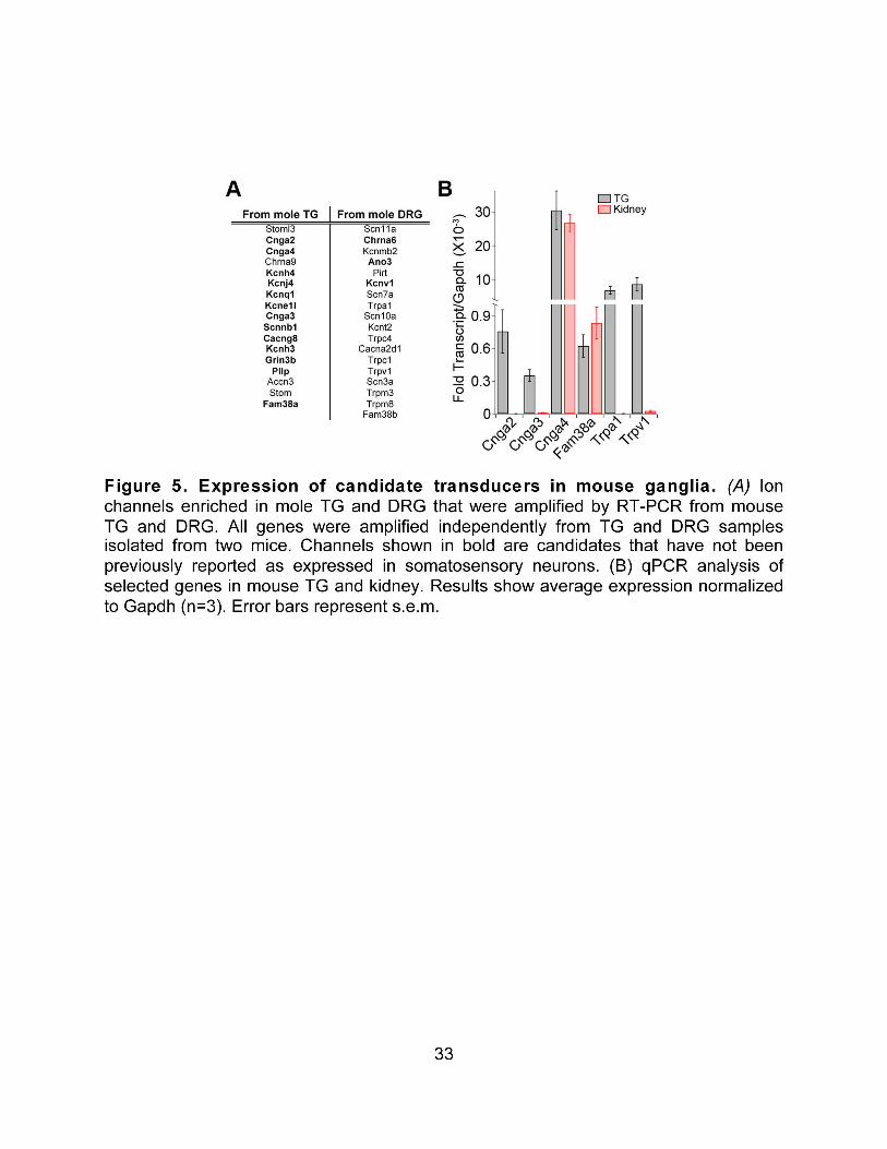


36
Table S1. Primers for amplification of star-nosed mole enriched genes in mouse.
Gene Forward Primer Reverse Primer Stoml3 CGTTACCTTCCCAATCTCAG ACATCTTTGATTTCCACCCG Cnga2 GGCGAGGGAAAGGGCACCAA AAGCCTGTGCGCAGCCGAAT Cnga4 GGAACATGTCTGGAAACCGA CTCCATCCTTGGACGTTCCTT Chrna9 GTGGGTGAGAGCTGCCTTAG ACCAACCCACTCCTCCTCTT Kcnh4 GACCTCCTTCTGTGCTCCTG AGACGAGGAGACCGTGAGAA Kcnj4 TAGTGCATGAAATCGACGAG TCTTCCTCCTGGCTCATAAG Kcnq1 CTCCTGTTTCTCTGTCTTCG GAAATGGGGTGTGCTTACTT Kcne1l AACTCTGGGGCCGTTTAACT ATGACTGGCCCTGAATGAAG Cnga3 TTGGACTACTCTGCAGATGT AAAGTAGATGCAGGCATTCC Scnn1b AGTCCTGCAATGACACCCAG GCTGGAAGCCAAAGTTGGTG Cacng8 GAGAGGGGTTTGTGGTGTGAA GATGGCGCTCAGGATAGGAA Kcnh3 TGAAGCAAGAAACACAGACA GTCTGAAGTGGAGGTCCATA Grin3b GCTGCAGTATTGGCTTCACA GCCGTGTAGTTCCTCAGCTC
Pllp AGTTCCCGTCGAAAGTGA AGGCGATAAAGGCAGTAATG Catsper1 AGGCGCCTGGTACATCATAC CGCTGCTTCACTGTCATGTT
Accn3 TTTCACTCGAATGGGGCAAT GAACCAGTACATTCTCCCCAGAG Stom TGCCTCGTTCTTTTTCGTAA TTTGAGACAGGTTCTTGGTG
Fam38a GCAGCCAGATGAGGAAGAGG TAGCTCCTCCTCCAGCTCCA Scn11a TGAGGCAACACTACTTCACCAATG AGCCAGAAACCAAGGTACTAATGATG Chrna6 TGGAACTGTGGGGCTATTTC AGCAGGAGCTTCGGATACAA Kcnmb2 GTCAGGGCCAGCTATCATGT TTAAAACCAACCCCCTTTCC
Ano3 GAGGAGGAAACACTTCGCCC TGCCAGTGCTGTTTGACGAA Pirt CCAAGGCCCTGGAGGTAGAC ACAGGAAGGCAGGCCCTATC
Kcnv1 ATTGCGCTCACTTGGGATGA AGAGCTTCACGCTGTCTCAC Scn7a ACCTTACCCACTGTGAGCGT CAGGGCCCGGTACTGTTTTT Trpa1 GGAGCAGACATCAACAGCAC GCAGGGGCGACTTCTTATC
Scn10a ACCGACAATCAGAGCGAGGAG ACAGACTAGAAATGGACAGAATCACC Kcnt2 AAGGCTGAGCAGAAAAGGGC AGGACAGTGGATCTGGTCGG Trpc4 CGTGGAGCTACTGCTGAACC CTGGACCGTGAATGCCTGAG
Cacna2d1 AGTGGACCTGGTGCGTATGA CTCAACTGCCTCAAGGAGCC Trpc1 GTACACTGCCCCCACCTTTC GGCGCAGTTCATTGAGGTTC Trpv1 TCACCGTCAGCTCTGTTGTC GGGTCTTTGAACTCGCTGTC Scn3a GGGTGTTGGGTGAGAGTGGAG AATGTAGTAGTGATGGGCTGATAAGAG Trpm3 GCCAGGCAAGCCATTCTCTT CCAGAAGGATGTTCGCCACC Trpm8 GGCTACACGGTAGGCATTGT TAAACCGATGCCTCATCTCC
Fam38b TCGCCGTGTTCCAGTTCATC TTGAAGCAAGTTCTCCCGGC

37
CHAPTER III: CNGA2 plays a modulatory role in mammalian mechanotransduction. This chapter is not yet in print. My experimental contributions were to Figures 1A-D, 2, and 3A-B and D-G. For this paper I performed cellular imaging, immunohistochemistry, and PCR experiments, made figures, and wrote the manuscript. Maurizio Pellegrino contributed to Figure 2A-C. Makoto Tsunozaki contributed to Figure 4. Wehong Lin contributed to Figure 1E. SUMMARY Little is known about the molecular mechanisms underlying mammalian touch transduction. We recently identified Cyclic nucleotide-gated channel A2 (Cnga2), as a candidate mechanotransduction channel. This channel acts as part of the transduction machinery in the olfactory system, but its expression in the somatosensory system has not been previously described. Here we suggest a role for Cnga2 in mechanotransduction in mice. First, Cnga2 is expressed in a subset of medium diameter neurons in the mouse trigeminal and dorsal root ganglia. Second, neurons isolated from mice lacking the CNGA2 channel display increased mechanical sensitivity. Third, CNGA2-deficient mice display a significant increase in tactile discrimination as compared to their wild type littermates, but no differences in sensitivity to noxious temperature or pressure. Together, these data indicate that CNGA2 channels play a modulatory role in tactile discrimination in vivo. This represents the second description of a of a single gene knockout mouse that displays altered tactile behavior in vivo. INTRODUCTION Despite much interest and study in recent years, the molecular mechanisms that underlie mechanotransduction in the mammalian somatosensory system remain unclear. We recently performed transcriptome sequencing to identify transcripts that are enriched in the somatosensory neurons that innervate the ultra-sensitive light touch organ of the star-nosed mole, the star. This screen yielded a range of channels, including several members of the cyclic-nucleotide gated channel (CNG) family, the most enriched of which was Cnga2 (211). CNG channels play a variety of roles in sensory transduction. They are required for photoresponses in rods and cones (212, 213), play a role in olfactory transduction (214), modulate inflammatory pain hypersensitivity in the somatosensory system (200), and affect chemosensation in C. elegans (215, 216). Cnga2 is activated by cAMP and cGMP and acts as a non-specific cation channel. In the olfactory system this channel is activated downstream of the olfactory receptors (ORs) and is required for OR mediated excitability of olfactory sensory neurons (214). As such, mice lacking Cnga2 are largely anosmic and display a range of deficits in olfactory mediated behaviors, including suckling behavior in infants (217), mating behavior in adult males (218), and odor associated learning (219). Here we examine the role of Cnga2 in somatosensation in mice. We show that Cnga2 is expressed in a subset of medium diameter somatosensory neurons. Somatosensory

38
neurons from mice lacking Cnga2 show an increase in the prevalence of stretch-sensitive neurons in vitro. Additionally, mice lacking Cnga2 show altered texture preference, suggesting a difference in tactile sensitivity. Taken together, these data suggest that Cnga2 may modulate mechanotransduction in the mammalian somatosensory system. RESULTS CNG channels are functionally expressed in mouse sensory ganglia. We have previously shown that Cnga2, Cnga3, and Cnga4 are expressed in sensory ganglia in the star-nosed mole. We confirmed expression of these transcripts in mouse sensory ganglia using qPCR. Cnga2 and Cnga3 expression is higher in sensory ganglia than non-neuronal tissue, while Cnga4 is expressed to a similar extent in kidney and DRG (Figure 1A). We also tested the effect of 8cpt-cGMP and forskolin, both general activators of CNG channels, on dissociated sensory neurons. These activators triggered Ca2+ influx in a subset of mouse DRG neurons (Figure 1B-C). The activated neurons had an average diameter of 22 µm (size ranged from 11 to 51 µm), indicating that they are a subset of medium-diameter neurons. Medium diameter neurons include Aδ fibers that function either as nociceptors that terminate as free-nerve endings, or as light touch receptors that associate with down hairs in the skin. There are no molecular markers to distinguish between these subtypes in culture, thus future skin-nerve recordings will help elucidate the exact subtype of neurons activated by cyclic nucleotides. Overall, these data show that the mammalian somatosensory system contains functional CNG channels in a distinct subset of sensory neurons. These experiments do not delineate which subtypes of CNG channels are active within somatosensory neurons, as 8cpt-cGMP and forskolin activate both CNGA2 and CNGA3 homomeric and heteromeric channels (220). In addition, the lack of specific inhibitors for distinct CNG channel subtypes necessitates the analysis of mice lacking Cnga2, Cnga3, and/or Cnga4 to assess the role of each channel in somatosensation. We next focused our analysis on the role of Cnga2, the most highly enriched channel from our transcriptomics screen (211). We used single cell PCR to validate Cnga2 expression in isolated sensory neurons rather than in whole ganglia that contain non-neuronal cells. We collected cytoplasmic contents from pools of neurons of small (≤20 µm), medium (20-35 µm), and large diameter (≥35 µm). Consistent with our imaging data, Cnga2 was only amplified from 1/3 pools of medium diameter neurons, 1/3 pools of small diameter neurons, and 0/3 pools of large diameter cells (average diameter Cnga2+ cells=22.4±3.5 µm, Figure 1D). We also observed a small number of GFP+ neurons in sensory ganglia from mice that express a GFP-fusion protein under the control of the Cnga2 promoter ((221) Figure 1E). Further experiments will be needed elucidate the exact subset of sensory neurons that express Cnga2. Cnga2 is not directly activated by mechanical stimuli.

39
Since Cnga2 is expressed in sensory neurons, we hypothesized that Cnga2 could act as a mechanosensitive channel. To determine if CNGA2 activity is directly altered by mechanical force, we heterologously expressed CNGA2 in HEK293 cells. CNGA2 overexpression did not alter the number of stretch sensitive cells or the amplitude of the stretch responses (Figure 2A-B). CNGA2 overexpression did strongly increase the response to 8cpt-cGMP, indicating that the channel was functionally expressed at high levels. CNGA2 overexpression also failed to alter cellular response to suction, unlike the mechanically gated channel Piezo1 (Figure 2C). Application of 8cpt-cGMP to patches from CNGA2 expressing cells showed increased channel activity within the patch but this activity was not altered by suction (data not shown). Taken together these data indicate that CNGA2 is not directly activated or inhibited by mechanical stimuli. However, given that other mechanosensitive channels require multiple components to display mechanical responses (80), it is possible that Cnga2 is still gated by mechanical forces in sensory neurons. Cnga2 does not effect development or innervation of sensory neurons. We also wanted to examine other possible mechanisms by which Cnga2 could alter mechanosensitivity There is evidence to suggest that CNG channels are expressed early in development in a number of neuronal cell types, and it has been suggested that CNGs may play a role in neuronal differentiation (222, 223). If Cnga2 plays a role in the differentiation or cell survival of some types of somatosensory neurons, it could alter mechanical sensitivity in the mouse by altering the number of mechanosensitive cells that innervate the skin. To determine whether loss of Cnga2 appreciably effects the distributions of subpopulations of sensory neurons, we stained for common markers of sensory neuron subtypes in DRG sections from mice lacking Cnga2 (Cnga2-/Y mice) (214). We did not observe any difference in staining for NF200, a marker of large diameter myelinated neurons, peripherin, a marker of small diameter neurons, or CGRP, a marker of peptidergic pain sensitive neurons (Figure 2D), indicating that Cnga2 does not grossly effect the distribution of DRG sensory neuron subtypes. Given the lack of difference between staining in wild type and Cnga2-/Y mice and that GFP is observable in sensory neurons in Cnga2tau::EGFP/Y mice which lack functional Cnga2 (Figure 1E), it is likely that Cnga2 is not required for cell survival. However, further molecular and functional characterization of Cnga2-expressing sensory neurons will be required to determine if Cnga2 plays a role in terminal differentiation. Since CNG channels have been implicated in both neurite guidance in Xenopus and the innervation pattern of sensory neurons in C. elegans, we also looked in the skin to determine if loss of Cnga2 affected sensory neuron innervation in mice (215, 216, 224). Using the pan-neuronal marker PGP9.5, we saw no apparent differences in the degree of innervation of the hindpaw glabrous skin between wild type (Figure 2E) and Cnga2-/Y mice (Figure 2E’). We also examined the innervation of non-neuronal somatosensory structures by somatosensory neurons. Merkel cells formed normal complexes with NF200 positive fibers in both wild type (Figure 2F) and Cnga2-/Y mice (Figure 2F’), and we observed no difference in the number of Merkel cells between wild type and hemizygous mice (Figure 2G). This suggests that Cnga2 is not grossly required for

40
sensory neuron populations. However, we cannot rule out the possibility that a small subset of fibers is affected by the loss of Cnga2. Cnga2 knockout mice show altered cellular responses to mechanical stimuli. To probe the physiological role of Cnga2 in somatosensation, we examined responses in isolated sensory neurons from mice lacking Cnga2. qPCR shows strong Cnga2 expression in wild type (WT) sensory neurons and no expression in Cnga2-/Y littermates (Cnga2 is X-linked; Figure 2A), confirming loss of Cnga2 at the transcriptional level. Ca2+ imaging was employed to measure the sensitivity of DRG neurons to chemical and mechanical stimuli. As previously shown (190) approximately 45% of neurons cultured from WT DRG showed robust responses to radial stretch (Figure 2B; white). Surprisingly, Neurons from Cnga2-/Y mice (Figure 2B; white) displayed a higher percentage of stretch-sensitive cells than neurons isolated from WT littermates (Figure 2B). We did not observe any differences in response amplitudes to radial stretch (Figure 2C). This suggests that Cnga2 either inhibits mechanosensitivity in some population of neurons in wild-type animals or it reduces the prevalence of mechanosensitive neurons by some developmental or cell survival mechanism. Surprisingly, the percentage of 8cpt-cGMP- and forskolin-activated neurons did not significantly differ between WT and Cnga2-/Y mice, suggesting that other CNG subunits contribute to these responses (Figure 2D). Cnga2-/Y neurons also displayed normal sensitivity to capsaicin, and no differences in capsaicin-evoked response amplitudes (Figure 2E-F). These data suggest that loss of CNGA2 channels does not simply enhance excitability, but rather has a specific effect on mechanical response properties. Loss of Cnga2 alters texture preference. We continued our characterization of the effect of Cnga2 on mechanosensation by testing somatosensory behaviors in Cnga2-/Y mice. To assess non-noxious tactile behaviors in Cnga2-/Y mice we utilized a texture discrimination task that measures preference for a rough versus smooth floor surface (Figure 4A). As previously reported, WT male mice display no preference for a rough surface (53, 99). Surprisingly, Cnga2-/Y mice spent significantly more time exploring the rough surface (Figure 4B) than their WT littermates. Previous studies have shown that unlike males, female mice prefer exploring rough surfaces (53), similar to the Cnga2-/Y mice. Because Cnga2 is x-linked and Cnga2-/Y mice do not mate, we are unable to probe tactile sensitivity of Cnga2-/-. The avoidance of the smooth surface was not due to general deficits in locomotion as these mice displayed normal exploratory behaviors and spent equal time exploring all surfaces when presented with identical stimuli (Figure 4B, left). Likewise, normal grooming and operant conditioning were previously reported (214, 218, 219). We found that Cnga2-/Y mice also displayed normal behavioral sensitivity to noxious mechanical and thermal stimuli. Cnga2-/Y mice and their WT littermates displayed characteristic mechanical thresholds for eliciting paw withdrawal (Figure 4D) in the Von Frey assay (225) and similar avoidance responses to noxious heat (Figure 4C) in a temperature preference test (20). These experiments show that CNGA2 channels are not required for nocifensive behaviors, but rather contribute to non-noxious tactile behaviors.
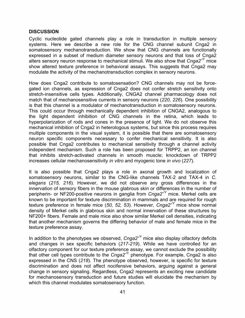
41
DISCUSSION Cyclic nucleotide gated channels play a role in transduction in multiple sensory systems. Here we describe a new role for the CNG channel subunit Cnga2 in somatosensory mechanotransduction. We show that CNG channels are functionally expressed in a subset of medium diameter sensory neurons and that loss of Cnga2 alters sensory neuron response to mechanical stimuli. We also show that Cnga2-/Y mice show altered texture preference in behavioral assays. This suggests that Cnga2 may modulate the activity of the mechanotransduction complex in sensory neurons. How does Cnga2 contribute to somatosensation? CNG channels may not be force-gated ion channels, as expression of Cnga2 does not confer stretch sensitivity onto stretch-insensitive cells types. Additionally, CNGA2 channel pharmacology does not match that of mechanosensitive currents in sensory neurons (220, 226). One possibility is that this channel is a modulator of mechanotransduction in somatosensory neurons. This could occur through mechanically dependent inhibition of CNGA2, analogous to the light dependent inhibition of CNG channels in the retina, which leads to hyperpolarization of rods and cones in the presence of light. We do not observe this mechanical inhibition of Cnga2 in heterologous systems, but since this process requires multiple components in the visual system, it is possible that there are somatosensory neuron specific components necessary to confer mechanical sensitivity. It is also possible that Cnga2 contributes to mechanical sensitivity through a channel activity independent mechanism. Such a role has been proposed for TRPP2, an ion channel that inhibits stretch-activated channels in smooth muscle; knockdown of TRPP2 increases cellular mechanosensitivity in vitro and myogenic tone in vivo (227). It is also possible that Cnga2 plays a role in axonal growth and localization of somatosensory neurons, similar to the CNG-like channels TAX-2 and TAX-4 in C. elegans (215, 216). However, we did not observe any gross differences in the innervation of sensory fibers in the mouse glabrous skin or differences in the number of peripherin- or NF200-positive neurons in ganglia from Cnga2-/Y mice. Merkel cells are known to be important for texture discrimination in mammals and are required for rough texture preference in female mice (50, 52, 53). However, Cnga2-/Y mice show normal density of Merkel cells in glabrous skin and normal innervation of these structures by NF200+ fibers. Female and male mice also show similar Merkel cell densities, indicating that another mechanism governs the differing behavior of male and female mice in the texture preference assay. In addition to the phenotypes we observed, Cnga2-/Y mice also display olfactory deficits and changes in sex specific behaviors (217-219). While we have controlled for an olfactory component for our texture preference assay, we cannot exclude the possibility that other cell types contribute to the Cnga2-/Y phenotype. For example, Cnga2 is also expressed in the CNS (218). The phenotype observed, however, is specific for texture discrimination and does not affect nocifensive behaviors, arguing against a general change in sensory signaling. Regardless, Cnga2 represents an exciting new candidate for mechanosensory transduction and future studies will elucidate the mechanism by which this channel modulates somatosensory function.

42
EXPERIMENTAL PROCEDURES Animals: Female Cnga2-/+ mice were obtained from Dr. Diego Restrepo. Tissue from Cnga2tau::EGFP/Y mice was obtained from Dr. Weihong Lin. For all experiments Cnga2-/Y mice were compared to Cnga2+/Y littermates. All procedures followed guidelines for the care and use of laboratory animals from the National Institutes of Health and were approved by the UC Berkeley Animal Care and Use Committee. RNA extraction: Mouse tissues were removed and homogenized in Trizol (Invitrogen). Total RNA was extracted as per the manufacturer’s instructions. Total RNA was DNAsed, re-purified (RNAeasy, Quiagen), and reverse transcribed (SuperScriptIII, Invitrogen) for qPCR or RT-PCR. qPCR: RNA was extracted from wild-type and Cnga2-/Y mice as described above. qPCR was performed using SYBR GreenER (Invitrogen) on a StepOnePlus™ Real-Time PCR system (Applied Biosystems). qPCR reactions were run in 10 µL using 1 µL of product from the RT reaction with the primers listed below. CT values were calculated as the average of 2 wells. Fold expression for each was calculated assuming 100% efficiency. Final values were calculated as an average of the biological replicates for each tissue type. Primers used are listed below: Cnga2:Fwd CTGGAACTCTGCTCGTGTGG, Rev GGGGTTGACACCTGGGTTTA Cnga3:Fwd CTTTGAATTCTTTGACCGCACA, Rev AGGTGTTTGGATAGACCCAGGA Cnga4:Fwd AGCACACACACATGCACACACT, Rev TCCTATACCAGCGACCTCCTTT Gapdh:Fwd ACATGGCCTCCAAGGAGTAAGA, Rev GGTGCAGCGAACTTTATTGATG Cell Culture: DRG were cultured as previously described (189). Briefly, mice were euthanized and dorsal root ganglia were dissected. Ganglia were digested for 20 minutes in 7 mg/mL collagenase P (Roche) then for 2 minutes in 0.25% trypsin. Cells were dissociated manually by trituration and plated on glass cover slides coated with 1 mg/mL Poly-D-Lysine and 0.1 mg/mL laminin. Cells were cultured overnight at 37 °C before imaging. HEK293T cells were cultured in DMEM (high glucose) supplemented with 10% FBS, penicillin-streptomycin (5,000 U/mL), and L-glutamine (10 mM). Cells (at 75-80% confluency) were transfected with Lipofectamine 2000 (Invitrogen) in OPTI-MEM solution (Invitrogen) using 0.8-1 µg mouse pCDNA3-Cnga2, pCNDA3-Fam38a, or vector. Calcium Imaging: Calcium imaging was performed as previously described for mouse neurons (20). Cells were loaded for an hour at room temperature with Fura-2AM (10 µM, Invitrogen) in Ringer’s solution (140 mM NaCl, 5 mM KCl, 2 mM CaCl2, 1 mM MgCl2, 10 mM D-glucose, 10 mM HEPES) supplemented with 0.2% pluronic acid (Invitrogen). Ratiometric calcium imaging was performed using alternating 340 nm and 380 nm excitation filters with images collected every three seconds. Responsive cells were defined as those whose calcium concentration increased more than 15% above the baseline after application of the stimulus. Stretch was applied using a StageFlexer/FX-3000 system (Flexcell), as previously described (190).

43
Immunohistochemistry: Mice were anesthetized with 250 mg/kg i.p. injection of sodium pentobarbital and perfused with 1X PBS (pH 7.3), followed by 4% paraformaldehyde (PFA) in PBS. Dorsal root ganglia and hindpaw skin were collected. Tissue was post-fixed for >4 hours in 4% PFA and incubated overnight in 20% sucrose at 4 °C. Tissue was embedded in Tissue-Tek optimal, sectioned at 12 µm, and mounted onto glass coverslips for staining. Sections were blocked for 90 minutes in PBST supplemented with 10% normal goat serum and incubated overnight at 4 °C with primary antibody. Primary antibodies were diluted as follows: mouse anti-NF200 (Sigma) and rabbit anti-peripherin 1:1000 (Millipore), Rabbit anti-CGRP (Milipore) and Rabbit anti-PGP9.5 (Milipore) 1:500, Rat anti-TROMA1 1:100 (Developmental Studies Hybridoma Bank). Sections were washed and incubated for one hour at room temperature in 1:3000 secondary antibody. Secondary antibodies (Invitrogen) were used as follows: anti-mouse Alexa 488 for NF200, anti-rabbit Alexa 568 for peripherin, CGRP, and PGP9.5, and anti-rat Alexa 488 for TROMA-1. Images were captured using MetaMorph (Molecular Devices). Cells were defined as being positive for a specific marker if the average intensity of the cell was more than 50% above background. Single-cell PCR: RNA was isolated from pools of 8-12 sensory neurons. Cell diameter was determined by bright-field imaging using MetaMorph Software. Cells from each size category were aspirated into a large–diameter glass electrode filled with lysis buffer (50 mM Tris-Cl, pH 8.3, 75 mM KCl, 3 mM MgCl2, 5 U–1 RNasin (Promega)) and were flash frozen. Reverse transcription was performed using murine Moloney leukemia virus and avian reverse transcriptases at 37 °C for 1 h. The product was diluted 1:5 and used as the template for PCR experiments. Primers for PCR were: Cnga2: Fwd CTGGAACTCTGCTCGTGTGG, Rev GGGGTTGACACCTGGGTTTA β-actin: Fwd CTGGTCGTCGACAACGGCTCC, Rev GCCAGATCTTCTCCATG Cnga2 cloning: Cnga2 was amplified from mouse TG and cloned in pCDNA3 using the ligation independent cloning using the following primers: Fwd TACTTCCAATCCAATGCCACCATGATGACCGAAAAATCCAAC Rev TTATCCACTTCCAATGCTATTCAGCAACAGCTGGC. Suction recordings: Suctions recordings were performed as previously described (129). Briefly, patch pipettes with a resistance of 2-5MΩ were used. Pipette solution contained (in mM): 130 NaCl, 5 KCl, 10 HEPES, 1 CaCl2, 1 MgCl2, 10 TEA-Cl (pH 7.3 with NaOH). After seals were formed, an external solution was used to zero the membrane potential (in mM: 140 KCl, 10 HEPES, 1 MgCl2, 10 glucose, pH 7.3 with KOH). Patches were held at -80 mV during suction recordings. Suction was applied to the pipette using a high speed pressure clamp (ALA scientific) from 0-50 mmHg in 10 mmHg steps with a step duration of 200 ms. Data were collected at 10 kHz and filtered at 2 kHz (Axopatch 200B, PClamp software).

44
Behavioral assays: In the texture preference assay, animals were acclimated to the two-chamber preference arena without sandpaper for two days prior to the preference test. On the day of the preference test, the bottom of each chamber was covered with either a coarse-textured, large grit size sandpaper (Wetordry 431Q 80C, 3M, St Paul, MN) or fine-textured small grit size sandpaper (Wetordry 413Q 200A, 3M). The animals were allowed to roam freely in the arena for ten minutes in the dark, and their behavior was monitored by an infrared camera and a custom-made laser tripwire localization device. Control experiments were carried out using the smooth-textured paper backing of fresh large and small grit sandpapers in each preference chamber. Video data was analyzed manually and location data was analyzed using custom scripts in MATLAB (Mathworks, Natick, MA). Temperature preference and Von Frey assays were carried out as previously described (20, 228). All assays were carried out blind to genotype. Statistics: All values are presented as average ± standard error of the mean (s.e.m.). Unless otherwise noted, all p-values were calculated using a one-way ANOVA with a Tukey’s Post Hoc analysis.


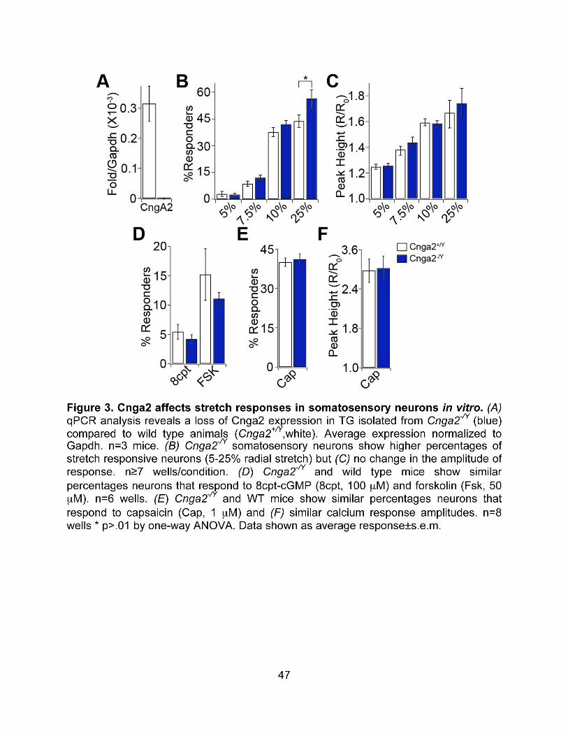


49
SUMMARY: There remains a great deal to learn about transduction mechanisms in the somatosensory system. Even for well-characterized receptors with distinct ligands and known expression profiles, such as TRPV1, new modulators of activity and roles in sensory transduction are still being discovered (229, 230). The work presented here represents a step forward in describing some of the pathways underlying somatosensory transduction. Our work suggests that TRPA1 plays a role in mediating histamine-independent itch and that as a convergence point for multiple itch related signaling pathways, it is a promising target for pharmacological intervention in clinically relevant itch. Indeed, recent studies have shown that TRPA1 is required for multiple models of chronic itch (231, 232). Further studies will elucidate which chronic itch conditions are TRPA1 dependent and whether TRPA1 inhibitors can alleviate itch in humans. Our work also suggests that Cnga2 plays a role in modulating the response to innocuous mechanical stimuli. With the work presented here, we cannot determine if Cnga2 interacts directly with the mechanically activated channel as part of the mechanotransduction complex or if it modulates cellular responses to mechanical force in a more indirect mechanism. Histological profiling of Cnga2 positive neurons in Cnga2tau::EGFP/Y mice should allow us to better characterize the response properties of this sensory neuronal subtype in wild type and mice lacking Cnga2 in vitro. We also hope to characterize the mechanical responses of sensory fibers in Cnga2-/Y mice using the ex-vivo skin-nerve preparation (233). This would allow us to directly compare the mechanical responses in texture sensitive Merkel fibers to determine if lack of Cnga2 alters mechanical thresholds. Lastly, we are continuing to adapt new behavioral paradigms that measure tactile sensitivity in vivo, including measuring tactile startle responses (234) and utilizing textured objects for novel object recognition tasks (235). In addition to helping us to define the role of Cnga2 in mechanosensitivity, these assays will also be important for both defining the role for other CNG channels and other candidate mechanotransducers. In addition to CNG channels, a number of other channel families have been implicated in governing cellular mechanical responses. We found several members of these families enriched in the TG of the star nosed mole. For example, the piezo protein family member Piezo1, which acts as a mechanosensitive channel but has not been implicated in somatosensory mechanotransduction (129), is enriched in the mole TG, We also found several members of the Deg/ENaC family of sodium channels enriched in the mole TG: ASIC3, ASIC4, and ENaCβ. In contrast, none of TMC family members, which appear to act as mechanically gated channels in hair cells, show differential expression (138). There is also a wealth (210 genes) of uncharacterized transmembrane domain-containing proteins that are enriched in the TG, any of which could act as mechanosensitive channels. We will continue to characterize the expression and activity of these candidate mechanotransducers in the years to come.

50
REFERENCES: 1. S.Gobel,W.M.Falls,E.Humphrey,Morphologyandsynapticconnectionsofultrafine
primaryaxonsinlaminaIofthespinaldorsalhorn:candidatesfortheterminalaxonalarborsofprimaryneuronswithunmyelinated(C)axons.JNeurosci1,1163(Oct,1981).
2. G.M.Storyetal.,ANKTM1,aTRP‐likechannelexpressedinnociceptiveneurons,isactivatedbycoldtemperatures.Cell112,819(Mar21,2003).
3. M.Bandelletal.,NoxiouscoldionchannelTRPA1isactivatedbypungentcompoundsandbradykinin.Neuron41,849(Mar25,2004).
4. D.M.Bautistaetal.,PungentproductsfromgarlicactivatethesensoryionchannelTRPA1.ProcNatlAcadSciUSA102,12248(Aug23,2005).
5. Y.Nakamuraetal.,Activationoftransientreceptorpotentialankyrin1evokesnociceptionthroughsubstancePreleasefromprimarysensoryneurons.Journalofneurochemistry120,1036(Mar,2012).
6. D.M.Bautistaetal.,TRPA1mediatestheinflammatoryactionsofenvironmentalirritantsandproalgesicagents.Cell124,1269(Mar24,2006).
7. K.Y.Kwanetal.,TRPA1contributestocold,mechanical,andchemicalnociceptionbutisnotessentialforhair‐celltransduction.Neuron50,277(Apr20,2006).
8. C.R.McNamaraetal.,TRPA1mediatesformalin‐inducedpain.ProcNatlAcadSciUSA104,13525(Aug14,2007).
9. S.R.Eidetal.,HC‐030031,aTRPA1selectiveantagonist,attenuatesinflammatory‐andneuropathy‐inducedmechanicalhypersensitivity.MolPain4,48(2008).
10. M.J.Caterinaetal.,Thecapsaicinreceptor:aheat‐activatedionchannelinthepainpathway.Nature389,816(Oct23,1997).
11. M.J.Caterinaetal.,Impairednociceptionandpainsensationinmicelackingthecapsaicinreceptor.Science288,306(Apr14,2000).
12. I.Shimizuetal.,EnhancedthermalavoidanceinmicelackingtheATPreceptorP2X3.Pain116,96(Jul,2005).
13. A.M.Peieretal.,Aheat‐sensitiveTRPchannelexpressedinkeratinocytes.Science296,2046(Jun14,2002).
14. A.D.Guleretal.,Heat‐evokedactivationoftheionchannel,TRPV4.JNeurosci22,6408(Aug1,2002).
15. M.J.Caterina,T.A.Rosen,M.Tominaga,A.J.Brake,D.Julius,Acapsaicin‐receptorhomologuewithahighthresholdfornoxiousheat.Nature398,436(Apr1,1999).
16. S.M.Huang,X.Li,Y.Yu,J.Wang,M.J.Caterina,TRPV3andTRPV4ionchannelsarenotmajorcontributorstomouseheatsensation.MolPain7,37.
17. U.Parketal.,TRPvanilloid2knock‐outmicearesusceptibletoperinatallethalitybutdisplaynormalthermalandmechanicalnociception.JNeurosci31,11425(Aug10).
18. J.Vriensetal.,TRPM3isanociceptorchannelinvolvedinthedetectionofnoxiousheat.Neuron70,482(May12).
19. D.D.McKemy,W.M.Neuhausser,D.Julius,IdentificationofacoldreceptorrevealsageneralroleforTRPchannelsinthermosensation.Nature416,52(Mar7,2002).
20. D.M.Bautistaetal.,ThementholreceptorTRPM8istheprincipaldetectorofenvironmentalcold.Nature448,204(Jul12,2007).

51
21. W.M.Knowlton,R.L.Daniels,R.Palkar,D.D.McCoy,D.D.McKemy,PharmacologicalblockadeofTRPM8ionchannelsalterscoldandcoldpainresponsesinmice.PLoSOne6,e25894.
22. K.Zimmermannetal.,Transientreceptorpotentialcationchannel,subfamilyC,member5(TRPC5)isacold‐transducerintheperipheralnervoussystem.ProcNatlAcadSciUSA108,18114(Nov1).
23. P.Sikand,S.G.Shimada,B.G.Green,R.H.LaMotte,Similaritchandnociceptivesensationsevokedbypunctatecutaneousapplicationofcapsaicin,histamineandcowhage.Pain144,66(Jul,2009).
24. P.Sikand,S.G.Shimada,B.G.Green,R.H.LaMotte,Sensoryresponsestoinjectionandpunctateapplicationofcapsaicinandhistaminetotheskin.Pain152,2485(Nov,2011).
25. Y.G.Sun,Z.F.Chen,Agastrin‐releasingpeptidereceptormediatestheitchsensationinthespinalcord.Nature448,700(Aug9,2007).
26. K.Rossbachetal.,HistamineH1,H3andH4receptorsareinvolvedinpruritus.Neuroscience190,89(Sep8,2011).
27. K.Tsujii,T.Andoh,J.B.Lee,Y.Kuraishi,Activationofproteinase‐activatedreceptorsinducesitch‐associatedresponsethroughhistamine‐dependentand‐independentpathwaysinmice.Journalofpharmacologicalsciences108,385(Nov,2008).
28. J.Liang,T.Kawamata,W.Ji,Molecularsignalingofpruritusinducedbyendothelin‐1inmice.ExpBiolMed(Maywood)235,1300(Nov,2010).
29. D.S.McQueen,M.A.Noble,S.M.Bond,Endothelin‐1activatesETAreceptorstocausereflexscratchinginBALB/cmice.Britishjournalofpharmacology151,278(May,2007).
30. P.G.Trentin,M.B.Fernandes,P.D'Orleans‐Juste,G.A.Rae,Endothelin‐1causespruritusinmice.ExpBiolMed(Maywood)231,1146(Jun,2006).
31. Q.Liuetal.,Sensoryneuron‐specificGPCRMrgprsareitchreceptorsmediatingchloroquine‐inducedpruritus.Cell139,1353(Dec24,2009).
32. S.K.Han,V.Mancino,M.I.Simon,PhospholipaseCbeta3mediatesthescratchingresponseactivatedbythehistamineH1receptoronC‐fibernociceptiveneurons.Neuron52,691(Nov22,2006).
33. N.Imamachietal.,TRPV1‐expressingprimaryafferentsgeneratebehavioralresponsestopruritogensviamultiplemechanisms.ProcNatlAcadSciUSA106,11330(Jul7,2009).
34. M.Trevisanietal.,4‐Hydroxynonenal,anendogenousaldehyde,causespainandneurogenicinflammationthroughactivationoftheirritantreceptorTRPA1.ProcNatlAcadSciUSA104,13519(Aug14,2007).
35. Q.Liuetal.,Moleculargeneticvisualizationofararesubsetofunmyelinatedsensoryneuronsthatmaydetectgentletouch.Natureneuroscience10,946(Aug,2007).
36. R.P.Sealetal.,Injury‐inducedmechanicalhypersensitivityrequiresC‐lowthresholdmechanoreceptors.Nature462,651(Dec3,2009).
37. L.Lietal.,Thefunctionalorganizationofcutaneouslow‐thresholdmechanosensoryneurons.Cell147,1615(Dec23,2011).

52
38. L.Kruger,E.R.Perl,M.J.Sedivec,Finestructureofmyelinatedmechanicalnociceptorendingsincathairyskin.TheJournalofcomparativeneurology198,137(May1,1981).
39. A.R.Light,E.R.Perl,Reexaminationofthedorsalrootprojectiontothespinaldorsalhornincludingobservationsonthedifferentialterminationofcoarseandfinefibers.TheJournalofcomparativeneurology186,117(Jul15,1979).
40. J.J.Lawson,S.L.McIlwrath,C.J.Woodbury,B.M.Davis,H.R.Koerber,TRPV1unlikeTRPV2isrestrictedtoasubsetofmechanicallyinsensitivecutaneousnociceptorsrespondingtoheat.Thejournalofpain:officialjournaloftheAmericanPainSociety9,298(Apr,2008).
41. R.C.Lennertz,M.Tsunozaki,D.M.Bautista,C.L.Stucky,Physiologicalbasisoftinglingparesthesiaevokedbyhydroxy‐alpha‐sanshool.JNeurosci30,4353(Mar24,2010).
42. L.Minichielloetal.,PointmutationintrkBcauseslossofNT4‐dependentneuronswithoutmajoreffectsondiverseBDNFresponses.Neuron21,335(Aug,1998).
43. C.L.Stucky,M.Koltzenburg,Thelow‐affinityneurotrophinreceptorp75regulatesthefunctionbutnottheselectivesurvivalofspecificsubpopulationsofsensoryneurons.JNeurosci17,4398(Jun1,1997).
44. K.M.Morrison,G.R.Miesegaes,E.A.Lumpkin,S.M.Maricich,MammalianMerkelcellsaredescendedfromtheepidermallineage.Developmentalbiology336,76(Dec1,2009).
45. A.Iggo,A.R.Muir,Thestructureandfunctionofaslowlyadaptingtouchcorpuscleinhairyskin.TheJournalofphysiology200,763(Feb,1969).
46. H.Haeberle,L.A.Bryan,T.J.Vadakkan,M.E.Dickinson,E.A.Lumpkin,Swelling‐activatedCa2+channelstriggerCa2+signalsinMerkelcells.PLoSOne3,e1750(2008).
47. H.Haeberleetal.,MolecularprofilingrevealssynapticreleasemachineryinMerkelcells.ProcNatlAcadSciUSA101,14503(Oct5,2004).
48. I.S.Hitchcock,P.G.Genever,P.M.Cahusac,EssentialcomponentsforaglutamatergicsynapsebetweenMerkelcellandnerveterminalinrats.Neuroscienceletters362,196(May27,2004).
49. M.S.Leung,C.C.Wong,ExpressionsofputativeneurotransmittersandneuronalgrowthrelatedgenesinMerkelcell‐neuritecomplexesoftherats.Lifesciences66,1481(2000).
50. S.M.Maricichetal.,Merkelcellsareessentialforlight‐touchresponses.Science324,1580(Jun19,2009).
51. P.M.Cahusac,S.S.Senok,I.S.Hitchcock,P.G.Genever,K.I.Baumann,AreunconventionalNMDAreceptorsinvolvedinslowlyadaptingtypeImechanoreceptorresponses?Neuroscience133,763(2005).
52. K.O.Johnson,G.D.Lamb,Neuralmechanismsofspatialtactilediscrimination:neuralpatternsevokedbybraille‐likedotpatternsinthemonkey.TheJournalofphysiology310,117(Jan,1981).
53. S.M.Maricich,K.M.Morrison,E.L.Mathes,B.M.Brewer,RodentsrelyonMerkelcellsfortexturediscriminationtasks.JNeurosci32,3296(Mar7,2012).

53
54. F.L.Rice,E.Kinnman,H.Aldskogius,O.Johansson,J.Arvidsson,TheinnervationofthemystacialpadoftheratasrevealedbyPGP9.5immunofluorescence.TheJournalofcomparativeneurology337,366(Nov15,1993).
55. B.T.Fundinetal.,Comprehensiveimmunofluorescenceandlectinbindinganalysisofintervibrissalfurinnervationinthemystacialpadoftherat.TheJournalofcomparativeneurology385,185(Aug25,1997).
56. M.Koltzenburg,C.L.Stucky,G.R.Lewin,Receptivepropertiesofmousesensoryneuronsinnervatinghairyskin.Journalofneurophysiology78,1841(Oct,1997).
57. M.Pare,R.Elde,J.E.Mazurkiewicz,A.M.Smith,F.L.Rice,TheMeissnercorpusclerevised:amultiafferentedmechanoreceptorwithnociceptorimmunochemicalproperties.JNeurosci21,7236(Sep15,2001).
58. A.Iggo,H.Ogawa,Correlativephysiologicalandmorphologicalstudiesofrapidlyadaptingmechanoreceptorsincat'sglabrousskin.TheJournalofphysiology266,275(Apr,1977).
59. V.G.Macefield,C.Hager‐Ross,R.S.Johansson,Controlofgripforceduringrestraintofanobjectheldbetweenfingerandthumb:responsesofcutaneousafferentsfromthedigits.Experimentalbrainresearch.ExperimentelleHirnforschung.Experimentationcerebrale108,155(Feb,1996).
60. M.Sato,ResponseofPaciniancorpusclestosinusoidalvibration.TheJournalofphysiology159,391(Dec,1961).
61. W.R.Loewenstein,M.Mendelson,ComponentsofReceptorAdaptationinaPacinianCorpuscle.TheJournalofphysiology177,377(Apr,1965).
62. M.R.Chambers,K.H.Andres,M.vonDuering,A.Iggo,ThestructureandfunctionoftheslowlyadaptingtypeIImechanoreceptorinhairyskin.Quarterlyjournalofexperimentalphysiologyandcognatemedicalsciences57,417(Oct,1972).
63. B.B.Edin,Quantitativeanalysisofstaticstrainsensitivityinhumanmechanoreceptorsfromhairyskin.Journalofneurophysiology67,1105(May,1992).
64. B.B.Edin,N.Johansson,Skinstrainpatternsprovidekinaestheticinformationtothehumancentralnervoussystem.TheJournalofphysiology487(Pt1),243(Aug15,1995).
65. S.S.Schafer,Regularityinthegenerationofdischargepatternsbyprimaryandsecondarymusclespindleafferents,asrecordedunderaramp‐and‐holdstretch.Experimentalbrainresearch.ExperimentelleHirnforschung.Experimentationcerebrale102,198(1994).
66. R.Kleinetal.,Disruptionoftheneurotrophin‐3receptorgenetrkCeliminateslamuscleafferentsandresultsinabnormalmovements.Nature368,249(Mar17,1994).
67. N.Levinaetal.,ProtectionofEscherichiacolicellsagainstextremeturgorbyactivationofMscSandMscLmechanosensitivechannels:identificationofgenesrequiredforMscSactivity.TheEMBOjournal18,1730(Apr1,1999).
68. S.I.Sukharev,P.Blount,B.Martinac,F.R.Blattner,C.Kung,Alarge‐conductancemechanosensitivechannelinE.coliencodedbymscLalone.Nature368,265(Mar17,1994).

54
69. S.I.Sukharev,B.Martinac,V.Y.Arshavsky,C.Kung,TwotypesofmechanosensitivechannelsintheEscherichiacolicellenvelope:solubilizationandfunctionalreconstitution.Biophysicaljournal65,177(Jul,1993).
70. P.Blountetal.,MembranetopologyandmultimericstructureofamechanosensitivechannelproteinofEscherichiacoli.TheEMBOjournal15,4798(Sep16,1996).
71. G.Chang,R.H.Spencer,A.T.Lee,M.T.Barclay,D.C.Rees,StructureoftheMscLhomologfromMycobacteriumtuberculosis:agatedmechanosensitiveionchannel.Science282,2220(Dec18,1998).
72. C.C.Cruickshank,R.F.Minchin,A.C.LeDain,B.Martinac,Estimationoftheporesizeofthelarge‐conductancemechanosensitiveionchannelofEscherichiacoli.Biophysicaljournal73,1925(Oct,1997).
73. E.Perozo,D.M.Cortes,P.Sompornpisut,A.Kloda,B.Martinac,OpenchannelstructureofMscLandthegatingmechanismofmechanosensitivechannels.Nature418,942(Aug29,2002).
74. R.B.Bass,P.Strop,M.Barclay,D.C.Rees,CrystalstructureofEscherichiacoliMscS,avoltage‐modulatedandmechanosensitivechannel.Science298,1582(Nov22,2002).
75. P.Moe,P.Blount,Assessmentofpotentialstimuliformechano‐dependentgatingofMscL:effectsofpressure,tension,andlipidheadgroups.Biochemistry44,12239(Sep13,2005).
76. B.Martinac,J.Adler,C.Kung,MechanosensitiveionchannelsofE.coliactivatedbyamphipaths.Nature348,261(Nov15,1990).
77. E.Perozo,A.Kloda,D.M.Cortes,B.Martinac,Physicalprinciplesunderlyingthetransductionofbilayerdeformationforcesduringmechanosensitivechannelgating.Naturestructuralbiology9,696(Sep,2002).
78. M.Chalfie,M.Au,GeneticcontrolofdifferentiationoftheCaenorhabditiseleganstouchreceptorneurons.Science243,1027(Feb24,1989).
79. M.Chalfie,J.Sulston,DevelopmentalgeneticsofthemechanosensoryneuronsofCaenorhabditiselegans.Developmentalbiology82,358(Mar,1981).
80. K.Hong,M.Driscoll,AtransmembranedomainoftheputativechannelsubunitMEC‐4influencesmechanotransductionandneurodegenerationinC.elegans.Nature367,470(Feb3,1994).
81. M.Huang,M.Chalfie,GeneinteractionsaffectingmechanosensorytransductioninCaenorhabditiselegans.Nature367,467(Feb3,1994).
82. R.O'Hagan,M.Chalfie,M.B.Goodman,TheMEC‐4DEG/ENaCchannelofCaenorhabditiseleganstouchreceptorneuronstransducesmechanicalsignals.Natureneuroscience8,43(Jan,2005).
83. M.B.Goodmanetal.,MEC‐2regulatesC.elegansDEG/ENaCchannelsneededformechanosensation.Nature415,1039(Feb28,2002).
84. D.S.Cheluretal.,ThemechanosensoryproteinMEC‐6isasubunitoftheC.eleganstouch‐celldegenerinchannel.Nature420,669(Dec12,2002).
85. M.Huang,G.Gu,E.L.Ferguson,M.Chalfie,Astomatin‐likeproteinnecessaryformechanosensationinC.elegans.Nature378,292(Nov16,1995).
86. T.B.Huberetal.,PodocinandMEC‐2bindcholesteroltoregulatetheactivityofassociatedionchannels.ProcNatlAcadSciUSA103,17079(Nov14,2006).

55
87. H.Du,G.Gu,C.M.William,M.Chalfie,ExtracellularproteinsneededforC.elegansmechanosensation.Neuron16,183(Jan,1996).
88. C.Savageetal.,mec‐7isabeta‐tubulingenerequiredfortheproductionof15‐protofilamentmicrotubulesinCaenorhabditiselegans.Genes&development3,870(Jun,1989).
89. T.Fukushigeetal.,MEC‐12,analpha‐tubulinrequiredfortouchsensitivityinC.elegans.Journalofcellscience112(Pt3),395(Feb,1999).
90. J.G.Cueva,A.Mulholland,M.B.Goodman,NanoscaleorganizationoftheMEC‐4DEG/ENaCsensorymechanotransductionchannelinCaenorhabditiseleganstouchreceptorneurons.JNeurosci27,14089(Dec19,2007).
91. L.Zhong,R.Y.Hwang,W.D.Tracey,PickpocketisaDEG/ENaCproteinrequiredformechanicalnociceptioninDrosophilalarvae.Currentbiology:CB20,429(Mar9,2010).
92. M.P.Priceetal.,TheDRASICcationchannelcontributestothedetectionofcutaneoustouchandacidstimuliinmice.Neuron32,1071(Dec20,2001).
93. M.P.Priceetal.,ThemammaliansodiumchannelBNC1isrequiredfornormaltouchsensation.Nature407,1007(Oct26,2000).
94. A.J.Pageetal.,DifferentcontributionsofASICchannels1a,2,and3ingastrointestinalmechanosensoryfunction.Gut54,1408(Oct,2005).
95. L.J.Drewetal.,Acid‐sensingionchannelsASIC2andASIC3donotcontributetomechanicallyactivatedcurrentsinmammaliansensoryneurones.TheJournalofphysiology556,691(May1,2004).
96. A.A.Staniland,S.B.McMahon,Micelackingacid‐sensingionchannels(ASIC)1or2,butnotASIC3,showincreasedpainbehaviourintheformalintest.EurJPain13,554(Jul,2009).
97. S.Kangetal.,SimultaneousdisruptionofmouseASIC1a,ASIC2andASIC3genesenhancescutaneousmechanosensitivity.PLoSOne7,e35225(2012).
98. R.Raoufetal.,Sodiumchannelsandmammaliansensorymechanotransduction.MolPain8,21(2012).
99. C.Wetzeletal.,Astomatin‐domainproteinessentialfortouchsensationinthemouse.Nature445,206(Jan11,2007).
100. C.Martinez‐Salgadoetal.,Stomatinandsensoryneuronmechanotransduction.Journalofneurophysiology98,3802(Dec,2007).
101. J.M.Kaplan,H.R.Horvitz,AdualmechanosensoryandchemosensoryneuroninCaenorhabditiselegans.ProcNatlAcadSciUSA90,2227(Mar15,1993).
102. H.A.Colbert,T.L.Smith,C.I.Bargmann,OSM‐9,anovelproteinwithstructuralsimilaritytochannels,isrequiredforolfaction,mechanosensation,andolfactoryadaptationinCaenorhabditiselegans.JNeurosci17,8259(Nov1,1997).
103. M.A.Hilliardetal.,InvivoimagingofC.elegansASHneurons:cellularresponseandadaptationtochemicalrepellents.TheEMBOjournal24,63(Jan12,2005).
104. S.L.Geffeneyetal.,DEG/ENaCbutnotTRPchannelsarethemajormechanoelectricaltransductionchannelsinaC.elegansnociceptor.Neuron71,845(Sep8,2011).

56
105. W.Liedtke,D.M.Tobin,C.I.Bargmann,J.M.Friedman,MammalianTRPV4(VR‐OAC)directsbehavioralresponsestoosmoticandmechanicalstimuliinCaenorhabditiselegans.ProcNatlAcadSciUSA100Suppl2,14531(Nov25,2003).
106. M.Suzuki,A.Mizuno,K.Kodaira,M.Imai,ImpairedpressuresensationinmicelackingTRPV4.TheJournalofbiologicalchemistry278,22664(Jun20,2003).
107. U.Parketal.,TRPvanilloid2knock‐outmicearesusceptibletoperinatallethalitybutdisplaynormalthermalandmechanicalnociception.JNeurosci31,11425(Aug10,2011).
108. R.G.Walker,A.T.Willingham,C.S.Zuker,ADrosophilamechanosensorytransductionchannel.Science287,2229(Mar24,2000).
109. T.Effertz,B.Nadrowski,D.Piepenbrock,J.T.Albert,M.C.Gopfert,DirectgatingandmechanicalintegrityofDrosophilaauditorytransducersrequireTRPN1.Natureneuroscience15,1198(Jul29,2012).
110. M.C.Gopfert,J.T.Albert,B.Nadrowski,A.Kamikouchi,SpecificationofauditorysensitivitybyDrosophilaTRPchannels.Natureneuroscience9,999(Aug,2006).
111. S.Sidi,R.W.Friedrich,T.Nicolson,NompCTRPchannelrequiredforvertebratesensoryhaircellmechanotransduction.Science301,96(Jul4,2003).
112. W.Li,Z.Feng,P.W.Sternberg,X.Z.Xu,AC.elegansstretchreceptorneuronrevealedbyamechanosensitiveTRPchannelhomologue.Nature440,684(Mar30,2006).
113. L.Kang,J.Gao,W.R.Schafer,Z.Xie,X.Z.Xu,C.elegansTRPfamilyproteinTRP‐4isapore‐formingsubunitofanativemechanotransductionchannel.Neuron67,381(Aug12,2010).
114. J.Howard,S.Bechstedt,Hypothesis:ahelixofankyrinrepeatsoftheNOMPC‐TRPionchannelisthegatingspringofmechanoreceptors.Currentbiology:CB14,R224(Mar23,2004).
115. D.P.Coreyetal.,TRPA1isacandidateforthemechanosensitivetransductionchannelofvertebratehaircells.Nature432,723(Dec9,2004).
116. Y.Leeetal.,PyrexiaisanewthermaltransientreceptorpotentialchannelendowingtolerancetohightemperaturesinDrosophilamelanogaster.Naturegenetics37,305(Mar,2005).
117. M.Rosenzweigetal.,TheDrosophilaorthologofvertebrateTRPA1regulatesthermotaxis.Genes&development19,419(Feb15,2005).
118. W.D.Tracey,Jr.,R.I.Wilson,G.Laurent,S.Benzer,painless,aDrosophilageneessentialfornociception.Cell113,261(Apr18,2003).
119. Y.Sunetal.,TRPAchannelsdistinguishgravitysensingfromhearinginJohnston'sorgan.ProcNatlAcadSciUSA106,13606(Aug11,2009).
120. R.Y.Hwang,N.A.Stearns,W.D.Tracey,TheankyrinrepeatdomainoftheTRPAproteinpainlessisimportantforthermalnociceptionbutnotmechanicalnociception.PLoSOne7,e30090(2012).
121. K.S.Kindtetal.,CaenorhabditiselegansTRPA‐1functionsinmechanosensation.Natureneuroscience10,568(May,2007).
122. M.Chatzigeorgiouetal.,SpecificrolesforDEG/ENaCandTRPchannelsintouchandthermosensationinC.elegansnociceptors.Natureneuroscience13,861(Jul,2010).

57
123. S.M.Brierleyetal.,TRPA1contributestospecificmechanicallyactivatedcurrentsandsensoryneuronmechanicalhypersensitivity.TheJournalofphysiology589,3575(Jul15,2011).
124. D.Vilceanu,C.L.Stucky,TRPA1mediatesmechanicalcurrentsintheplasmamembraneofmousesensoryneurons.PLoSOne5,e12177(2010).
125. K.Y.Kwan,J.M.Glazer,D.P.Corey,F.L.Rice,C.L.Stucky,TRPA1modulatesmechanotransductionincutaneoussensoryneurons.JNeurosci29,4808(Apr15,2009).
126. F.Maingret,M.Fosset,F.Lesage,M.Lazdunski,E.Honore,TRAAKisamammalianneuronalmechano‐gatedK+channel.TheJournalofbiologicalchemistry274,1381(Jan15,1999).
127. A.J.Pateletal.,Amammaliantwoporedomainmechano‐gatedS‐likeK+channel.TheEMBOjournal17,4283(Aug3,1998).
128. A.Allouietal.,TREK‐1,aK+channelinvolvedinpolymodalpainperception.TheEMBOjournal25,2368(Jun7,2006).
129. B.Costeetal.,Piezo1andPiezo2areessentialcomponentsofdistinctmechanicallyactivatedcationchannels.Science330,55(Oct1,2010).
130. B.Costeetal.,Piezoproteinsarepore‐formingsubunitsofmechanicallyactivatedchannels.Nature483,176(Mar8,2012).
131. B.J.McHughetal.,IntegrinactivationbyFam38AusesanovelmechanismofR‐Rastargetingtotheendoplasmicreticulum.Journalofcellscience123,51(Jan1,2010).
132. C.M.Hackney,D.N.Furness,Thecompositionandroleofcrosslinksinmechanoelectricaltransductioninvertebratesensoryhaircells.Journalofcellscience126,1721(Apr15,2013).
133. K.Kurimaetal.,Dominantandrecessivedeafnesscausedbymutationsofanovelgene,TMC1,requiredforcochlearhair‐cellfunction.Naturegenetics30,277(Mar,2002).
134. G.Keresztes,H.Mutai,S.Heller,TMCandEVERgenesbelongtoalargernovelfamily,theTMCgenefamilyencodingtransmembraneproteins.BMCgenomics4,24(Jun17,2003).
135. W.Marcotti,A.Erven,S.L.Johnson,K.P.Steel,C.J.Kros,Tmc1isnecessaryfornormalfunctionalmaturationandsurvivalofinnerandouterhaircellsinthemousecochlea.TheJournalofphysiology574,677(Aug1,2006).
136. M.Chatzigeorgiou,S.Bang,S.W.Hwang,W.R.Schafer,tmc‐1encodesasodium‐sensitivechannelrequiredforsaltchemosensationinC.elegans.Nature494,95(Feb7,2013).
137. Y.Kawashimaetal.,Mechanotransductioninmouseinnerearhaircellsrequirestransmembranechannel‐likegenes.JClinInvest121,4796(Dec,2011).
138. B.Panetal.,TMC1andTMC2AreComponentsoftheMechanotransductionChannelinHairCellsoftheMammalianInnerEar.Neuron,(Jul17,2013).
139. A.Ikoma,M.Steinhoff,S.Stander,G.Yosipovitch,M.Schmelz,Theneurobiologyofitch.NatRevNeurosci7,535(Jul,2006).
140. A.A.Ajayi,A.Oluokun,O.Sofowora,A.Akinleye,A.T.Ajayi,Epidemiologyofantimalarial‐inducedpruritusinAfricans.EurJClinPharmacol37,539(1989).

58
141. V.B.Reddy,A.O.Iuga,S.G.Shimada,R.H.LaMotte,E.A.Lerner,Cowhage‐evokeditchismediatedbyanovelcysteineprotease:aligandofprotease‐activatedreceptors.JNeurosci28,4331(Apr23,2008).
142. L.M.Johaneketal.,AroleforpolymodalC‐fiberafferentsinnonhistaminergicitch.JNeurosci28,7659(Jul23,2008).
143. B.Nameretal.,Separateperipheralpathwaysforpruritusinman.Journalofneurophysiology100,2062(Oct,2008).
144. N.Sardana,C.Santos,E.Lehman,T.Craig,Acomparisonofintranasalcorticosteroid,leukotrienereceptorantagonist,andtopicalantihistamineinreducingsymptomsofperennialallergicrhinitisasassessedthroughtheRhinitisSeverityScore.AllergyAsthmaProc31,5(Jan,2010).
145. R.A.Nathan,Managementofpatientswithallergicrhinitisandasthma:literaturereview.SouthMedJ102,935(Sep,2009).
146. M.Steinhoffetal.,Proteinase‐activatedreceptor‐2mediatesitch:anovelpathwayforpruritusinhumanskin.JNeurosci23,6176(Jul16,2003).
147. R.Rukwied,G.Lischetzki,F.McGlone,G.Heyer,M.Schmelz,Mastcellmediatorsotherthanhistamineinducepruritusinatopicdermatitispatients:adermalmicrodialysisstudy.BrJDermatol142,1114(Jun,2000).
148. K.Tsujii,T.Andoh,H.Ui,J.B.Lee,Y.Kuraishi,InvolvementofTryptaseandProteinase‐ActivatedReceptor‐2inSpontaneousItch‐AssociatedResponseinMiceWithAtopy‐likeDermatitis.Journalofpharmacologicalsciences109,388(Mar,2009).
149. P.H.Howarth,M.Salagean,D.Dokic,Allergicrhinitis:notpurelyahistamine‐relateddisease.Allergy55Suppl64,7(2000).
150. S.Davidsonetal.,Theitch‐producingagentshistamineandcowhageactivateseparatepopulationsofprimatespinothalamictractneurons.JNeurosci27,10007(Sep12,2007).
151. M.Schmelzetal.,ChemicalresponsepatternofdifferentclassesofC‐nociceptorstopruritogensandalgogens.Journalofneurophysiology89,2441(May,2003).
152. Y.G.Sunetal.,Cellularbasisofitchsensation.Science325,1531(Sep18,2009).153. M.G.Leeetal.,AgonistsoftheMAS‐relatedgene(Mrgs)orphanreceptorsasnovel
mediatorsofmastcell‐sensorynerveinteractions.JImmunol180,2251(Feb15,2008).
154. S.E.Jordtetal.,MustardoilsandcannabinoidsexcitesensorynervefibresthroughtheTRPchannelANKTM1.Nature427,260(Jan15,2004).
155. G.C.ParsonsME,Histamineanditsreceptors..BritJPharmacol147,S127(2006).156. D.Julius,A.B.MacDermott,R.Axel,T.M.Jessell,Molecularcharacterizationofa
functionalcDNAencodingtheserotonin1creceptor.Science241,558(Jul29,1988).157. M.J.Zylka,X.Dong,A.L.Southwell,D.J.Anderson,Atypicalexpansioninmiceofthe
sensoryneuron‐specificMrgGprotein‐coupledreceptorfamily.ProcNatlAcadSciUSA100,10043(Aug19,2003).
158. S.K.Hanetal.,OrphanGprotein‐coupledreceptorsMrgA1andMrgC11aredistinctivelyactivatedbyRF‐amide‐relatedpeptidesthroughtheGalphaq/11pathway.ProcNatlAcadSciUSA99,14740(Nov12,2002).

59
159. P.C.Kerstein,D.delCamino,M.M.Moran,C.L.Stucky,PharmacologicalblockadeofTRPA1inhibitsmechanicalfiringinnociceptors.MolPain5,19(2009).
160. B.M.Kim,S.H.Lee,W.S.Shim,U.Oh,Histamine‐inducedCa(2+)influxviathePLA(2)/lipoxygenase/TRPV1pathwayinratsensoryneurons.Neuroscienceletters361,159(May6,2004).
161. W.S.Shimetal.,TRPV1mediateshistamine‐induceditchingviatheactivationofphospholipaseA2and12‐lipoxygenase.JNeurosci27,2331(Feb28,2007).
162. S.Wangetal.,PhospholipaseCandproteinkinaseAmediatebradykininsensitizationofTRPA1:amolecularmechanismofinflammatorypain.Brain131,1241(May,2008).
163. C.Montell,InsearchoftheholygrailforDrosophilaTRP.Neuron58,825(Jun26,2008).
164. N.Dascal,Ion‐channelregulationbyGproteins.TrendsEndocrinMet12,391(Nov,2001).
165. I.Rishal,Y.Porozov,D.Yakubovich,D.Varon,N.Dascal,Gbetagamma‐dependentandGbetagamma‐independentbasalactivityofGprotein‐activatedK+channels.TheJournalofbiologicalchemistry280,16685(Apr29,2005).
166. Y.Zhou,J.Sondek,T.K.Harden,ActivationofhumanphospholipaseC‐eta2byGbetagamma.Biochemistry47,4410(Apr15,2008).
167. E.Bianchi,M.Norcini,A.Smrcka,C.Ghelardini,SupraspinalGbetagamma‐dependentstimulationofPLCbeta(3)originatingfromGinhibitoryprotein‐muopioidreceptor‐couplingisnecessaryformorphineinducedacutehyperalgesia.Journalofneurochemistry111,171(Oct,2009).
168. S.G.Shimada,R.H.LaMotte,Behavioraldifferentiationbetweenitchandpaininmouse.Pain139,681(Oct31,2008).
169. G.Yosipovitch,A.Fleisher,Itchasscociatedwithskindisease:Advancesinpathophysiologyandemergingtherapies.AmJClinDermatol4,617(2003).
170. B.Abila,I.C.Ezeamuzie,P.S.Igbigbi,A.W.Ambakederemo,L.Asomugha,EffectsoftwoantihistaminesonchloroquineandhistamineinducedwealandflareinhealthyAfricanvolunteers.AfrJMedMedSci23,139(Jun,1994).
171. S.Zurborg,B.Yurgionas,J.A.Jira,O.Caspani,P.A.Heppenstall,DirectactivationoftheionchannelTRPA1byCa2+.Natureneuroscience10,277(Mar,2007).
172. Y.Y.Wang,R.B.Chang,H.N.Waters,D.D.McKemy,E.R.Liman,ThenociceptorionchannelTRPA1ispotentiatedandinactivatedbypermeatingcalciumions.TheJournalofbiologicalchemistry283,32691(Nov21,2008).
173. J.L.Mathews,A.V.Smrcka,J.M.Bidlack,ANovelGbetagamma‐SubunitInhibitorSelectivelyModulatesmu‐Opioid‐DependentAntinociceptionandAttenuatesAcuteMorphine‐InducedAntinociceptiveToleranceandDependence.JournalofNeuroscience28,12183(Nov19,2008).
174. M.Rousset,T.Cens,A.Gouin‐Charnet,F.Scamps,P.Charnet,Ca2+andphosphatidylinositol4,5‐bisphosphatestabilizeaGbetagamma‐sensitivestateofCa(V)2Ca2+channels.JournalofBiologicalChemistry279,14619(Apr9,2004).
175. Y.Guanetal.,Mas‐relatedG‐protein‐coupledreceptorsinhibitpathologicalpaininmice.ProcNatlAcadSciUSA107,15933(Sep7,2010).

60
176. S.Davidson,X.Zhang,S.G.Khasabov,D.A.Simone,G.J.Giesler,Jr.,Reliefofitchbyscratching:state‐dependentinhibitionofprimatespinothalamictractneurons.Natureneuroscience12,544(May,2009).
177. S.E.Rossetal.,LossofinhibitoryinterneuronsinthedorsalspinalcordandelevateditchinBhlhb5mutantmice.Neuron65,886(Mar25,2010).
178. V.Andriantsoanirina,D.Menard,L.Tuseo,R.Durand,HistoryandcurrentstatusofPlasmodiumfalciparumantimalarialdrugresistanceinMadagascar.ScandJInfectDis42,22(2010).
179. F.L.KremeyerB,JamesJ.Cox,AliakmalMomin,FrancoisRugiero,SteveMarsh,C.GeoffreyWoods,NicholasG.Jones,KathrynJ.Paterson,FlorenceR.Fricker,AndrésVillegas,NataliaAcosta,NicolásG.Pineda‐Trujillo,JuanDiegoRamírez,JuliánZea,Mari‐WynBurley,GabrielBedoya,DavidL.H.Bennett,JohnN.Woodsendemail,AndrésRuiz‐Linaressendemail,AGain‐of‐FunctionMutationinTRPA1CausesFamilialEpisodicPainSyndrome.Neuron66,671(2010).
180. E.A.Lumpkin,K.L.Marshall,A.M.Nelson,Thecellbiologyoftouch.JCellBiol191,237(Oct18,2010).
181. K.C.Catania,Structureandinnervationofthesensoryorgansonthesnoutofthestar‐nosedmole.TheJournalofcomparativeneurology351,536(Jan23,1995).
182. K.C.Catania,UltrastructureoftheEimer'sorganofthestar‐nosedmole.TheJournalofcomparativeneurology365,343(Feb12,1996).
183. K.C.Catania,F.E.Remple,Asymptoticpreyprofitabilitydrivesstar‐nosedmolestotheforagingspeedlimit.Nature433,519(Feb3,2005).
184. E.A.Lumpkin,M.J.Caterina,Mechanismsofsensorytransductionintheskin.Nature445,858(Feb22,2007).
185. S.N.Lawson,P.J.Waddell,Somaneurofilamentimmunoreactivityisrelatedtocellsizeandfibreconductionvelocityinratprimarysensoryneurons.TheJournalofphysiology435,41(Apr,1991).
186. S.N.Lawson,B.A.Crepps,E.R.Perl,RelationshipofsubstancePtoafferentcharacteristicsofdorsalrootganglionneuronesinguinea‐pig.TheJournalofphysiology505(Pt1),177(Nov15,1997).
187. M.J.Zylka,F.L.Rice,D.J.Anderson,TopographicallydistinctepidermalnociceptivecircuitsrevealedbyaxonaltracerstargetedtoMrgprd.Neuron45,17(Jan6,2005).
188. C.F.Marfurt,R.E.Kingsley,S.E.Echtenkamp,Sensoryandsympatheticinnervationofthemammaliancornea.Aretrogradetracingstudy.InvestOphthalmolVisSci30,461(Mar,1989).
189. M.Tominagaetal.,Theclonedcapsaicinreceptorintegratesmultiplepain‐producingstimuli.Neuron21,531(Sep,1998).
190. M.R.Bhattacharyaetal.,Radialstretchrevealsdistinctpopulationsofmechanosensitivemammaliansomatosensoryneurons.ProcNatlAcadSciUSA105,20015(Dec16,2008).
191. G.Lunter,M.Goodson,Stampy:AstatisticalalgorithmforsensitiveandfastmappingofIlluminasequencereads.GenomeRes21,936(Jun,2010).
192. H.Li,R.Durbin,FastandaccurateshortreadalignmentwithBurrows‐Wheelertransform.Bioinformatics25,1754(Jul15,2009).

61
193. S.Anders,W.Huber,Differentialexpressionanalysisforsequencecountdata.GenomeBiol11,R106(2010).
194. T.Guoetal.,AnevolvingNGF‐Hoxd1signalingpathwaymediatesdevelopmentofdivergentneuralcircuitsinvertebrates.Natureneuroscience14,31(Jan,2011).
195. B.Abrahamsenetal.,Thecellandmolecularbasisofmechanical,cold,andinflammatorypain.Science321,702(Aug1,2008).
196. L.Djouhrietal.,TheTTX‐resistantsodiumchannelNav1.8(SNS/PN3):expressionandcorrelationwithmembranepropertiesinratnociceptiveprimaryafferentneurons.TheJournalofphysiology550,739(Aug1,2003).
197. J.Vriensetal.,TRPM3isanociceptorchannelinvolvedinthedetectionofnoxiousheat.Neuron70,482(May12,2011).
198. S.E.Kim,B.Coste,A.Chadha,B.Cook,A.Patapoutian,TheroleofDrosophilaPiezoinmechanicalnociception.Nature483,209(Mar8,2012).
199. X.Grosmaitre,L.C.Santarelli,J.Tan,M.Luo,M.Ma,Dualfunctionsofmammalianolfactorysensoryneuronsasodordetectorsandmechanicalsensors.Natureneuroscience10,348(Mar,2007).
200. S.Heineetal.,CNGA3:atargetofspinalnitricoxide/cGMPsignalingandmodulatorofinflammatorypainhypersensitivity.JNeurosci31,11184(Aug3,2011).
201. M.Biel,Cyclicnucleotide‐regulatedcationchannels.TheJournalofbiologicalchemistry284,9017(Apr3,2009).
202. L.Belluscio,G.H.Gold,A.Nemes,R.Axel,MicedeficientinG(olf)areanosmic.Neuron20,69(Jan,1998).
203. S.T.Wongetal.,DisruptionofthetypeIIIadenylylcyclasegeneleadstoperipheralandbehavioralanosmiaintransgenicmice.Neuron27,487(Sep,2000).
204. P.R.Senthilanetal.,Drosophilaauditoryorgangenesandgenetichearingdefects.Cell150,1042(Aug31,2012).
205. A.Krogh,TheProgressofPhysiology.Science70,200(Aug30,1929).206. J.C.Marioni,C.E.Mason,S.M.Mane,M.Stephens,Y.Gilad,RNA‐seq:anassessment
oftechnicalreproducibilityandcomparisonwithgeneexpressionarrays.GenomeRes18,1509(Sep,2008).
207. J.D.Bendtsen,H.Nielsen,G.vonHeijne,S.Brunak,Improvedpredictionofsignalpeptides:SignalP3.0.JMolBiol340,783(Jul16,2004).
208. T.Hirokawa,S.Boon‐Chieng,S.Mitaku,SOSUI:classificationandsecondarystructurepredictionsystemformembraneproteins.Bioinformatics14,378(1998).
209. A.Krogh,B.Larsson,G.vonHeijne,E.L.Sonnhammer,PredictingtransmembraneproteintopologywithahiddenMarkovmodel:applicationtocompletegenomes.JMolBiol305,567(Jan19,2001).
210. K.Hofmann,W.Stoffel,TMbase‐Adatabaseofmembranespanningproteinssegments.Biol.Chem.Hoppe‐Seyler347,166(1993).
211. K.A.Gerholdetal.,Thestar‐nosedmolerevealscluestothemolecularbasisofmammaliantouch.PLoSOne8,e55001(2013).
212. E.Claesetal.,MorphologicalcharacterizationoftheretinaoftheCNGA3(‐/‐)Rho(‐/‐)mutantmouselackingfunctionalconesandrods.InvestOphthalmolVisSci45,2039(Jun,2004).

62
213. L.Leconte,C.J.Barnstable,ImpairmentofrodcGMP‐gatedchannelalpha‐subunitexpressionleadstophotoreceptorandbipolarcelldegeneration.InvestOphthalmolVisSci41,917(Mar,2000).
214. L.J.Brunet,G.H.Gold,J.Ngai,Generalanosmiacausedbyatargeteddisruptionofthemouseolfactorycyclicnucleotide‐gatedcationchannel.Neuron17,681(Oct,1996).
215. C.M.Coburn,C.I.Bargmann,Aputativecyclicnucleotide‐gatedchannelisrequiredforsensorydevelopmentandfunctioninC.elegans.Neuron17,695(Oct,1996).
216. E.L.Peckol,J.A.Zallen,J.C.Yarrow,C.I.Bargmann,SensoryactivityaffectssensoryaxondevelopmentinC.elegans.Development126,1891(May,1999).
217. D.W.Loganetal.,Learnedrecognitionofmaternalsignatureodorsmediatesthefirstsucklingepisodeinmice.Currentbiology:CB22,1998(Nov6,2012).
218. V.S.Mandiyan,J.K.Coats,N.M.Shah,DeficitsinsexualandaggressivebehaviorsinCnga2mutantmice.Natureneuroscience8,1660(Dec,2005).
219. W.Lin,J.Arellano,B.Slotnick,D.Restrepo,Odorsdetectedbymicedeficientincyclicnucleotide‐gatedchannelsubunitA2stimulatethemainolfactorysystem.JNeurosci24,3703(Apr7,2004).
220. U.B.Kaupp,R.Seifert,Cyclicnucleotide‐gatedionchannels.PhysiolRev82,769(Jul,2002).
221. H.Zhao,R.R.Reed,XinactivationoftheOCNC1channelgenerevealsaroleforactivity‐dependentcompetitionintheolfactorysystem.Cell104,651(Mar9,2001).
222. T.Leinders‐Zufall,H.Rosenboom,C.J.Barnstable,G.M.Shepherd,F.Zufall,Acalcium‐permeablecGMP‐activatedcationconductanceinhippocampalneurons.Neuroreport6,1761(Sep11,1995).
223. F.Zufall,G.M.Shepherd,C.J.Barnstable,CyclicnucleotidegatedchannelsasregulatorsofCNSdevelopmentandplasticity.Currentopinioninneurobiology7,404(Jun,1997).
224. K.Togashietal.,CyclicGMP‐gatedCNGchannelsfunctioninSema3A‐inducedgrowthconerepulsion.Neuron58,694(Jun12,2008).
225. S.R.Chaplan,F.W.Bach,J.W.Pogrel,J.M.Chung,T.L.Yaksh,Quantitativeassessmentoftactileallodyniaintheratpaw.JNeurosciMethods53,55(Jul,1994).
226. D.M.Bautista,E.A.Lumpkin,Perspectiveson:informationandcodinginmammaliansensoryphysiology:probingmammaliantouchtransduction.JGenPhysiol138,291(Sep,2011).
227. R.Sharif‐Naeinietal.,Polycystin‐1and‐2dosageregulatespressuresensing.Cell139,587(Oct30,2009).
228. M.Tsunozakietal.,A'toothachetree'alkylamideinhibitsAmechanonociceptorstoalleviatemechanicalpain.TheJournalofphysiology591,3325(Jul1,2013).
229. T.S.McDowell,Z.Y.Wang,R.Singh,D.Bjorling,CB1cannabinoidreceptoragonistpreventsNGF‐inducedsensitizationofTRPV1insensoryneurons.Neuroscienceletters,(Jul12,2013).
230. Z.Wu,Q.Yang,R.J.Crook,R.G.O'Neil,E.T.Walters,TRPV1channelsmakemajorcontributionstobehavioralhypersensitivityandspontaneousactivityinnociceptorsafterspinalcordinjury.Pain,(Jun27,2013).

63
231. B.Liuetal.,TRPA1controlsinflammationandpruritogenresponsesinallergiccontactdermatitis.FASEBjournal:officialpublicationoftheFederationofAmericanSocietiesforExperimentalBiology,(May30,2013).
232. S.R.Wilsonetal.,TheionchannelTRPA1isrequiredforchronicitch.JNeurosci33,9283(May29,2013).
233. K.Zimmermannetal.,Phenotypingsensorynerveendingsinvitrointhemouse.Natureprotocols4,174(2009).
234. N.S.Simons‐Weidenmaier,M.Weber,C.F.Plappert,P.K.Pilz,S.Schmid,Synapticdepressionandshort‐termhabituationarelocatedinthesensorypartofthemammalianstartlepathway.BMCneuroscience7,38(2006).
235. H.P.Wu,J.C.Ioffe,M.M.Iverson,J.M.Boon,R.H.Dyck,Novel,whisker‐dependenttexturediscriminationtaskformice.Behaviouralbrainresearch237,238(Jan15,2013).