expression and characterization of recombinant human factor v and a mutant lacking a major portion...
TRANSCRIPT

6762 Biochemistry 1990, 29, 6762-6768
A1 A2 B A3 C1
Expression and Characterization of Recombinant Human Factor V Lacking a Major Portion of the Connecting Region?
C2
and a Mutant
A1 A2 A3
William H. Kane,*vI Denise Devore-Carter, and Thomas L. Ortels
C1 C2
Division of Hematology- Oncology, Department of Medicine, Box 3656, Duke University Medical Center, Durham, North Carolina 27710
Received April 27, 1990; Revised Manuscript Received May 22, 1990
ABSTRACT: Human coagulation factor V is a protein cofactor that is an essential component of the pro- thrombinase complex. A full-length factor V c D N A has been subcloned into the mammalian expression vector pDX and used to transfect COS cells. Approximately 95 f 4% of the recombinant human factor V (rHFV) synthesized in COS cells is secreted into the culture medium. Forty-eight hours after transfection rHFV antigen levels in the conditioned medium were 70 f 15 ng/mL. Factor V activity determined by fibrometer assay increased approximately 5-fold from 0.027 f 0.012 to 0.124 f 0.044 uni t /mL following activation by the factor V activating enzyme from Russell's viper venom (RVV-V). A chromogenic assay specific for factor Va indicated that recombinant factor V had 3.8 f 1.3% of the activity of the activated protein. The estimated specific activity of the recombinant factor Va was approximately 1800 f 500 units/mg, which is similar to the specific activity of purified plasma factor Va of 1700-2000 units/mg. Immuno- precipitation of [35S] methionine-labeled r H F V revealed a single high molecular mass component (- 330 kDa). Treatment of rHFV with thrombin or RVV-V resulted in the formation of proteolytic products that were similar to those seen with plasma factor V. We have also expressed a mutant, rHFV-des-B811-1441, that lacks a large portion of the highly glycosylated connecting region that is present in factor V. Im- munoprecipitation of [35S] methionine-labeled rHFV-des-Bgl revealed a single-chain polypeptide with M , - 230 kDa. This mutant constitutively expressed 38 f 7% of the activity of the RVV-V-activated protein. These results suggest that one of the functions of the large connecting region in factor V is to inhibit constitutive procoagulant activity.
F a c t o r v is a protein cofactor that is an essential component of the prothrombinase complex (Kane & Davie, 1988). This complex consists of factor Va, factor Xa, calcium, and a phospholipid surface. The generation of thrombin is a critical reaction during hemostasis. Thrombin catalyzes a number of important reactions including the activation of platelets, the activation of factors V, V111, and XIII, the conversion of fibrinogen to fibrin, and the activation of protein C. Human factor V circulates in plasma as a high molecular weight single-chain glycoprotein (Dahlback, 1980; Kane & Majerus, 198 1 ; Katzmann et al., 198 I ) . Factor V appears to have little or no intrinsic procoagulant activity (Nesheim et al., 1979b). It is converted to the active species factor Va through limited proteolysis by thrombin (Nesheim et al., 1979a; Esmon, 1979; Kane & Majerus, 1981; Katzmann et al., 1981; Suzuki et al., 1982) or factor Xa (Foster et al., 1983; Monkovic & Tracy, 1990). Factor Va is composed of a heavy chain ( M , 105 kDa) and a light chain ( M , 73 kDa) derived from the amino- and carboxyl-terminal regions of factor V , respectively. Two carbohydrate-rich fragments from the central connecting re- gion are released during the activation of the molecule by thrombin (Esmon, 1980; Kane & Majerus, 1981). The heavy and light chains remain as a noncovalent complex held together by calcium ions and hydrophobic interactions (Esmon, 1979; Guinto & Esmon, 1982; Laue et al., 1989; Krishnaswamy et ai., 1989). Activation of factor V unmasks binding sites in
'This work was supported by a Clinician Scientist Award (86-415) from the American Heart Association and E. R. Squibb and Sons Inc. with funds contributed in part by the North Carolina Affiliate of the American Heart Association and by a Searle Scholar Award (8943-130).
t AHA-Squibb Clinician Scientist Awardee; 1989 Searle Scholar. $Supported by Blood Banking Sciences and Related Areas Training
Grant 5T32 HL-07057.
0006-2960/90/0429-6762$02.50/0
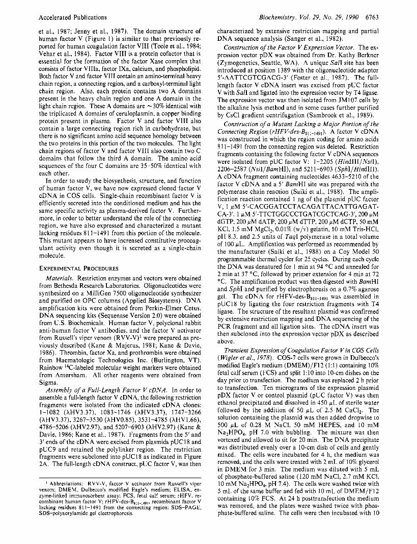
Heavy Chain Connecting Reglon Light Chain
Factor V
rHFV Des B811.149,
FIGURE 1 : Domain structure of human factor V and rHFV-des- B811-1491. The location of the heavy chain, connecting region, and light chain sequences are indicated. Domains are indicated by the letters in the boxes. Proteolytic cleavage sites determined for plasma factor V are indicated by the arrows. Thrombin cleaves after arginines 709, 1018, and 1545. The factor V activating enzyme from Russell's viper venom cleaves after arginine 1545. Factor Xa cleaves after arginine 1018 and at an undetermined site in the heavy chain region.
the molecule for prothrombin and factor Xa (Esmon et al., 1973; Suzuki et al., 1982). The prothrombin binding site appears to be contained within the heavy chain (Guinto & Esmon, 1984). Both the heavy chain and the light chain of factor Va appear to contribute to the binding site for factor Xa (Tucker et al., 1983; Annamalai et al., 1987). The light chain of factor Va contains the binding site for cell and phospholipid surfaces (Higgins & Mann, 1983; Tracy & Mann, 1983). Surface-bound factor Va accelerates pro- thrombin activation 4 orders of magnitude by shifting the reaction pathway to an ordered sequential reaction in which meizothrombin is the sole intermediate (Krishnaswamy et al., 1987).
Recently, the complete primary structure of human factor V was determined by a combination of cDNA cloning and protein sequence analysis (Kane & Davie, 1986, 1988; Kane
0 1990 American Chemical Society
Accelerated Publications Biochemistry, Vol. 29, No. 29, 1990 6763
characterized by extensive restriction mapping and partial D N A sequence analysis (Sanger et al., 1982).
Construction of the Factor V Expression Vector. The ex- pression vector pDX was obtained from Dr. Kathy Berkner (Zymogenetics, Seattle, WA). A unique Sal1 site has been introduced at position 1389 with the oligonucleotide adaptor 5’-AATTCGTCGACG-3’ (Foster et al., 1987). The full- length factor V cDNA insert was excised from pUC factor V with Sal1 and ligated into the expression vector by T4 ligase. The expression vector was then isolated from JM107 cells by the alkaline lysis method and in some cases further purified by CsCl gradient centrifugation (Sambrook et al., 1989).
Construction of a Mutant Lacking a Major Portion of the Connecting Region (rHFV-des-Bsll_1491). A factor V cDNA was constructed in which the region coding for amino acids 81 1-1491 from the connecting region was deleted. Restriction fragments containing the following factor V cDNA sequences were isolated from pUC factor V: 1-2205 (HindIIIINsiI), 2206-2587 (NsiIIBamHI), and 521 1-6903 (SphIIHindIII). A cDNA fragment containing nucleotides 4633-5210 of the factor V cDNA and a 5’ BamHI site was prepared with the polymerase chain reaction (Saiki et al., 1988). The ampli- fication reaction contained 1 ng of the plasmid pUC factor V, 1 p M 5’-CACGGATCCTACAGATTACATTGAGAT- CA-3‘, 1 pM 5’-TTCTGGCCCTGATCGCTCAG-3’, 200 pM dGTP, 200 p M dATP, 200 pM dTTP, 200 pM dCTP, 50 mM KCI, 1.5 mM MgCI,, 0.01% (w/v) gelatin, 10 mM Tris-HCI, pH 8.3, and 2.5 units of TaqI polymerase in a total volume of 100 pL. Amplification was performed as recommended by the manufacturer (Saiki et al., 1988) on a Coy Model 50 programmable thermal cycler for 25 cycles. During each cycle the DNA was denatured for 1 min a t 94 “ C and annealed for 2 min a t 37 “C, followed by primer extension for 4 min at 72 “C. The amplification product was then digested with BamHI and SphI and purified by electrophoresis on a 0.7% agarose gel. The cDNA for rHFV-des-Bsll-149, was assembled in pUC18 by ligating the four restriction fragments with T4 ligase. The structure of the resultant plasmid was confirmed by extensive restriction mapping and DNA sequencing of the PCR fragment and all ligation sites. The cDNA insert was then subcloned into the expression vector pDX as described above.
Transient Expression of Coagulation Factor V in COS Cells (Wigler et al., 1978). COS-7 cells were grown in Dulbecco’s modified Eagle’s medium (DMEM)/F12 (1:l) containing 10% fetal calf serum (FCS) and split 1:lO into IO-cm dishes on the day prior to transfection. The medium was replaced 2 h prior to transfection. Ten micrograms of the expression plasmid pDX factor V or control plasmid (pUC factor V) was then ethanol precipitated and dissolved in 450 pL of sterile water followed by the addition of 50 p L of 2.5 M CaCI,. The solution containing the plasmid was then added dropwise to 500 p L of 0.28 M NaCI, 50 mM HEPES, and 10 m M Na2HP04, pH 7.0 with bubbling. The mixture was then vortexed and allowed to sit for 20 min. The DNA precipitate was distributed evenly over a 10-cm dish of cells and gently mixed. The cells were incubated for 4 h, the medium was removed, and the cells were treated with 2 mL of 10% glycerol in DMEM for 3 min. The medium was diluted with 5 mL of phosphate-buffered saline (120 m M NaCI, 2.7 m M KCI, 10 mM Na2HP04, pH 7.4). The cells were washed twice with 5 mL of the same buffer and fed with 10 mL of DMEM/F12 containing 10% FCS. At 24 h posttransfection the medium was removed, and the plates were washed twice with phos- phate-buffered saline. The cells were then incubated with 10
et al., 1987; Jenny et al., 1987). The domain structure of human factor V (Figure 1) is similar to that previously re- ported for human coagulation factor VI11 (Toole et al., 1984; Vehar et al., 1984). Factor VI11 is a protein cofactor that is essential for the formation of the factor Xase complex that consists of factor VIIIa, factor IXa, calcium, and phospholipid. Both factor V and factor VI11 contain an amino-terminal heavy chain region, a connecting region, and a carboxyl-terminal light chain region. Also, each protein contains two A domains present in the heavy chain region and one A domain in the light chain region. These A domains are -30% identical with the triplicated A domains of ceruloplasmin, a copper binding protein present in plasma. Factor V and factor VI11 also contain a large connecting region rich in carbohydrate, but there is no significant amino acid sequence homology between the two proteins in this portion of the two molecules. The light chain regions of factor V and factor VI11 also contain two C domains that follow the third A domain. The amino acid sequences of the four C domains are 35-50% identical with each other.
I n order to study the biosynthesis, structure, and function of human factor V, we have now expressed cloned factor V cDNA in COS cells. Single-chain recombinant factor V is efficiently secreted into the conditioned medium and has the same specific activity as plasma-derived factor V. Further- more, in order to better understand the role of the connecting region, we have also expressed and characterized a mutant lacking residues 8 1 1-1 49 1 from this portion of the molecule. This mutant appears to have increased constitutive procoag- ulant activity even though it is secreted as a single-chain molecule.
EXPERIMENTAL PROCEDURES Materials. Restriction enzymes and vectors were obtained
from Bethesda Research Laboratories. Oligonucleotides were synthesized on a MilliGen 7500 oligonucleotide synthesizer and purified on O P C columns (Applied Biosystems). DNA amplification kits were obtained from Perkin-Elmer Cetus. DNA sequencing kits (Sequenase Version 2.0) were obtained from U S . Biochemicals. Human factor V, polyclonal rabbit anti-human factor V antibodies, and the factor V activator from Russell’s viper venom (RVV-V)’ were prepared as pre- viously described (Kane & Majerus, 1981; Kane & Davie, 1986). Thrombin, factor Xa, and prothrombin were obtained from Haematologic Technologies Inc. (Burlington, VT). Rainbow I4C-labeled molecular weight markers were obtained from Amersham. All other reagents were obtained from Sigma.
Assembly of a Full-Length Factor V cDNA. In order to assemble a full-length factor V cDNA, the following restriction fragments were isolated from the indicated cDNA clones: 1-1 082 (XHV3.37), 1083-1746 (XHV3.37), 1747-3266 (XHV3.37), 3267-3530 (XHV0.85), 3531-4785 (XHV1.66), 4786-5206 (XHV2.97), and 5207-6903 (XHV2.97) (Kane & Davie, 1986; Kane et al., 1987). Fragments from the 5’ and 3’ ends of the cDNA were excised from plasmids pUCl8 and pUC9 and retained the polylinker region. The restriction fragments were subcloned into pUC18 as indicated in Figure 2A. The full-length cDNA construct, pUC factor V, was then
I Abbreviations: RVV-V, factor V activator from Russell’s viper venom; DMEM, Dulbecco’s modified Eagle’s medium; ELISA, en- zyme-linked immunosorbent assay; FCS, fetal calf serum; rHFV, re- combinant human factor v ; rHFV-des-B,,,-,,9,, recombinant factor V lacking residues 81 1-1491 from the connecting region; SDS-PAGE, SDS-polyacrylamide gel electrophoresis.

6764
mL of serum-free medium (DMEM/FI 2 supplemented with 5 mM CaCI2 and 5 mg/mL bovine serum albumin) for 24 h. The conditioned medium was then harvested and assayed for factor V coagulant activity and antigen. Cells lysates were prepared by treating the plates with 1 mL of serum-free medium containing 0.1% Triton X-100 or by three cycles of freezing and thawing.
Factor V Activity Assays. Factor V coagulant activity was measured with a fibrometer assay as described by Bloom et al. (1979). Standard curves were prepared from dilutions of pooled human plasma. I n the fibrometer assay, 1 unit is defined as the amount of factor V activity present in 1 mL of unactivated pooled human plasma. Factor Va activity was measured with the chromogenic assay described by Baruch et al. (1 986) except that the assay was initiated by the addition of factor Xa and 8% (v/v) rabbit brain cephalin was substi- tuted for phospholipid vesicles. After a 2-min incubation, the amount of thrombin generated was quantitated by measuring the rate of hydrolysis of the chromogenic substrate S-2238. The absorbance a t 405 nm was measured with a V,,, mi- crotiter plate reader (Molecular Devices Inc.) interfaced with a Macintosh SE computer using A-soft software (Biometallics, Inc.). Standard curves were prepared from dilutions of pooled human plasma that had been activated with 3.75 pg/mL RVV-V for 15 min at 37 OC. In the chromogenic assay, 1 unit is defined as the amount of factor V activity present in 1 m L of activated pooled human plasma.
ELlSA Assay for Factor V. A sandwich ELISA was de- veloped to measure factor V antigen with a sensitivity in the 1 ng/mL range. Microtiter plates were coated with 50 pL of affinity-purified rabbit anti-human factor V polyclonal anti- body ( 2 pg/mL in phosphate-buffered saline) and incubated overnight at 4 O C . The coating solution was removed, and the wells were blocked with 0.2 mL of diluting buffer (phos- phate-buffered saline containing 0.1% Tween-20 and 0.25% bovine serum albumin). A 50-pL aliquot of diluted test sample was then added to the well and incubated at 37 OC for 30 min. The plate was washed twice with 0.1% Tween-20 in phos- phate-buffered saline. Biotinylated rabbit anti-human factor V antibody was then added to each well (50 pL of 2.5 pg/mL stock in diluting buffer) and incubated at 37 O C for 30 min. The plate was then washed three times with wash buffer and incubated with 50 pL of 5 pg/mL alkaline phosphatase/ streptavidin conjugate in diluting buffer. The plate was then washed three times with wash buffer, and 50 pL of disodium p-nitrophenyl phosphate (1 mg/mL in 0.1 M Tris-HCI, pH 8.8, 5 mM MgCI,) was added and incubated for 30 min a t 37 OC. Color development was terminated with the addition of 150 pL of 0. I N sodium hydroxide. Absorbance at 405 nm was measured with a V,,, microtiter plate reader (Molecular Devices lnc.). Standard curves were determined from purified plasma derived human factor V.
Immunoprecipitation of Recombinant Factor V. COS cells were transfected with the pDX expression vector by calcium phosphate precipitation as described above. Twenty-four hours following transfection the cells were washed twice with phosphate-buffered saline and then changed to methionine- deficient DMEM (serum free). After a 2-h incubation the methionine-deficient medium was replaced, and 500 pCi of [35S]methionine was added to the medium. Following a 6-h labeling, the medium was harvested and stored a t -70 "C. Recombinan t factor V was activated with thrombin ( 1 unit/mL) or RVV-V (3.75 pg/mL) for 15 min a t 37 OC. The conditioned medium (1-mL volume) was then cleared by in- cubation with gelatin agarose (60 pL) twice for 90 min. The
Biochemistry, Vol. 29, No. 29, 1990 Accelerated Publications
A.
t w wm c m H E E S
B. Nco I 467
Ue pDX Factor V
6522 Nco I 6404 Xho I
5867 EcoR BstE'II 5082 FIGURE 2: Assembly of the factor V expression vector. (A) Restriction fragments of the factor V cDNAs were isolated from preparative restriction enzyme digests by agarose gel electrophoresis. The re- striction enzymes generating the fragment and the first and last nucleotides a t the end of each fragment are identified. Restriction fragments with compatible ends were ligated together into pUCl8 by T 4 ligase. The final construct pUC factor V contained the full-length factor V cDNA. Portions of the pUC18 and pUC9 po- lylinker are indicated by the boxes. Restriction sites in the plylinker are indicated: H, HindIII; E, EcoRI; S, SalI. (B) The features in expression vector pDX factor V are as follows: (a) SV-40 promoter and enhancer; (b) adenovirus major late promoter and leader sequence; (c) splice donor/acceptor; (d) factor V cDNA; (e) polyadenylation signal. The locations of selected restriction endonuclease sites are indicated.
medium was then cleared again with 20 pL of protein A- Sepharose beads and 2 pg of control rabbit IgG (Sigma) for 90 min. Recombinant factor V was then immunoprecipitated with 2 pg of affinity-purified polyclonal anti-factor V antibody in the presence of 20 p L of protein A-Sepharose beads. Precipitated proteins were then denatured in the presence of SDS and 0-mercaptoethanol and analyzed by SDS-PAGE on 5% acrylamide gels (Laemmli, 1970) followed by autoradi- ography.
RESULTS Assembly of the Factor V Expression Vector. We had
previously isolated a series of overlapping clones that comprised a full-length factor V cDNA sequence. Assembly of a full- length cDNA constuct was complicated because the factor V cDNA contained multiple EcoRI sites and because one of our clones (XHV2.97) contained an in-frame deletion of 297 nu- cleotides (Kane et al., 1987; Kane & Davie, 1986). The method we used to assemble the full-length factor V cDNA is shown in Figure 2A. Extensive restriction mapping and partial DNA sequence analysis indicated that no artifacts were introduced during assembly of the construct. Polylinker se- quences on the 5' and 3' ends of the cDNA make it possible to excise the full-length cDNA insert with the restriction
Accelerated Publications
enzyme Sal1 or Smal. The expression vector pDX has been previously used to express a number of coagulation factors and growth factors in mammalian cells (Busby et al., 1987: Berkner et al., 1986; Foster et al., 1987: Kaushansky et al., 1986: Powell et al., 1986). In order to use this expression vector to express the factor V cDNA, we inserted a Sal1 restriction site at position 1389. The restriction map for the pDX factor V expression vector is shown in Figure 2B. This construct contains the adenovirus major late promoter, the SV40 en- hancer, the adenovirus tripartite leader sequences upstream of the cloning site followed by a polyadenylation site, and the SV-40 origin of replication downstream of the cloning site.
Expression of Recombinant Factor V. Recombinant factor V was expressed when pDX factor V was utilized to transfect COS cells (Figure 28). Levels of factor V in the conditioned medium peaked 48 h after transfection, at which time factor V antigen levels were 70 f 15 ng/mL (mean f S D of seven experiments). Factor V antigen was not detected in the conditioned medium when cells were transfected with a control plasmid (pUC factor V). In order to determine whether re- combinant factor V was efficiently secreted from the COS cells, we measured the amount of factor V antigen present in conditioned media and in cell lysates at either 24 or 48 h after changing to serum-free media. These results indicate that 95 f 4% (mean f S D of 16 experiments) of the factor V syn- thesized in COS cells is secreted into the culture medium. The amount of recombinant factor V in the conditioned medium is reduced approximately 7-fold to 11 ng/mL when 5 mg/mL albumin is not added to the serum-free medium. This re- quirement is probably due to the tendency of human factor V to adsorb to plastic at low protein concentrations (Kane & Majerus, 1981).
The factor V activity in the conditioned medium determined by fibrometer assay increased approximately 5-fold from 0.027 f 0.012 to 0.124 f 0.044 unit/mL (mean f SD of seven experiments) following activation by RVV-V, indicating that recombinant factor V was secreted largely as the prmofactor form of the molecule. With use of the fibrometer assay, the activity of purified plasma factor V increased -20-25-fold following activation with thrombin or RVV-V. The fact that recombinant factor V appears to only activate -5-fold in the fibrometer assay may be in part due to the presence of small amounts of activated cofactor in the conditioned media but is also probably a reflection of the sensitivity of the fibrometer assay (see below). The activity of control conditioned media was -0.002 unit/mL before and after activation with RVV-V. This value is near the lower limit of sensitivity for the fi- brometer assay. The estimated specific activity of the re- combinant factor Va was approximately 1800 f 500 units/mg (mean f SD of seven experiments), which is similar to the specific activity of purified plasma factor Va of 17OCb2000 units/mg (Kane & Majerus, 1981). The specific activity of plasma factor Va activated with either thrombin or RVV-V is identical (Kane & Majerus. 1981).
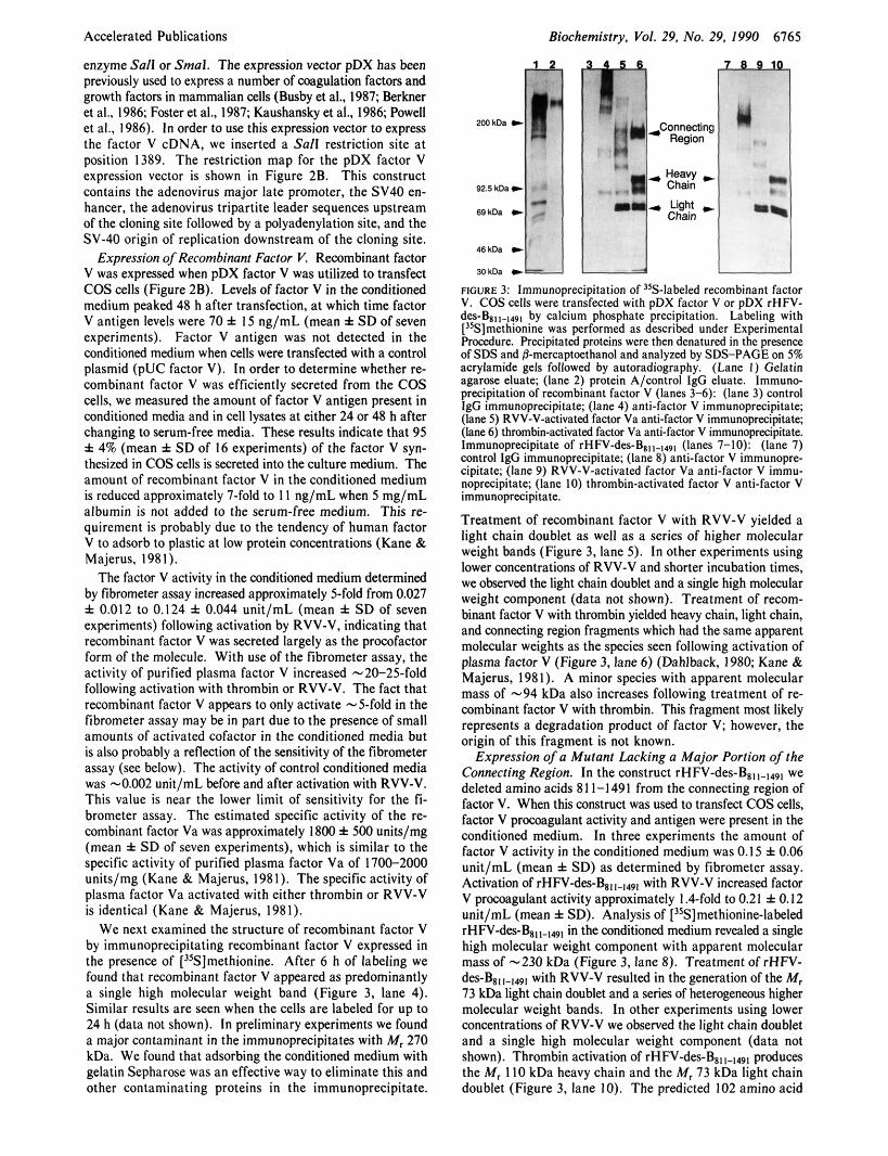
We next examined the structure of recombinant factor V by immunoprecipitating recombinant factor V expressed in the presence of ["S]methionine. After 6 h of labeling we found that recombinant factor V appeared as predominantly a single high molecular weight band (Figure 3, lane 4). Similar results are seen when the cells are labeled for up to 24 h (data not shown). In preliminary experiments we found a major contaminant in the immunoprecipitates with M, 270 kDa. We found that adsorbing the conditioned medium with gelatin Sepharose was an effective way to eliminate this and other contaminating proteins in the immunoprecipitate.
Biochemistry. Val. 29, No. 29, 1990 6165
9 2 5 k D a - 1 1 1 . ::]"I Light * 6911- + . -
46kDa * MkDs *
Chain
FIGURE 3: Immunoprecipitation of 'sS-labeled recombinant factor V. COS cells were transfected with pDX factor V or pDX rHFV- des-B811.149, by calcium phosphate precipitation. Labeling with ["S]methionine was performed as described under Experimental Procedure. F'recipitated proteins were then denatured in the presence of SDS and 0-mercaptcethanol and analyzed by SDS-PAGE on 5% acrylamide gels fallowed by autoradiography. (Lane I ) Gelatin agarose eluate; (lane 2) protein A/wntrol IgG eluate. Immuno- precipitation of recombinant factor V (lanes 3-6): (lane 3) control IgG immunoprecipitate; (lane 4) anti-factor V immunoprecipitate; (lane 5 ) RVV-V-activated factor Va anti-factor V immunoprecipitate; (lane 6 ) thrombin-activated factor Va anti-factor V immunoprecipitate. Immunoprecipitate of rHFV-des-Bs,,.l,9, (lanes 7-10): (lane 7) control IgG immunoprecipitate; (lane 8) anti-factor V immunopre- cipitate; (lane 9) RVV-V-activated factor Va anti-factor V immu- noprecipitate; (lane IO) thrombin-activated factor V anti-factor V immunoprecipitate.
Treatment of rewmbinant factor V with RVV-V yielded a light chain doublet as well as a series of higher molecular weight bands (Figure 3. lane 5) . In other experiments using lower concentrations of RVV-V and shorter incubation times, we observed the light chain doublet and a single high molecular weight component (data not shown). Treatment of recom- binant factor V with thrombin yielded heavy chain, light chain, and connecting region fragments which had the same apparent molecular weights as the species seen following activation of plasma factor V (Figure 3, lane 6) (Dahlback, 1980; Kane & Majerus, 1981). A minor species with apparent molecular mass of -94 kDa also increases following treatment of re- combinant factor V with thrombin. This fragment most likely represents a degradation product of factor V; however, the origin of this fragment is not known.
Expression of a Mutant Lacking a Major Portion of the Connecting Region. In the construct rHFV-des-B8,,.,,9, we deleted amino acids 81 1-1491 from the connecting region of factor V. When this C O I N ~ N ~ ~ was used to transfect COS cells, factor V procoagulant activity and antigen were present in the conditioned medium. In three experiments the amount of factor V activity in the conditioned medium was 0.15 f 0.06 unit/mL (mean f SD) as determined by fibrometer assay. Activation of rHM-des--$, with RVV-V increased factor V procoagulant activity approximately 1.4-fold to 0.21 f 0.12 unit/mL (mean f SD). Analysis of [%]methionine-labeled rHM-des-B,,,.,,, in the conditioned medium revealed a single high molecular weight component with apparent molecular mass of -230 kDa (Figure 3, lane 8). Treatment of rHFV- des-B8,,-,49, with RVV-V resulted in thegeneration of the M, 73 kDa light chain doublet and a series of heterogeneous higher molecular weight bands. In other experiments using lower concentrations of RVV-V we observed the light chain doublet and a single high molecular weight component (data not shown). Thrombin activation of rHFV-des-B,,,~,,9, produces the M, 1 IO kDa heavy chain and the M, 73 kDa light chain doublet (Figure 3, lane IO). The predicted 102 amino acid

6766 Biochemistry, Vol. 29, No. 29, 1990 Accelerated Publications
1 2 3 4
F i c u R E 4: Activation of recombinant factor v. For each recombinant protein, conditioned medium from four transfections was assayed before and after activation with RVV-V. Each activity measurement was based on 9-1 2 independent determinations. The activity following activation with RVV-V was 27 & 10 milliunits of factor Va/mL for recombinant factor V and 1 1 1 f 45 milliunits of factor Va/mL for rHFV-des-B,, 1-1491. The activity of the unactivated and activated cofactors is expressed as per cent of the corresponding RVV-V-ac- tivated recombinant protein. Error bars indicate the standard error of the mean. ( I ) Recombinant factor V; (2) RVV-V-activated re- combinant factor v; (3) ~ H F V - ~ ~ S - B , , ~ - ~ ~ ~ ~ ; (4) RVV-V-activated rHFV-des-B,,
fragment from the truncated connecting region is not visualized because it does not contain any methionine residues. The concentration of rHFV-des-B,, in the conditioned medium was estimated to be 80 ng/mL by use of the ELISA for factor V. This is only an approximate concentration since we have not yet standardized the ELISA with purified rHFV-des- Bsl Immunoprecipitation of 35S-labeled recombinant proteins indicates that deletion of residues 81 1-1491 from the connecting region results in a 2-5-fold increase in the ex- pression of the recombinant protein. The clotting activity of the RVV-V-activated 35S-labeled recombinant proteins sug- gests that the specific activity of rHFV-des-Bsll-1491 is similar (approximately 70%) to that of native factor Va (data not shown).
Procoagulant Activity of Factor V-des-B,, 1-1491. Prelimi- nary results indicated that treatment of rHFV-des-B,, with thrombin or RVV-V resulted in less than a 2-fold increase in factor V procoagulant activity as measured by fibrometer assay. The activity of single chain factor V in clotting assays is believed to be due to activation of the procofactor during the assay (Nesheim et al., 1979b). We used a chromogenic assay (Baruch et al., 1986) that is more sensitive and specific for the activated cofactor to further characterize the pro- coagulant activity of recombinant factor V. In this assay factor V present in unactivated plasma had 1-2% of the activity of the activated cofactor. With use of the chromogenic assay, the procoagulant activity of recombinant human factor V was 3.8 f 1.3% of that of the activated cofactor (Figure 4). In contrast, rHFV-des-B,, constitutively expressed 38 f 7% of the activity of the RVV-V-activated protein (p < 0.005). We considered the possibility that rHFV-des-B,, was more sensitive to proteolysis by thrombin or factor Xa than native factor V. When 35S-labeled single-chain recombinant factor V and rHFV-des-B,, were treated with increasing concentrations of thrombin (0-8.5 nM) or factor Xa/rabbit brain cephalin (0-5.2 nM) for 2 min, there was no significant difference in the extent of proteolysis of the two recombinant proteins as judged by immunoprecipitation and SDS-poly- acrqlamide gel electrophoresis (data not shown).
Drscussro~ In order to characterize the functional domains of the factor
V molecule, we have developed a mammalian cell expression system utilizing the cloned factor V cDNA and COS cells.
We find that single-chain recombinant factor V is efficiently secreted into the conditioned medium. Activation of recom- binant factor V by thrombin results in the formation of the factor Va heavy chain and light chain as well as a large fragment from the connecting region. the estimated specific activity of recombinant factor Va is the same as the specific activity of plasma-derived factor Va. Our results suggest that recombinant factor V is structurally and functionally very similar to plasma-derived factor V.
We have previously determined the primary structure of human factor V by cDNA cloning (Kane & Davie, 1986; Kane et al., 1987). Factor V contains four types of internal repeating sequences. The three A domains present in factor V are approximately 30% identical with the A domains present in ceruloplasmin and factor VI11 (Takahashi et al., 1984; Toole et a]., 1984; Vehar et al., 1984). The two C domains located a t the carboxyl terminus are -40% identical with the C do- mains present in factor VIII. The connecting region or B domain in factor V is comprised of 836 amino acids (amino acids 7 10-1 545) located between the second and third A do- mains. The connecting region of factor V is not homologous to the B domain of factor VI11 nor to the sequence of any other known protein. The connecting region is also unique in that it contains two tandem repeats of a 17 amino acid sequence as well as 3 1 tandem repeats of a 9 amino acid sequence. The connecting region appears to be heavily glycosylated (Esmon, 1980) and contains 25 potential N-linked glycosylation sites and 15% serine and threonine.
Little is known about the molecular mechanism of factor V activation and the role of the connecting region in this process. When factor V is activated by thrombin, the pro- teolytic fragments derived from the connecting region are released from factor Va (Esmon, 1979; Suzuki et al., 1982; Kane & Majerus, 1982). Thrombin-activated factor Va is a calcium-dependent heterodimer composed of one heavy chain and one light chain (Esmon, 1979). Factor V procoagulant activity can be reconstituted by mixing isolated heavy chain and light chain fragments in the presence of divalent cations such as calcium or manganese (Esmon, 1979; Suzuki et al., 1982). These results indicate that the connecting region is not required for procoagulant activity. However, cleavage of Arg,545Ser1546 by the factor V activator in Russell’s viper venom (RVV-V) appears to be sufficient for complete acti- vation of factor V (Esmon, 1980; Kane & Majerus, 1981). Recently, Monkovic and Tracy (1990) have proposed that cleavage of Arg,o,,Thr,o,9 by factor Xa may also be sufficient for complete activation of human factor V (Figure 1). Thus, when factor V is activated by RVV-V or factor Xa, the con- necting region fragments are not released from the activated molecule. In contrast, nonspecific proteolysis within the connecting region of factor V by platelet proteases does not appear to activate the molecule (Baruch et al., 1986; Viskup et al., 1987).
In order to assess the influence of the connecting region on the expression and activity of recombinant factor V, we have expressed a mutant, ~ H F V - ~ ~ S - B ~ ~ ~ - ~ ~ ~ , , that lacks a large portion of the connecting region. Deletion of this portion of the connecting region results in a 2-5-fold increase in the expression of the recombinant protein. This mutant expresses factor V procoagulant activity. In contrast to recombinant factor V, however, we find that rHFV-des-B811-1491 appears to possess significant procoagulant activity without prior ac- tivation by thrombin or RVV-V. Our initial characterization of the rHFV-des-B8,,-,491 mutant suggests that this protein has some interesting properties. Several aspects will require
Accelerated Publications Biochemistry, Vol. 29, No. 29, 1990 6767
further study. While it appears that the specific activity and proteolytic processing of this mutant are similar to those of plasma-derived factor V, purification of the protein will be required to determine the precise specific activity and pro- teolytic cleavage sites. In addition, absolute proof that sin- gle-chain rHFV-des-B8, 1-149, has procoagulant activity in the absence of thrombin activation will require mutation of the two thrombin cleavage sites. These experiments are currently in progress.
Comparison of our results with recombinant factor V to previously reported studies on the expression of recombinant factor VI11 reveals a number of important differences. First, we find that recombinant factor V is efficiently secreted (95 f 4%) from COS cells. In contrast, Kaufman has reported that only 10% of recombinant factor VI11 is secreted from C H O cells and the remaining 90% is sequestered in the Golgi bound to an immunoglobulin binding protein (Kaufman et al., 1988). Second, we find that recombinant factor V is secreted as a single-chain polypeptide and that it is resistant to pro- teolysis in conditioned medium. The polypeptide structure of recombinant factor VIII, however, is heterogeneous due to proteolysis within the connecting region or B domain (Eaton et al., 1987; Kaufman et al., 1988). Third, we find that rHFV-des-B811-1491 does not appear to be more susceptible than recombinant factor V to proteolysis by thrombin or factor Xa. I n contrast, rHFVIIl-des-B797-1652 was reported to be signif- icantly more sensitive to thrombin activation than plasma- derived factor VllI (Eaton et al., 1986). Finally, our data suggest that deletion of the connecting region in recombinant factor V markedly increases the procoagulant activity of the unactivated molecule. In contrast, deletion of the connecting region in factor VlIl ( ~ H F V I I I - ~ ~ S - B , , , - , , ~ ~ ) resulted in a molecule that could still be activated 15-20-fold by thrombin, which was identical with plasma-derived factor VI11 (Eaton et al., 1986).
Our results underscore the fact that although there are significant similarities in the amino acid sequences of factor V and factor VIII, there are also many important differences between these two molecules. The molecular mechanisms that lead to cofactor activation are not understood. The relative inactivity of the procofactor may be due to the fact that the heavy chain and light chain domains are physically separated by the connecting region. Alternatively, the heavy chain and light chain may be associated in the procofactor, but the en- zyme and substrate binding sites may be sterically blocked by the connecting region. Further studies on the expression and mutagenesis of factor V and factor VI11 will be helpful in addressing these issues.
ACKNOWLEDGMENTS
We thank Dr. Earl Davie (University of Washington) for his support and encouragement and Drs. Charles S. Greenberg, William A. Dittman, David C. Sane, and Larry D. Cripe for useful discussions.
REFERENCES
Annamalai, A. E., Rao, A. K., Chiu, H. C., Wang, D., Dutta Roy, A. K., Walsh, P. N., & Colman, R. W. (1987) Blood
Baruch, D., Hemker, H. C., & Lindhout, T. (1986) Eur. J . Biochem. 154, 21 3-21 8.
Berkner, K. L., Busby, S . , Davie, E., Hart, C., Insley, M., Kisiel, W., Kumar, A,, Murray, M., O’Hara, P., Woodbury, R., & Hagen, F. (1986) Cold Spring Harbor Symp. Quant. Biol. 51, 531-541.
70, 139-146.
Bloom, J. W., Nesheim, M. E., & Mann, K. G. (1979j Thromb. Res. 15, 595-599.
Busby, S . , Kumar, A., Joseph, A., Halfpap, L., Insley, M., Berkner, K., Kurachi, K., & Woodbury, R. (1985) Naure (London) 316, 271-273.
Dahlback, B. (1980) J . Clin. Invest. 66, 583-591. Eaton, D. L., Wood, W. I . , Eaton, D., Hass, P. E., Holl-
ingshead, P., Wion, K. L., Mather, J., Lawn, R. M., Vehar, G. A,, & Gorman, C. (1986) Biochemistry 25, 8343-8347.
Eaton, D. L., Hass, P. E., Riddle, L., Mather, J., Wiebe, M., Gregory, T., & Vehar, G. A. (1987) J . Biol. Chem. 262, 3285-3290.
Esmon, C. T. (1979) J . Biol. Chem. 254, 964-973. Esmon, C. T. (1980) in The Regulation of Coagulation
(Mann, K. G., & Taylor, F. B., Eds.) pp 137-143, Elsev- ier/North-Holland, New York.
Esmon, C. T., Owen, W. G., Duiguid, D. L., & Jackson, C. M. (1973) Biochim. Biophys. Acta 310, 289.
Foster, D. C., Rudinski, M. S . , Schach, B. G., Berkner, K. L., Kumar, A. A., Hagen, F. S . , Sprecher, C. A,, Insley, M. Y., & Davie, E. W. (1987) Biochemistry 26, 7003-701 1.
Foster, W. B., Nesheim, M. E., & Mann, K. G. (1983) J . Biol. Chem. 258, 13970-1 3977.
Guinto, E. R., & Esmon, C. T. (1982) J . Biol. Chem. 257,
Guinto, E. R., & Esmon, C. T. (1984) J . Biol. Chem. 259,
Higgins, D. L., & Mann, K. G. (1983) J . Biol. Chem. 258,
Jenny, R. J., Pittman, D. D., Toole, J . J., Kriz, R. W., Aldape, R. A., Hewick, R. M., Kaufman, R. J., & Mann, K. G. (1987) Proc. Natl. Acad. Sci. U.S.A. 84, 4846-4850.
Kane, W. H., & Majerus, P. W. (1981) J . Biol. Chem. 256,
Kane, W. H., & Majerus, P. W. (1982) J . Biol. Chem. 257,
Kane, W. H., & Davie, E. W. (1986) Proc. Natl. Acad. Sci.
Kane, W. H., & Davie, E. W. (1988) Blood 71, 539-555. Kane, W. H., Ichinose, A., Hagen, F. S., & Davie, E. W.
(1987) Biochemistry 26, 6508-6514. Kane, W. H., Lindhout, M. J., Jackson, C. M., & Majerus,
P. W. (1980) J . Biol. Chem. 255, 1170-1 174. Katzmann, J. A., Nesheim, M. E., Hibbard, L. S . , & Mann,
K. G. (1981) Proc. Natl. Acad.Sci . U.S.A. 78, 162-166. Kaufman, R. J., Wasley, L. C., & Dorner, A. J. (1988) J. Biol.
Chem. 263, 6352-6362. Kaushansky, K., O’Hara, P. J., Berkner, K., Segal, G. M.,
Hagen, F. S., & Adamson, J. W. (1986) Proc. Natl. Acad. Sci. U.S.A. 83, 3101-3105.
Krishnaswamy, S . , Church, W. R., Nesheim, M. E., & Mann, K. G. (1987) J . Biol. Chem. 262, 3291-3299.
Krishnaswamy, S . , Russell, G. D., & Mann, K. G. (1989) J . Biol. Chem. 264, 3160-3168.
Laemmli, U. K. (1970) Nature (London) 227, 680-685. Laue, T. M., Lu, R., Krieg, U. C., Esmon, C. T., & Johnson,
Monkovic, D. D., & Tracy, P. B. (1990) Biochemistry 29,
Nesheim, M. E., Myrmel, K. H., Hibbard, L., & Mann, K.
Nesheim, M. E., Taswell, J. B., & Mann, K. G. (1979b) J .
10038-1 0043.
13986-13992.
6503-6508.
1002-1007.
3963-3969.
U.S.A. 83, 6800-6804.
A. E. (1 989) Biochemistry 28, 4762-477 1.
I 1 18-1 128.
G. (1979a) J . Biol. Chem. 254, 508-517.
Biol. Chem. 254, 10952-10962,

6768 Biochemistry 1990, 29, 6768-6777
Powell, J. S., Berkner, K. L., Lebo, R. V., & Adamson, J. W. ( 1 986) Proc. Natl. Acad. Sci. U.S.A. 83, 6465-6469.
Saiki, R. K., Gelfand, D. H., Stoffel, S., Scharf, S. J., Higuchi, R., Horn, G. T., Mullis, K. B., & Erlich, H. A. (1988) Science 239, 487-491.
Sambrook, J., Fritsch, E. F., & Maniatis, T. (1989) Molecular Cloning: A Laboratory Manual, Cold Spring Harbor Laboratory Press, Cold Spring Harbor, NY.
Sanger, F., Coulson, A. R., Hong, G. F., Hill, D. F., & Pet- ersen, G. B. (1982) Proc. Natl . Acad. Sci. U.S.A. 79,
Suzuki, K., Dahlback, B., & Stenflo, J. (1982) J . Biol. Chem.
Takahashi, N. , Ortel, T . L., & Putnam, F. W. (1984) Proc. Natl. Acad. Sci. U.S.A. 81, 390-394.
Toole, J . J., Knopf, J . L., Wozney, J. M., Sultzman, L. A., Buecker, J . L., Pittman, D. D., Kaufman, R. J., Brown, E.,
729-773.
257, 6556-6564.
Art ides
Shoemaker, C . , Orr, E. C., et al. (1984) Nature 312,
Toole, J. J., Pittman, D. D., Orr, E. C., Murtha, P., Wasley, L. C., & Kaufman, R. J. (1986) Proc. Natl. Acad. Sci.
Tracy, P. B., & Mann, K. G. (1983) Proc. Natl. Acad. Sci.
Tucker, M. M., Foster, W. B., Katzmann, J. A., & Mann, K. G. (1983) J . Biol. Chem. 258, 1210-1214.
Vehar, G. A,, Keyt, B., Eaton, D. L., Rodriguez, H., OBrien, D. P., Rotblat, F., Oppermann, H., Keck, R., Wood, W. I., Harkins, R. N., Tuddenham, E. G. D., Lawn, R. M., & Capon, D. J. (1984) Nature 312, 337-342.
Viskup, R. W., Tracy, P. B., & Mann, K. G. (1987) Blood
Wigler, M., Pellicer, A., Silverstein, S., & Axel, R. (1978)
342-347.
U.S.A. 83, 5939-5942.
U.S.A. 80, 2380-2384.
69, 1188-1 195.
Cell 14, 725-731.
Identification and Characterization of a 43-Kilodalton Laminin Fragment from the “A” Chain (Long Arm) with High-Affinity Heparin Binding and Mammary
Epithelial Cell Adhesion-Spreading Activities+
C. N . Rao* and Nicholas A. Kefalides Connective Tissue Research Institute and Department of Medicine, University of Pennsylvania and University City Science
Center, 3624 Market Street, Philadelphia, Pennsylvania 191 04 Received February 6, 1990; Revised Manuscript Received April 18, 1990
ABSTRACT: A recently described procedure of reduction and carboxymethylation followed by heparin- Sepharose chromatography [Arumugham et al. (1988) Connect. Tissue Res. 18, 135-1471 was used to characterize high-affinity heparin binding fragments of the laminin “A” chain. Two laminin fragments of MI 53K and 43K selectively bound to the heparin-Sepharose column from the chymotrypsin digest of laminin, indicating that these fragments originate from the “A” chain. Without reduction and carboxy- methylation but in the presence of 2.0 M urea, the heparin-Sepharose-bound material from the chymotrypsin laminin digest contains all the attachment-promoting activity for normal mouse mammary epithelial cells. The reduced 200-kDa intact three short a r m fragment, fragments of M, 70K-160K obtained either from laminin or from the reduced 200-kDa three short a r m fragment, and the 53-kDa heparin binding fragment were all inactive in promoting the adhesion of mouse mammary epithelial cells. The mammary epithelial cell adhesion and spreading properties of laminin are associated with the high-affinity heparin binding 43-kDa fragment. The mammary epithelial cells attach to the 43-kDa fragment substrate and synthesize laminin, collagen type IV, and desmoplankins I and I1 as are the cells attached to laminin substrate and to the cells grown on tissue culture dishes. The biologically active 43-kDa fragment is generated from laminin, but not from the three short a r m fragment. These results suggest that normal mouse mammary epithelial cells interact with laminin through a single site which is present in the 43-kDa heparin binding fragment located on the long a r m of the “A” chain.
x e glycoprotein laminin ( M , 850K) is the most abundant component of specialized extracellular matrices known as basement membranes (Chung et al., 1979; Timpl et al., 1979). Laminin consists of three chains designated “A” (MI 400K), “BI” (MI 220K), and “B2“ ( M , 200K); all three have been
‘This work was supported by NIH Grants AR 20553, HL 29492, and
* Address correspondence to this author at Cancer Diagnostic Inc., AR 07490.
2101 E. Jefferson St., Suite 310, Rockville, MD 20852.
0006-2960/90/0429-6768$02.50/0
cloned and sequenced to full length (Sasaki & Yamada, 1987; Sasaki et al., 1987, 1988; Pikkarainen et al., 1987). By ro- tary-shadowing electron microscopy, laminin has the shape of an asymmetric cross with one long arm and three short arms (Engel et al., 1981). Laminin is synthesized by a variety of cell types which include endothelial, epithelial, muscle, and Schwann cells and is involved in numerous biological functions (Kleinman et al., 1985; Liotta et al., 1986; Martin & Timpl, 1986; Timpl, 1989; Panayotou et al., 1989). By interaction with components of basement membrane macromolecules such
0 1990 American Chemical Society