extensive molecular dynamics ... -...
TRANSCRIPT

Extensive Molecular Dynamics Simulations Showing That Canonical G8 and ProtonatedA38H+ Forms Are Most Consistent with Crystal Structures of Hairpin Ribozyme
Vojtech Mlynsky,† Pavel Banas,†,‡ Daniel Hollas,† Kamila Reblova,‡ Nils G. Walter,§
Jirı Sponer,*,†,‡ and Michal Otyepka*,†,‡
Department of Physical Chemistry, Faculty of Science, Palacky UniVersity Olomouc, tr. 17. listopadu 12,771 46 Olomouc, Czech Republic, Institute of Biophysics, Academy of Sciences of the Czech Republic,KraloVopolska 135, 612 65 Brno, Czech Republic, and Department of Chemistry, Single Molecule AnalysisGroup, UniVersity of Michigan, 930 North UniVersity AVenue, Ann Arbor, Michigan 48109-1055
ReceiVed: January 6, 2010; ReVised Manuscript ReceiVed: April 12, 2010
The hairpin ribozyme is a prominent member of the group of small catalytic RNAs (RNA enzymes orribozymes) because it does not require metal ions to achieve catalysis. Biochemical and structural data haveimplicated guanine 8 (G8) and adenine 38 (A38) as catalytic participants in cleavage and ligation catalyzedby the hairpin ribozyme, yet their exact role in catalysis remains disputed. To gain insight into dynamics inthe active site of a minimal self-cleaving hairpin ribozyme, we have performed extensive classical, explicit-solvent molecular dynamics (MD) simulations on time scales of 50-150 ns. Starting from the available X-raycrystal structures, we investigated the structural impact of the protonation states of G8 and A38, and theinactivating A-1(2′-methoxy) substitution employed in crystallography. Our simulations reveal that a canonicalG8 agrees well with the crystal structures while a deprotonated G8 profoundly distorts the active site. ThusMD simulations do not support a straightforward participation of the deprotonated G8 in catalysis. Bycomparison, the G8 enol tautomer is structurally well tolerated, causing only local rearrangements in theactive site. Furthermore, a protonated A38H+ is more consistent with the crystallography data than a canonicalA38. The simulations thus support the notion that A38H+ is the dominant form in the crystals, grown at pH6. In most simulations, the canonical A38 departs from the scissile phosphate and substantially perturbs thestructures of the active site and S-turn. Yet, we occasionally also observe formation of a stable A-1(2′-OH) · · ·A38(N1) hydrogen bond, which documents the ability of the ribozyme to form this hydrogen bond,consistent with a potential role of A38 as general base catalyst. The presence of this hydrogen bond is, however,incompatible with the expected in-line attack angle necessary for self-cleavage, requiring a rapid transition ofthe deprotonated 2′-oxyanion to a position more favorable for in-line attack after proton transfer from A-1(2′-OH) to A38(N1). The simulations revealed a potential force field artifact, occasional but irreversible formationof “ladder-like”, underwound A-RNA structure in one of the external helices. Although it does not affect thecatalytic center of the hairpin ribozyme, further studies are under way to better assess possible influence ofsuch force field behavior on long RNA simulations.
Introduction
The hairpin ribozyme is a self-cleaving and -ligating catalyticRNA motif classified as a small RNA enzyme or ribozyme(Figure 1). It is found in the minus strand of the satellite RNAassociated with the Tobacco Ringspot Virus, where it promotesdouble-rolling circle replication.1-3 It can also be engineeredto catalyze reversible, site-specific phosphodiester bond cleavageon an external, complementary RNA substrate.4 The hairpinribozyme achieves rate acceleration similar to that for otherribozymes,5,6 and it does not require a specific metal ion toachieve full catalytic efficiency (cleavage rate ∼0.5 min-1).4,7
Although the folded hairpin ribozyme features an active sitepocket of deep negative potential, similar to that of the hepatitisdelta virus (HDV) ribozyme, once formed, this pocket appearsto be secluded from solvent,8 in sharp contrast to the open pocket
of the HDV ribozyme.9 The HDV ribozyme catalytic pocket isknown to interact with divalent ions. If divalents are not present,the pocket is immediately (in molecular dynamics simulationson nanoseconds time scale)9,10 soaked by monovalent ions thatare likely to interfere with the deep negative potential. Thehairpin ribozyme, as it avoids interactions with ions sterically,is thus exposing RNA functional groups and water moleculesin the active site to a largely uncompensated negative electro-static potential for long time periods. Previous moleculardynamics (MD) simulations suggested that long-residency watermolecules in the active site may thus become activated toparticipate in catalysis.8,11-13 The established lack of a catalyticmetal ion requirement makes the hairpin ribozyme an especiallyuseful model system in which to probe the direct role ofnucleobases in RNA catalysis as a major remaining challengein the field.14
A range of structural and biochemical data have implicatedguanine 8 (G8) and adenine 38 (A38) as direct participants incatalysis of cleavage and ligation (Figure 2). Mutation ordeletion of the conserved G8 in loop A near the scissilephosphate diminishes activity by ∼1000-fold without signifi-
* Corresponding authors. M.O.: tel, +420 585 634 756; fax, +420 585634 761; e-mail, [email protected], J.S.: tel, +420 541 517 133;e-mail, [email protected].
† Palacky University Olomouc.‡ Academy of Sciences of the Czech Republic.§ University of Michigan.
J. Phys. Chem. B 2010, 114, 6642–66526642
10.1021/jp1001258 2010 American Chemical SocietyPublished on Web 04/26/2010

cantly disrupting the global structure of the ribozyme.15-17 Theposition of G8 near the 2′-OH attacking nucleophile, as observedin crystal structures, first suggested the possibility that an N1unprotonated G8- may act as general base during catalysis(Figure 2A).11,15,18,19 Recent experiments measuring the ioniza-tion state of an 8-azaguanosine substitution at this position,however, do not provide support for the G8- general basemechanism as the pKa of G8 was estimated to be 9.5,20 nearthe unperturbed pKa value of guanine, making deprotonationunlikely.21 Exogenous nucleobase rescue experiments suggestedthat the catalytic role of G8 rather lies in charge stabilizationof the transition state (TS) and/or alignment of the reactivegroups.13,14,16,22 Recent molecular dynamics (MD) and quantummechanical/molecular mechanical (QM/MM) calculations areconsistent with the latter model, as they suggest that G8 couldfacilitate catalysis through stabilizing both the developing chargeon the scissile phosphate and the strained backbone conforma-tions adopted along the reaction pathway.23-25 A direct com-parison as to what extent the two possible G8 protonation statesare structurally compatible with the crystal structures is stilllacking.
Compared to that case for G8, a substantially strongerinhibition is affected by abasic substitution of A38, whichimpairs catalysis more than 10 000-fold. Furthermore, exogenousnucleobase rescue experiments indicate that the protonation stateof A38(N1) plays a direct role in catalysis.26,27 Crystal structuresof TS analogs place A38(N1) near the 5′-oxygen leaving group,implicating A38 as the general acid.19,28-30 In accordance, recentcrystallographic and molecular dynamics studies support in-volvement of the A38(N1) imino group in catalysis.23,31 Ramanspectroscopy shows that the pKa of A38 is shifted towardneutrality, implying that A38 might be protonated underphysiological pH ∼7 prior to cleavage.32 This shift in pKa andthe resulting protonation of A38 is expected to be facilitated
by the pocket of negative electrostatic potential in the solvent-shielded active site.8,32 An alternative role of A38 in alignmentof reactive groups and electrostatic stabilization of negativecharge in the TS was also suggested.23,26,27 A possible involve-ment of A38 in catalysis has been studied by QM/MM, withthe conclusion that two mechanisms are plausible involvingeither A38 in electrostatic stabilization of the TS or theprotonated A38H+ in general acid catalysis (Figure 2B).24,25
Finally, on the basis of recent MD simulations a third reactionmechanism has been suggested, where A38 acts as a protonshuttle (Figure 2C).23
Despite all experimental and computational efforts, a con-sensus on the protonation states and catalytic roles of G8 andA38 has not been reached.6 In the present study, we use classicalMD simulations on 50-150 ns time scales (more than 1.1 µsin total, as summarized in Table 1) to explicitly address theprotonation states of G8 and A38 prior to cleavage. Our resultssuggest that the crystal structures most likely harbor a canonicalG8 (or possibly the G8 enol tautomer) and a protonated A38H+.Additionally, we document in detail the marked structural impacton active site architecture that a catalysis-blocking A-1(2′-methoxy) modification has, often used for convenient crystallization.
Molecular Dynamics
Starting structures of a minimal, junction-less hairpin ri-bozyme for MD simulations (Table 1) were derived from acrystal structure grown at 6.0 pH and determined at (2.05 Å)resolution (PDB code 2OUE, original PDB code 1ZFR).11 Thesimulated systems were neutralized with Na+ counterions andimmersed in a rectangular water box with an at least 10.0 Åthick layer of TIP3P water molecules all around the RNA solute.The solute-solvent system was minimized prior to MD simula-tion as follows. Minimization of the ribozyme hydrogen atoms
Figure 1. Structure of the junction-less hairpin ribozyme. (A) Three-dimensional structure of the hairpin ribozyme, the double helical A-RNAstems (H1-H4) and loops are shown in different colors. The black arrow indicates the cleavage site. (B) Sequence and secondary structure of thehairpin ribozyme. The colors of helical stems and loops match those in panel A. S-turn and E-loop motifs in loop B are indicated by gray andyellow boxes, respectively. Base pairs are annotated using standard classification.72 The black arrow indicates the cleavage site.
Molecular Dynamics Simulations of Hairpin Ribozyme J. Phys. Chem. B, Vol. 114, No. 19, 2010 6643

was followed by minimization of counterions and watermolecules. Subsequently, the ribozyme was frozen and solvent
molecules with counterions were allowed to move during a 10ps long MD run to relax the density in the box. The nucleobaseswere allowed to relax in several minimization runs withdecreasing force constants applied to the backbone phosphateatoms. After full relaxation, the system was slowly heated to298.15 K over 100 ps using 2 fs time steps and NpT conditions.The simulations were performed under periodic boundaryconditions in the NpT ensemble (298.15 K, 1 atm) with 2 fstime steps. The particle-mesh Ewald method was used tocalculate electrostatic interactions and a 10.0 Å cutoff wasapplied for Lennard-Jones interactions. The SHAKE algorithmwas applied to all bonds containing hydrogen atoms. TheSANDER module of AMBER 9.033 with the Cornell et al. forcefield parm9934 was used for all simulations, except for tworeference MD simulations (Table 1) where instead the recentparmbsc0 version35 of the Cornell et al. force field was used.Two additional MD simulations (marked as “ES” in Table 1)were run in KCl salt excess with a 10.0 Å thick layer of SPC/Ewater molecules (parameters for KCl were taken from ref 36).The parameters for all nonstandard residues (Supporting Infor-mation) were derived according to the Cornell et al. procedure.37
Partial atomic charges were determined using restrained elec-trostatic potential (RESP) fits.38 The ab initio calculationsrequired for the parametrization of the protonated adenine, 2′-methoxy-adenine, deprotonated guanine, and guanine-N1,O6-enol tautomer were carried out using the Gaussian03 program39
at the HF/6-31G(d) level of theory (see Supporting Informationfor details of this parametrization).
Results
O2′-Methoxy Group of A-1 Distorts the Active Site of theHairpin Ribozyme. The 2′-methoxy modification of the activesite A-1 nucleotide was recently used by Wedekind and co-workers to solve the crystal structure of a minimal hairpinribozyme in a precleavage state at the highest resolution obtainedfor this ribozyme yet, 2.05 Å11 (see a recent review6 for asummary of all crystal structures of the hairpin ribozyme).Previous MD studies and recent X-ray crystallography datasuggested that this 2′-methoxy group distorts the conformationof the A-1 nucleotide,8,23,31 but so far no MD simulationincluding this modification has emerged. Thus we comparedtwo simulations with the catalytically inactivating 2′-methoxygroup at A-1 (denoted as OMe simulations, one with a canonicalA38, labeled as OMe/G8/A38, and the other with a protonatedA38H+, labeled as OMe/G8/A38H+; see Table 1) alongsidecorresponding simulations carrying the native 2′-hydroxyl group(labeled as WT simulations; see Table 1).
Among all OMe and WT simulations, the lowest root-mean-square deviation (rmsd) of the active site from that in the crystalstructure was observed in the OMe/G8/A38H+ simulation withan rmsd of 1.07 Å (average 49-50 ns into the simulation),suggesting that this simulation is in best agreement with thecrystallographic geometry (Supporting Information Figure S9).
We observed that the 2′-methoxy group of A-1 remainedstable in its crystallographic position (Figure 3A) in both OMesimulations (OMe/G8/A38 and OMe/G8/A38H+). Most notably,the sugar moiety of the 2′-methoxylated A-1 preserved itscrystallographic C2′-endo sugar pucker, whereas the G8(N1H)(occasionally alternating with G8(N2H) in the OMe/G8/A38H+
simulation) remained in hydrogen bonding contact with theoxygen of the 2′-methoxy group (Figure 3D). A slight adjust-ment of the scissile phosphate was the only deviation in thestructural arrangement of the A-1 and G+1 nucleotides withrespect to the crystal structure, which occurred in both OMe
Figure 2. Three catalytic strategies proposed for phosphodiestercleavage by the hairpin ribozyme. (A) Mechanism with G8- as thegeneral base accepting a proton from the A-1(2′-OH) nucleophile. (B)Mechanism in which A38H+ acts as the general acid protonating theleaving G+1(O5′) alcoholate. (C) Mechanism in which A38 acts as aproton shuttle accepting a proton from the A-1(2′-OH) nucleophile andtransferring it to the leaving group G+1(O5′).
TABLE 1: Overview of Performed MD Simulations
simulation label A-1 G8 A38 time (ns) force field
WT/G8-/A38 2′-OH G8- A38 74 Parm99WT/G8-/A38H+ 2′-OH G8- A38H+ 50 Parm99WT/G8t/A38 2′-OH G8tb A38 66 Parm99WT/G8t/A38H+ 2′-OH G8tb A38H+ 69 Parm99OMe/G8/A38 2′-OMea G8 A38 50 Parm99OMe/G8/A38H+ 2′-OMe G8 A38H+ 100 Parm99WT1/G8/A38 2′-OH G8 A38 50 Parm99WT2/G8/A38 2′-OH G8 A38 150 Parm99WT1/G8/A38H+ 2′-OH G8 A38H+ 50 Parm99WT2/G8/A38H+ 2′-OH G8 A38H+ 150 Parm99WT/G8/A38/ES 2′-OH G8 A38 80c Parm99WT/G8/A38H+/ES 2′-OH G8 A38H+ 80c Parm99WT/G8/A38/bsc0 2′-OH G8 A38 150 Parmbsc0WT/G8/A38H+/bsc0 2′-OH G8 A38H+ 150 Parmbsc0
a 2′-OMe stands for 2′-methoxy. b G8t stands for guanine-N1,O6-enoltautomer. c Simulation with excess salt (KCl).
6644 J. Phys. Chem. B, Vol. 114, No. 19, 2010 Mlynsky et al.
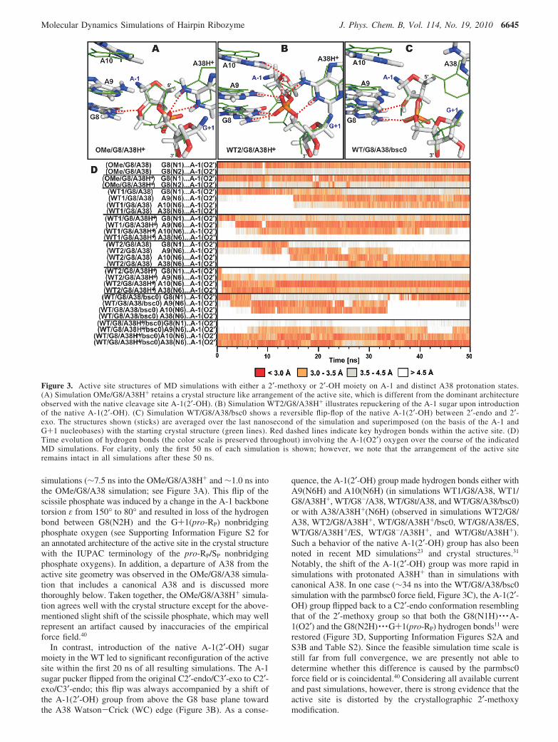
simulations (∼7.5 ns into the OMe/G8/A38H+ and ∼1.0 ns intothe OMe/G8/A38 simulation; see Figure 3A). This flip of thescissile phosphate was induced by a change in the A-1 backbonetorsion ε from 150° to 80° and resulted in loss of the hydrogenbond between G8(N2H) and the G+1(pro-RP) nonbridgingphosphate oxygen (see Supporting Information Figure S2 foran annotated architecture of the active site in the crystal structurewith the IUPAC terminology of the pro-RP/SP nonbridgingphosphate oxygens). In addition, a departure of A38 from theactive site geometry was observed in the OMe/G8/A38 simula-tion that includes a canonical A38 and is discussed morethoroughly below. Taken together, the OMe/G8/A38H+ simula-tion agrees well with the crystal structure except for the above-mentioned slight shift of the scissile phosphate, which may wellrepresent an artifact caused by inaccuracies of the empiricalforce field.40
In contrast, introduction of the native A-1(2′-OH) sugarmoiety in the WT led to significant reconfiguration of the activesite within the first 20 ns of all resulting simulations. The A-1sugar pucker flipped from the original C2′-endo/C3′-exo to C2′-exo/C3′-endo; this flip was always accompanied by a shift ofthe A-1(2′-OH) group from above the G8 base plane towardthe A38 Watson-Crick (WC) edge (Figure 3B). As a conse-
quence, the A-1(2′-OH) group made hydrogen bonds either withA9(N6H) and A10(N6H) (in simulations WT1/G8/A38, WT1/G8/A38H+, WT/G8-/A38, WT/G8t/A38, and WT/G8/A38/bsc0)or with A38/A38H+(N6H) (observed in simulations WT2/G8/A38, WT2/G8/A38H+, WT/G8/A38H+/bsc0, WT/G8/A38/ES,WT/G8/A38H+/ES, WT/G8-/A38H+, and WT/G8t/A38H+).Such a behavior of the native A-1(2′-OH) group has also beennoted in recent MD simulations23 and crystal structures.31
Notably, the shift of the A-1(2′-OH) group was more rapid insimulations with protonated A38H+ than in simulations withcanonical A38. In one case (∼34 ns into the WT/G8/A38/bsc0simulation with the parmbsc0 force field, Figure 3C), the A-1(2′-OH) group flipped back to a C2′-endo conformation resemblingthat of the 2′-methoxy group so that both the G8(N1H) · · ·A-1(O2′) and the G8(N2H) · · ·G+1(pro-RP) hydrogen bonds11 wererestored (Figure 3D, Supporting Information Figures S2A andS3B and Table S2). Since the feasible simulation time scale isstill far from full convergence, we are presently not able todetermine whether this difference is caused by the parmbsc0force field or is coincidental.40 Considering all available currentand past simulations, however, there is strong evidence that theactive site is distorted by the crystallographic 2′-methoxymodification.
Figure 3. Active site structures of MD simulations with either a 2′-methoxy or 2′-OH moiety on A-1 and distinct A38 protonation states.(A) Simulation OMe/G8/A38H+ retains a crystal structure like arrangement of the active site, which is different from the dominant architectureobserved with the native cleavage site A-1(2′-OH). (B) Simulation WT2/G8/A38H+ illustrates repuckering of the A-1 sugar upon introductionof the native A-1(2′-OH). (C) Simulation WT/G8/A38/bsc0 shows a reversible flip-flop of the native A-1(2′-OH) between 2′-endo and 2′-exo. The structures shown (sticks) are averaged over the last nanosecond of the simulation and superimposed (on the basis of the A-1 andG+1 nucleobases) with the starting crystal structure (green lines). Red dashed lines indicate key hydrogen bonds within the active site. (D)Time evolution of hydrogen bonds (the color scale is preserved throughout) involving the A-1(O2′) oxygen over the course of the indicatedMD simulations. For clarity, only the first 50 ns of each simulation is shown; however, we note that the arrangement of the active siteremains intact in all simulations after these 50 ns.
Molecular Dynamics Simulations of Hairpin Ribozyme J. Phys. Chem. B, Vol. 114, No. 19, 2010 6645

Canonical Form of G8 Is Consistent with the CrystalStructure. We carried out a set of simulations to compare thestructural dynamics of three possible protonation states of G8,i.e., the canonical guanine (G8), the N1,O6-enol tautomer (G8t),and the N1-deprotonated form (G8-), in the presence of thenative A-1(2′-OH) group (Table 1; see Supporting InformationFigure S1 for the structures of the G8 protonation forms). It isworth noting that the protonation state of G8 influenced bothA38 and A38H+ simulations similarly. Likewise, the A-1 sugarrepuckering induced by A-1(2′-OH) described in the previousparagraph was observed in all simulations independent of theG8 protonation state.
Both simulations carrying the deprotonated G8- form (i.e.,WT/G8-/A38 and WT/G8-/A38H+) showed expulsion of theG8- from the active site during the initial equilibration. Boththe G8(N2H) · · ·G+1(pro-RP) and the G8(N1) · · ·A-1(2′-OH)hydrogen bonds observed in the crystal structure were disruptedand not reestablished over the entire simulation (Figure 4A,D).A similar expulsion of the G8 nucleobase occurred during theequilibration of both simulations with the G8t tautomer (WT/G8t/A38 and WT/G8t/A38H+); however, G8 returned to itsoriginal position in the active site during the first 10 ns in bothsimulations and reestablished the G8t(N2H) · · ·G+1(pro-RP)hydrogen bond (Figure 4D). This G8t(N2H) · · ·G+1(pro-RP)hydrogen bond was finally disrupted and immediately replacedby a newly formed G8t(O6H) · · ·G+1(pro-RP) hydrogen bond30-35 ns into both simulations, which remained a stable bindingpattern of G8t to the scissile phosphate over the rest of thesimulations (Figure 4B,D). Therefore, while G8t causes a local,minor rearrangement of the active site compared to the crystal
structure, it remained quite compatible with the overall hairpinribozyme structure.
By comparison, G8 stayed in more stable contact with thescissile phosphate in all simulations harboring the canonical G8form (i.e., WT1/G8/A38, WT2/G8/A38, WT1/G8/A38H+, andWT2/G8/A38H+; Figure 4D), making base-phosphate (BPh)41
contacts to the G+1(pro-RP) or (pro-SP) nonbridging oxygens.These BPh interactions between G8 and the scissile phosphatefluctuated among 3BPh, 4BPh, and 5BPh binding patterns(Figure 4C,D).41 The 5BPh contact of G8 to the G+1(pro-RP)nonbridging oxygen was accompanied by a G8(N1H) · · ·A-1(O2′) hydrogen bond in simulation WT/G8/A38/bsc0. Thisdistinct arrangement was caused by the reversion of the C2′-endo-to-C3′-endo repuckering induced by the native A-1(2′-OH) and discussed above (see Figure 3C).
Protonated A38H+, but Not A38, Is Consistent with theCrystal Structure. We observed significant differences inthe behavior of canonical A38 and protonated A38H+ forms. Theprotonated A38H+ remained tightly bound in its crystal-likeposition in the active site, while the canonical A38 form wasexpelled from the active site. The A38H+(N1) · · ·G+1(O5′)hydrogen bond, which has been suggested to play a key role incatalysis,18,19,23,28-30 remained stable in all simulations carryingthe protonated form of A38H+ (Figure 5D). Note that OMe/G8/A38H+ is the only simulation where the A38H+(N1H) · · ·G+1(O5′) contact was temporarily interrupted and departureof A38H+ from the scissile phosphate occurred. However, areturn of A38H+ and subsequent restoration of theA38H+(N1H) · · ·G+1(O5′) hydrogen bond was observed ∼36ns into this simulation (Figure 5D).
Figure 4. Active site structures of MD simulations with distinct G8 (and A38) protonation states. (A) Simulation WT/G8-/A38H+, where G8-
leaves the proximity of the scissile phosphate (black arrow). (B) Simulation WT/G8t/A38H+ documents a contact between G8t and the scissilephosphate with a G8t(O6H) · · ·G+1(pro-RP) hydrogen bond re-formed after an initial expulsion of G8t (black arrow). (C) Simulation WT1/G8/A38H+ shows the typical G8 · · ·G+1 4BPh contact in simulations with a canonical G8. The structures shown (sticks) are averaged over the lastnanosecond of the simulation and superimposed (on the basis of the A-1 and G+1 nucleobases) with the starting crystal structure (green lines). (D)Time evolution of interatomic distances (the color scale is the same as in Figure 3) between G8 and the G+1 pro-RP and pro-SP oxygen atomsduring the first 50 ns of each MD simulation.
6646 J. Phys. Chem. B, Vol. 114, No. 19, 2010 Mlynsky et al.

In sharp contrast to the protonated A38H+, the canonical A38typically shifted away from the scissile phosphate; the distancebetween A38(N1) and G+1(O5′) gradually increased up to 7-8Å, the pairing with A24 was interrupted and, eventually, A38left the active site in simulations WT1/G8/A38, OMe/G8/A38,WT/G8/A38/bsc0, WT/G8t/A38 and WT/G8-/A38 (Figure5A,B,D). Alternatively, in simulations WT2/G8/A38 and WT/G8/A38/ES the A38(N1) · · ·G+1(O5′) distance similarly in-creased to 5 Å, but then A38 established an A38(N6H) · · ·A-1(O2′) hydrogen bond (in WT2/G8/A38 at ∼26 ns and in WT/G8/A38/ES simulation at ∼2 ns, Supporting Information FigureS3A), which was followed by A-1(2′-OH) · · ·A38(N1) hydrogenbond formation (in WT2/G8/A38 at ∼34 ns and in WT/G8/A38/ES simulation at ∼2 ns; Figure 5C,E). The A-1(2′-OH) · · ·A38(N1) hydrogen bond remained stable until the endof both simulations (Supporting Information Figure S3B). Theformation of this A-1(2′-OH) · · ·A38(N1) hydrogen bond cor-responds to the recently described interaction between A-1(2′-OH) and A38(N1), which was suggested to be catalyticallyrelevant (Figure 2C).23 Notably, the formation of the A-1(2′-OH) · · ·A38(N1) hydrogen bond was inevitably accompaniedby a loss of the O2′-P-O5′ in-line attack configurationinvolving A-1(2′-OH) (Supporting Information Figure S4A).
To further validate the observed differences in A38 andA38H+ behavior, we performed two additional, 150-ns-long MD
simulations of the hairpin ribozyme with native A-1(2′-OH) andparmbsc0 force field and two 80-ns-long MD simulations withexcess KCl salt, in both cases one simulation with a canonicalA38, the other with protonated A38H+. Parmbsc0 is the latestvariant of the parm99 force field, which features modified R/γtorsion profiles that suppress γ-trans geometries.35 Parmbsc0leads to a decisive improvement of B-DNA simulations,35,42,43
while both force fields seem to perform equally well for RNAsimulations.35,44-48 In the excess salt simulations we replacedthe Na+ counterions by twice the amount of K+ ions and Cl-
ions for charge neutralization. Net neutralization results in asodium concentration of ∼0.30 M, while the latter simulationscontain ∼0.65 M K+. Such excess KCl salt conditions in MDsimulations were recently shown to cause a modest sequence-dependent compaction of canonical A-RNA double helices.48
Significantly, the distinct behavior of A38 and A38H+ simula-tions was reproduced with both the parm99 and parmbsc0 forcefield as well as in the presence of neutralizing Na+ counterionsand excess KCl salt. We conclude that the compact binding ofthe protonated A38H+ to G+1(O5′), the expulsion of thecanonical A38 from the active site, and the alternative, lesscommon shift of A38 toward the A-1(2′-OH) are robust resultsreflective of the A38 protonation state and independent of otherdetails of the MD simulation.
Figure 5. Canonical A38 either loses pairing with the A24 nucleobase while leaving the active site during the majority of MD simulations or,alternatively, establishes an A-1(2′-OH) · · ·A38(N1) hydrogen bond. (A) Stick representation of the average structure of the last nanosecond ofsimulation WT1/G8/A38 illustrating the A38-A24 base pair disruption. While the crystal geometry is represented by green lines, yellow and orangelines represent snapshots of consecutive A38 positions during its expulsion (black arrow) from the active site. (B) Same as panel A, but from adifferent viewpoint. (C) Average structure of the active site (taken from the last nanosecond of simulation WT2/G8/A38 and overlaid with thecrystal structure in green) showing A38(N6H) · · ·A-1(O2′) and A-1(2′-OH) · · ·A38(N1) hydrogen bond formation. (D) and (E) Time evolution ofthe A38(N1) · · ·G+1(O5′) and A38(N1) · · ·A-1(O2′) distances in all simulations.
Molecular Dynamics Simulations of Hairpin Ribozyme J. Phys. Chem. B, Vol. 114, No. 19, 2010 6647

S-Turn Behavior. The crystal structures suggest that BPhcontacts may contribute to the stability of the S-turn regionlocated between helices H3 and H4 (Figure 1). Accordingly,we observed a strong correlation between S-turn behavior andthe presence of the BPh contact between A38(N1) and G+1(O5′).This interaction, which places the A38(N1) atom within 2.6-2.8Å of G+1(O5′) and is also observed in the oxo-vanadium TS-mimic X-ray structures,19,28 is very unique since it does notcorrespond to any established BPh interaction pattern asclassified by Zirbel et al.41 In fact, there are no more than twocandidates for such an interaction found in the availableribosomal structures and even these cases may be artificial dueto resolution limits.41 Since the relative orientation of A38 withrespect to the G+1 phosphate closely mimics the 4BPhinteraction of guanine-phosphate, where the N1 nitrogen ofguanine is protonated, this unique arrangement of A38 · · ·G+1provides a very strong indication that the adenine A38 isprotonated in the crystal structure, which was grown at pH 6.As discussed above, we observed this unique 4BPh interactionbetween A38H+ and G+1 phosphate to be stable in all MDsimulations carrying the protonated A38H+.
The conformation of S-turns bearing the protonated A38H+
were well preserved in all MD simulations (Figure 6, seeSupporting Information for the behavior of S-turn backbonetorsions). Thus we suggest that a tight and stable A38H+ · · ·G+1BPh contact (Figure 5D) is important for stabilizing (anchoring)the S-turn conformation as well as for proper arrangement ofthe active site. In contrast, S-turns bearing the canonical A38shifted away from the scissile phosphate and underwentdeformations in most MD simulations (Figure 6), due to the
loss of the A38-G+1 4BPh contact (Figures 5B, 5D). It isworth noting that the S-turn region maintained its fold specif-ically in simulations WT2/G8/A38 and WT/G8/A38/ES, whereA38 established instead a hydrogen bond with the A-1(2′-OH)attacking nucleophile, as discussed above. The changes observedin the S-turn region affected also base pairing in helix H4. Inparticular, the G36 nucleobase often lost its base pair with A26and became either stacked between residues A26 and C27 orformed a new cis WC base-pair with C27 (displacing the G35nucleobase) (Supporting Information Figure S5).
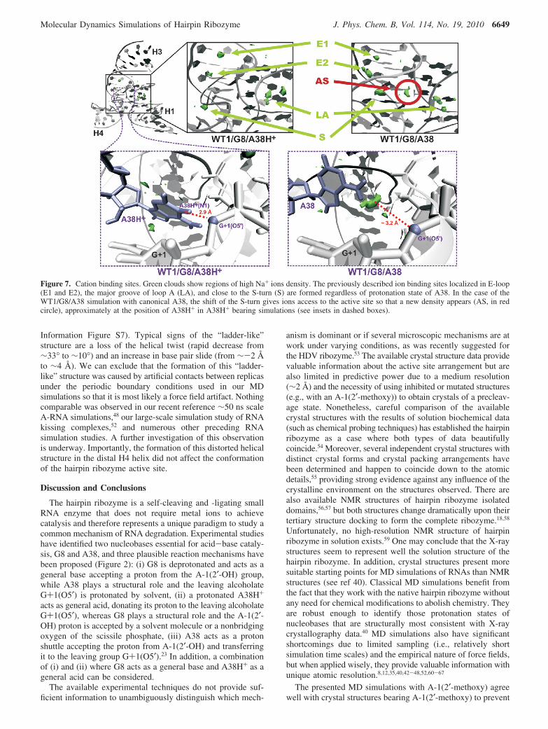
Cation Binding Sites. Monovalent cation binding sitesidentified in the MD simulations presented here in general agreewith those determined in previous MD simulations.23 Ion bindingsites of highest Na+ density include two sites within the E-loop(E1 and E2, Figure 7), a site along the major groove of loop A(LA, Figure 7), and a site near the S-turn region (S, Figure 7).These ion binding sites were observed in all simulationsregardless of the protonation state of A38. Still, we identifiedsome differences between structures containing either canonicalA38 or protonated A38H+ adenine. In particular, expulsion ofthe canonical A38 from the active site results in opening of theS-turn. Consequently, in simulations with a canonical A38 anadditional Na+ ion density appears inside the S-turn, close tothe scissile phosphate of the active site (AS spot on Figure 7)in the pocket between the U-2/A-1 sugar-phosphate backboneand the A38 nucleotide. This additional Na+ ion density wasdetected in the position occupied in the X-ray structures insteadby the WC edge of A38. This active site cation density wasonly observed when the catalytic core was disrupted and openedup toward solvent and therefore did not occur in the twosimulations with canonical A38 (WT2/G8/A38, WT/G8/A38/ES) where A38 formed interactions with A-1(2′-OH). Whenthe core remained closed as in the crystal structures, the activesite cavity remained inaccessible to cations, as describedpreviously.8
Transition of A-RNA Stem to a “Ladder-Like” Structure.It is well established that, while MD simulations of nucleic acidsare very insightful, their accuracy is limited by force fieldapproximations, especially on longer simulation time scales.35,40,49-51
The present simulations reveal one such possible artifact, which,however, does not affect our main conclusions. The A-type helixH4 occasionally formed a distorted structure, named here the“ladder-like” conformation (Figure 8 and Supporting Informa-tion Figure S6). Transition of a double helix to the “ladder-like” structure was observed for both force fields (parm99 andparmbsc0) and with different protonation states of A38 and G8,in altogether 4 out of 14 simulations with Na+ counterions (WT/G8t/A38H+, OMe/G8/A38, OMe/G8/A38H+, and WT/G8/A38/bsc0). The “ladder-like” structures were not observed in the two80 ns excess KCl salt simulations; however, we cannot ruleout that such simulations would also provide this artifact. Thetransition of helix H4 to its “ladder-like” conformation wasirreversible at the present time scale (tens to hundreds ofnanoseconds). In individual simulations the “laddering” of helixH4 was initiated within the first 30 ns (see SupportingInformation Table S4). The “ladder-like” structure was char-acterized by a shift of the glycosidic � angle from -160° to∼-90°, a small decrease in � (from ∼-65° to ∼-85°), and anincrease of the δ (from ∼80° to ∼110°) and ε (from ∼-160°to ∼-150°) torsions. Sugar puckers of nucleotides in the“ladder-like” structure changed from the initial C3′-endo (A-RNA form) to C2′-exo. The transition was also accompaniedby a slight shift of the first peak in the P-P radial distributionfunction by 0.2 Å toward higher values (see Supporting
Figure 6. Ribbon diagrams showing the average structures from thelast nanosecond (orange ribbon) superimposed over the crystal structure(green) of the minimal junction-less hairpin ribozyme with helixesH1-H4 indicated. (A) Simulations with canonical A38 (here OMe/G8/A38) show S-turn degradation (black box). (B) Simulations withprotonated A38H+ (here OMe/G8/A38H+) preserve the crystallographicS-turn conformation.
6648 J. Phys. Chem. B, Vol. 114, No. 19, 2010 Mlynsky et al.
Information Figure S7). Typical signs of the “ladder-like”structure are a loss of the helical twist (rapid decrease from∼33° to ∼10°) and an increase in base pair slide (from ∼-2 Åto ∼4 Å). We can exclude that the formation of this “ladder-like” structure was caused by artificial contacts between replicasunder the periodic boundary conditions used in our MDsimulations so that it is most likely a force field artifact. Nothingcomparable was observed in our recent reference ∼50 ns scaleA-RNA simulations,48 our large-scale simulation study of RNAkissing complexes,52 and numerous other preceding RNAsimulation studies. A further investigation of this observationis underway. Importantly, the formation of this distorted helicalstructure in the distal H4 helix did not affect the conformationof the hairpin ribozyme active site.
Discussion and Conclusions
The hairpin ribozyme is a self-cleaving and -ligating smallRNA enzyme that does not require metal ions to achievecatalysis and therefore represents a unique paradigm to study acommon mechanism of RNA degradation. Experimental studieshave identified two nucleobases essential for acid-base cataly-sis, G8 and A38, and three plausible reaction mechanisms havebeen proposed (Figure 2): (i) G8 is deprotonated and acts as ageneral base accepting a proton from the A-1(2′-OH) group,while A38 plays a structural role and the leaving alcoholateG+1(O5′) is protonated by solvent, (ii) a protonated A38H+
acts as general acid, donating its proton to the leaving alcoholateG+1(O5′), whereas G8 plays a structural role and the A-1(2′-OH) proton is accepted by a solvent molecule or a nonbridgingoxygen of the scissile phosphate, (iii) A38 acts as a protonshuttle accepting the proton from A-1(2′-OH) and transferringit to the leaving group G+1(O5′).23 In addition, a combinationof (i) and (ii) where G8 acts as a general base and A38H+ as ageneral acid can be considered.
The available experimental techniques do not provide suf-ficient information to unambiguously distinguish which mech-
anism is dominant or if several microscopic mechanisms are atwork under varying conditions, as was recently suggested forthe HDV ribozyme.53 The available crystal structure data providevaluable information about the active site arrangement but arealso limited in predictive power due to a medium resolution(∼2 Å) and the necessity of using inhibited or mutated structures(e.g., with an A-1(2′-methoxy)) to obtain crystals of a precleav-age state. Nonetheless, careful comparison of the availablecrystal structures with the results of solution biochemical data(such as chemical probing techniques) has established the hairpinribozyme as a case where both types of data beautifullycoincide.54 Moreover, several independent crystal structures withdistinct crystal forms and crystal packing arrangements havebeen determined and happen to coincide down to the atomicdetails,55 providing strong evidence against any influence of thecrystalline environment on the structures observed. There arealso available NMR structures of hairpin ribozyme isolateddomains,56,57 but both structures change dramatically upon theirtertiary structure docking to form the complete ribozyme.18,58
Unfortunately, no high-resolution NMR structure of hairpinribozyme in solution exists.59 One may conclude that the X-raystructures seem to represent well the solution structure of thehairpin ribozyme. In addition, crystal structures present moresuitable starting points for MD simulations of RNAs than NMRstructures (see ref 40). Classical MD simulations benefit fromthe fact that they work with the native hairpin ribozyme withoutany need for chemical modifications to abolish chemistry. Theyare robust enough to identify those protonation states ofnucleobases that are structurally most consistent with X-raycrystallography data.40 MD simulations also have significantshortcomings due to limited sampling (i.e., relatively shortsimulation time scales) and the empirical nature of force fields,but when applied wisely, they provide valuable information withunique atomic resolution.8,12,35,40,42-48,52,60-67
The presented MD simulations with A-1(2′-methoxy) agreewell with crystal structures bearing A-1(2′-methoxy) to prevent
Figure 7. Cation binding sites. Green clouds show regions of high Na+ ions density. The previously described ion binding sites localized in E-loop(E1 and E2), the major groove of loop A (LA), and close to the S-turn (S) are formed regardless of protonation state of A38. In the case of theWT1/G8/A38 simulation with canonical A38, the shift of the S-turn gives ions access to the active site so that a new density appears (AS, in redcircle), approximately at the position of A38H+ in A38H+ bearing simulations (see insets in dashed boxes).
Molecular Dynamics Simulations of Hairpin Ribozyme J. Phys. Chem. B, Vol. 114, No. 19, 2010 6649

self-cleavage. In contrast, MD simulations with the nativeA-1(2′-OH) show rapid changes in A-1(2′-OH) group positionand A-1 sugar pucker more consistent with the active sitearchitecture observed in crystals bearing a 2′,5′-phosphodiesterat the cleavage site.28 The differences between MD simulationsof the hairpin ribozyme with and without a 2′-methoxy groupat A-1 explicitly show that the methoxy group distorts the activesite. This observation agrees with previous MD simulations,where changes in A-1 sugar pucker and repositioning of its 2′-OH were observed.8,23 The same changes were also describedin a recent crystal structure of a hairpin ribozyme mutant wherethe native A-1(2′-OH) group was present.31 This finding clearlyimplies that mechanistic interpretations based on 2′-methoxymodified RNA structures are not straightforward.
We further observed that the canonical G8 form is structurallyconsistent with crystallographic data, while the deprotonatedG8- form causes large structural distortions of the active site.The deprotonated G8- base quickly leaves the active site, likelydue to electrostatic repulsion with the scissile phosphate. Thisobservation is not consistent with a catalytic role of G8 as thegeneral base. Similarly, a recent experimental study estimatedthe pKa of G8 to 9.5,20 implying that G8 is largely in thecanonical (protonated) form under physiological conditions. Inour MD simulations, the G8 enol tautomer remains in contactwith the active site and might be considered for a potentialstructural role in catalysis. However, the ∼19 kcal/mol higher∆G‡ expected for the self-cleavage in the presence of the G8tautomeric form (relative to a canonical G8) calls a role for theG8 enol tautomer during catalysis into question.25 Thus, withinthe limits of classical MD simulations we suggest that only thecanonical G8 is structurally and energetically feasible for themechanism of self-cleavage. We observed that the G8(N1) iminogroup of the canonical G8 forms stable hydrogen bonds with
the G+1(pro-Sp) or G+1(pro-Rp) nonbridging oxygens and/ora hydrogen bond with A-1(O2′). This finding together with priorcomputational data suggests that G8 likely helps to arrangeparticipating functional groups for catalysis and electrostaticallystabilizes the transition states.23-25
In contrast to G8, our simulations highlight that the protonatedrather than the canonical form of A38 is most consistent withthe available crystal structures. Three types of behavior wereobserved for the unprotonated canonical A38: (i) A38 departsfrom the scissile phosphate, which leads to large structuralchanges in the S-turn bearing the A38 base (simulations WT1/G8/A38 and OMe/G8/A38). (ii) A38 retracts from the scissilephosphate but remains at an ∼7 Å distance after losing its basepairing with A24 (simulations WT/G8/A38/bsc0, WT/G8-/A38,and WT/G8t/A38). (iii) A38 establishes a hydrogen bondingcontact with A-1(2′-OH) and remains close to the scissilephosphate (simulations WT2/G8/A38 and WT/G8/A38/ES).Once established, the contact between the A-1(2′-OH) nucleo-phile and the A38 base remains stable until the end of the MDsimulation. This contact might be catalytically relevant becauseA38 was suggested as a potential shuttle capable of acceptinga proton from the nucleophile and transferring it to theG+1(O5′) oxygen of the leaving alcoholate.23 Notably, forma-tion of the A-1(2′-OH) · · ·A38(N1) hydrogen bond is in conflictwith the favorable in-line attack angle expected during cleavage,as previously observed,23 suggesting that proton exchange wouldhave to be followed by a rapid transition of the deprotonated2′-oxyanion to a position more favorable to in-line attack.
Notably, all simulations with the protonated A38H+ generallyagree well with the crystal structure data. Recent Ramanspectroscopy analysis yielded direct evidence for an elevatedpKa of A38, suggesting that it can become protonated underphysiological conditions.32 Analysis of the A38 interaction withthe scissile phosphate in the crystal structures shows that thecorresponding BPh (base-phosphate) interaction does not fallinto the classified range of adenine-specific BPh contacts41 andrather mimics a 4BPh interaction, which is typical only for aguanine nucleobase bearing a protonated N1 nitrogen atom. Thismay implicate a noncanonical protonation state of A38 to enablethis new and rare 4BPh contact. All of these findings suggestthat A38 is protonated in the functional hairpin ribozyme. Thatis, a protonated A38H+ is structurally important to stabilize thereactive fold of the hairpin ribozyme; the S-turn conformationis stabilized by a strong ionic interaction between the scissilephosphate group and the WC edge of the A38H+ nucleobase,which can be classified as a new and rare 4BPh interaction ofadenine specific for the N1-protonated adenine.
The protonation of A38 can be also rationalized on the basisof a pocket of very deep negative electrostatic potential (ESP)inside the active site of the hairpin ribozyme,8,68 which isstructurally isolated from solvent cations so that the active sitenucleobases are directly exposed to an unsaturated ESP poten-tial.8 This ESP becomes (partially) saturated by the positivecharge of a protonated A38H+. We did not observe penetrationof Na+ ions into the closed active site cavity, in agreement withpreviously published MD simulations.8,23 Na+ ions enter theactive site only in case of a canonical A38, which reconfiguresthe S-turn and opens the active site to solvent (Figure 7). Theentering Na+ ions then take approximately the position of theA38 WC edge to saturate the negative ESP of the cavity. Thisfinding attests to a tendency of the active site to neutralize thedeep negative ESP and suggests another role for A38H+, i.e.,neutralization of the active site ESP. Conversely, the largenegative ESP inside the occluded active site of the hairpin
Figure 8. Distortion of the A-type helix H4. (A) A snapshot from theend of the OMe/G8/A38H+ simulation shows a “ladder-like” conforma-tion of helix H4 (red), with a more detailed view in panel (B), whichwe consider a force field artifact. (C) Time evolution of the glycosidicdihedral � profile of nucleotide G33 inside the H4 RNA stem duringsimulation OMe/G8/A38H+. Formation of a “ladder-like” stem structureis accompanied by a shift of � above -90°.
6650 J. Phys. Chem. B, Vol. 114, No. 19, 2010 Mlynsky et al.

ribozyme perturbs the pKa of A38 toward neutrality, as directlyobserved69,70 and suggested on the basis of structural data.31 Thisrepresents one of the major differences in utilization of anegative ESP pocket and cations binding between the hairpinand HDV ribozymes.9,44
We here also have identified a potential artifact of theAMBER parm99 and parmbsc0 force fields in the formation ofa “ladder-like”, underwound RNA duplex structure instead ofthe canonical A-type RNA helix (Figure 8). This “ladder-like”structure appears on a tens-of-nanoseconds time scale and seemsto be irreversible on the accessible 50-150 ns time scale. Theartifact occurred only in a minority of simulations, affected onlythe peripheral H4 helix, and did not propagate into thecatalytically relevant parts of the simulated structures. However,it clearly appears to have the potential to accumulate in longerMD simulations. Excess KCl salt may slow down or preventformation of this distorted structure in an A-RNA stem, althoughmore simulations would be needed for validation. A detailedanalysis of this behavior is ongoing. We did not notice such abehavior in any of our published RNA studies but detectedfurther cases in unpublished simulations mainly on small RNAmodel systems. The “ladder-like” helix distortion significantlyaffects glycosidic � torsion angles, which leads us to hypothesizethat the � torsion parameters are imbalanced. However, pre-liminary observations imply that the recently suggested forcefield with modified � torsion parameters71 does not preventformation of the “ladder-like” structural artifact but may ratherspeed it up.
Acknowledgment. This study was supported by grantsLC512 (to M.O.), LC06030 (to J.S.), and MSM6198959216 (toM.O.) from the Ministry of Education of the Czech Republic,grants 203/09/1476 (to J.S.) and 203/09/H046 (to M.O. and J.S.)from the Grant Agency of the Czech Republic, grantsIAA400040802 (to M.O. and J.S.), KJB400040901 (to J.S.),and 1QS500040581 (to J.S.) from the Grant Agency of theAcademy of Sciences of the Czech Republic, grants AV0Z50040507(to J.S.) and AV0Z50040702 (to J.S.) from the Academy ofSciences of the Czech Republic, and NIH grant GM62357 (toN.G.W.).
Supporting Information Available: Parameters of non-standard residues for MD simulations, torsion angles of theS-turn region, evolution of in-line attack angle, and supportingfigures. This material is available free of charge via the Internetat http://pubs.acs.org.
References and Notes
(1) Buzayan, J. M.; Hampel, A.; Bruening, G. Nucleic Acids Res. 1986,14, 9729.
(2) Buzayan, J. M.; Feldstein, P. A.; Segrelles, C.; Bruening, G. NucleicAcids Res. 1988, 16, 4009.
(3) van Tol, H.; Buzayan, J. M.; Feldstein, P. A.; Eckstein, F.; Bruening,G. Nucleic Acids Res. 1990, 18, 1971.
(4) Walter, N. G.; Burke, J. M. Curr. Opin. Chem. Biol. 1998, 2, 24.(5) Fedor, M. J. J. Mol. Biol. 2000, 297, 269.(6) Fedor, M. J. Annu. ReV. Biophys. 2009, 38, 271.(7) Murray, J. B.; Seyhan, A. A.; Walter, N. G.; Burke, J. M.; Scott,
W. G. Chem. Biol. 1998, 5, 587.(8) Rhodes, M. M.; Reblova, K.; Sponer, J.; Walter, N. G. Proc. Natl.
Acad. Sci. U.S.A. 2006, 103, 13380.(9) Krasovska, M. V.; Sefcikova, J.; Reblova, K.; Schneider, B.; Walter,
N. G.; Sponer, J. Biophys. J. 2006, 91, 626.(10) Krasovska, M. V.; Sefcikova, J.; Spackova, N.; Sponer, J.; Walter,
N. G. J. Mol. Biol. 2005, 351, 731.(11) Salter, J.; Krucinska, J.; Alam, S.; Grum-Tokars, V.; Wedekind,
J. E. Biochemistry 2006, 45, 686.(12) Park, H.; Lee, S. J. Chem. Theory Comput. 2006, 2, 858.(13) Walter, N. G. Mol. Cell 2007, 28, 923.
(14) Cochrane, J. C.; Strobel, S. A. Acc. Chem. Res. 2008, 41, 1027.(15) Pinard, R.; Hampel, K. J.; Heckman, J. E.; Lambert, D.; Chan, P. A.;
Major, F.; Burke, J. M. EMBO J. 2001, 20, 6434.(16) Kuzmin, Y. I.; Da Costa, C. P.; Fedor, M. J. J. Mol. Biol. 2004,
340, 233.(17) Ditzler, M. A.; Rueda, D.; Mo, J. J.; Hakansson, K.; Walter, N. G.
Nucleic Acids Res. 2008, 36, 7088.(18) Rupert, P. B.; Ferre-D’Amare, A. R. Nature 2001, 410, 780.(19) Rupert, P. B.; Massey, A. P.; Sigurdsson, S. T.; Ferre-D’Amare,
A. R. Science 2002, 298, 1421.(20) Liu, L.; Cottrell, J. W.; Scott, L. G.; Fedor, M. J. Nat. Chem. Biol.
2009, 5, 351.(21) Bevilacqua, P. C.; Brown, T. S.; Nakano, S.; Yajima, R. Biopoly-
mers 2004, 73, 90.(22) Lebruska, L. L.; Kuzmine, I. I.; Fedor, M. J. Chem. Biol. 2002, 9,
465.(23) Ditzler, M. A.; Sponer, J.; Walter, N. G. RNA 2009, 15, 560.(24) Nam, K.; Gao, J. L.; York, D. M. RNA 2008, 14, 1501.(25) Nam, K. H.; Gao, J. L.; York, D. M. J. Am. Chem. Soc. 2008, 130,
4680.(26) Kuzmin, Y. I.; Da Costa, C. P.; Cottrell, J. W.; Fedor, M. J. J.
Mol. Biol. 2005, 349, 989.(27) Cottrell, J. W.; Kuzmin, Y. I.; Fedor, M. J. J. Biol. Chem. 2007,
282, 13498.(28) Torelli, A. T.; Krucinska, J.; Wedekind, J. E. RNA 2007, 13, 1052.(29) Macelrevey, C.; Salter, J. D.; Krucinska, J.; Wedekind, J. E. RNA
2008, 14, 1600.(30) Torelli, A. T.; Spitale, R. C.; Krucinska, J.; Wedekind, J. E.
Biochem. Biophys. Res. Commun. 2008, 371, 154.(31) Spitale, R. C.; Volpini, R.; Heller, M. G.; Krucinska, J.; Cristalli,
G.; Wedekind, J. E. J. Am. Chem. Soc. 2009, 131, 6093.(32) Guo, M.; Spitale, R. C.; Volpini, R.; Krucinska, J.; Cristalli, G.;
Carey, P. R.; Wedekind, J. E. J. Am. Chem. Soc. 2009, 131, 12908.(33) Case, D. A.; Darden, T. A.; Cheatham, T. E.; Simmerling, C. L.;
Wang, J.; Duke, R. E.; Luo, R.; Merz, K. M.; Pearlman, D. A.; Crowley,M.; Walker, R. C.; Zhang, W.; Wang, B.; Hayik, S.; Roitberg, A.; Seabra,G.; Wong, K. F.; Paesani, F.; Wu, X.; Brozell, S.; Tsui, V.; Gohlke, H.;Yang, L.; Tan, C.; Mongan, J.; Hornak, V.; Cui, G.; Beroza, P.; Mathews,D. H.; Schafmeister, C.; RossW. S., Kollman, P. A. University of California:San Francisco, 2006.
(34) Wang, J. M.; Cieplak, P.; Kollman, P. A. J. Comput. Chem. 2000,21, 1049.
(35) Perez, A.; Marchan, I.; Svozil, D.; Sponer, J.; Cheatham, T. E.;Laughton, C. A.; Orozco, M. Biophys. J. 2007, 92, 3817.
(36) Joung, I. S.; Cheatham, T. E. J. Phys. Chem. B 2008, 112, 9020.(37) Cornell, W. D.; Cieplak, P.; Bayly, C. I.; Gould, I. R.; Merz, K. M.;
Ferguson, D. M.; Spellmeyer, D. C.; Fox, T.; Caldwell, J. W.; Kollman,P. A. J. Am. Chem. Soc. 1995, 117, 5179.
(38) Cornell, W. D.; Cieplak, P.; Bayly, C. I.; Kollman, P. A. J. Am.Chem. Soc. 1993, 115, 9620.
(39) Frisch, M. J., Trucks, G. W.; Schlegel, H. B.; Scuseria, G. E.; Robb,M. A.; Cheeseman, J. R.; Montgomery, J. A., Jr.; Vreven, T.; Kudin, K. N.;Burant, J. C.; Millam, J. M.; Iyengar, S. S.; Tomasi, J.; Barone, V.;Mennucci, B.; Cossi, M.; Scalmani, G.; Rega, N.; Petersson, G. A.;Nakatsuji, H.; Hada, M.; Ehara, M.; Toyota, K.; Fukuda, R.; Hasegawa, J.;Ishida, M.; Nakajima, T.; Honda, Y.; Kitao, O.; Nakai, H.; Klene, M.; Li,X.; Knox, J. E.; Hratchian, H. P.; Cross, J. B.; Bakken, V.; Adamo, C.;Jaramillo, J.; Gomperts, R.; Stratmann, R. E.; Yazyev, O.; Austin, A. J.;Cammi, R.; Pomelli, C.; Ochterski, J. W.; Ayala, P. Y.; Morokuma, K.;Voth, G. A.; Salvador, P.; Dannenberg, J. J.; Zakrzewski, V. G.; Dapprich,S.; Daniels, A. D.; Strain, M. C.; Farkas, O.; Malick, D. K.; Rabuck, A. D.;Raghavachari, K.; Foresman, J. B.; Ortiz, J. V.; Cui, Q.; Baboul, A. G.;Clifford, S.; Cioslowski, J.; Stefanov, B. B.; Liu, G.; Liashenko, A.; Piskorz,P.; Komaromi, I.; Martin, R. L.; Fox, D. J.; Keith, T.; Al-Laham, M. A.;Peng, C. Y.; Nanayakkara, A.; Challacombe, M.; Gill, P. M. W.; Johnson,B.; Chen, W.; Wong, M. W.; Gonzalez, C.; Pople, J. A. Gaussian 03;Gaussian, Inc.: Pittsburgh, 2003.
(40) Ditzler, M. A.; Otyepka, M.; Sponer, J.; Walter, N. G. Acc. Chem.Res. 2010, 43, 40.
(41) Zirbel, C. L.; Sponer, J. E.; Sponer, J.; Stombaugh, J.; Leontis,N. B. Nucleic Acids Res. 2009, 37, 4898.
(42) Perez, A.; Lankas, F.; Luque, F. J.; Orozco, M. Nucleic Acids Res.2008, 36, 2379.
(43) Perez, A.; Luque, F. J.; Orozco, M. J. Am. Chem. Soc. 2007, 129,14739.
(44) McDowell, S. E.; Spackova, N.; Sponer, J.; Walter, N. G.Biopolymers 2007, 85, 169.
(45) Sponer, J.; Spackova, N. Methods 2007, 43, 278.(46) Spackova, N.; Sponer, J. Nucleic Acids Res. 2006, 34, 697.(47) Reblova, K.; Lankas, F.; Razga, F.; Krasovska, M. V.; Koca, J.;
Sponer, J. Biopolymers 2006, 82, 504.(48) Besseova, I.; Otyepka, M.; Reblova, K.; Sponer, J. Phys. Chem.
Chem. Phys. 2009, 11, 10701.
Molecular Dynamics Simulations of Hairpin Ribozyme J. Phys. Chem. B, Vol. 114, No. 19, 2010 6651

(49) Fadrna, E.; Spackova, N.; Stefl, R.; Koca, J.; Cheatham, T. E.;Sponer, J. Biophys. J. 2004, 87, 227.
(50) Banas, P.; Jurecka, P.; Walter, N. G.; Sponer, J.; Otyepka, M.Methods 2009, 49, 202.
(51) Fadrna, E.; Spackova, N.; Sarzynska, J.; Koca, J.; Orozco, M.;Cheatham, T. E.; Kulinski, T.; Sponer, J. J. Chem. Theory Comput. 2009,5, 2514.
(52) Reblova, K.; Fadrna, E.; Sarzynska, J.; Kulinski, T.; Kulhanek, P.;Ennifar, E.; Koca, J.; Sponer, J. Biophys. J. 2007, 93, 3932.
(53) Banas, P.; Rulisek, L.; Hanosova, V.; Svozil, D.; Walter, N. G.;Sponer, J.; Otyepka, M. J. Phys. Chem. B 2008, 112, 11177.
(54) Ryder, S. P.; Strobel, S. A. Nucleic Acids Res. 2002, 30, 1287.(55) Alam, S.; Grum-Tokars, V.; Krucinska, J.; Kundracik, M. L.;
Wedekind, J. E. Biochemistry 2005, 44, 14396.(56) Cai, Z. P.; Tinoco, I. Biochemistry 1996, 35, 6026.(57) Butcher, S. E.; Allain, F. H. T.; Feigon, J. Nat. Struct. Biol. 1999,
6, 212.(58) Hampel, K. J.; Burke, J. M. Biochemistry 2001, 40, 3723.(59) Buck, J.; Li, Y. L.; Richter, C.; Vergne, J.; Maurel, M. C.; Schwalbe,
H. ChemBioChem 2009, 10, 2100.(60) Auffinger, P.; Hashem, Y. Curr. Opin. Struct. Biol. 2007, 17, 325.
(61) Orozco, M.; Noy, A.; Perez, A. Curr. Opin. Struct. Biol. 2008, 18,185.
(62) Cheatham, T. E. Curr. Opin. Struct. Biol. 2004, 14, 360.(63) Mackerell, A. D. J. Comput. Chem. 2004, 25, 1584.(64) Hall, K. B. Curr. Opin. Chem. Biol. 2008, 12, 612.(65) Chen, A. A.; Draper, D. E.; Pappu, R. V. J. Mol. Biol. 2009, 390,
805.(66) Huang, W.; Kim, J.; Jha, S.; Aboul-Ela, F. Nucleic Acids Res. 2009,
37, 6528.(67) Romanowska, J.; Setny, P.; Trylska, J. J. Phys. Chem. B 2008,
112, 15227.(68) Chin, K.; Sharp, K. A.; Honig, B.; Pyle, A. M. Nat. Struct. Biol.
1999, 6, 1055.(69) Tang, C. L.; Alexov, E.; Pyle, A. M.; Honig, B. Biophys. J. 2002,
82, 131a.(70) Tang, C. L.; Alexov, E.; Pyle, A. M.; Honig, B. J. Mol. Biol. 2007,
366, 1475.(71) Ode, H.; Matsuo, Y.; Neya, S.; Hoshino, T. J. Comput. Chem. 2008,
29, 2531.(72) Leontis, N. B.; Westhof, E. RNA 2001, 7, 499.
JP1001258
6652 J. Phys. Chem. B, Vol. 114, No. 19, 2010 Mlynsky et al.