facial nerve schwannoma: the rare/great mimicker of
TRANSCRIPT

Facial Nerve Schwannoma Pathapati et al.THIEME
510
Facial Nerve Schwannoma: The Rare/Great Mimicker of Vestibular Schwannoma/NeuromaDeepthi Pathapati1 Kiran Barla1 Monal Dayal2 Rajitha Gati1 Praveen Kumar Lakota1
1Department of Radiodiagnosis, Omega Hospitals, Hyderabad, Telangana, India
2Department of Pathology, Omega Hospitals, Hyderabad, Telangana, India
published onlineJuly 27, 2021
Address for correspondence Deepthi Pathapati, Omega Hospitals, Banjara Hills, Hyderabad 500034, Telangana, India (e-mail: [email protected]).
Schwannomas are benign tumors arising from Schwann cells which are a protective casing of nerves, composing myelin sheath and can develop in any nerve where Schwann cells are present. Most common are vestibulocochlear nerve schwan-nomas. Facial nerve schwannomas (FNSs) are uncommon tumors involving seventh nerve of which geniculate ganglion involvement is most common. Clinical presenta-tions and the imaging appearances of FNSs are influenced by the topographical anat-omy of the facial nerve and vary according to the segments involved. We report a case of 73-year-old man presenting with right side facial weakness of lower motor neuron type involvement. Computed tomography and magnetic resonance imaging are clinching the diagnosis. An early diagnosis is important in containing the disease facilitating early surgical intervention.
Abstract
DOI https://doi.org/ 10.1055/s-0041-1734356 ISSN 0971-3026
© 2021. Indian Radiological Association.This is an open access article published by Thieme under the terms of the Creative Commons Attribution-NonDerivative-NonCommercial-License, permitting copying and reproduction so long as the original work is given appropriate credit. Contents may not be used for commercial purposes, or adapted, remixed, transformed or built upon. (https://creativecommons.org/licenses/by-nc-nd/4.0/).Thieme Medical and Scientific Publishers Private Ltd. A-12, Second Floor, Sector -2, NOIDA -201301, India
Introduction
Facial nerve schwannomas (FNSs) are rare slow-growing tumors, accounting for less than 1% of all tempo-ral bone tumors. They are typically solitary, unilateral, and sporadic in nature. FNSs may be bilateral as part of neurofibromatosis-2 spectrum. Rarely, multiple schwan-nomas may involve peripheral branches of the facial nerve. Age of presentation varies from 5 to 84 years. No gender or side predilection is observed.1 They are rare benign tumors, which originate along the facial nerve. On imaging, they pres-ent as an enhancing cerebellopontine (CP) angle mass which is difficult to distinguish from vestibular schwannoma (also termed acoustic neuroma) and meningiomas. Facial nerve arises in the pons travel through the internal acoustic meatus and enters the facial nerve canal (or fallopian canal) which is Z-shaped structure. The bony canal has three segments: labyrinthine, tympanic, and mastoid (►Figs. 1 and 2).
The labyrinthine segment runs from the fundus of the internal auditory canal (IAC) to the geniculate ganglion. It is both the narrowest (<0.7 mm diameter) and the shortest (3–5 mm length) segment of the facial nerve. The labyrinthine segment travels anterolaterally from the IAC and superior to the cochlea until it reaches the geniculate ganglion. The first branch of the facial nerve, the greater superficial petrosal nerve (GSPN), exits anteriorly from the geniculate ganglion and runs along the anterior surface of the temporal bone into the pterygoid canal (vidian canal).2 Geniculate ganglion contains cell bodies associated with facial nerve-specialized taste and general somatic sensory fibers. Unless pathologi-cal, geniculate ganglion is not observed even after contrast administration.3 At the geniculate ganglion, the facial nerve makes a 75-degree turn posteriorly to become the tympanic segment (the first or anterior genu).
The tympanic segment runs from the geniculate ganglion to the second (or posterior) genu. Within the tympanic cavity,
Indian J Radiol Imaging 2021;31:510–513.
Keywords ► CP angle ► CT ► contrast- enhanced MRI ► facial nerve ► geniculate ganglion ► schwannoma
Case Report
Published online: 2021-07-27

511Facial Nerve Schwannoma Pathapati et al.
Indian Journal of Radiology and Imaging Vol. 31 No. 2/2021 © 2021. Indian Radiological Association.
the facial nerve passes medial to the incus. It runs posterosu-perior to the cochleariform process, superior and lateral to the oval window, and then inferior to the lateral semicircular canal. At the pyramidal process, the tympanic segment turns interiorly at a 95- to 125-degree angle (at the second genu) to become the mastoid or vertical segment. The mastoid segment of the facial nerve runs posteromedially along the external auditory canal to its exit from the temporal bone at the stylomastoid foramen (13 mm in adults). The nerve exits the temporal bone at the stylomastoid foramen, entering the substance of the parotid gland.2
FNSs commonly present with peripheral facial neurop-athy and/or various otologic symptoms including sensori-neural and conducting hearing loss. Facial paralysis is often seen at a later stage or may be totally absent. The reason for this is attributed to the neuronal tolerance induced by the extremely slow growth of the tumor, abundant tumor vascu-larity, and commonly associated dehiscence of adjacent bone. Occasionally, FNSs may present as an intraparotid mass or as an intracranial lesion.1
Case ReportA 73-year-old man presented with sudden onset of right-sided facial weakness of 1-month duration. He also had a sudden onset of right-sided tinnitus without notice-able hearing impairment or dizziness. Computed tomogra-phy (CT) revealed an isodense to hyperdense lesion in the right CP angle with erosive extension into the petrous part of temporal bone (►Fig. 3). Magnetic resonance imaging (MRI) revealed an extra-axial altered signal intensity lesion which is Tl hypointense and T2 heterogeneously hyperintense lesion in the right CP angle with lateral extension into IAC causing its expansion (►Fig. 4) and another similar signal intensity lesion in the right temporal region near the petrous part of the temporal bone (►Fig. 5). Semicircular canals involvement was absent; however, there was a slight abutment of cochlea on the right side (►Fig. 6A). Heterogeneous enhancement of both the lesions was noted on the contrast study (►Fig. 6B).
Based on imaging findings and clinical history, diagnosis of FNS was thus conceivable. Other differential considered
Fig. 1 Diagram of the course of the infratemporal facial nerve from the fundus of the IAC to the stylomastoid foramen. IAC, internal audi-tory canal.
Fig. 2 (A–C) Axial temporal bone CT images demonstrate the intracanalicular (solid arrow in [A]), labyrinthine (double-lined arrow in [A]), geniculate ganglion (double-lined arrow in [B]), tympanic (solid arrow in [B]), and mastoid (arrow in [C]) segments of the facial nerve. CT, computed tomography.
Fig. 3 (A, B) Axial and coronal reformatted images of CT in soft tissue window showing hyperdense lesion in the temporal region. (C, and D) Axial and sagittal reformatted images of CT in bone win-dow showing widening of IAC and erosion of petrous part of the tem-poral bone. CT, computed tomography; IAC, internal auditory canal.
Fig. 4 (A–C) Axial T1, T2, and FLAIR images of MRI of the brain showing a well-defined extra-axial altered signal intensity lesion in the right CP angle extending into IAC and the higher sections show-ing similar signal intensity lesion in the temporal region with CSF cleft sign. CP, cerebellopontine; CSF, cerebrospinal fluid; FLAIR, fluid-attenuated inversion recovery; IAC, internal auditory canal; MRI, magnetic resonance imaging.
512
Indian Journal of Radiology and Imaging Vol. 31 No. 2/2021 © 2021. Indian Radiological Association.
Facial Nerve Schwannoma Pathapati et al.
based on imaging was hemangioma. Cranial nerves (CNs) VII and VIII schwannomas may have identical imaging appear-ances if they are confined to the CP angle–IAC (CPA-IAC) complex. Hence, it is important to evaluate the findings for extended enhancement or expansion of the labyrinthine seg-ment and geniculate ganglion of the facial nerve, as this is a subtle clue to a VII cranial than VIII nerve origin. Facial nerve decompression was done and excised tumor histopathology was reported as schwannoma (►Fig. 7).
DiscussionSchwannoma is an ectodermal benign encapsulated tumor arising from Schwann cells.4 FNSs are rare lesions that can arise anywhere along the course of the facial nerve, from its origin in the CP angle to its extracranial ramifications in the parotid space of the extracranial head and neck. Additional distinct imaging appearances were seen along the course of the facial nerve. CPA-IAC FNSs are indistinguishable from acoustic schwannomas if they did not enter the labyrin-thine segment of the facial nerve canal. When extensive, a CPA-IAC FNS can present with an unusual but distinctive imaging appearance, a “dumbbell” shape due to extension from the IAC fundus through the labyrinthine segment and into the geniculate fossa. This configuration along with Tl MR enhancement and sharply scalloped, or fusiform, enlarge-ment of the facial nerve canal is diagnostic of an FNS.
FNSs centered in the geniculate fossa most commonly conformed to the classic tubular description of the lesion or were seen as round masses enlarging only the fossa itself. When an FNS extended along the GSPN, a round middle cra-nial fossa extra-axial mass was seen. Lesions arising from the geniculate ganglion will have “bulbous” enlargement at the geniculate fossa itself. FNSs emanating from the GSPN scallop the anterior margin of the geniculate fossa and the adjacent bony petrous apex.
A tympanic segment FNS often lobulated into the middle ear cavity, losing its tubular configuration. The tympanic segment of the facial nerve lacks the thick surrounding bony architecture of the labyrinthine segment of the facial nerve canal. FNSs involving this segment often do not have fusi-form morphology but instead are multilobular. Mastoid seg-ment FNS could break into adjacent mastoid air cells, thereby appearing as an aggressive tumor, especially on MRI. The mastoid segment of the facial nerve is surrounded by fragile, thin-walled septations separating the mastoid air cells. When the mastoid segment of the facial nerve is involved, irregular, “invasive” tumor margins may be seen on MRI. CT explains these margins by showing the FNS breaking into surrounding mastoid air cells.5
One study of 600 temporal bone studies reported FNS inci-dence of 0.8%. The incidence of infratemporal facial neuromas was 0.8% in a cadaveric study, although this figure is higher than the rate of clinical presentation.6 Clinical presentation not only differs on the basis of site but also by the variability of structures getting involved. Facial nerve paralysis, hearing loss, facial nerve pain, and hemifacial spasm decreased lacri-mation. Slowly progressive or sudden facial weakness, often preceded by facial twitching, is a common complaint.7 In ~5% of patients with Bell’s palsy, facial nerve neuroma was found to be the cause8 and normal facial nerve function was report-edly found in 27% of patients.9
Other differentials considered in the evaluation of intra-temporal facial nerve lesions are cholesteatoma, facial nerve hemangioma, and perineural parotid malignancy. In these lesions, CT shows enlargement of the intratemporal facial nerve canal. Congenital or invasive acquired cholesteatoma that involves the facial nerve canal does not enhance on postcontrast-enhanced Tl MRI, which easily distinguishes
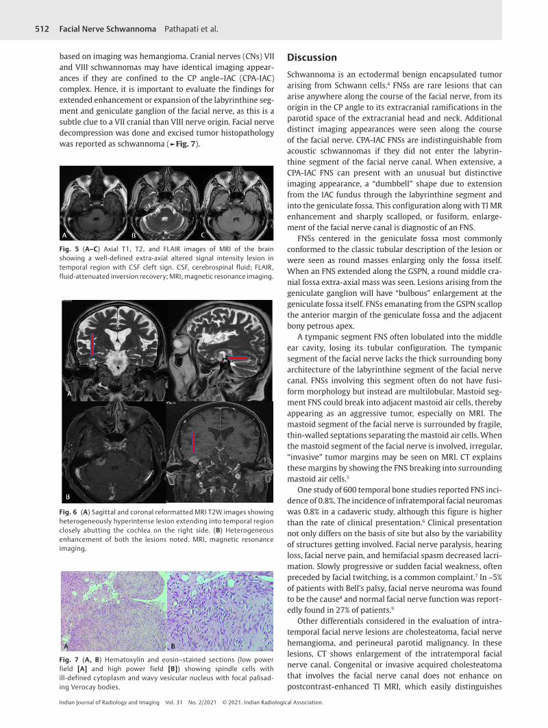
Fig. 5 (A–C) Axial T1, T2, and FLAIR images of MRI of the brain showing a well-defined extra-axial altered signal intensity lesion in temporal region with CSF cleft sign. CSF, cerebrospinal fluid; FLAIR, fluid-attenuated inversion recovery; MRI, magnetic resonance imaging.
Fig. 6 (A) Sagittal and coronal reformatted MRI T2W images showing heterogeneously hyperintense lesion extending into temporal region closely abutting the cochlea on the right side. (B) Heterogeneous enhancement of both the lesions noted. MRI, magnetic resonance imaging.
Fig. 7 (A, B) Hematoxylin and eosin–stained sections (low power field [A] and high power field [B]) showing spindle cells with ill-defined cytoplasm and wavy vesicular nucleus with focal palisad-ing Verocay bodies.

513Facial Nerve Schwannoma Pathapati et al.
Indian Journal of Radiology and Imaging Vol. 31 No. 2/2021 © 2021. Indian Radiological Association.
themselves from FNS. Facial nerve hemangioma involves both the facial nerve canal and adjacent bone, with more aggressive bony changes seen as irregular margins and/or a “moth-eaten” appearance. When an internal calcified hon-eycomb matrix is present (50%), ossifying hemangioma is quite distinctive. Parotid malignancy with perineural spread can be seen extending cephalad from an intraparotid invasive mass from distal to proximal along an enlarged facial nerve canal.5
Diagnostic workup includes audiometry (audiological test), auditory brain stem evoked response audiometry, CT, and contrast-enhanced MRI.
Treatment includes surgical removal. Approach depends on the site of tumor, size of tumor, and hearing loss. Timing for surgery is controversial because facial nerve neuromas almost always grow slowly10 and waiting to pursue surgery until the facial nerve either shows progressive deterioration or becomes paralyzed in patients with normal facial nerve function has been advocated.11,12 To delay the need for com-plete resection further, wide tumor decompression to allow the tumor to expand outside the natural bony confines has also been advocated.10 However, some authors believe that as the surgical approach becomes more difficult, the likeli-hood of postoperative complications increases, and recovery of facial function is poorer if the tumor continues to grow. The risk of surrounding structures such as the inner ear being invaded increases and eventually the brain stem may become compressed. Surgical resection of facial nerve neuromas is indicated without delay for patients with progressive facial palsy or paralysis, for large CP angle tumors compressing the brain stem or producing hydrocephalus, and for tumors invading the inner ear.10
ConclusionEvaluation of the FNS requires a combined approach of cor-relating accurate clinical information with CT and MRI find-ings. Awareness of the imaging anatomy of the facial nerve and the characteristic CT and MRI appearances of FNS involv-ing different facial nerve segments is crucial to arrive at the
correct diagnosis. However, the possible imaging differen-tials at various locations must be borne in mind so as to avoid potential diagnostic pitfalls.
Financial Support and SponsorshipNil.
Conflicts of InterestNone declared.
References
1 Mundada P, Purohit BS, Kumar TS, Tan TY. Imaging of facial nerve schwannomas: diagnostic pearls and potential pitfalls. Diagn Interv Radiol 2016;22(1):40–46
2 Gupta S, Mends F, Hagiwara M, Fatterpekar G, Roehm PC. Imaging the facial nerve: a contemporary review. Radiol Res Pract 2013;2013:248039
3 Chen M-C, Tseng T-M, Hung S-H, Chen P-Y. Facial nerve schwannoma: a case report and review of the literature. Oncol Lett 2014;8(6):2787–2789
4 Symon L, Cheesman AD, Kawauchi M, Bordi L. Neuromas of the facial nerve: a report of 12 cases. Br J Neurosurg 1993;7(1):13–22
5 Wiggins RH III, Harnsberger HR, Salzman KL, Shelton C, Kertesz TR, Glastonbury CM. The many faces of facial nerve schwannoma. AJNR Am J Neuroradiol 2006;27(3):694–699
6 Saito H, Baxter A. Undiagnosed intratemporal facial nerve neu-rilemomas. Arch Otolaryngol 1972;95(5):415–419
7 Latack JT, Gabrielsen TO, Knake JE, et al. Facial nerve neuro-mas: radiologic evaluation. Radiology 1983;149(3):731–739
8 Okabe Y, Nagayama I, Takiguchi T, Furukawa M. Intratemporal facial nerve neurinoma without facial paralysis. Auris Nasus Larynx 1992;19(4):223–227
9 Sataloff RT, Frattali MA, Myers DL. Intracranial facial neuromas: total tumor removal with facial nerve preservation: a new surgical technique. Ear Nose Throat J 1995;74(4):244–246, 248–256
10 Angeli SI, Brackmann DE. Is surgical excision of facial nerve schwannomas always indicated. ? Otolaryngol Head Neck Surg 1997;117(6):S144–S147
11 King TT, Morrison AW. Primary facial nerve tumors within the skull. J Neurosurg 1990;72(1):1–8
12 Lipkin AF, Coker NJ, Jenkins HA, Alford BR. Intracranial and intratemporal facial neuroma. Otolaryngol Head Neck Surg 1987;96(1):71–79