fallopian tube radiology - dr. sumit sharma
TRANSCRIPT
Dr. Sumit Sharma (PG)Dept. of Radiodiagnosis
SLIMS, Puducherry
Female Genital System-
The Fallopian Tubes
Fallopian Tube
The eponymous name, the Fallopian tube, is named after Gabriel Fallopius. He was Italian anatomist (1523-62), the same anatomist who gave his name to the Fallopian ligament and the Fallopian canal.
Fallopian Tube - Introduction
The uterine tube, also known as Fallopian tube, is approximately 10-12 cm long and 1-4 mm in diameter. It bridges the gap between the ovary laterally, and the uterus medially. Through it, the ovum passes into the uterine cavity. If conception occurs, it does so within the tube. The peritoneal reflection draping over the salpinges forms the mesosalpinx.
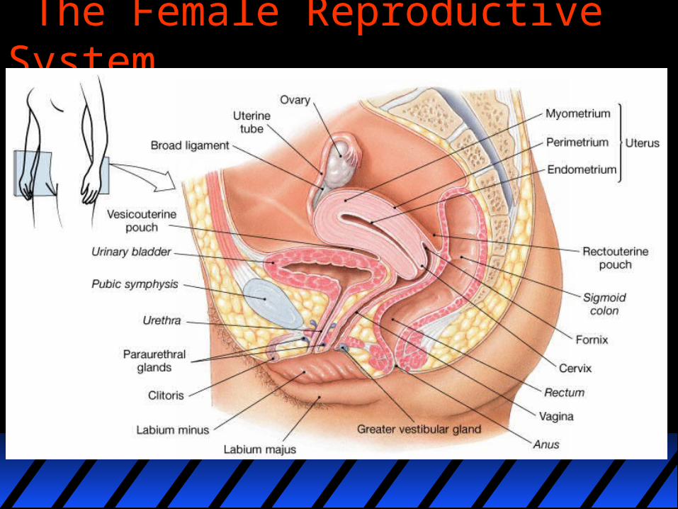
The Female Reproductive System
Development of the Reproductive Systems
Like the kidney apparatus:
•the gonads develop in a RETROPERITONEAL position next to the dorsal body wall.•they are derived from intermediate mesoderm.
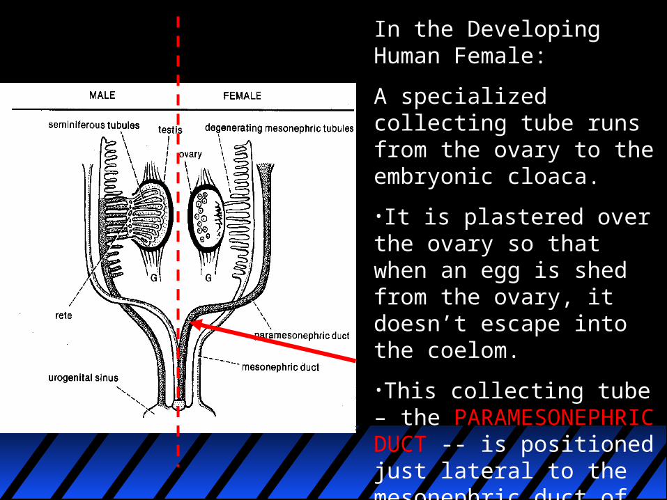
In the Developing Human Female:
A specialized collecting tube runs from the ovary to the embryonic cloaca.
•It is plastered over the ovary so that when an egg is shed from the ovary, it doesn’t escape into the coelom.
•This collecting tube – the PARAMESONEPHRIC DUCT -- is positioned just lateral to the mesonephric duct of the developing kidney.
Developing Human Female (Continued):
•The caudal ends of the right and left paramesonephric ducts fuse near their entrance into the embryonic cloaca to become the UTERUS AND VAGINA.•The remaining unfused parts are then known as the UTERINE TUBES, or more commonly the FALLOPIAN TUBES.
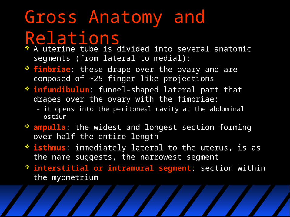
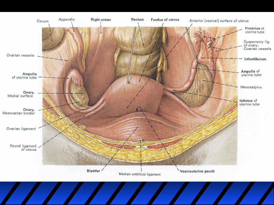
Gross Anatomy and Relations A uterine tube is divided into several anatomic
segments (from lateral to medial): fimbriae: these drape over the ovary and are composed of
~25 finger like projections infundibulum: funnel-shaped lateral part that drapes over
the ovary with the fimbriae:– it opens into the peritoneal cavity at the abdominal ostium
ampulla: the widest and longest section forming over half the entire length
isthmus: immediately lateral to the uterus, is as the name suggests, the narrowest segment
interstitial or intramural segment: section within the myometrium
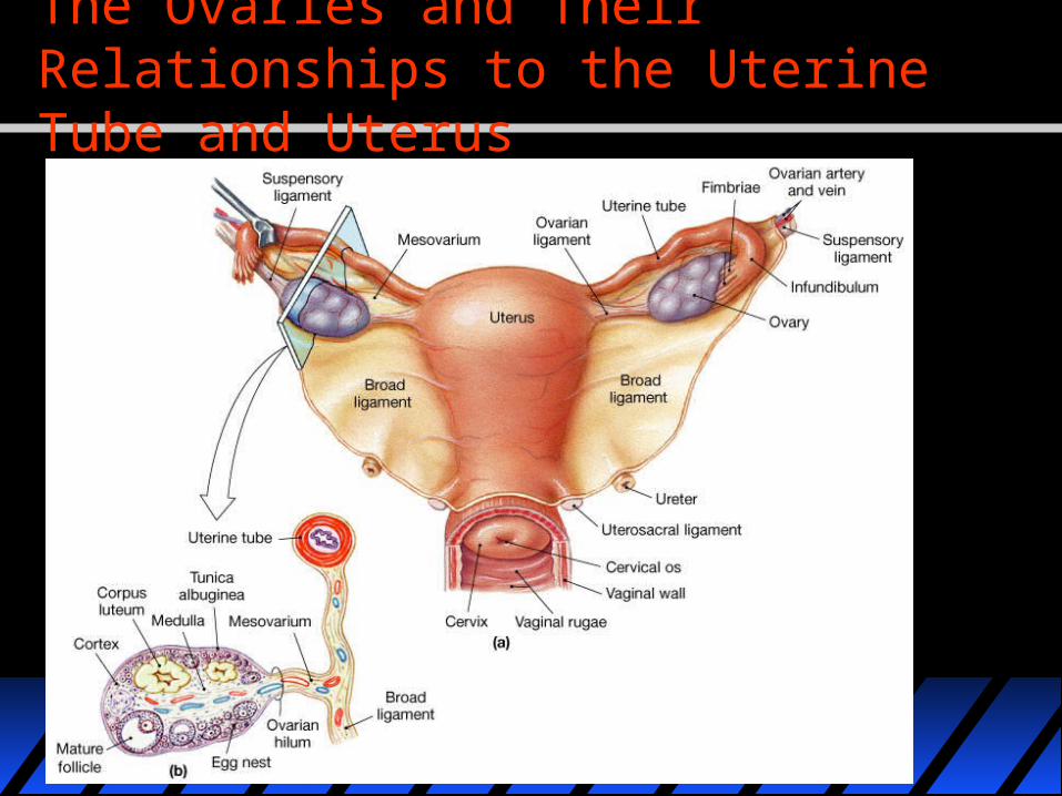
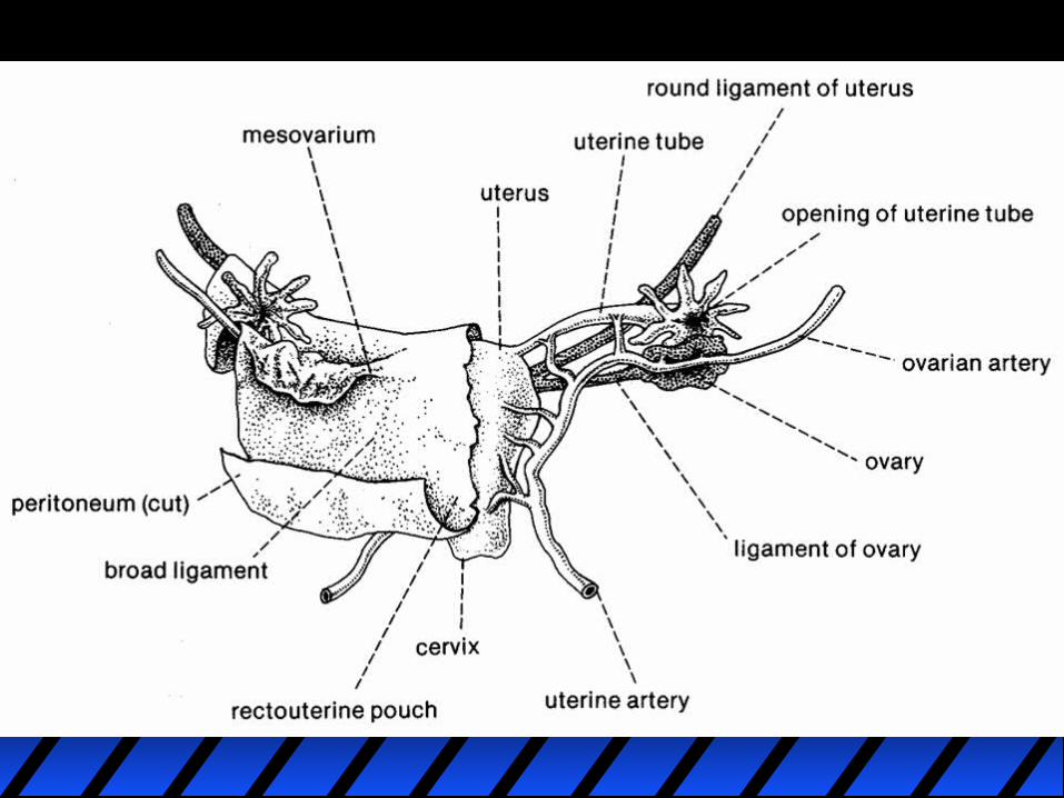
The Ovaries and Their Relationships to the Uterine Tube and Uterus
SUPPORTING LIGAMENTS OF THE OVARY AND UTERUS
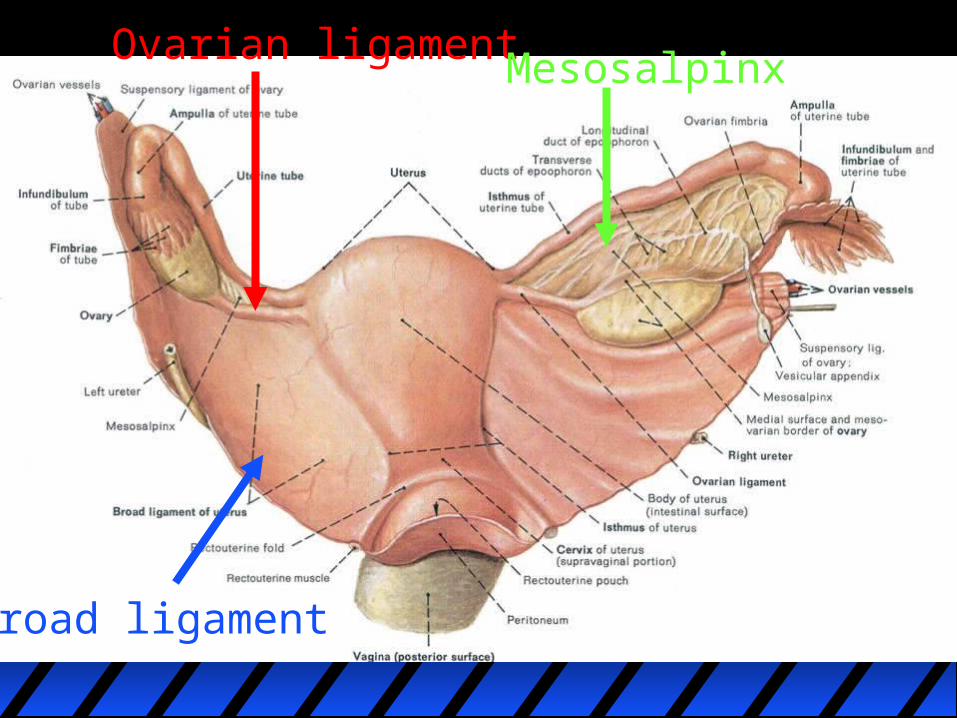
BROAD LIGAMENT – Sheet of connective tissue supporting uterus laterally, as well as fallopian tube and ovary out to lateral body wall.
OVARIAN LIGAMENT – connective tissue strap/band anchoring ovary to lateral uterine wall.
MESOSALPINX – connective tissue sheet spanning distance between ovarian ligament and fallopian tube.
Ovarian ligament
Broad ligament
Mesosalpinx
Blood supply
arterial supply: tubal branch of the ovarian artery and terminal (tubal) branch of the uterine artery
venous drainage: similarly named veins
Lymphatic supply
Lymph drainage is predominantly laterally and up to the para-aortic lymph nodes (like the ovaries).
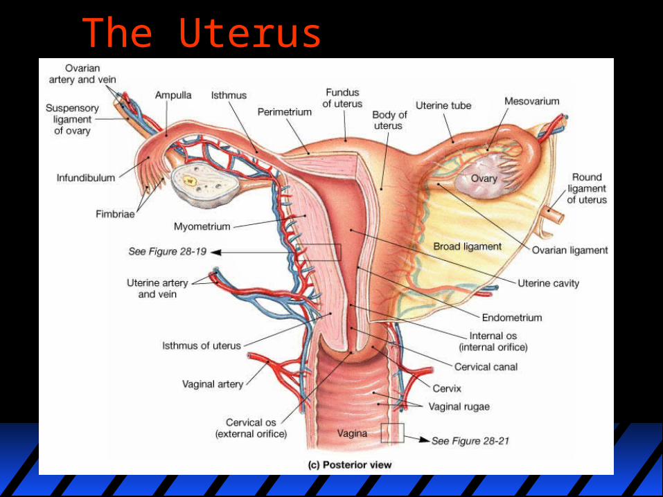
The Uterus
Nerve supply
ovarian and uterine plexuses (from T11 - L1)
Histology
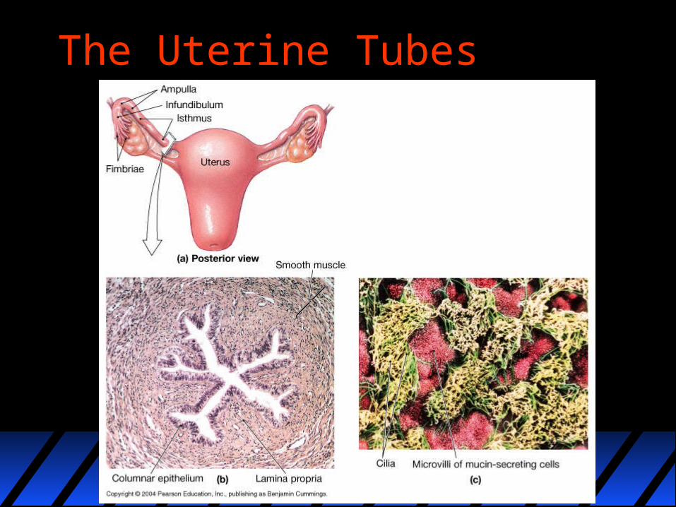
Like many other muscular hollow tubes it has two layers of muscle (inner circular, outer longitudinal), and is lined by columnar epithelium, a mixture of ciliated and non ciliated. It is the former that 'beat' the ovum towards the uterus.
The Uterine Tubes
Radiological appearance The normal uterine tubes are not visualized at
cross-sectional imaging unless they are outlined by fluid.
In the presence of peritoneal fluid or contrast material, the uterine tubes appear as paired, thin, serpentine juxta-uterine structures extending either anteriorly or posteriorly into the cul-de-sac.

Plain films
Contrast studies can be completed by performing a hysterosalpingogram (HSG)
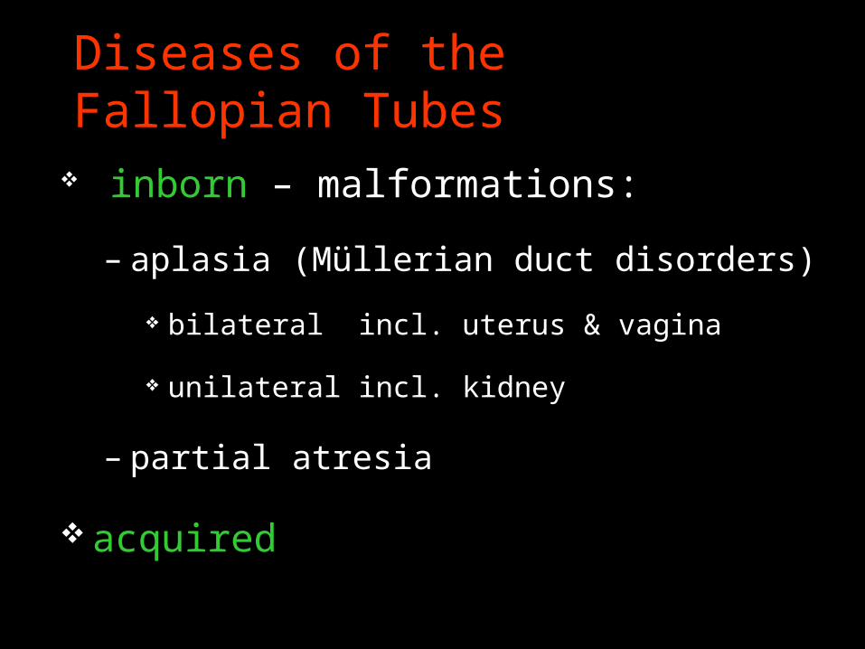
Diseases of the Fallopian Tubes
inborn – malformations:
– aplasia (Müllerian duct disorders)
bilateral incl. uterus & vagina
unilateral incl. kidney
– partial atresia
acquired
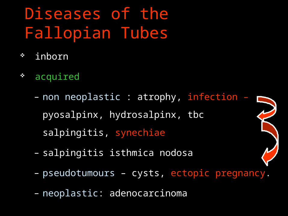
Diseases of the Fallopian Tubes
inborn
acquired
– non neoplastic : atrophy, infection – pyosalpinx,
hydrosalpinx, tbc salpingitis, synechiae
– salpingitis isthmica nodosa
– pseudotumours – cysts, ectopic pregnancy.
– neoplastic: adenocarcinoma
Diseases of the Fallopian Tubes & Ovaries
PID – pelvic inflammatory disease (chronic
salpingooophoritis)
tubal sterility

Hysterosalpingogram
Hysterosalpingogram (HSG) is a fluoroscopic examination of the uterus and the Fallopian tubes, most commonly used in the investigation of infertility or recurrent spontaneous abortions.
Technique the procedure should be performed during the
proliferative phase of the patient’s menstrual cycle (days 6-12), when the endometrium is thinnest – this improves visualisation of the uterine cavity, and also
minimises the possibility that the patient may be pregnant – if there is any uncertainty about the patient’s pregnancy
status, a bHCG is warranted prior to commencing.
After an antiseptic clean of the external genital area, a vaginal speculum is inserted with the patient in the lithotomy position ; the cervix is cleaned with an aseptic solution.
Catheterisation of the cervix is then performed ; the type of device used depends on local practice preferences– e.g. 6 Fr Foley catheter with balloon inflation, or– any one of a range of available HSG catheters or metal
cannulas
water soluble iodinated contrast is subsequently injected slowly under fluoroscopic guidance
A typical fluoroscopic examination includes preliminary frontal view of the pelvis, as well as subsequent spot images that demonstrate uterine endometrial contour, filled fallopian tubes and bilateral intraperitoneal spill of contrast, to establish tubal patency
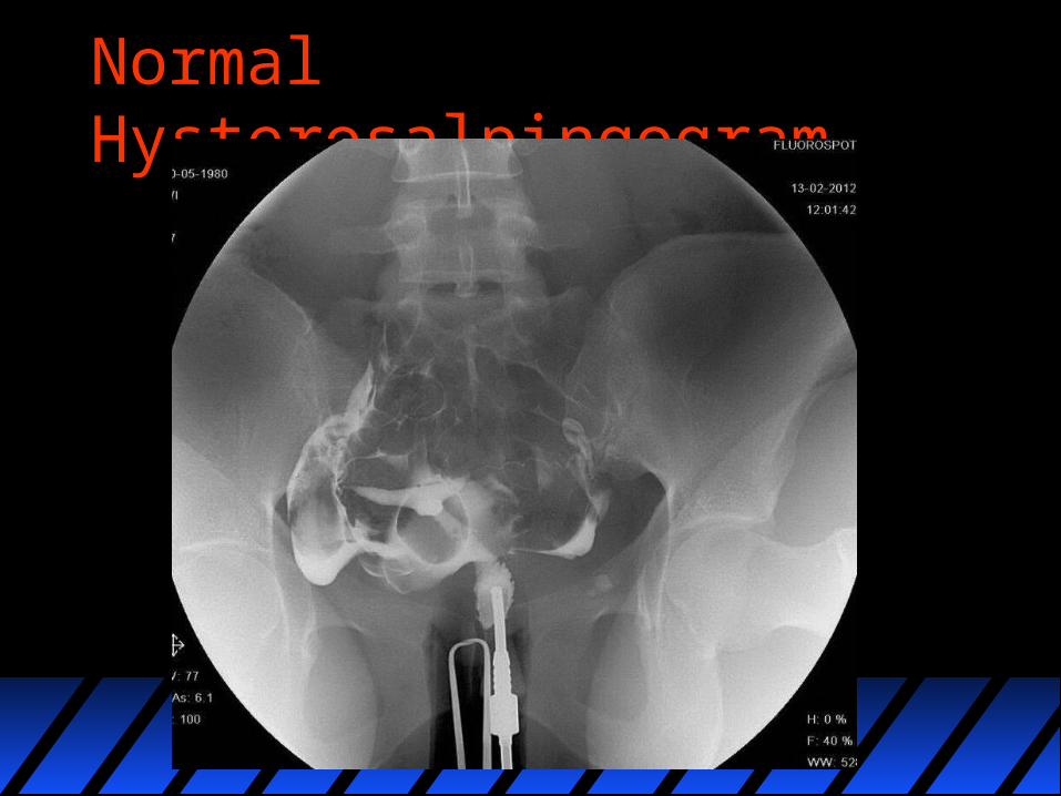
Normal Hysterosalpingogram
Contraindications and ComplicationsContraindicationspregnancyactive pelvic infectionrecent uterine or tubal surgery
Complications
Common but self limitingabdominal crampingPV spotting
Rare but seriouspelvic infectioncontrast reaction
Detectable pathology
Conditions which may be detected with HSG include Tubal obliteration of fallopian tubes : usually secondary to previous
pelvic inflammation. It must be differentiated from incomplete tubal opacification due to tubal spasm, or underfilling of the uterus with contrast
tubal polyps tubal malignancy hydrosalpinx salpingitis isthmica nodosa (SIN) tubal spasm can be physiological
Fallopian tube polyp A Fallopian tube polyp refers to a small focal lesion of ectopic
endometrial tissue located at the intramural portion of the fallopian tube.
Epidemiology The reported incidence is 1- 2.5% on hysterosalpingograms
performed for assessment of infertility 3
Clinical presentation Most patients with tubal polyps are asymptomatic and polyps
are usually an incidental finding at hysterosalpingography.
Radiographic features
They can be unilateral or bilateral, and they usually measure less than 1 cm in diameter.
Hysterosalpingogram - HSG
Tubal polyps appear as smooth, rounded or oval filling defects which are not associated to tubal dilatation or obstruction, with free flow of contrast medium to the peritoneal cavity.
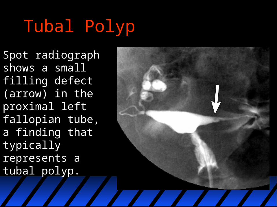
Tubal Polyp
Spot radiograph shows a small filling defect (arrow) in the proximal left fallopian tube, a finding that typically represents a tubal polyp.
Salpingitis isthmica nodosa Salpingitis isthmica nodosa (SIN) (sometimes also referred to
as perisalpingitis isthmica nodosa - PIN) refers to nodular scarring of the fallopian tubes. In very early stages, the tubes may appear almost normal. As scarring and nodularity progresses, the changes become more radiographically apparent.
Aetiology the aetiology of SIN has remained a matter of controversy since
its first description the prevailing theories include an inflammatory (salpingitis), a
congenital, and more recently an acquired (but not post-inflammatory) aetiology
Location It can involve the entire fallopian tube, but usually involves the
proximal two-thirds of the fallopian tube.
Radiographic features
An accurate radiographic diagnosis is important due to its strong association with infertility and ectopic pregnancy.
Hysterosalpingography (HSG)A characteristic finding is multiple nodular diverticular spaces involving the fallopian tubes (usually involving the proximal two-thirds of the fallopian tubes).HSG of the nodular area with severe SIN shows several pockets (diverticulae) containing the introduced contrast material 5.It is observed that no dominant channel is seen as the contrast flows through the tube. This means there appears to be no direct pathway for sperm to travel which increases the chances of a tubal pregnancy.
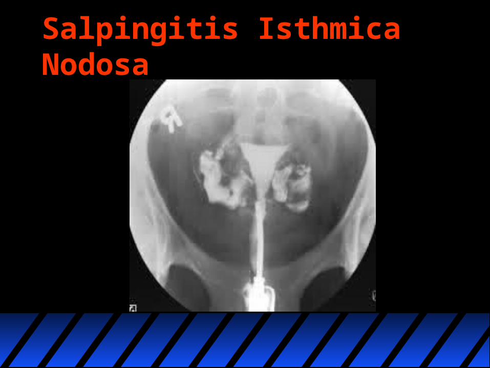
Salpingitis Isthmica Nodosa
Complicationsincreased risk of tubal ectopic pregnancy 4
Subfertility
EtymologyThe term is derived from salpingitis for inflammation of the salpinx (tube), isthmica for involvement of the proximal isthmic portion of fallopian tube and nodosa for its nodular appearance
Differential diagnosisConsiderations includetubal tuberculosis
often has multiple constrictions along the course of fallopian tube can form because of scarring and give rise to "beaded" appearance to the tubes on HSG can also have adnexal calcification
Hydrosalpinx Hydrosalpinx is a descriptive term and refers to a fluid
filled dilatation of the fallopian tube. Clinical presentation Patients may be asymptomatic or may present with
pelvic pain or infertility. Pathology One or both fallopian tubes may be affected. A
hydrosalpinx results from an accumulation of secretions when the tube is occluded at its distal end (obstruction of the ampullary segment) or both ends. On rare occasions, transient distention of the fallopian tubes occurs because of retrograde passage of blood from the uterus without complete distal occlusion.
Causes endometriosis ovulation induction pelvic inflammatory disease (e.g chlamydial or gonococcal
infection): a hydrosalpinx is most commonly a sequela of adhesions from pelvic inflammatory disease
post hysterectomy (without salpingo-oophorectomy)– unilateral or bilateral hydrosalpinx may also occur in women after
hysterectomy when only the fallopian tubes are left to protect the blood supply to the ovary
– this is from accumulation of tubal secretions caused by surgical blockage proximally and adhesion-related blockage distally
tubal ligation tubal malignancy: primary or secondary tumours of the fallopian
tubes
Radiographic featuresUltrasound May be seen as a thin-or thick-walled (in chronic cases), elongated or
folded, tubular, C shaped or S shaped fluid-filled structure that is distinct from the uterus and ovary.
Longitudinal folds that are present in a normal fallopian tube may become thickened in the presence of a hydrosalpinx.
The folds may produce a characteristic “cogwheel” appearance when imaged in cross section. These folds are pathognomonic of a hydrosalpinx.
Incomplete septae may also give a "beads on a string" sign. A significantly scarred hydrosalpinx may present as a multi-locular
cystic mass with multiple septa (often incomplete) creating multiple compartments. These septa are generally incomplete, and the compartments can be connected. However, with more pronounced scarring, differentiation from an ovarian mass may not be possible.
HysterosalpingogramWill classically show a dilated fallopian tube filling with contrast with absence of free spillage.
CTA hydrosalpinx may be seen incidentally at CT as a fluid-attenuation tubular juxta-uterine structure that is separate from the ovary. A simple hydrosalpinx is not accompanied by pelvic inflammation. The tubal wall may enhance following contrast.
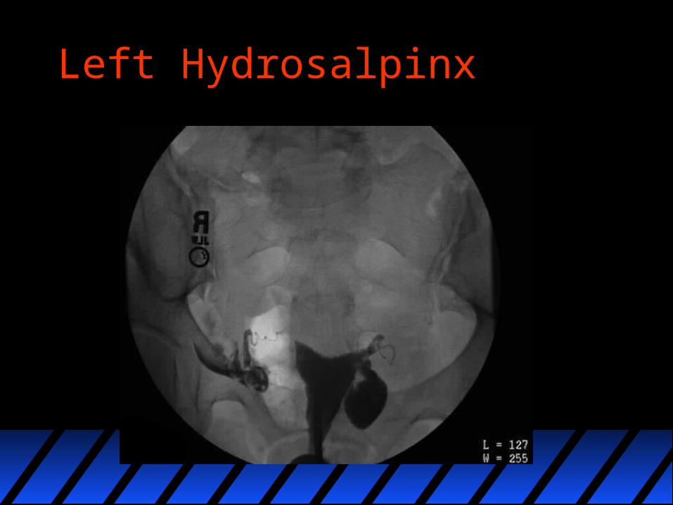
Left Hydrosalpinx
MRI MR imaging is the modality of choice for the characterisation
and localisation of adnexal masses that are inadequately evaluated with ultrasound. A dilated fallopian tube is interposed between the uterus and ovary and demonstrates fluid signal intensity. Incomplete septa or folds can be seen. The mucosal plicae are usually effaced, and the tube wall is uniformly smooth and thin.
Signal characteristics of the dilated tube(s) include: T1: typically hypo-intense although can be hyper-intense if there
is proteinaceous fluid T1 C+ (Gd): the the mucosal plicae and the tube walls may
show mild enhancement T2: hyper-intense
Complications
tubal torsion: can be a late complication
Differential diagnosisGeneral imaging differential considerations include: elongated para ovarian cystcystic ovarian neoplasm(s) bowel obstruction dilated pelvic veins elongated pelvic perineural cyst
Fallopian tube spasm
It is a transient functional anomaly that can mimic a true mechanical tubal occlusion.
At radiography, tubal spasm cannot be distinguished from a tubal occlusion. Administration of spasmolytic agents such as Glucagon can occasionally result in uterine muscle relaxation and consequent tube opacification, thereby helping differentiate a spasm from true occlusion .
Delayed radiography may also be performed to help differentiate tubal spasm from a true tubal occlusion.

Fallopian Tube Spasm
Ectopic pregnancy Ectopic pregnancy refers to the implantation of a
fertilised ovum outside of the uterine cavity.
Epidemiology
The overall incidence has increased over the last few decades and is currently thought to affect 1-2% of pregnancies. There is an increased incidence in in-vitro fertilisation pregnancies (IVF).
Clinical presentation
Presentation is often with abdominal pain or bleeding. If unrecognised haemorrhage can be life threatening.

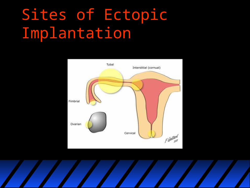
Location of ectopics
tubal ectopic: 93-97%– ampullary ectopic: most common ~70% of tubal
ectopics and ~ 65% of all ectopics– isthmal ectopic: ~12% of tubal ectopics and ~11%
of all ectopics– fimbrial ectopic: ~11% of tubal ectopics and ~10%
of all ectopics
Sites of Ectopic Implantation
Markers
• serum beta HCG levels tend to increase at a slower rate
• serum progesterone levels can be not as elevated as for an intrauterine pregnancy; 5-25 ng/ml range although not absolute

Radiographic features
It is essential to know a quantitative beta HCG prior to scanning as this will determine what you expect to see. At levels below 1000 IU a normal early pregnancy may well not be visible, and therefore should the scan prove negative, a repeat scan in a couple of days (along with a repeat beta HCG) is necessary (beta HCG should normally double approximately every two days).
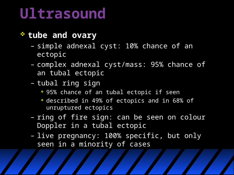
Ultrasound tube and ovary
– simple adnexal cyst: 10% chance of an ectopic– complex adnexal cyst/mass: 95% chance of an
tubal ectopic– tubal ring sign
95% chance of an tubal ectopic if seen described in 49% of ectopics and in 68% of unruptured
ectopics
– ring of fire sign: can be seen on colour Doppler in a tubal ectopic
– live pregnancy: 100% specific, but only seen in a minority of cases
It is of utmost importance not to be reassured by the presence of a live intrauterine pregnancy, as this may delay the important diagnosis of a co-existing ectopic pregnancy (i.e. heterotopic pregnancy).
This life-threatening condition for both mother and intrauterine child necessitates a high level of clinical suspicion, especially in cases of assisted reproduction (e.g. in-vitro fertilisation) or former tubal surgery
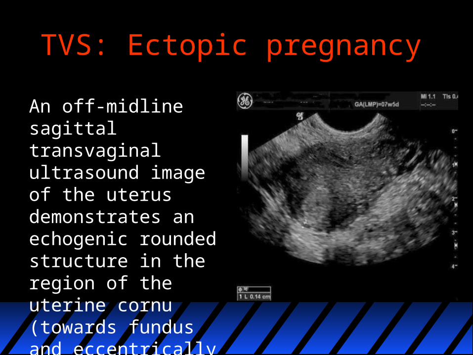
TVS: Ectopic pregnancy
An off-midline sagittal transvaginal ultrasound image of the uterus demonstrates an echogenic rounded structure in the region of the uterine cornu (towards fundus and eccentrically placed).
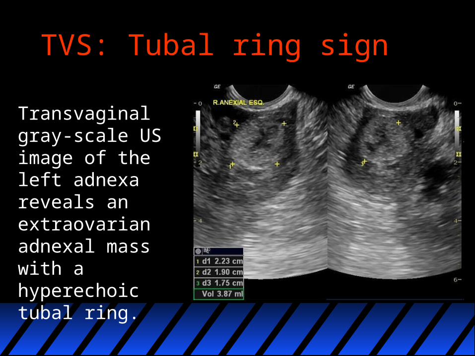
TVS: Tubal ring sign
Transvaginal gray-scale US image of the left adnexa reveals an extraovarian adnexal mass with a hyperechoic tubal ring.
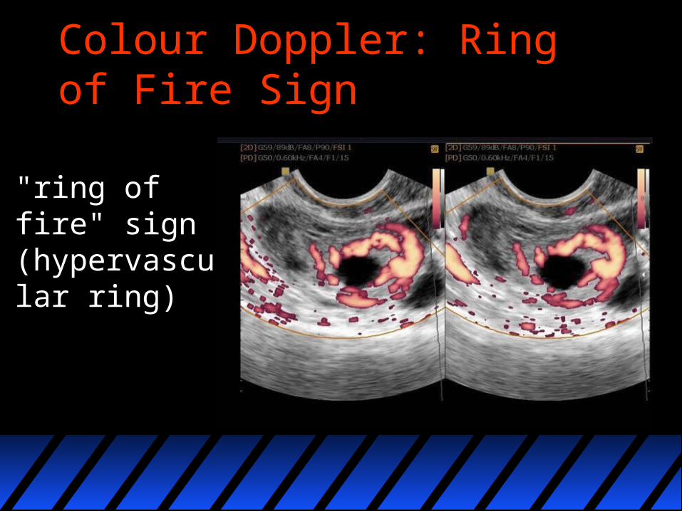
Colour Doppler: Ring of Fire Sign
"ring of fire" sign (hypervascular ring)

Ring of Fire Sign
Complications
Complications somewhat depend on the type of ectopic. General complications for a typical (tubal) ectopic pregnancy include
tubal rupture: 15-20%

Management
In general the options are: Surgical: (in the case of tubal ectopics with open or
laparoscopic salpingectomy or salpingotomy) Medical:
– methotrexate (a folate antagonist) either administered systemically or by direct ultrasound guided injection or potassium chloride (direct injection only obviously)
– usually considered if size small (e.g <4 cm) and if no complication
– the gestational mass can paradoxically increase in size following methotrexate on subsequent scanning and does not necessarily imply failure of methotrexate therapy
Management
Conservative or expectant management is being recognised as an option for those ectopics where rupture has not occurred (i.e. no haemoperitoneum ) and fetal demise has already taken place.
Differential diagnosis
The differential diagnosis of abdominal pain in a pregnant patient is broad. An ectopic pregnancy must be excluded with ultrasound. Other common diagnoses in this setting include:
ruptured corpus luteum appendicitis