familial amyloid polyneuropathy: receptor for advanced glycation
TRANSCRIPT
Familial Amyloid Polyneuropathy: Receptor for Advanced GlycationEnd Products-Dependent Triggering of Neuronal Inflammatory andApoptotic Pathways
Monica Mendes Sousa,1 Shi Du Yan,2 Rui Fernandes,1 Antonio Guimaraes,3 David Stern,2 andMaria Joao Saraiva1,4
1Institute for Cellular and Molecular Biology, 2Departments of Pathology, Surgery, and Physiology and Cellular Biophysics,Columbia University, New York, New York 10032, 3Hospital Geral de Santo Antonio, Porto 4150-180, Portugal, and4Instituto de Ciencias Biomedicas Abel Salazar, Porto 4099-003, Portugal
Familial amyloid polyneuropathy (FAP) is a neurodegenerativedisorder associated with extracellular deposition of mutanttransthyretin (TTR) amyloid fibrils, particularly in the peripheralnervous system. We have hypothesized that binding of TTRfibrils to the receptor for advanced glycation end products(RAGE) on critical cellular targets is associated with a destruc-tive stress response underlying peripheral nerve dysfunction.Analysis of nerve biopsy samples from patients with FAP (n �16) at different stages of disease (0–3), compared with age-matched controls (n � 4), by semiquantitative immunohistologyand in situ hybridization showed increased levels of RAGE,beginning at the earliest stages of the disease (FAP 0; p � 0.02)and especially localized in axons. Upregulation of proinflamma-tory cytokines (tumor necrosis factor-� and interleukin-1�) (ap-proximately threefold; p � 0.02) and the inducible form of nitricoxide synthase (iNOS) (�2.5-fold; p � 0.04) was also observedin a distribution overlapping RAGE expression. Tyrosine nitra-
tion and increased activated caspase-3 in axons from FAPpatients ( p � 0.03) were apparent. Although these data sug-gest the presence of ongoing neuronal stress, there was noupregulation of neurotrophins (nerve growth factor andneurotrophin-3) in FAP nerves. Studies on cultured neuronal-like, Schwann, and endothelial cells incubated with TTR fibrilsdisplayed RAGE-dependent expression of cytokines and iNOSat early times (6 and 12 hr, respectively), followed by later (24 hr)activation of caspase-3 and DNA fragmentation. We proposethat the interaction of TTR fibrils with RAGE may contribute tocellular stress and toxicity in FAP. Furthermore, there is anapparent lack of responsiveness of Schwann cells in FAP nerveto provide neurotrophic factors.
Key words: familial amyloidotic polyneuropathy; amyloid;transthyretin; RAGE; caspase-3; inducible nitric oxide synthase;inflammatory cytokine
Familial amyloid polyneuropathy (FAP) is a neurodegenerativedisorder characterized by the extracellular deposition of amyloidfibrils composed of mutant forms of transthyretin (TTR) inseveral tissues, particularly the peripheral nervous system (Coim-bra and Andrade, 1971a,b). Among the point mutations in TTRpromoting amyloidogenesis (Saraiva, 2001), the most common isa substitution of Val for Met at position 30 (V30M) (Saraiva et al.,1983). TTR amyloid deposits are distributed diffusely in theperipheral nervous system, involving nerve trunks, plexuses, andsensory and autonomic ganglia (Coimbra and Andrade, 1971b).Amyloid deposits in peripheral nerves occur especially in theendoneurium, where they appear close to Schwann cells (SCs)and collagen fibrils (Coimbra and Andrade, 1971a). In severelyaffected nerves, endoneurial contents are replaced by amyloid,and few nerve fibers retain viability. By contrast, the CNS in FAPis relatively spared, except for the ependymal lining and lepto-
meninges. Outside the nervous system, extensive amyloid depos-its have been observed throughout connective tissue in a perivas-cular distribution.
The accumulation of extracellular, crossed �-sheet fibrils is ahallmark of amyloidoses. Pathologically, fibrillar accumulationappears to be closely linked to dysfunction of the surroundingcells and vasculature. This view of FAP pathogenesis wouldsuggest that nerve fiber degeneration results from multifocalcompression by amyloid deposits (Said et al., 1984). However,unmyelinated fibers (UFs) that are primarily affected in FAP aremore resistant to compression than myelinated fibers (MFs).Similarly, despite the presence of vascular amyloid, evidence ofcompromised blood flow sufficient to adversely affect organ func-tion has not been demonstrated in FAP (Fujimura et al., 1991).
Local cellular activation, ultimately resulting in cell dysfunc-tion and death, may contribute to the pathogenesis of amyloid-related disorders. In Alzheimer’s disease (AD), the close associ-ation between expression of inflammatory mediators (cytokines,chemokines, complement proteins, acute phase reactants), acti-vated astrocytes and microglia, and neuritic plaques has suggesteda prominent role for immune/inflammatory pathways in the neu-rodegenerative process (Mrak et al., 1995; Smith et al., 1997;Veerhuis et al., 1999). Among the candidate mechanisms throughwhich pathogenic forms of amyloidogenic molecules might per-turb cellular properties is engagement of cellular receptors. Thereceptor for advanced glycation end products (RAGE) displays
Received May 9, 2001; revised July 18, 2001; accepted July 19, 2001.This work was supported by grants from PRAXIS XXI (35785/99 and 35735/99),
Portugal, from the United States Public Health Service (AG17490, AG16223), andfrom the Surgical Research Fund. M.M.S. is the recipient of a postdoctoral fellow-ship (PRAXIS XXI/BPD/22027/99), and R.F. is the recipient of a BTI Fellowship(PRAXIS XXI/BTI /PL021902) from Fundacao para a Ciencia e Tecnologia,Portugal. We thank Paul Moreira for the production and purification of recom-binant TTR.
Correspondence should be addressed to Maria Joao Saraiva, IBMC-Amyloid Unit,R. Campo Alegre, 823, 4150-180 Porto, Portugal. E-mail: [email protected] © 2001 Society for Neuroscience 0270-6474/01/217576-11$15.00/0
The Journal of Neuroscience, October 1, 2001, 21(19):7576–7586
increased expression in FAP tissues and has been shown to bindfibrillar TTR triggering nuclear factor (NF)-�B expression(Sousa et al., 2000). The possible involvement of RAGE in thebiology of amyloidoses was strengthened by the recent demon-stration that RAGE bound amyloid A, and that blockade ofRAGE suppressed splenic expression of proinflammatory cyto-kines, NF-�B activation, and accumulation of amyloid in a modelof systemic amyloidosis (Yan et al., 2000). RAGE is a member ofthe immunoglobulin superfamily with a broad repertoire of li-gands in addition to amyloid-associated macromolecules, includ-ing products of nonenzymatic glycoxidation (advanced glycationend products), proinflammatory mediators (S100/calgranulins),and amphoterin (Hori et al., 1995; Yan et al., 1996, 1999, 2000;Hofmann et al., 1999). In each case, the receptor recruits signaltransduction mechanisms, often resulting in a sustained andpathogenic inflammatory response (Yan et al., 1996; Schmidt etal., 1999; Hofmann et al., 2001).
In the present work we initiated a systematic evaluation of therelationship between RAGE and FAP, on the basis of the hy-pothesis that expression of the receptor in FAP tissue at earlytimes might contribute to the pathogenesis of evolving cellulardysfunction.
MATERIALS AND METHODSFAP patients. Sural nerve biopsy specimens from FAP patients (n � 16)and normal controls (n � 4; near-relatives of FAP patients who ulti-mately turned out not to have mutations in TTR) were available in theHospital Geral de Santo Antonio (Porto, Portugal), because this materialwas obtained as part of the clinical diagnosis and evaluation of polyneu-ropathy in this region of Portugal, before the current use of less invasivemethods. Initial characterization of clinical material included morpho-metric studies of nerve fiber density and abundance of amyloid deposits.Patients were scanned for mutations in TTR by immunoblotting (Saraivaet al., 1985). The general characterization of the patients under study isshown in Table 1.
Morphometric studies were performed on sural nerve biopsy tissuefixed in glutaraldehyde (2.5%) in 0.1 M cacodylate buffer, pH 7.4, post-fixed in osmium tetroxide, and embedded in Epon. Quantitation ofmyelinated fibers in semithin sections was performed in an area of atleast 0.1 mm 2 at a magnification of 1000�. Myelinated fibers werecounted, their diameters were measured, and the density was calculated.Unmyelinated fibers were counted from thin sections in an area of atleast 0.005 mm 2, and their densities were calculated. A scoring system ofpatient material was based on morphometry of MFs and UFs: FAP 0(n � 5), no reduction in the number of fibers (MFs �10,000 fibers/mm 2;UFs �50,000 fibers/mm 2); FAP 1 (n � 4), discrete reduction (MFsvaried between 7000 and 2000 fibers/mm 2; UFs varied between 4000 and40,000 fibers/mm 2); FAP 2 (n � 4), evident reduction (MFs �1000fibers/mm 2; UFs � 10,000 fibers/mm 2); and FAP 3 (n � 3), severereduction (MFs � 1000 fibers/mm 2; UFs absent). Standard values forcontrol nerves ranged from 7000 to 11,000 fibers/mm 2 for MFs and from30,000 to 80,000 fibers/mm 2 for UFs (see Table 1).
Amyloid deposition was assessed by Congo red staining and in mostcases was inversely proportional to nerve fiber density (Coimbra andAndrade, 1971b). In FAP 0 patients, amyloid was absent (by Congo redstaining), but immunohistochemical reaction of TTR was detected. Asemiquantitative index of amyloid deposition in endoneurium andepineurium included the following: 0, no amyloid; 1, scarce amyloiddeposition; 2, several amyloid deposits; and 3, abundant amyloid. Allhistologic measurements were made independently by two pathologistsunaware of the patient’s clinical diagnosis. The presence of Congored-positive areas was closely correlated with immunoreactivity withTTR.
Immunohistochemistry. For immunohistochemical analysis, paraffinsections were deparaffinated, dehydrated in a modified alcohol series,and incubated in blocking buffer [1% bovine serum albumin (BSA) and4% horse serum] for 30 min at 37°C in a moist chamber. Incubation withprimary antibody, at the appropriate dilution in blocking buffer, wasperformed overnight at 4°C. Primary antibodies were rabbit polyclonal
anti-TTR IgG (Dako, Glostrup, Denmark; 1:300), murine monoclonalanti-human RAGE (1:25), polyclonal rabbit anti-RAGE IgG (Yan et al.,2000) (1:300), goat anti-human interleukin (IL)-1� IgG and goat anti-human tumor necrosis factor (TNF)-� IgG (Santa Cruz Biotechnology,Santa Cruz, CA; 1:25), rabbit anti- inducible form of nitric oxide synthase(iNOS) IgG (Chemicon, Temecula, CA; 1:2500), rabbit anti-endothelialNOS (eNOS) IgG (Chemicon; 1:100), rabbit anti-neuronal NOS (nNOS)IgG (Chemicon; 1:1000), rabbit anti-3-nitrotyrosine IgG (Chemicon;1:1000), rabbit anti-activated caspase-3 IgG (PharMingen, San Diego,CA; 1:25), monoclonal anti-human CD68 IgG1k (Dako; 1:10), monoclo-nal anti-human CD11b, Mac1 IgG1 (Chemicon; 1:10), monoclonal anti-myelin basic protein IgG1 (Chemicon; 1:300), rabbit anti-factor VIII IgG(Sigma, St. Louis, MO; 1:200), monoclonal anti-smooth muscle �-actinIgG2a (Sigma; 1:100), rabbit anti-neurofilament 200 IgG (Sigma; 1:200),rabbit anti-brain-derived neurotrophic factor (BDNF) IgG (Chemicon;1:1000), sheep anti-nerve growth factor-� (NGF�) (Chemicon; 1:500),and rabbit anti-neurotrophin 3 IgG (NT3) (Chemicon; 1:1000). Antigenvisualization was performed with either biotin–extravidin–alkalinephosphatase or biotin–extravidin–peroxidase kits (Sigma), using FastRed (Sigma) or 3-amino-9-ethyl carbazole (Sigma), respectively, as sub-strates. On parallel control sections, primary antibody was replaced byblocking buffer. Double staining was accomplished by decolorizing thefirst stain with 95% ethanol and blocking peroxidase activity with hydro-gen peroxide (3%)/methanol for 15 min. Subsequent immunohistochem-istry was performed with either affinity-purified alkaline phosphatase- orperoxidase-conjugated secondary antibody (Sigma). Semiquantitativeanalysis of immunohistochemical images was performed with the Uni-versal Imaging system (NIH). The results shown represent percentage(�SD) of the image area occupied by a particular immunoreactivity.
In situ hybridization. Digoxigenin-labeled probes were transcribed fromthe plasmid B379–2A (Brett et al., 1993) containing a 1406 bp fragmentof bovine RAGE and PCRII containing a 470 bp fragment of mouseIL-1� (a kind gift of Dr Shi Fang Yan, Columbia University, New York),using the DIG RNA labeling kit (Roche, Indianapolis, IN). Antisenseprobe for RAGE was transcribed from the T3 promoter with the plasmidlinearized with XhoI, and control sense probe was transcribed from theT7 promoter with the plasmid linearized with XbaI. Antisense probe forIL-1� was transcribed from the T7 promoter with the plasmid linearizedwith SpeI, and a control sense probe was transcribed from the SP6promoter with the plasmid linearized with XhoI. The digoxigenin-labeled RNA probes were hybridized to cellular mRNA by standardprocedures and detected with anti-digoxigenin alkaline phosphatase-conjugated antibody. Antibody was visualized with X-phosphate andnitroblue tetrazolium salt.
Proteins. Human soluble RAGE (sRAGE) was expressed using thebaculovirus system and purified to homogeneity (Yan et al., 1996).Recombinant TTR was purified from Escherichia coli BL21 transformedwith plasmids carrying wild-type TTR cDNA as described (Almeida etal., 1997). TTR fibrils were generated from soluble wild-type TTR (2mg/ml) by incubation with 0.05 M sodium acetate, pH 3.6, for 48 hr atroom temperature (Bonifacio et al., 1996). Formation of amyloid fibrilswas tested by thioflavin T fluorescence. The lipopolysaccharide (LPS)content of protein preparations was tested using the Limulus amebocyteassay (Sigma). At a protein concentration of 2 mg/ml, sRAGE hadno detectable LPS. Similarly, TTR at a concentration of 1 mg/ml wasfree of LPS.
Cell lines and cell culture. RN22 (rat Schwann cell line), PC12 (ratadrenal cell line with a neuronal-like phenotype), and BAE-1 (bovineaortic endothelium cell line) were from the European Collection of CellCultures. Wild-type mouse embryonic fibroblasts (MEF1s) were kindlyprovided by Dr. Miguel Seabra (Imperial College, London). All cell lineswere propagated in 10 cm dishes in monolayers and maintained at 37°Cin a humidified atmosphere of 95% and 5% CO2. Cells were grown inDMEM (Life Technologies, Gaithersburg, MD) supplemented with 10%fetal bovine serum (FBS) (Life Technologies) and 100 U/ml penicillin(Life Technologies) (complete media). Primary cultures from dorsal rootganglion (DRG) were prepared from �250 DRGs from neonatal to3-week-old mice. DRGs were suspended in DMEM containing 10%collagenase/dispase stock solution (Sigma) and incubated at 37°C for 1hr. After dissociation of the tissue, the suspension was centrifuged at1000 rpm for 10 min, and the resulting pellet was washed with DMEMthree times to remove excess collagenase. For primary SCs, cells wereresuspended in 10 ml per �20 DRGs in DMEM supplemented with 0.1%BSA, 20% FBS, 30 mM glucose, and 2 mM glutamine and plated in 10 cm
Sousa et al. • Neurodegeneration in FAP J. Neurosci., October 1, 2001, 21(19):7576–7586 7577
dishes (10 ml per dish) coated with laminin (Sigma) and incubated for 2.5hr at 37°C. During this period, nearly all non-neuronal cells attach,leaving neurons in the culture supernatant. Cell culture media wasaspirated and replaced by fresh media. SCs were grown to a confluentmonolayer over �7 d. To obtain neuron DRG, cells were plated inMatrigel (Sigma) in neuronal growth media: DMEM supplemented with100 ng/ml nerve growth factor (Life Technologies), 5% FBS, and 1% B27supplement (Life Technologies). The purity of primary cultures used forexperiments was �85% neurons and �80% SCs, based on staining withantibody to neurofilament 200 and myelin basic protein in neuronal andSchwann cell cultures, respectively.
For RT-PCR and caspase-3 assays, cultured cells were grown in com-plete media in six-well dishes. For cell death detection, cells were grownin 96-well plates. When �80% confluence was reached, cells were washedwith HBSS and incubated with assay buffer (DMEM containing 1%dialyzed FBS) with the indicated amount of either soluble or fibrillarTTR for the time periods shown for each experiment. For studies usingspecific antibodies, nonimmune (NI) IgG or sRAGE, cells were prein-cubated for 3 hr with the indicated amount of antibody, NI IgG, orprotein (10 �g/ml, unless stated otherwise).
RNA extraction and RT-PCR. Total RNA extraction was performedusing Trizol reagent (Life Technologies) according to the manufacturer’sinstructions. The concentration of total RNA was determined spectro-photometrically in RNase-free water. Reverse transcription was per-formed using 10 �g total RNA and Superscript II (Life Technologies),primed by oligo-dT following the manufacturer’s instructions. PCR (94°C,3 min, 1 cycle; 94°C, 20 sec, 56°C, 45 sec, 72°C, 1 min, 30 cycles; 72°C, 6 min,1 cycle) was performed using 1⁄10 of the obtained cDNA. The followingprimers were used: for IL-1�, sense (5�-ATGGCAACTGTTCCT-GAACTCAACT-3�) and antisense (5�-CAGGACAGG TATAGATTCT-TTCCTTT-3�); for mouse and rat TNF-�, sense (5�-TTCTGT CTACT-GAACTTCGGGGTGATCGGTCC-3�) and antisense (5�-GTATGAG-A TAGCAAATCGGCTGACGGTGTGGG-3�); for bovine TNF-�, sense(5�-TTCTGTCTACTGAACTTCGGGGTGATTGGTCC-3�) and anti-sense (5�-GTATGAAATGGCAAACCGGCTGACGGTGTGGG-3�); formouse and rat iNOS, sense (5�-GGAACCTACCAGCTCACTCTG-3�)and antisense (5�-CCACTTC AGCCCGAGCTCCTG-3�); for bovineRAGE, sense (5�-AGCGGCTGGAATGGAAA CTGAACA-3�) and an-tisense (5�-GAAGGGGCAAGGGCACA CCATC-3�); for mouse and ratRAGE, sense (5�-ATGGCAGCAGCGTGTCGGAGC-3�) and antisense(5�-GGGACCCTTAGCTGGCACTTAGAT-3�); for BDNF, sense (5�-CGGCCCAACG AAGAAAACC-3�) and antisense (5�-TAAGGGCCCG-AACATACGATTGG-3�); for NGF, sense (5�-GGCCCATGGTACAAT-CCCTTTCA-3�) and antisense (5�-TCA GCCTCTTCTTGTAGCCT-TCCT-3�); and for NT3, sense (5�-AGCCGATTGCA ACAGACAC-3�)and antisense (5�-CCAGCGCCAGCCTACGAGT-3�). To normalize andcontrol differences in total RNA concentration among samples, mRNAlevels for �-actin were determined by RT-PCR using the following primersfor mouse and rat �-actin: sense (5�-GTGGGCCGCTCTAGGCACCAA-3�) and antisense (5�-CTCTTTGATGTC ACGCACGATTTC-3�); and forbovine �-actin, sense (5�-CTATCCCTGT ACGCCTCTGGC-3�) and an-tisense (5�-GGCGTAGAGGTC TTTGCGGATG-3�). All PCR productswere visualized in 1% agarose gels by ethidium bromide staining.
Caspase-3 assays. Activation of caspase-3 was measured using theCaspACE fluorometric 96-well-plate assay system (Promega, Madison,WI) following the manufacturer’s instructions. Briefly, 80% confluentcells were exposed for 24 hr to 1 �M TTR (either soluble or fibrils) in thepresence or absence of �RAGE or NI IgG. Subsequently each well wastrypsinized, and the cell pellet was lysed in 100 �l hypotonic lysis buffer(Promega) by four cycles of freeze/thawing. Forty microliters of each celllysate were used in duplicates for determination of caspase-3 activation.The remaining cell lysate was used to measure total cellular proteinconcentration with the Bio-Rad protein assay kit (Bio-Rad, Hercules,CA), using BSA as standard. Values shown are the mean of duplicates ofthree independent experiments.
Cell death detection. Cell death detection was performed using the celldeath detection ELISAplus kit (Roche) following the manufacturer’sinstructions. Briefly, after incubation with 2 �M TTR (either soluble orfibrils) for 24 hr, in the presence or absence of �RAGE or NI IgG, 60 �llysis buffer (Roche) was added to each well and 40 �l was used in theassay. Results presented are the mean of triplicate wells of two indepen-dent experiments.
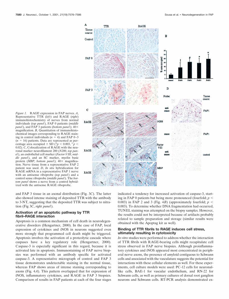
RESULTSIncreased expression of RAGE in peripheral nervefrom early FAP patientsWe reported previously that FAP patients had increased expres-sion of RAGE in affected peripheral nerves (Sousa et al., 2000).These results led us to perform a more detailed analysis ofRAGE expression in FAP by analyzing sural nerve biopsies from16 patients spanning different stages of FAP (Table 1). Patientswere scored using a morphometric scale based on nerve fiberdensity (0–3, the latter representing the greatest loss of nervefibers; see Materials and Methods). FAP 0 patients possess theV30M mutant form of TTR, normal nerve fiber density, anddeposition of TTR in nerve tissue, but amyloid is not evident (i.e.,immunoreactive deposits are negative by Congo red staining). Itshould be noted that at all other stages of FAP (1–3), immuno-reactive TTR and Congo red-positive deposits were highly cor-related. RAGE expression was enhanced in nerves of FAP 0patients (Fig. 1A). Although RAGE was present in several celltypes, our initial impression was that the staining pattern wasmost consistent with increased expression of the receptor inaxons (see below). Analysis of images of five biopsies of FAP 0patients, compared with normal age-matched controls (n � 4),demonstrated an approximately threefold increase in area occu-pied by immunoreactive RAGE in the patients (Fig. 1B) ( p �0.02). As the course of FAP evolved, there was a tendency forRAGE expression to increase. Semiquantitative analysis showeda statistically significant difference between all stages of FAP(0–3) and controls ( p � 0.003). Although there was no statisti-cally significant difference between FAP 1 and FAP 2, there wasa significant difference between FAP 0 and FAP 3 (Fig. 1B) ( p �0.01).
Neuronal expression of RAGE in FAPBecause of the likely presence of RAGE in several cell types innerve tissue from patients with FAP, a more detailed analysis tomatch cells expressing RAGE with those displaying markers forneurons, vascular endothelium, and Schwann cells was under-taken. Neurons showed increased RAGE staining based on co-localization with the neuron-specific marker neurofilament 200(Fig. 1C, top panels). Vascular endothelium stained positively forRAGE and the endothelial marker Factor VIII (Fig. 1C, middlepanels). It was apparent that RAGE was also present in vascularsmooth muscle cells. In some cases, we found that RAGE immu-noreactivity coincided with Schwann cells (Fig. 1C, bottom pan-els). Based on our survey of cells bearing RAGE antigen in FAPnerves, axons were the most abundant cellular structure display-ing positive staining. Consistent with this impression, in situhybridization using an antisense RAGE probe showed transcriptsof the receptor to be predominately localized to axons of FAPnerves (Fig. 1D, top panel). Control hybridizations with either asense RAGE probe and FAP tissue (Fig. 1D, middle panel) or anantisense RAGE probe in nerve from a control individual (Fig.1D, bottom panel) resulted in only background staining.
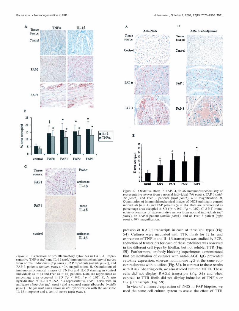
Expression of inflammatory cytokines in FAP nervePrevious studies demonstrated RAGE-dependent activation ofNF-�B by TTR fibrils in vitro and nuclear staining for NF-�Bp50in FAP tissues (Sousa et al., 2000). Because nuclear translocationof NF-�B might underlie increased expression of proinflamma-tory cytokines, we analyzed the presence of two key mediators,IL-1� and TNF-�, in affected nerves from FAP patients inrelation to deposition of TTR. Semiquantitative analysis of im-
7578 J. Neurosci., October 1, 2001, 21(19):7576–7586 Sousa et al. • Neurodegeneration in FAP
munohistochemical images for immunoreactive TNF-� andIL-1� in FAP nerve biopsies, compared with age-matched controlindividuals, demonstrated expression of these cytokines that co-incided with the pattern observed for RAGE; namely, they wereespecially localized to the endoneurial axons (Fig. 2A). Althoughnormal nerve showed virtually no detectable antigen, FAP 0individuals (i.e., before amyloid was present) already displayedincreased TNF-� and IL-1� antigens (Fig. 2A,B). Increasedlevels of these cytokines were also evident in FAP 1–3 individu-als, in which case the pattern of TNF-� and IL-1� appearedjuxtaposed to deposits of TTR. In each case, the level ofcytokine appeared to increase by approximately threefold,compared with controls, and was statistically significant (Fig.2 B). In situ hybridization was performed to determine theactual site of IL-1� mRNA synthesis. Antisense probe hybrid-ized with FAP 1 tissue demonstrated a clear signal, consistentwith axons as the principal site of IL-1� transcripts (Fig. 2C).In contrast, study of FAP 1 tissue with the sense IL-1� probeand control tissue with the antisense IL-1� probe displayedonly background staining.
Local expression of TNF-� and IL-1� in peripheral nervesuggested the possible recruitment of leukocytes, lymphocytes,and mononuclear phagocytes to FAP lesions. However, our stud-ies using antibodies reactive with markers on polymorphonuclearleukocytes (CD68), lymphocytes (CD11b, Mac1), and mononu-clear phagocytes (F4/80) did not confirm the presence of thesesubpopulations of white cells. An important consideration in theinterpretation of these data is that our samples were preserved inglutaraldehyde, paraffin-embedded, and stored for prolongedtimes. Thus, it is possible that loss of immunoreactivity could
account for some of these results. However, in view of the numberof antibodies tested and the absence of cellular infiltrates in FAPnerve tissue, at the level of general histologic analysis, it seemslikely that there is a relative paucity of white cells in these lesions.
Oxidant stress in FAPOxidant stress attributable to generation of reactive oxygen andnitrogen species is likely to have an important role in neurode-generative and neuroinflammatory disorders (Calabrese et al.,2000). High levels of NO, produced by iNOS, are known to exertmultiple toxic effects on cells (Combs et al., 2001). In view of thewell known link between cytokines and expression of iNOS, weprobed FAP nerves for iNOS antigen. Increased levels of iNOSwere observed in FAP nerves, especially in axons, in a distribu-tion overlapping that for both RAGE and proinflammatory cy-tokines in FAP 0 and FAP 3 (Fig. 3A). On the basis of oursemiquantitative immunohistologic evaluation of the differentstages of FAP, enhanced expression of iNOS was evident in FAP0 patients (approximately twofold) compared with controls ( p �0.04) (Fig. 3B). Furthermore, this increase was also seen in laterstages of the disease (FAP 1–3), where it approached �2.5-fold( p � 0.02). In contrast to these data regarding iNOS expression,no increase in immunoreactive eNOS or nNOS was observedwhen the same sections were analyzed (data not shown).
The reaction of NO with superoxide produces the peroxynitriteanion (Radi et al., 2001). Peroxynitrite is a powerful oxidant thatcan nitrate aromatic amino acid residues such as tyrosine to form3-nitrotyrosine (3-NT). Thus, 3-NT is a useful marker of per-oxynitrite production. Immunostaining of FAP nerves with anti-body to 3-NT demonstrated immunoreactive material in FAP 0
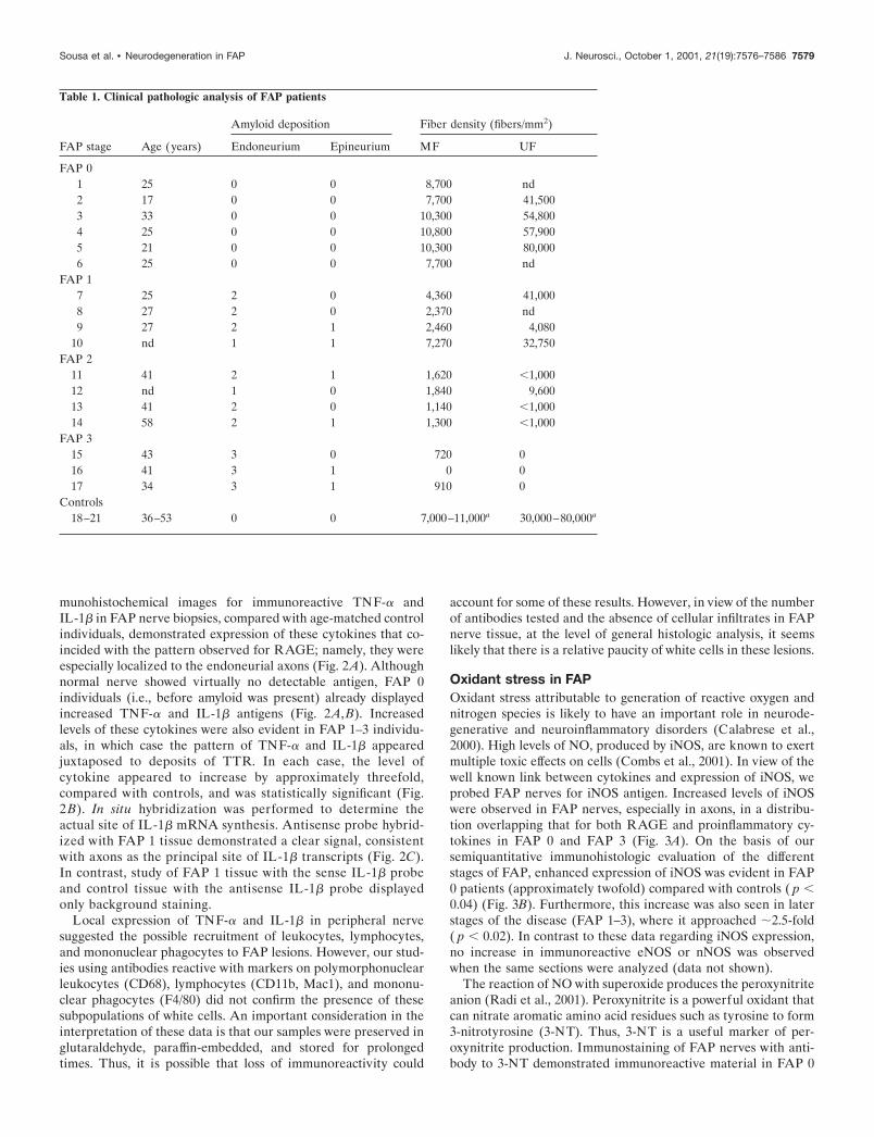
Table 1. Clinical pathologic analysis of FAP patients
FAP stage Age (years)
Amyloid deposition Fiber density (fibers/mm2)
Endoneurium Epineurium MF UF
FAP 01 25 0 0 8,700 nd2 17 0 0 7,700 41,5003 33 0 0 10,300 54,8004 25 0 0 10,800 57,9005 21 0 0 10,300 80,0006 25 0 0 7,700 nd
FAP 17 25 2 0 4,360 41,0008 27 2 0 2,370 nd9 27 2 1 2,460 4,080
10 nd 1 1 7,270 32,750FAP 2
11 41 2 1 1,620 �1,00012 nd 1 0 1,840 9,60013 41 2 0 1,140 �1,00014 58 2 1 1,300 �1,000
FAP 315 43 3 0 720 016 41 3 1 0 017 34 3 1 910 0
Controls18–21 36–53 0 0 7,000–11,000a 30,000–80,000a
Sousa et al. • Neurodegeneration in FAP J. Neurosci., October 1, 2001, 21(19):7576–7586 7579
and FAP 3 tissue in an axonal distribution (Fig. 3C). The latteralso showed intense staining of deposited TTR with the antibodyto 3-NT, suggesting that the deposited TTR was subject to nitra-tion (Fig 3C, right panel).
Activation of an apoptotic pathway by TTRfibril–RAGE interactionApoptosis is a common mechanism of cell death in neurodegen-erative disorders (Hengartner, 2000). In the case of FAP, localexpression of cytokines and iNOS in neurons suggested evenmore strongly that programmed cell death might be triggered.Apoptosis involves the activation of a proteolytic cascade wherecaspases have a key regulatory role (Hengartner, 2000).Caspase-3 is especially significant in this regard, because it isactivated late in apoptosis. Immunostaining of FAP nerve biop-sies was performed with an antibody specific for activatedcaspase-3. A representative micrograph of control and FAP 3tissue demonstrates undetectable staining in the normal tissue,whereas FAP shows areas of intense staining corresponding toaxons (Fig. 4A). This pattern overlapped that for expression ofiNOS, inflammatory cytokines, and RAGE in FAP 3 biopsies.Comparison of results in FAP patients at each of the four stages
indicated a tendency for increased activation of caspase-3, start-ing in FAP 0 patients but being more pronounced (fourfold; p �0.003) in FAP 2 and 3 (Fig. 4B) (approximately fourfold; p �0.003). To determine whether DNA fragmentation had occurred,TUNEL staining was attempted on the biopsy samples. However,the results could not be interpreted because of artifacts probablyrelated to sample preparation and storage (similar results wereobtained with the Apoptag kit as well).
Binding of TTR fibrils to RAGE induces cell stress,ultimately resulting in cytotoxicityIn vitro studies were performed to address whether the interactionof TTR fibrils with RAGE-bearing cells might recapitulate cellstress observed in FAP nerve biopsies. Although proinflamma-tory cytokines and iNOS appeared most concentrated in periph-eral nerve axons, the presence of amyloid contiguous to Schwanncells and associated with the vasculature suggests the potential forinteractions with these cellular elements as well. For these exper-iments, cell culture models were used: PC-12 cells for neuronal-like cells, BAE-1 for vascular endothelium, and RN-22 forSchwann cells, as well as primary cultures of dorsal root ganglionneurons and Schwann cells. RT-PCR analysis demonstrated ex-
Figure 1. RAGE expression in FAP nerves. A,Representative TTR (lef t) and RAGE (right)immunohistochemistry of nerves from normalindividuals (top panel ), FAP 0 patients (middlepanel ), and FAP 3 patients (bottom panel ); 40�magnification. B, Quantitation of immunohisto-chemical images corresponding to RAGE stain-ing in control individuals (n � 4) and FAP 0–3(n � 16) patients. Data are represented as per-centage area occupied � SD (*p � 0.003, ‡ p �0.02). C, Colocalization of RAGE with the neu-ronal marker neurofilament 200 (N200, top pan-el ), an endothelial cell marker (Factor VIII, mid-dle panel ), and an SC marker, myelin basicprotein (MBP, bottom panel ); 40� magnifica-tion. Nerve tissue from a representative FAP 2patient was used. D, In situ hybridization forRAGE mRNA in a representative FAP 1 nervewith an antisense riboprobe (top panel ) and acontrol sense riboprobe (middle panel ). The bot-tom panel shows a nerve from a control hybrid-ized with the antisense RAGE riboprobe.
7580 J. Neurosci., October 1, 2001, 21(19):7576–7586 Sousa et al. • Neurodegeneration in FAP
pression of RAGE transcripts in each of these cell types (Fig.5A). Cultures were incubated with TTR fibrils for 12 hr, andexpression of TNF-� and IL-1� transcripts was studied by PCR.Induction of transcripts for each of these cytokines was observedin the different cell types by fibrillar, but not soluble, TTR (Fig.5B). Furthermore, antibody blocking experiments demonstratedthat preincubation of cultures with anti-RAGE IgG preventedcytokine expression, whereas nonimmune IgG at the same con-centration was without effect (Fig. 5B). In contrast to these resultswith RAGE-bearing cells, we also studied cultured MEF1. Thesecells did not display RAGE transcripts (Fig. 5A) and whenexposed to TTR fibrils did not display induction of TNF-� orIL-1� transcripts (Fig. 5B).
In view of enhanced expression of iNOS in FAP biopsies, weused the same cell culture system to assess the effect of TTR
Figure 2. Expression of proinflammatory cytokines in FAP. A, Repre-sentative TNF-� (lef t) and IL-1� (right) immunohistochemistry of nervesfrom normal individuals (top panel ), FAP 0 patients (middle panel ), andFAP 3 patients (bottom panel ); 40� magnification. B, Quantitation ofimmunohistochemical images of TNF-� and IL-1� staining in controlindividuals (n � 4) and FAP (n � 16) patients. Data are represented aspercentage area occupied � SD (*p � 0.01, ‡ p � 0.02). C, In situhybridization of IL-1� mRNA in a representative FAP 1 nerve with anantisense riboprobe (lef t panel ) and a control sense riboprobe (middlepanel ). The far right panel shows in situ hybridization with the antisenseIL-1� riboprobe and a control nerve (right panel ).
Figure 3. Oxidative stress in FAP. A, iNOS immunohistochemistry ofrepresentative nerves from a normal individual (lef t panel ), FAP 0 (mid-dle panel ), and FAP 3 patients (right panel ); 40� magnification. B,Quantitation of immunohistochemical images of iNOS staining in controlindividuals (n � 4) and FAP patients (n � 16). Data are represented aspercentage area occupied � SD (*p � 0.01, ‡ p � 0.02). C, 3-NT immu-nohistochemistry of representative nerves from normal individuals (lef tpanel ), an FAP 0 patient (middle panel ), and an FAP 3 patient (rightpanel ); 40� magnification.
Sousa et al. • Neurodegeneration in FAP J. Neurosci., October 1, 2001, 21(19):7576–7586 7581
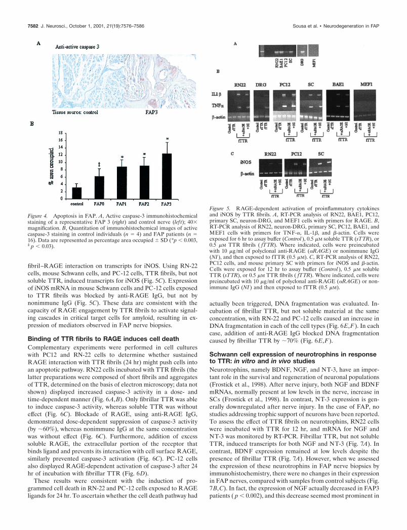
fibril–RAGE interaction on transcripts for iNOS. Using RN-22cells, mouse Schwann cells, and PC-12 cells, TTR fibrils, but notsoluble TTR, induced transcripts for iNOS (Fig. 5C). Expressionof iNOS mRNA in mouse Schwann cells and PC-12 cells exposedto TTR fibrils was blocked by anti-RAGE IgG, but not bynonimmune IgG (Fig. 5C). These data are consistent with thecapacity of RAGE engagement by TTR fibrils to activate signal-ing cascades in critical target cells for amyloid, resulting in ex-pression of mediators observed in FAP nerve biopsies.
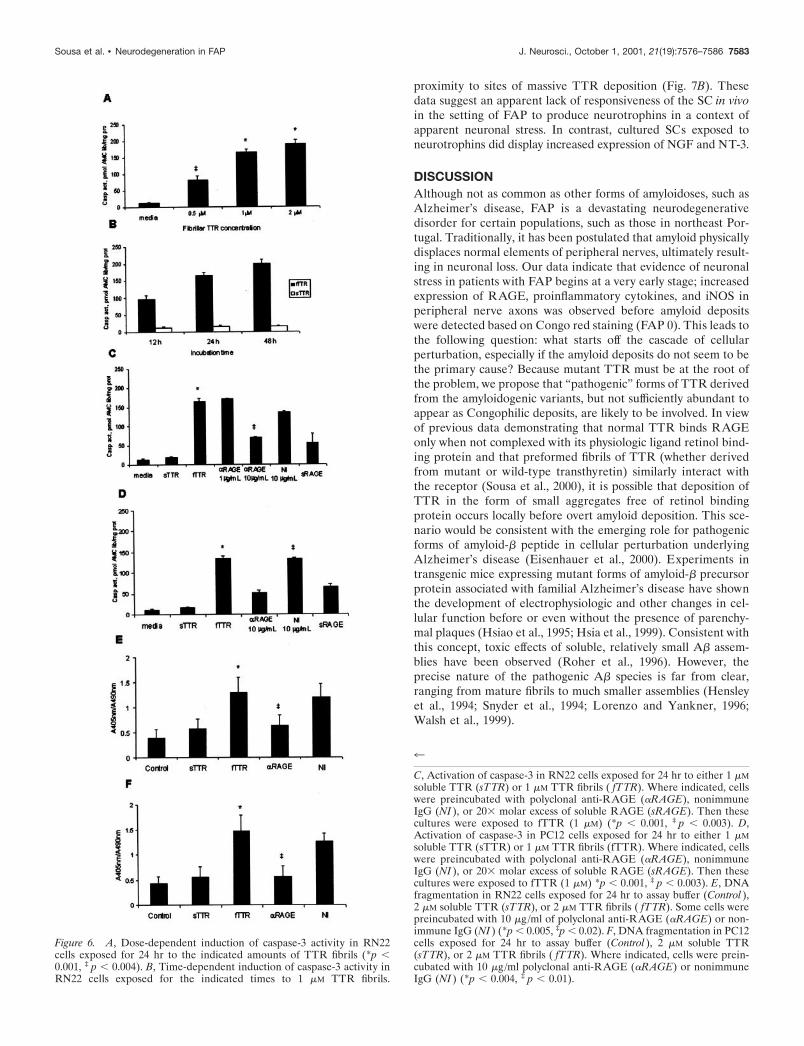
Binding of TTR fibrils to RAGE induces cell deathComplementary experiments were performed in cell cultureswith PC12 and RN-22 cells to determine whether sustainedRAGE interaction with TTR fibrils (24 hr) might push cells intoan apoptotic pathway. RN22 cells incubated with TTR fibrils (thelatter preparations were composed of short fibrils and aggregatesof TTR, determined on the basis of electron microscopy; data notshown) displayed increased caspase-3 activity in a dose- andtime-dependent manner (Fig. 6A,B). Only fibrillar TTR was ableto induce caspase-3 activity, whereas soluble TTR was withouteffect (Fig. 6C). Blockade of RAGE, using anti-RAGE IgG,demonstrated dose-dependent suppression of caspase-3 activity(by �60%), whereas nonimmune IgG at the same concentrationwas without effect (Fig. 6C). Furthermore, addition of excesssoluble RAGE, the extracellular portion of the receptor thatbinds ligand and prevents its interaction with cell surface RAGE,similarly prevented caspase-3 activation (Fig. 6C). PC-12 cellsalso displayed RAGE-dependent activation of caspase-3 after 24hr of incubation with fibrillar TTR (Fig. 6D).
These results were consistent with the induction of pro-grammed cell death in RN-22 and PC-12 cells exposed to RAGEligands for 24 hr. To ascertain whether the cell death pathway had
actually been triggered, DNA fragmentation was evaluated. In-cubation of fibrillar TTR, but not soluble material at the sameconcentration, with RN-22 and PC-12 cells caused an increase inDNA fragmentation in each of the cell types (Fig. 6E,F). In eachcase, addition of anti-RAGE IgG blocked DNA fragmentationcaused by fibrillar TTR by �70% (Fig. 6E,F).
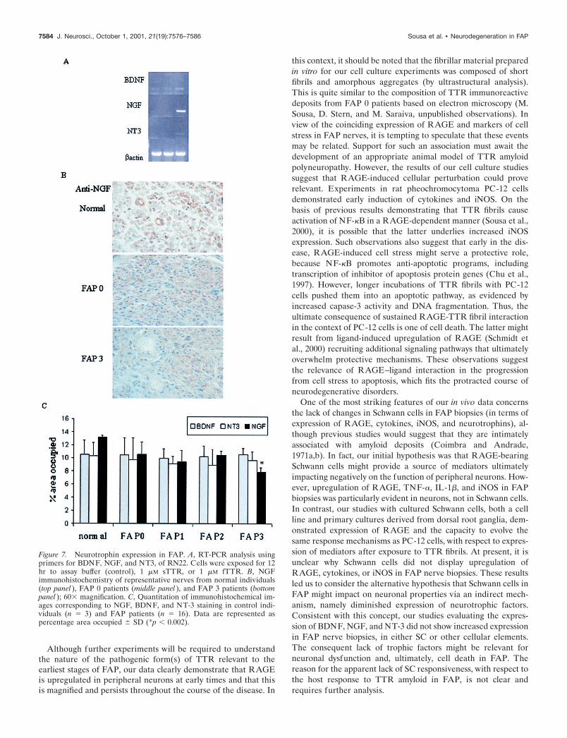
Schwann cell expression of neurotrophins in responseto TTR: in vitro and in vivo studiesNeurotrophins, namely BDNF, NGF, and NT-3, have an impor-tant role in the survival and regeneration of neuronal populations(Frostick et al., 1998). After nerve injury, both NGF and BDNFmRNAs, normally present at low levels in the nerve, increase inSCs (Frostick et al., 1998). In contrast, NT-3 expression is gen-erally downregulated after nerve injury. In the case of FAP, nostudies addressing trophic support of neurons have been reported.To assess the effect of TTR fibrils on neurotrophins, RN22 cellswere incubated with TTR for 12 hr, and mRNA for NGF andNT-3 was monitored by RT-PCR. Fibrillar TTR, but not solubleTTR, induced transcripts for both NGF and NT-3 (Fig. 7A). Incontrast, BDNF expression remained at low levels despite thepresence of fibrillar TTR (Fig. 7A). However, when we assessedthe expression of these neurotrophins in FAP nerve biopsies byimmunohistochemistry, there were no changes in their expressionin FAP nerves, compared with samples from control subjects (Fig.7B,C). In fact, the expression of NGF actually decreased in FAP3patients ( p � 0.002), and this decrease seemed most prominent in
Figure 4. Apoptosis in FAP. A, Active caspase-3 immunohistochemicalstaining of a representative FAP 3 (right) and control nerve (lef t); 40�magnification. B, Quantitation of immunohistochemical images of activecaspase-3 staining in control individuals (n � 4) and FAP patients (n �16). Data are represented as percentage area occupied � SD (*p � 0.003,‡ p � 0.03).
Figure 5. RAGE-dependent activation of proinflammatory cytokinesand iNOS by TTR fibrils. A, RT-PCR analysis of RN22, BAE1, PC12,primary SC, neuron-DRG, and MEF1 cells with primers for RAGE. B,RT-PCR analysis of RN22, neuron-DRG, primary SC, PC12, BAE1, andMEF1 cells with primers for TNF-�, IL-1�, and �-actin. Cells wereexposed for 6 hr to assay buffer (Control ), 0.5 �M soluble TTR (sTTR), or0.5 �M TTR fibrils ( fTTR). Where indicated, cells were preincubatedwith 10 �g/ml of polyclonal anti-RAGE (�RAGE) or nonimmune IgG(NI ), and then exposed to fTTR (0.5 �M). C, RT-PCR analysis of RN22,PC12 cells, and mouse primary SC with primers for iNOS and �-actin.Cells were exposed for 12 hr to assay buffer (Control ), 0.5 �M solubleTTR (sTTR), or 0.5 �M TTR fibrils ( fTTR). Where indicated, cells werepreincubated with 10 �g/ml of polyclonal anti-RAGE (�RAGE) or non-immune IgG (NI ) and then exposed to fTTR (0.5 �M).
7582 J. Neurosci., October 1, 2001, 21(19):7576–7586 Sousa et al. • Neurodegeneration in FAP
proximity to sites of massive TTR deposition (Fig. 7B). Thesedata suggest an apparent lack of responsiveness of the SC in vivoin the setting of FAP to produce neurotrophins in a context ofapparent neuronal stress. In contrast, cultured SCs exposed toneurotrophins did display increased expression of NGF and NT-3.
DISCUSSIONAlthough not as common as other forms of amyloidoses, such asAlzheimer’s disease, FAP is a devastating neurodegenerativedisorder for certain populations, such as those in northeast Por-tugal. Traditionally, it has been postulated that amyloid physicallydisplaces normal elements of peripheral nerves, ultimately result-ing in neuronal loss. Our data indicate that evidence of neuronalstress in patients with FAP begins at a very early stage; increasedexpression of RAGE, proinflammatory cytokines, and iNOS inperipheral nerve axons was observed before amyloid depositswere detected based on Congo red staining (FAP 0). This leads tothe following question: what starts off the cascade of cellularperturbation, especially if the amyloid deposits do not seem to bethe primary cause? Because mutant TTR must be at the root ofthe problem, we propose that “pathogenic” forms of TTR derivedfrom the amyloidogenic variants, but not sufficiently abundant toappear as Congophilic deposits, are likely to be involved. In viewof previous data demonstrating that normal TTR binds RAGEonly when not complexed with its physiologic ligand retinol bind-ing protein and that preformed fibrils of TTR (whether derivedfrom mutant or wild-type transthyretin) similarly interact withthe receptor (Sousa et al., 2000), it is possible that deposition ofTTR in the form of small aggregates free of retinol bindingprotein occurs locally before overt amyloid deposition. This sce-nario would be consistent with the emerging role for pathogenicforms of amyloid-� peptide in cellular perturbation underlyingAlzheimer’s disease (Eisenhauer et al., 2000). Experiments intransgenic mice expressing mutant forms of amyloid-� precursorprotein associated with familial Alzheimer’s disease have shownthe development of electrophysiologic and other changes in cel-lular function before or even without the presence of parenchy-mal plaques (Hsiao et al., 1995; Hsia et al., 1999). Consistent withthis concept, toxic effects of soluble, relatively small A� assem-blies have been observed (Roher et al., 1996). However, theprecise nature of the pathogenic A� species is far from clear,ranging from mature fibrils to much smaller assemblies (Hensleyet al., 1994; Snyder et al., 1994; Lorenzo and Yankner, 1996;Walsh et al., 1999).
Figure 6. A, Dose-dependent induction of caspase-3 activity in RN22cells exposed for 24 hr to the indicated amounts of TTR fibrils (*p �0.001, ‡ p � 0.004). B, Time-dependent induction of caspase-3 activity inRN22 cells exposed for the indicated times to 1 �M TTR fibrils.
4
C, Activation of caspase-3 in RN22 cells exposed for 24 hr to either 1 �Msoluble TTR (sTTR) or 1 �M TTR fibrils ( fTTR). Where indicated, cellswere preincubated with polyclonal anti-RAGE (�RAGE), nonimmuneIgG (NI ), or 20� molar excess of soluble RAGE (sRAGE). Then thesecultures were exposed to fTTR (1 �M) (*p � 0.001, ‡ p � 0.003). D,Activation of caspase-3 in PC12 cells exposed for 24 hr to either 1 �Msoluble TTR (sTTR) or 1 �M TTR fibrils (fTTR). Where indicated, cellswere preincubated with polyclonal anti-RAGE (�RAGE), nonimmuneIgG (NI ), or 20� molar excess of soluble RAGE (sRAGE). Then thesecultures were exposed to fTTR (1 �M) *p � 0.001, ‡ p � 0.003). E, DNAfragmentation in RN22 cells exposed for 24 hr to assay buffer (Control ),2 �M soluble TTR (sTTR), or 2 �M TTR fibrils ( fTTR). Some cells werepreincubated with 10 �g/ml of polyclonal anti-RAGE (�RAGE) or non-immune IgG (NI ) (*p � 0.005, ‡p � 0.02). F, DNA fragmentation in PC12cells exposed for 24 hr to assay buffer (Control ), 2 �M soluble TTR(sTTR), or 2 �M TTR fibrils ( fTTR). Where indicated, cells were prein-cubated with 10 �g/ml polyclonal anti-RAGE (�RAGE) or nonimmuneIgG (NI ) (*p � 0.004, ‡ p � 0.01).
Sousa et al. • Neurodegeneration in FAP J. Neurosci., October 1, 2001, 21(19):7576–7586 7583
Although further experiments will be required to understandthe nature of the pathogenic form(s) of TTR relevant to theearliest stages of FAP, our data clearly demonstrate that RAGEis upregulated in peripheral neurons at early times and that thisis magnified and persists throughout the course of the disease. In
this context, it should be noted that the fibrillar material preparedin vitro for our cell culture experiments was composed of shortfibrils and amorphous aggregates (by ultrastructural analysis).This is quite similar to the composition of TTR immunoreactivedeposits from FAP 0 patients based on electron microscopy (M.Sousa, D. Stern, and M. Saraiva, unpublished observations). Inview of the coinciding expression of RAGE and markers of cellstress in FAP nerves, it is tempting to speculate that these eventsmay be related. Support for such an association must await thedevelopment of an appropriate animal model of TTR amyloidpolyneuropathy. However, the results of our cell culture studiessuggest that RAGE-induced cellular perturbation could proverelevant. Experiments in rat pheochromocytoma PC-12 cellsdemonstrated early induction of cytokines and iNOS. On thebasis of previous results demonstrating that TTR fibrils causeactivation of NF-�B in a RAGE-dependent manner (Sousa et al.,2000), it is possible that the latter underlies increased iNOSexpression. Such observations also suggest that early in the dis-ease, RAGE-induced cell stress might serve a protective role,because NF-�B promotes anti-apoptotic programs, includingtranscription of inhibitor of apoptosis protein genes (Chu et al.,1997). However, longer incubations of TTR fibrils with PC-12cells pushed them into an apoptotic pathway, as evidenced byincreased capase-3 activity and DNA fragmentation. Thus, theultimate consequence of sustained RAGE-TTR fibril interactionin the context of PC-12 cells is one of cell death. The latter mightresult from ligand-induced upregulation of RAGE (Schmidt etal., 2000) recruiting additional signaling pathways that ultimatelyoverwhelm protective mechanisms. These observations suggestthe relevance of RAGE–ligand interaction in the progressionfrom cell stress to apoptosis, which fits the protracted course ofneurodegenerative disorders.
One of the most striking features of our in vivo data concernsthe lack of changes in Schwann cells in FAP biopsies (in terms ofexpression of RAGE, cytokines, iNOS, and neurotrophins), al-though previous studies would suggest that they are intimatelyassociated with amyloid deposits (Coimbra and Andrade,1971a,b). In fact, our initial hypothesis was that RAGE-bearingSchwann cells might provide a source of mediators ultimatelyimpacting negatively on the function of peripheral neurons. How-ever, upregulation of RAGE, TNF-�, IL-1�, and iNOS in FAPbiopsies was particularly evident in neurons, not in Schwann cells.In contrast, our studies with cultured Schwann cells, both a cellline and primary cultures derived from dorsal root ganglia, dem-onstrated expression of RAGE and the capacity to evolve thesame response mechanisms as PC-12 cells, with respect to expres-sion of mediators after exposure to TTR fibrils. At present, it isunclear why Schwann cells did not display upregulation ofRAGE, cytokines, or iNOS in FAP nerve biopsies. These resultsled us to consider the alternative hypothesis that Schwann cells inFAP might impact on neuronal properties via an indirect mech-anism, namely diminished expression of neurotrophic factors.Consistent with this concept, our studies evaluating the expres-sion of BDNF, NGF, and NT-3 did not show increased expressionin FAP nerve biopsies, in either SC or other cellular elements.The consequent lack of trophic factors might be relevant forneuronal dysfunction and, ultimately, cell death in FAP. Thereason for the apparent lack of SC responsiveness, with respect tothe host response to TTR amyloid in FAP, is not clear andrequires further analysis.
Figure 7. Neurotrophin expression in FAP. A, RT-PCR analysis usingprimers for BDNF, NGF, and NT3, of RN22. Cells were exposed for 12hr to assay buffer (control), 1 �M sTTR, or 1 �M fTTR. B, NGFimmunohistochemistry of representative nerves from normal individuals(top panel ), FAP 0 patients (middle panel ), and FAP 3 patients (bottompanel ); 60� magnification. C, Quantitation of immunohistochemical im-ages corresponding to NGF, BDNF, and NT-3 staining in control indi-viduals (n � 3) and FAP patients (n � 16). Data are represented aspercentage area occupied � SD (*p � 0.002).
7584 J. Neurosci., October 1, 2001, 21(19):7576–7586 Sousa et al. • Neurodegeneration in FAP
Another striking feature of FAP nerve biopsies was the lack ofan immune/inflammatory infiltrate despite the production ofIL-1� and TNF-� by axons. Although it might be argued that thecytokines are not effectively released into the nerve bed (be-cause staining was largely restricted to axons), it is more likelythat free polypeptides diffused away from the axon and werenot adequately visualized by immunostaining. Furthermore,we would predict that other mechanisms must be operative toprevent recruitment of immune/inflammatory effector cells.This pathologic picture is certainly different from what isobserved in the CNS. Neuritic plaques in Alzheimer’s diseaseare well known for their association with activated microgliaand astrocytes, which might contribute to protective or patho-genic mechanisms (Akiyama et al., 2000). In this context, it ispossible that if a humoral immune response selective for patho-genic TTR species could be elicited in humans (by analogywith A� immunization studies in mice) (Schenk et al., 1999;Janus et al., 2000; Morgan et al., 2000), resulting in attractionof mononuclear phagocytes to sites of amyloid deposition,these cells might accelerate amyloid clearance. Consideringthe apparent absence of mononuclear phagocytes in FAP le-sions, the results of an influx of such cells for removing amyloidcould be considerable, although the ultimate outcome for neu-ronal function might be complex (especially if products ofinflammatory cells perturbed neuronal function).
The results of our experiments provide support for a possiblelink between RAGE and neuronal dysfunction underlying FAP.Although future studies will be required to establish whether thislink reflects a cause–effect relationship, our studies have alreadyprovided insights into selective neuronal perturbation in FAPbiopsies occurring before overt amyloid deposition.
REFERENCESAkiyama H, Arai T, Kondo H, Tanno E, Haga C, Ikeda K (2000) Cell
mediators of inflammation in the Alzheimer disease brain. AlzheimerDis Assoc Disord 14[Suppl]1:S47–53.
Almeida MR, Damas AM, Lans MC, Brower A, Saraiva MJ (1997)Thyroxine binding to transthyretin Met 119. Comparative studies ofdifferent heterozygotic carriers and structural analysis. Endocrine6:309–315.
Bonifacio MJ, Sakaki Y, Saraiva MJ (1996) “In vitro” amyloid fibrilformation from transthyretin: the influence of ions and the amyloido-genicity of TTR variants. Biochim Biophys Acta 1316:35–42.
Brett J, Schmidt AM, Yan SD, Zou YS, Weidman E, Pinsky D, NowygrodR, Neeper M, Przysiecki C, Shaw A, Stern D (1993) Survey of thedistribution of a newly characterized receptor for advanced glycationend products in tissues. Am J Pathol 143:1699–1712.
Calabrese V, Bates TE, Stella AMG (2000) NO synthase and NO-dependent signal pathways in brain aging and neurodegenerative dis-orders: the role of oxidant/antioxidant balance. Neurochem Res25:1315–1341.
Chu ZL, McKinsey TA, Liu L, Gentry JJ, Malim MH, Ballard DW(1997) Suppression of tumor necrosis factor-induced cell death byinhibitor of apoptosis c-IAP2 is under NF-kappaB control. Proc NatlAcad Sci USA 94:10057–10062.
Coimbra A, Andrade C (1971a) Familial amyloid polyneuropathy: anelectron microscope study of peripheral nerve in five cases. I. Interstitialchanges. Brain 94:199–206.
Coimbra A, Andrade C (1971b) Familial amyloid polyneuropathy: anelectron microscope study of peripheral nerve in five cases. II. Nervefibril changes. Brain 94:207–212.
Combs CK, Karlo JC, Kao SC, Landreth GE (2001) �-Amyloid stimu-lation of microglia and monocytes results in TNF-�-dependent expres-sion of inducible nitric oxide synthase and neuronal apoptosis. J Neu-rosci 21:1179–1188.
Eisenhauer PB, Johnson RJ, Wells JM, Davies TA, Fine REJ (2000)Toxicity of various amyloid beta peptide species in cultured humanblood-brain barrier endothelial cells: increased toxicity of Dutch typemutant. Neurosci Res 60:804–810.
Frostick SP, Yin Q, Kemp GJ (1998) Schwann cells, neurotrophic fac-tors, and peripheral nerve regeneration. Microsurgery 18:397–405.
Fujimura H, Lacroix C, Said G (1991) Vulnerability of nerve fibres to
ischaemia. A quantitative light and electron microscope study. Brain114:1929–1942.
Hengartner MO (2000) The biochemistry of apoptosis. Nature407:685–687.
Hensley K, Carney JM, Mattson MP, Aksenova M, Harris M, Wu JF,Floyd RA, Butterfield DA (1994) A model for beta-amyloid aggrega-tion and neurotoxicity based on free radical generation by the peptide:relevance to Alzheimer disease. Proc Natl Acad Sci USA91:3270–3274.
Hofmann MA, Drury S, Fu C, Qu W, Taguchi A, Lu Y, Avila C,Kambham N, Bierhaus A, Nawroth P, Neurath MF, Slattery T, BeachD, McClary J, Nagashima M, Morser J, Stern D, Schmidt AM (1999)RAGE mediates a novel proinflammatory axis: a central cell surfacereceptor for S100/calgranulin polypeptides. Cell 97:889–901.
Hofmann MA, Lalla E, Lu Y, Gleason MR, Wolf BM, Tanji N, Ferran JrLJ, Kohl B, Rao V, Kisiel W, Stern DM, Schmidt AM (2001) Hyper-homocysteinemia enhances vascular inflammation and accelerates ath-erosclerosis in a murine model. J Clin Invest 107:675–683.
Hori O, Brett J, Slattery T, Cao R, Zhang J, Chen JX, Nagashima M,Lundh ER, Vijay S, Nitecki D, Stern D, Schmidt AM (1995) Thereceptor for advanced glycation end products (RAGE) is a cellularbinding site for amphoterin. Mediation of neurite outgrowth and co-expression of rage and amphoterin in the developing nervous system.J Biol Chem 270:25752–25761.
Hsia AY, Masliah E, McConlogue L, Yu GQ, Tatsuno G, Hu K, Kholo-denko D, Malenka RC, Nicoll RA, Mucke L (1999) Plaque-independent disruption of neural circuits in Alzheimer’s disease mousemodels. Proc Natl Acad Sci USA 96:3228–3233.
Hsiao KK, Borchelt DR, Olson K, Johannsdottir R, Kitt C, Yunis W, XuS, Eckman C, Younkin S, Price D (1995) Age-related CNS disorderand early death in transgenic FVB/N mice overexpressing Alzheimeramyloid precursor proteins. Neuron 15:1203–1218.
Janus C, Pearson J, McLaurin J, Mathews PM, Jiang Y, Schmidt SD,Chishti MA, Horne P, Heslin D, French J, Mount HT, Nixon RA,Mercken M, Bergeron C, Fraser PE, St. George-Hyslop P, Westaway D(2000) A beta peptide immunization reduces behavioural impairmentand plaques in a model of Alzheimer’s disease. Nature 408:979–982.
Lorenzo A, Yankner BA (1996) Amyloid fibril toxicity in Alzheimer’sdisease and diabetes. Ann NY Acad Sci 777:89–95.
Morgan D, Diamond DM, Gottschall PE, Ugen KE, Dickey C, Hardy J,Duff K, Jantzen P, DiCarlo G, Wilcock D, Connor K, Hatcher J, HopeC, Gordon M, Arendash GW (2000) A beta peptide vaccination pre-vents memory loss in an animal model of Alzheimer’s disease. Nature408:915–916.
Mrak RE, Sheng JG, Griffin WS (1995) Glial cytokines in Alzheimer’sdisease: review and pathogenic implications. Hum Pathol 26:816–823.
Radi R, Peluffo G, Alvarez MN, Naviliat M, Cayota A (2001) Unravel-ing peroxynitrite formation in biological systems. Free Radic Biol Med30:463–488.
Roher AE, Chaney MO, Kuo YM, Webster SD, Stine WB, HaverkampLJ, Woods AS, Cotter RJ, Tuohy JM, Krafft GA, Bonnell BS, Em-merling MR (1996) Morphology and toxicity of Abeta-(1–42) dimerderived from neuritic and vascular amyloid deposits of Alzheimer’sdisease. J Biol Chem 271:20631–20635.
Said G, Ropert A, Faux N (1984) Length-dependent degeneration offibers in Portuguese amyloid polyneuropathy: a clinicopathologic study.Neurology 34:1025–1032.
Saraiva MJ (2001) Transthyretin mutations in hyperthyroxinemia andamyloid diseases. Hum Mutat 17:493–503.
Saraiva MJ, Birken S, Costa PP, Goodman DS (1983) Amyloid fibrilprotein in familial amyloidotic polyneuropathy, Portuguese type. Def-inition of molecular abnormality in transthyretin (prealbumin). J ClinInvest 74:104–119.
Saraiva MJ, Costa PP, Goodman DS (1985) Biochemical marker infamilial amyloidotic polyneuropathy, Portuguese type. Family studieson the transthyretin (prealbumin)-methionine-30 variant. J Clin Invest76:2171–2177.
Schenk D, Barbour R, Dunn W, Gordon G, Grajeda H, Guido T, Hu K,Huang J, Wood K, Khan K, Kholodenko D, Lee M, Liao Z, Lieber-burg I, Motter R, Mutter L, Soriano F, Shopp G, Vasquez N, VandevertC, Walker S, Wogulis M, Yednock T, Games D, Seubert P (1999)Immunization with amyloid-beta attenuates Alzheimer-disease-like pa-thology in the PDAPP mouse. Nature 400:116–117.
Schmidt AM, Yan SD, Wautier JL, Stern D (1999) Activation of recep-tor for advanced glycation end products: a mechanism for chronicvascular dysfunction in diabetic vasculopathy and atherosclerosis. CircRes 84:489–497.
Schmidt AM, Yan SD, Yan SF, Stern DM (2000) The biology of thereceptor for advanced glycation end products and its ligands. BiochimBiophys Acta 1498:99–111.
Smith MA, Richey Harris PL, Sayre LM, Beckman JS, Perry G (1997)Widespread peroxynitrite-mediated damage in Alzheimer’s disease.J Neurosci 17:2653–2657.
Snyder SW, Ladror US, Wade WS, Wang GT, Barrett LW, MatayoshiED, Huffaker HJ, Krafft GA, Holzman TF (1994) Amyloid-beta ag-
Sousa et al. • Neurodegeneration in FAP J. Neurosci., October 1, 2001, 21(19):7576–7586 7585

gregation: selective inhibition of aggregation in mixtures of amyloidwith different chain lengths. Biophys J 67:1216–1228.
Sousa MM, Yan SD, Stern D, Saraiva MJ (2000) Interaction of thereceptor for advanced glycation end products (RAGE) with transthy-retin triggers nuclear transcription factor kB (NF-�B) activation. LabInvest 80:1101–1110.
Veerhuis R, Janssen I, De Groot CJ, Van Muiswinkel FL, Hack CE,Eikelenboom P (1999) Cytokines associated with amyloid plaques inAlzheimer’s disease brain stimulate human glial and neuronal cellcultures to secrete early complement proteins, but not C1-inhibitor.Exp Neurol 160:289–299.
Walsh DM, Hartley DM, Kusumoto Y, Fezoui Y, Condron MM, Lo-makin A, Benedek GB, Selkoe DJ, Teplow DB (1999) Amyloid beta-
protein fibrillogenesis. Structure and biological activity of protofibrillarintermediates. J Biol Chem 274:25945–25952.
Yan SD, Chen X, Fu J, Chen M, Zhu H, Roher A, Slattery T, Zhao L,Nagashima M, Morser J, Migheli A, Nawroth P, Stern D, Schmidt AM(1996) RAGE and amyloid-beta peptide neurotoxicity in Alzheimer’sdisease. Nature 382:685–691.
Yan SD, Roher A, Schmidt AM, Stern DM (1999) Cellular cofactors foramyloid beta-peptide-induced cell stress. Moving from cell culture to invivo. Am J Pathol 155:1403–1411.
Yan SD, Zhu H, Zhu A, Golabek A, Du H, Roher A, Yu J, Soto C,Schmidt AM, Stern D, Kindy M (2000) Receptor-dependent cellstress and amyloid accumulation in systemic amyloidosis. Nat Med6:643–651.
7586 J. Neurosci., October 1, 2001, 21(19):7576–7586 Sousa et al. • Neurodegeneration in FAP