fangfang yang, hua wang, bianbian yan, tong li, lulu min
TRANSCRIPT

Fangfang Yang, Hua Wang, Bianbian Yan, Tong Li, Lulu Min, Erfei Chen* and Jin Yang*
Decreased level of miR-1301 promotes colorectalcancer progression via activation of STAT3pathwayhttps://doi.org/10.1515/hsz-2020-0301Received September 4, 2020; accepted November 2, 2020;published online May 13, 2021
Abstract: The molecular pathogenesis of colorectal can-cer (CRC) has been widely investigated in recent years.Accumulating evidence has indicated that microRNA(miRNA) dysregulation participates in the processes ofdriving CRC initiation and progression. Aberrant expres-sion of miR-1301 has been found in various tumor types.However, its role in CRC remains to be elucidated. In thepresent study, we identified miR-1301 was enrichedin normal colorectal tissues and significantly down-regulated in CRC. Decreased level of miR-1301 stronglycorrelated with aggressive pathological characteristics,including advanced stage and metastasis. Bioinformaticsand dual luciferase assay demonstrated that STAT3 is adirect target of miR-1301. Gain and loss-of-function assaysshowed that miR-1301 had no effect on cell proliferation.Overexpression of miR-1301 suppressed cell migrationand invasion capacity of pSTAT3-positive LoVo cells, butnot pSTAT3-negative SW480 cells, while inhibition ofmiR-1301 consistently promoted cell migration and inva-sion in both cell lines. Additionally, miR-1301 inhibitionrestored the suppressed migration and invasion ofSTAT3-knockdown LoVo cells. MiR-1301 functioned as atumor suppressor to modulate the IL6/STAT3 signalingpathway. In summary, this study highlights the signifi-cant role of miR-1301/STAT3 axis in CRC metastasis.
Keywords: cell migration and invasion; colorectal cancer;metastasis; miRNA regulation; STAT3.
Introduction
Colorectal cancer (CRC) is the leading malignant tumors indeveloped countries with high incidence and mortality.Alteration ofdriver genes and epigenetic changes contributeto CRC initiation and progression (Chen et al. 2018c). As anessential way of epigenetic regulation, microRNAs are smallnoncoding RNAs that play essential roles in gene expressionregulation by binding to 3′-untranslated region (3′-UTR) oftargets (Wang et al. 2019). Aberrant miRNA expression hasbeen extensively reported in CRC using miRNA profiles orindependent validation group (Arndt et al. 2009; Chen et al.2014; Keller et al. 2014; Loo et al. 2015; Lu et al. 2018). The in-depth mechanism studies of dysregulated miRNA networkare issues to be solved. The number of direct target of eachmiRNA ranges from hundreds to thousands. Depending onmRNA targets they regulated,miRNA acts as oncogene or astumor suppressor gene.
MiR-1301 has been reported mostly as a tumor sup-pressor inmultiple cancer types (Fanget al. 2012;Wang et al.2017; Wen et al. 2019; Yang et al. 2017; Zhi et al. 2017), butinversely, in prostate cancer, it functioned as an oncogene(Bi et al. 2016; Song et al. 2018). As far as we are concerned,the importance of miR-1301 in CRC is still uncovered. In thisstudy, we utilized public data of 14 tumor types and foundthe expression of miR-1301 is highest in normal colorectaltissues and significantly down-regulated in CRC. Thedecreasing trend is only detected in CRC and pancreaticcancer, suggesting miR-1301 is important in digestive tract.Using bioinformatics analysis, we found miR-1301/STAT3axis may dominate in the network regulated by miR-1301.
STAT3 (Signal transducer and activator of tran-scription 3), a transcriptional mediator of oncogenicsignaling, is constitutively active in ∼70% of humancancers (Wei et al. 2019). Distant metastasis causes mostof the cancer-related morbidity and mortality afterinitial treatment (Chang et al. 2017). STAT3 activation inCRC is reported associated with metastasis (Ye et al.2017). Furthermore, increasing evidences demonstrated
*Corresponding authors: Erfei Chen and Jin Yang, Institute ofPreventive Genomic Medicine, School of Life Sciences, NorthwestUniversity, Xi’an 710069, China; and Key Laboratory of ResourceBiology and Biotechnology in Western China, Ministry of Education,School of Life Sciences, Northwest University, Xi’an 710069, China,E-mail: [email protected] (E. Chen), [email protected](J. Yang). https://orcid.org/0000-0001-5394-2366 ( J. Yang)Fangfang Yang, Hua Wang, Bianbian Yan, Tong Li and Lulu Min,Institute of Preventive Genomic Medicine, School of Life Sciences,Northwest University, Xi’an 710069, China; Key Laboratory ofResource Biology and Biotechnology in Western China, Ministry ofEducation, School of Life Sciences, Northwest University, Xi’an710069, China
Biol. Chem. 2021; 402(7): 805–813
Open Access. © 2020 Fangfang Yang et al., published by De Gruyter. This work is licensed under the Creative Commons Attribution 4.0International License.
that knocking down STAT3 expression could suppressthe growth of CRC cells in vitro and in vivo. Here,to further confirm the suppressor role of miR-1301is mainly via STAT3 pathway, we selected two miR-1301-expressing cell lines, LoVo (pSTAT3-positive) andSW480 (pSTAT3-negative) for function study. Our re-sults may provide a novel therapeutic target foradvanced CRC patients.
Results
MiR-1301 is significantly down-regulated intumor samples and correlated with CRCmetastasis
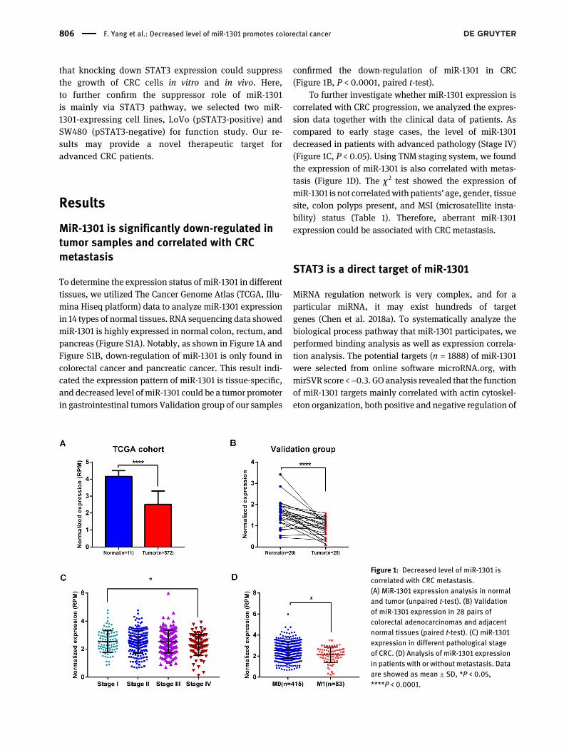
To determine the expression status of miR-1301 in differenttissues, we utilized The Cancer Genome Atlas (TCGA, Illu-mina Hiseq platform) data to analyze miR-1301 expressionin 14 types of normal tissues. RNA sequencing data showedmiR-1301 is highly expressed in normal colon, rectum, andpancreas (Figure S1A). Notably, as shown in Figure 1A andFigure S1B, down-regulation of miR-1301 is only found incolorectal cancer and pancreatic cancer. This result indi-cated the expression pattern of miR-1301 is tissue-specific,and decreased level of miR-1301 could be a tumor promoterin gastrointestinal tumors Validation group of our samples
confirmed the down-regulation of miR-1301 in CRC(Figure 1B, P < 0.0001, paired t-test).
To further investigate whether miR-1301 expression iscorrelated with CRC progression, we analyzed the expres-sion data together with the clinical data of patients. Ascompared to early stage cases, the level of miR-1301decreased in patients with advanced pathology (Stage IV)(Figure 1C, P < 0.05). Using TNM staging system, we foundthe expression of miR-1301 is also correlated with metas-tasis (Figure 1D). The χ 2 test showed the expression ofmiR-1301 is not correlatedwith patients’ age, gender, tissuesite, colon polyps present, and MSI (microsatellite insta-bility) status (Table 1). Therefore, aberrant miR-1301expression could be associated with CRC metastasis.
STAT3 is a direct target of miR-1301
MiRNA regulation network is very complex, and for aparticular miRNA, it may exist hundreds of targetgenes (Chen et al. 2018a). To systematically analyze thebiological process pathway that miR-1301 participates, weperformed binding analysis as well as expression correla-tion analysis. The potential targets (n = 1888) of miR-1301were selected from online software microRNA.org, withmirSVR score <−0.3. GO analysis revealed that the functionof miR-1301 targets mainly correlated with actin cytoskel-eton organization, both positive and negative regulation of
Figure 1: Decreased level of miR-1301 iscorrelated with CRC metastasis.(A) MiR-1301 expression analysis in normaland tumor (unpaired t-test). (B) Validationof miR-1301 expression in 28 pairs ofcolorectal adenocarcinomas and adjacentnormal tissues (paired t-test). (C) miR-1301expression in different pathological stageof CRC. (D) Analysis of miR-1301 expressionin patients with or without metastasis. Dataare showed as mean ± SD, *P < 0.05,****P < 0.0001.
806 F. Yang et al.: Decreased level of miR-1301 promotes colorectal cancer

cell proliferation, negative regulation of transcription orgene expression (Figure 2A). We further discovered thegenes with annotation in KEGG are mainly involved incancer related pathways (MAPK, Wnt, and Jak-STAT)(Figure 2B). Particularly, STAT3, a core effector of Jak-STAT pathway, attracted our attention. Though TCGA datashowed no correlation between miR-1301 and STAT3 RNAexpression, we found in metastasis cases (M1), miR-1301 isnegatively correlated with phosphor-Stat3 expression(P = 0.015, r = −0.323, Figure 2C). Two tightly binding site ofSTAT3 for miR-1301 were showed in Figure 2D and 2E. Toconfirm this physical interaction, a dual luciferase assaywas performed and the result showed that overexpression
of miR-1301 could significantly repress the luciferase ac-
tivity (wild-type 3′-UTRof STAT3). STAT3 is clearly reported
correlated with CRC metastasis, but the study of miR-1301/
STAT3 in CRC has not been reported. The similar expres-
sion pattern and binding assay confirmed STAT3 is one of
direct targets of miR-1301.
Table : Correlation of miR- expression to pathological fea-tures of TCGA CRC samples.
Variables Cases miR-expression
χ P Value
High Low
Age(years)> . .≤
GenderMale . .Female
Colon polyps presentYes . .No
Tissue siteColon . .Rectum
MSI statusMSS . .MSI
Figure 2: STAT3 is a direct target of miR-1301.(A) GOanalysis of 1888 target genes ofmiR-1301. (B) Pathway analysis ofmiR-1301 targets usingDAVID v6.7 software. (C) Pearson’s correlationanalysis of miR-1301 and pSTAT3 (Y705) expression in metastasis samples from TCGA CRC cohort. (D–E) Schematic diagrams of wild-type andmutant type 3′-UTR of STAT3. Dual luciferase reporter assaywas used to detect the reporter activity in SW480 and LoVo cells. Data were shownas mean ± SEM, **P < 0.01, ***P < 0.001, ****P < 0.0001.
F. Yang et al.: Decreased level of miR-1301 promotes colorectal cancer 807

Inhibition of miR-1301 promotes CRC cellmigration and invasion
MiR-1301 is expressed in both SW480 and LoVo cells, andwe performed overexpression and inhibition ofmiR-1301 inthese two cell lines to study its effect on cell phenotype.Transfection efficiency of miR-1301 mimics and inhibitorwere determined using QPCR in both cell lines (Figure S2).We firstly investigated cell proliferation using CCK8 assayand observed miR-1301 expression in SW480 cells had noeffect on cell proliferation (Figure S3). In LoVo cells,overexpression of miR-1301 could slightly decreasedcell proliferation ability. However, test of three indepen-dent experiments showed no statistic difference. These
demonstrated the effect of miR-1301on CRC proliferation isweak and limit.
To investigate whether miR-1301 affects cell migra-tion capacity, we applied wound scratch assay. InSW480 cells, miR-1301 inhibition enhanced the migrationcapacity as compared to negative controls, while over-expression of miR-1301 showed no significant effect(Figure 3A). In LoVo cells, gain of miR-1301 inhibitedmigration, and conversely, loss of miR-1301 promotes themigration capacity (Figure 3B). Furthermore, using aMatrigel invasion assay, we explored the effect ofmiR-1301 on the invasion capacity of CRC cells. Similarly,the results were consistent withmigration assay in SW480and LoVo cells (Figure 3C).
Figure 3: Inhibition of miR-1301 promotes CRC cell migration and invasion.(A–B) Wound scratch was performed to detect cell migration ability (A, SW480; B, LoVo). The images were under a microscope at amagnification of 100× and photographed at 0 and 48 h. (C) Cell invasion analysis in SW480 and LoVo cells, detected by Transwell chamberswith Matrigel. The images were under a microscope at a magnification of 100×.
808 F. Yang et al.: Decreased level of miR-1301 promotes colorectal cancer

MiR-1301 regulates CRC cell migration andinvasion through STAT3/MMPs axis
MiR-1301 directly targets STAT3, and we hypothesized themigration and invasion effect of miR-1301 was associatedwith STAT3 and its downstream targets. We then per-formed blots of STAT3, pSTAT3, and STAT3 targets MMP2and MMP9 to confirm our hypothesis (Figure 4A). BothTotal STAT3 andpSTAT3 level changed after transfection ofmiR-1301 in LoVo cells (pSTAT3-positive cells). STAT3 hasbeen confirmed a target for therapy in CRC. Block of STAT3
pathways could depress cell invasion abilities. To furtherclarify thatmiR-1301 affects CRC cell invasion by regulationSTAT3, LoVo cells were transfected with small-interferingRNA (siRNA) specific for STAT3 to inhibit its expression.
Knockdown of STAT3 could inhibit the expression of MMPs
(Figure 4B) as well as cell invasion phenotype (Figure 4C).
After addition of miR-1301 inhibitor in STAT3-knockdown
cells, this effect was attenuated.To further confirm the role of miR-1301/STAT3 axis in
CRC metastasis, we tested whether miR-1301 over-expression could inhibit IL-6-mediated phosphorylation of
Figure 4: MiR-1301 regulates CRC cell migration and invasion through STAT3/MMPs axis.(A) Western bolts of STAT3, pSTAT3, and MMPs afer transfection of miR-1301 mimics or inhibitors in LoVo cells. Lane 1: NC. Lane 2: miR-1301mimics. Lane 3: inhibitor NC. Lane 4: miR-1301 inhibitor. (B) Western blots of STAT3, pSTAT3, and MMPs in LoVo cells after cotransfection ofmiR-1301 inhibitor/inhibitor NC and STAT3 siRNA/siRNANC. (C) Cell invasion analysis in LoVo cells. (D–E)Western blots of STAT3, pSTAT3, andMMPs (D: SW480; E: LoVo). (F) Schematic model of miR-1301/STAT3 axis in CRC metastasis.
F. Yang et al.: Decreased level of miR-1301 promotes colorectal cancer 809

STAT3 and suppress MMPs expression. As a direct target ofmiR-1301, total STAT3 and pSTAT3 were down-regulatedafter overexpression of miR-1301 in pSTAT3-activated cells(Figure 4D and 4E). Phospho-STAT3 is absent in basalSW480 (Figure 4D), in which the suppression role ofmiR-1301 had no effect. Cell invasion phenotype as well asMMPs blot in SW480 cells confirmed our hypothesis thatmiR-1301 exerted its suppressor role through STAT3/MMPsaxis (Figure 4F).
Discussion
Substantial evidence has shown that aberrant miRNAexpression is common and involved in CRC initiation andprogression (De Robertis et al. 2018; Grassi et al. 2018; Liuet al. 2018). The miRNA-based targeted therapy hasbecome a promising approach for anti-tumor therapy(Sun et al. 2018). Therefore, identifying novelmarkers andsystematic investigation of the mechanisms underlyingCRC initiation and development may help to identifyeffective therapeutic targets for CRC therapy. In the pre-sent study, of the 14 organs tested, we identified theexpression of miR-1301 is enriched in colorectum andpancreas and significantly down-regulated in tumorpatients (P < 0.05 for pancreatic cancer and P < 0.0001 forCRC). Decreased level of miR-1301 is correlated with CRCprogression, including advanced stage and metastasis.Our function assays indicated that inhibition of miR-1301promoted cell migration and invasion in both LoVo andSW480 cells. Further in-depth mechanism uncoveredmiR-1301 regulating network in CRC metastasis. Notably,STAT3 was validated as an essential target of miR-1301.We confirmed miR-1301-mediated inhibition is mainly viaSTAT3 pathway, indicating that miR-1301/STAT3may be apromising therapeutic target for CRC.
Previous studies have reported that miR-1301 isdifferentially expressed and mostly presented as a tumorsuppressor in several types of human cancers. The bio-logical functions of miR-1301 depend on different target itbinds to. Yang et al. reported that miR-1301 directly targetsBCL9 and inhibits Wnt/β-catenin signaling thus to hepa-tocellular carcinoma (HCC) invasion and angiogenesis(Yang et al. 2017). Controversially, another study reportedmiR-1301 was highly expressed in HCC cell lines as well astumor samples and specifically targeted the tumor sup-pressive KLF6-FL. Functionally, enhancement of miR-1301promoted cell migration and angiogenesis (Liang et al.2014). In glioma cells, miR-1301 inhibited glioma cell pro-liferation and blocked the cell cycle to G1 by negativelyregulating N-Ras and its downstream targets, MEK-ERK1/2
(Zhi et al. 2017). However, in our proliferation test,miR-1301 showed limit effect on CRC cells. A better expla-nation could be found in our GO and pathway analysis.Though miR-1301 targets BCLs or N-Ras which couldpositively regulate cell proliferation, several targets unre-ported like CDKN1 and BAX exerting inhibitory roles in cellproliferation may be an offset. Recently, other researchteams have also discovered the novel miR-1301/STAT3 axisin cancer progression. LncRNAABHD11-AS1 could promotetumor progression in papillary thyroid carcinoma and actas a competitive endogenous RNA to enhance STAT3expression by sponging miR-1301 (Wen et al. 2019). Simi-larly in hepatocellular carcinoma, LINC01433 promotescancermetastasis and progression throughmodulating themiR-1301/STAT3 axis (Huang et al. 2019).
STAT3, a member of the STAT protein family, is typi-cally triggered by the binding of cytokines and growthfactors to their related receptors. Numerous studies haveindicated that activated IL-6/STAT3 signaling is one of thecrucial pathways involving in CRC. Hyperactivation ofSTAT3 pathway promotes the expression of genes thatinvolved in cell cycle arrest (e.g., Cyclin D1), survival (e.g.,Bcl2, survivin), and metastasis (e.g., vimentin, MMPs) thatcontribute to CRC initiation and progression. TargetingSTAT3 by RNAi or miRNA is an appealing anti-cancerstrategy. Full understanding of miRNA-STAT3 regulationwould be helpful for CRC therapy. MiR-124 and miR-874have been reported directly targeted STAT3 and inhibitedCRC proliferation (Zhang et al. 2013; Zhao and Dong 2016).In the present study, we found miR-1301 directly binds toSTAT3 and is negatively correlated with pSTAT3 (Y705)expression in metastasis patients. To confirm the tumorsuppressor role of miR-1301 is mainly via STAT3 pathway,we selected two CRC cell lines, LoVo (pSTAT3-positive,high metastatic capacity) and SW480 (pSTAT3-negative,low metastatic capacity). In LoVo cells, transfection ofmiR-1301 mimics inhibited total STAT3 and pSTAT3, andsuppressed cell migration and invasion capacity. Due tothe absence of pSTAT3 in SW480 cells, miR-1301 over-expression did not change pSTAT3 expression andinvasion capacity. These result indicated the inhibitoryeffect of miR-1301 on CRC migration and invasion isSTAT3-dependent.
Conclusions
In conclusion, our research revealed that miR-1301 isenriched in normal colorectal tissues and significantlydown-regulated in CRC. Decreased level of miR-1301 canpotently promote CRC cell migration and metastasis via
810 F. Yang et al.: Decreased level of miR-1301 promotes colorectal cancer

activation of STAT3 pathways. These results suggest thatmiR-1301 is a promising therapeutic target for advancedCRC patients.
Materials and methods
TCGA data access
Clinical data and gene expression data of 14 tumor types weredownloaded from UCSC Xena.
Tissue samples
CRC tissues and adjacent normal tissues from 28 patients were kindlyendowed from the Fourth Military Medical University.
RNA extraction and quantitative real time PCR
Total RNA was extracted using TRIzol reagent (Invitrogen, USA). ThemiRNA was then reverse transcribed to cDNA according to themanufacture of microRNA First Strand cDNA Sythesis (Poly A Tailing)Kit (Sangon, China). Quantitative real-time PCR amplifications wereperformed with the SYBR Premix Ex TaqTM II kit (TAKARA, Japan) andspecific primers for miR-1301 or U6 (Sangon, China, Table 2). The realtime PCR was detected by CFX96TM real-time PCR system. Relativeexpression was normalized according to formulas 2−ΔΔCt, and U6 wasset as an internal control.
GO and pathway analysis
Online software DAVID v6.7 was used for Gene Ontology (GO) andpathway analysis of miR-1301 targets.
Cell lines
Colorectal adenocarcinoma cells SW480 and LoVo were purchasedfrom ATCC (American Type Culture Collection) and cultured in RPMI1640 medium supplemented with 10% fetal bovine serum (FBS, GibcoUSA). Cells were maintained at 37 °C in atmosphere of 5% CO2.
Transfection
MiR-1301 mimics (Forward: UUGCAGCUGCCUGGGAGUGACUUC;Reverse: AGUCACUCCCAGGCAGCUGCAAUU), miR-1301 inhibitor(Sequence: GAAGUCACUCCCAGGCAGCUGCAA), and STAT3 siRNAwere synthesized and purified (GenePharma, China). Target sequence
of STAT3 siRNA was: UCCAGUUUCUUAAUUUGUUGACGGGUC.SW480 and LoVo Cells were reverse transfected with HiPerFecttransfection reagent (Qiagen, USA) according to the manuals. Formimics and siRNA, the final concentration was 10 nM, while theconcentration for inhibitor was 20 nM.
Luciferase reporter assay
The sequence of wild type or mutant type of STAT3-3′UTR (300 bp)were synthesized and constructed into the pmiR-GLO™ luciferasereporter plasmid (Promega, USA). CRC cells were cotransfected with500 ng of pmiR-STAT3-WT or pmiR-STAT3-MUT and 10 nM miR-1301mimics or negative controls in 24-well plates. After transfectionof 24 h,the luciferase activity was measured with a dual luciferase reporterassay system (Promega, USA).
Cell proliferation assay
Cell Counting Kit-8 (Dojindo Laboratories, Japan) was performed todetect cell viability according to a previously reported protocol (Wanget al. 2019). Briefly, the transfected cells were seeded into 96-well plateat a density of 2000/well with five replicates. At indicated time, 10 μlCCK-8 solution was added into each well. After incubation for 2–3 h,the viable cells were measured at a wavelength of 450 nm.
Cell migration and invasion assay
Wound scratch assay was performed to evaluate migration capacity,and detail methodwas described as previously (Chen et al. 2018b). Forcell invasion assay, the Transwell chambers (Corning, USA) withMatrigel (BD Biosciences, USA) were applied. Transfected cells weretreated with Mitomycin C (10 μg/ml) before plating to inhibit cellproliferation. 2 × 10⁴ cells in a total volume of 100 μl serum-free me-diumwere then plated in the upper chamber. The cells were incubatedat 37 °C for 24 h (LoVo) or 48 h (SW480). The invaded cells on the lowerchamber were fixed with 4% paraformaldehyde, stained with 0.1%crystal violet, and washed with PBS. The invasion capacities wereanalyzed by counting the number of invaded cells in five randomlypicked fields as observed under a microscope at a magnification of100×.
Western blotting
Protein extraction and quantification were carried out according to apreviously described protocal (Chen et al. 2018c). The antibodies usedin this study were as follows: mouse anti-Stat3 (#9139, 1:1000; CellSignaling Technology), rabbit anti-Phospho-Stat3 (Tyr705) (#9145,1:1000; Cell SignalingTechnology), rabbit anti-MMP9 (YT5357, 1:1000;ImmunoWay), rabbit anti-MMP2 (10373-2-AP, 1:2000; Proteintech),and mouse anti-GAPDH (YM3029, 1:5000; ImmunoWay).
Statistical analysis
GraphPad Prism (GraphPad Software, Inc.) was used for statisticalanalysis in this study. All experiments were repeated at least three
Table : Primer sequences for qRT-PCR.
Gene Primer sequence (′-′)
miR- F: CGAGCTGCCTGGGAGTGACU F: CTCGCTTCGGCAGCACA
F. Yang et al.: Decreased level of miR-1301 promotes colorectal cancer 811

times and the resultswere presented as themean±SD.AP value < 0.05was considered statistically significant.
Author contribution: All the authors have acceptedresponsibility for the entire content of this submittedmanuscript and approved submission.Research funding: The study was supported by a grantfrom Key Science and Technology Program of ShaanxiProvince (No. 2019ZDLSF02-05 and No. 2018ZDXM-SF-064).Research involving human participants: The work hasbeen performed in accordance with Declaration ofHelsinki, and all patients involved in this study hadgiven written informed consent.Conflict of interest statement: All authors declare thatthere are no conflicts of interest.
References
Arndt, G.M., Dossey, L., Cullen, L.M., Lai, A., Druker, R., Eisbacher, M.,Zhang, C., Tran, N., Fan, H., Retzlaff, K., Bittner, A., and Raponi,M. (2009). Characterization of global microRNA expressionreveals oncogenic potential of miR-145 in metastatic colorectalcancer. BMC Canc. 9: 374.
Bi, D., Ning, H., Liu, S., Que, X., and Ding, K. (2016). miR-1301promotes prostate cancer proliferation throughdirectly targetingPPP2R2C. Biomed. Pharmacother. 81: 25–30.
Chen, D.L., Wang, Z.Q., Zeng, Z.L., Wu, W.J., Zhang, D.S., Luo, H.Y.,Wang, F., Qiu,M.Z.,Wang,D.S., Ren, C., et al. (2014). Identificationof microRNA-214 as a negative regulator of colorectal cancer livermetastasis by way of regulation of fibroblast growth factorreceptor 1 expression. Hepatology 60: 598–609.
Chang, Y.C., Su, C.Y., Chen, M.H., Chen, W.S., Chen, C.L., and Hsiao,M. (2017). Secretory RAB GTPase 3C modulates IL6-STAT3pathway to promote colon cancer metastasis and is associatedwith poor prognosis. Mol. Canc. 16: 135.
Chen, E., Li, Q., Wang, H., Yang, F., Min, L., and Yang, J. (2018a).MiR-92a promotes tumorigenesis of colorectal cancer, atranscriptomic and functional based study. Biomed.Pharmacother. 106: 1370–1377.
Chen, E., Li, Q., Wang, H., Zhang, P., Zhao, X., Yang, F., and Yang, J.(2018b). MiR-32 promotes tumorigenesis of colorectal cancer bytargeting BMP5. Biomed. Pharmacother. 106: 1046–1051.
Chen, E., Yang, F., He, H., Li, Q., Zhang, W., Xing, J., Zhu, Z., Jiang, J.,Wang, H., Zhao, X., et al. (2018c). Alteration of tumor suppressorBMP5 in sporadic colorectal cancer: a genomic andtranscriptomic profiling based study. Mol. Canc. 17: 176.
De Robertis, M., Mazza, T., Fusilli, C., Loiacono, L., Poeta, M.L., Sanchez,M., Massi, E., Lamorte, G., Diodoro, M.G., Pescarmona, E., et al.(2018). EphB2 stem-related and EphA2 progression-relatedmiRNA-based networks in progressive stages of CRC evolution: clinicalsignificance and potential miRNA drivers. Mol. Canc. 17: 169.
Fang, L., Yang, N., Ma, J., Fu, Y., and Yang, G.S. (2012). microRNA-1301-mediated inhibition of tumorigenesis. Oncol. Rep. 27: 929–934.
Grassi, A., Perilli, L., Albertoni, L., Tessarollo, S., Mescoli, C., Urso,E.D.L., Fassan, M., Rugge, M., and Zanovello, P. (2018). Acoordinate deregulation of microRNAs expressed in mucosaadjacent to tumor predicts relapse after resection in localizedcolon cancer. Mol. Canc. 17: 17.
Huang, H., Bu, Y.Z., and Zhang, X.Y. (2019). LINC01433 promoteshepatocellular carcinoma progression via modulating the miR-1301/STAT3 axis. J. Cell. Physiol. 234: 6116–6124.
Keller, A., Leidinger, P., Vogel, B., Backes, C., ElSharawy, A., Galata,V., Mueller, S.C., Marquart, S., Schrauder, M.G., Strick, R., et al.(2014). miRNAs can be generally associated with humanpathologies as exemplified for miR-144. BMC Med. 12: 224.
Liang, W.C., Wang, Y., Xiao, L.J., Wang, Y.B., Fu, W.M., Wang, W.M.,Jiang, H.Q., Qi, W., Wan, D.C., Zhang, J.F., et al (2014).Identification of miRNAs that specifically target tumorsuppressive KLF6-FL rather than oncogenic KLF6-SV1 isoform.RNA Biol. 11: 845–854.
Liu, Y., Chen, X., Cheng, R., Yang, F., Yu,M.,Wang, C., Cui, S., Hong, Y.,Liang, H., Liu, M., et al. (2018). The Jun/miR-22/HuR regulatoryaxis contributes to tumourigenesis in colorectal cancer. Mol.Canc. 17: 11.
Loo, J.M., Scherl, A., Nguyen, A., Man, F.Y., Weinberg, E., Zeng, Z.,Saltz, L., Paty, P.B., and Tavazoie, S.F. (2015). Extracellularmetabolic energetics can promote cancer progression. Cell 160:393–406.
Lu, J.H., Zuo, Z.X., Wang, W., Zhao, Q., Qiu, M.Z., Luo, H.Y., Chen, Z.H.,Mo, H.Y., Wang, F., Yang, D.D., et al. (2018). A two-microRNA-based signature predicts first-line chemotherapy outcomes inadvanced colorectal cancer patients. Cell Death Dis. 4: 116.
Song, X.L., Huang, B., Zhou, B.W., Wang, C., Liao, Z.W., Yu, Y., andZhao, S.C. (2018). miR-1301-3p promotes prostate cancer stemcell expansion by targeting SFRP1 and GSK3beta. Biomed.Pharmacother. 99: 369–374.
Sun, Z., Shi, K., Yang, S., Liu, J., Zhou, Q., Wang, G., Song, J., Li, Z.,Zhang, Z., and Yuan, W. (2018). Effect of exosomal miRNA oncancer biology and clinical applications. Mol. Canc. 17: 147.
Wang, B., Wu, H., Chai, C., Lewis, J., Pichiorri, F., Eisenstat, D.D.,Pomeroy, S.L., and Leng, R.P. (2017). MicroRNA-1301suppresses tumor cell migration and invasion by targeting thep53/UBE4B pathway in multiple human cancer cells. Canc. Lett.401: 20–32.
Wang, H., Yan, B., Zhang, P., Liu, S., Li, Q., Yang, J., Yang, F., and Chen,E. (2019). MiR-496 promotes migration and epithelial-mesenchymal transition by targeting RASSF6 in colorectalcancer. J. Cell. Physiol. 235: 1469–1479.
Wei, N., Li, J., and Fang, C. (2019). Targeting colon cancer with thenovel STAT3 inhibitor bruceantinol. Oncogene 38: 1676–1687.
Wen, J., Wang, H., Dong, T., Gan, P., Fang, H., Wu, S., Li, J., Zhang, Y.,Du, R., and Zhu, Q. (2019). STAT3-induced upregulation of lncRNAABHD11-AS1 promotes tumour progression in papillary thyroidcarcinoma by regulating miR-1301-3p/STAT3 axis and PI3K/AKTsignalling pathway. Cell Prolif 52: e12569.
Yang, C., Xu, Y., Cheng, F., Hu, Y., Yang, S., Rao, J., andWang, X. (2017).miR-1301 inhibits hepatocellular carcinoma cell migration,invasion, and angiogenesis by decreasing Wnt/beta-cateninsignaling through targeting BCL9. Cell Death Dis. 8: e2999.
Ye, T.H., Yang, F.F., Zhu, Y.X., Li, Y.L., Lei, Q., Song, X.J., Xia, Y.,Xiong, Y., Zhang, L.D., Wang, N.Y., et al. (2017). Inhibition ofStat3 signaling pathway by nifuroxazide improves antitumor
812 F. Yang et al.: Decreased level of miR-1301 promotes colorectal cancer

immunity and impairs colorectal carcinoma metastasis. CellDeath Dis. 8: e2534.
Zhang, J., Lu, Y., Yue, X., Li, H., Luo, X., Wang, Y., Wang, K., andWan, J.(2013). MiR-124 suppresses growth of human colorectal cancerby inhibiting STAT3. PloS One 8: e70300.
Zhao, B., and Dong, A.S. (2016). MiR-874 inhibits cell growth andinduces apoptosis by targetingSTAT3 in human colorectal cancercells. Eur. Rev. Med. Pharmacol. Sci. 20: 269–277.
Zhi, T., Jiang, K., Zhang, C., Xu, X., Wu, W., Nie, E., Yu, T., Zhou, X.,Bao, Z., Jin, X., et al. (2017). MicroRNA-1301 inhibits proliferationof human glioma cells by directly targeting N-Ras. Am. J. Canc.Res. 7: 982–998.
Supplementary Material: The online version of this article offerssupplementary material https://doi.org/10.1515/hsz-2020-0301
F. Yang et al.: Decreased level of miR-1301 promotes colorectal cancer 813