fastkd2 is an rna-binding protein required for ... · fastkd2 is an rna-binding protein required...
TRANSCRIPT

FASTKD2 is an RNA-binding protein requiredfor mitochondrial RNA processing and translation
JOHANNES POPOW,1 ANNE-MARIE ALLEAUME,1 TOMAZ CURK,2 THOMAS SCHWARZL,1 SVEN SAUER,3
and MATTHIAS W. HENTZE11European Molecular Biology Laboratory (EMBL), 69117 Heidelberg, Germany2Faculty of Computer and Information Science, University of Ljubljana, 1000 Ljubljana, Slovenia3Division of Inherited Metabolic Diseases, Department of General Pediatrics, University Children’s Hospital Heidelberg,69120 Heidelberg, Germany
ABSTRACT
Mitochondrial RNA processing is an essential step for the synthesis of the components of the electron transport chain inall eukaryotic organisms, yet several aspects of mitochondrial RNA biogenesis and regulation are not sufficiently understood.RNA interactome capture identified several disease-relevant RNA-binding proteins (RBPs) with noncanonical RNA-binding architectures, including all six members of the FASTK (FAS-activated serine/threonine kinase) family of proteins. Amutation within one of these newly assigned FASTK RBPs, FASTKD2, causes a rare form of Mendelian mitochondrialencephalomyopathy. To investigate whether RNA binding of FASTKD2 contributes to the disease phenotype, we identified theRNA targets of FASTKD2 by iCLIP. FASTKD2 interacts with a defined set of mitochondrial transcripts including 16S ribosomalRNA (RNR2) and NADH dehydrogenase subunit 6 (ND6) messenger RNA. CRISPR-mediated deletion of FASTKD2 leads toaberrant processing and expression of RNR2 and ND6 mRNA that encodes a subunit of the respiratory complex I. Metabolicphenotyping of FASTKD2-deficient cells reveals impaired cellular respiration with reduced activities of all respiratorycomplexes. This work identifies key aspects of the molecular network of a previously uncharacterized, disease-relevant RNA-binding protein, FASTKD2, by a combination of genomic, molecular, and metabolic analyses.
Keywords: RNA-binding proteins; iCLIP; mitochondria; transcript processing; oxidative phosphorylation; Mendelian disease
INTRODUCTION
Mitochondria require a dedicated transcription and trans-lation machinery to synthesize a subset of the membraneproteins constituting the electron transport chain (ETC).Mitochondrial DNA (mtDNA) is transcribed bidirectionallygiving rise to polycistronic light-strand and heavy-strandtranscripts that mature by extensive nucleolytic processing.Typically, the polycistronic precursor transcripts are pro-cessed by the excision of tRNAs flanking most protein-cod-ing genes and the two ribosomal RNAs, a process that isreferred to as the tRNA punctuation model (Ojala et al.1981b). However, the maturation of some of the mitochon-drial transcripts, such as CYB, ND5, ND6, ATP8/6, andCOX3, also relies on activities other than the mitochondrialtRNA processing machinery (Brzezniak et al. 2011; Sanchezet al. 2011). As both nuclear and mitochondrial genes codefor components of the ETC, intricate coordination of theoutput from both genomes is required to ensure proper stoi-
chiometry of the respiratory enzyme complexes. Regulationof mitochondrial gene expression is exerted post-transcrip-tionally as the steady-state levels of transcripts derived fromcommon polycistronic precursors vary extensively in theirabundance (Piechota et al. 2006; Nagao et al. 2008). Approx-imately 1400 nuclear genes are essential for multiple aspectsof mitochondrial homeostasis including, among others, as-sembly of the ETC and mitochondrial transcript process-ing (Calvo et al. 2006; Pagliarini et al. 2008; Wolf andMootha 2014). The identification of proteins binding polya-denylated transcripts in human cells by interactome capturerevealed that many proteins lacking classical RNA-bindingdomains associate with RNA in human and mouse cells(Baltz et al. 2012; Castello et al. 2012; Kwon et al. 2013).Potentially, these previously unknown RBPs may includefactors that are relevant for mitochondrial RNA process-ing, as human mitochondrial RNAs bear poly(A) tails thatenable their oligo(dT)-mediated capture (Chang and Tong2012).
Corresponding author: [email protected] published online ahead of print. Article and publication date are at
http://www.rnajournal.org/cgi/doi/10.1261/rna.052365.115. Freely availableonline through the RNA Open Access option.
© 2015 Popow et al. This article, published in RNA, is available under aCreative Commons License (Attribution-NonCommercial 4.0 Interna-tional), as described at http://creativecommons.org/licenses/by-nc/4.0/.
REPORT
RNA 21:1873–1884; Published by Cold Spring Harbor Laboratory Press for the RNA Society 1873
Cold Spring Harbor Laboratory Press on November 24, 2015 - Published by rnajournal.cshlp.orgDownloaded from

Mitochondria have been implicated in a multitude of cel-lular pathways, including oxidative phosphorylation and ap-optosis, and contribute to diverse biological processes suchas aging or neurodegenerative diseases (Calvo and Mootha2010; Koopman et al. 2013). Defects of mitochondrial res-piration are associated with a wide spectrum of clinicalmanifestations and, given the dual genetic origin of the com-ponents of the ETC, elucidating their molecular causes oftenis not straightforward (Fernández-Vizarra et al. 2009).
Mutations within dozens of the RNA-binding proteins de-tected by interactome capture, including FASTKD2 (FAS-in-duced serine/threonine kinase domain containing protein 2),which lacks canonical RNA-binding domains, have been as-sociated with Mendelian diseases. Mutated FASTKD2 hasbeen identified as the likely cause of an atypical form of in-fantile mitochondrial encephalomyopathy in a consanguine-ous family of Bedouin origin (Ghezzi et al. 2008). FASTKD2is a member of the human FASTK protein family, formedby six architecturally related proteins named FASTK andFASTKD1-5 that localize to mitochondria (Ghezzi et al.2008; Pagliarini et al. 2008; Simarro et al. 2010). FASTKhas been proposed to act as a protein kinase (Tian et al.1995), but critical active site residues are not conserved with-in the family (Simarro et al. 2010). FASTK family proteinsharbor the FAST_1 and FAST_2 domains of currently un-known function as well as the RAP domain that, based onhomology modeling, may act as an RNA-binding module(Lee and Hong 2004). FASTKD3 is the first FASTK familymember shown to be required for mitochondrial function,but the underlying molecular mechanisms have remainedunexplored (Simarro et al. 2010). Taking the RNA-bindingactivity of FASTKD2 (Castello et al. 2012) and early workimplicating FASTK family proteins inmitochondrial physiol-ogy (Ghezzi et al. 2008; Simarro et al. 2010) as a basis, we de-cided to explore whether it functions in mitochondrial RNAmetabolism.
RESULTS AND DISCUSSION
FASTKD2 associates with a select spectrum ofmitochondrial transcripts
All members of the FASTK protein family have independent-ly been detected as RNA-binding proteins in mRNA interac-tome data sets obtained from cells of murine and humanorigin (Baltz et al. 2012; Castello et al. 2012; Kwon et al.2013). In particular, FASTKD2 and -4 were captured by poly-adenylated transcripts in all three mRNA interactome stud-ies published to date (Fig. 1A). With the ultimate goal ofgaining insight into the molecular functions of FASTKD2,potentially helping to understand the reported hereditaryform of encephalomyopathy associated with FASTKD2 mu-tations, we set out to identify its associated transcripts. To thisend, we used the green fluorescent protein (GFP)-nanotrapsystem (Rothbauer et al. 2008) to trace and stringently purify
FASTKD2 fused to a C-terminal GFP tag expected not tointerfere with the N-terminal mitochondrial localization sig-nal of FASTKD2 (Ghezzi et al. 2008). Although the initial val-idation of FASTKD2 as an RNA-binding protein confirmedthe copurification of an N-terminally GFP-tagged constructwith polyadenylated transcripts (Castello et al. 2012), themitochondrial localization of this construct was not tested.C-terminally tagged FASTKD2-GFP colocalizes with mito-chondria based on the overlapping staining pattern withMitotracker dye both in transfected MCF-7 and U2OS cells(Fig. 1B).To assess RNA-binding of FASTKD2-GFP, we purified
the protein from UV-irradiated stable cell lines, and radioac-tively labeled cross-linked transcripts by polynucleotide ki-nase treatment after gradual nucleolytic trimming. Analysisof the immunoprecipitate by gel electrophoresis and autora-diography revealed the crosslink-dependent copurificationof RNAwith FASTKD2-GFP (Fig. 1C). Near equal expressionlevels of endogenous and tagged FASTKD2 were achievedby careful titration of the tetracycline concentration used toinduce expression of the tagged protein in the stably transfect-ed cells (Fig. 2A). Tomaximize the purification of FASTKD2-associated transcripts and minimize the inclusion of RNAsbound to potentially interacting proteins, we applied high-ly stringent wash conditions and assessed the recovery ofFASTKD2 by gel electrophoresis (Fig. 2B). We then isolatedcross-linked transcripts from the immunoprecipitates ofthree individual single-cell clones using cell lines expressingthe mitochondrial localization signal (MLS) of FASTKD2(amino acids 17–68) fused to GFP as a background control.RNA was isolated from immunoprecipitates, reverse
transcribed and cloned by a modified version of the iCLIP(individual nucleotide resolution UV cross-linking and im-munoprecipitation) protocol (Konig et al. 2010). The circu-larization-based cDNA cloning procedure applied by iCLIPfacilitates the detection of the crosslink site position, thusachieving a high spatial resolution of the resulting RNA–pro-tein crosslinkmaps (Urlaub et al. 2002; Konig et al. 2010).Weobtained cDNA libraries from three independent cell clonesfor both FASTKD2-GFP and the MLS-EGFP control (Table1). These cDNA libraries were subsequently sequenced andPCR amplification artifacts removed with the help of therandom barcodes that were included in the library amplifica-tion primers using the iCount pipeline (Konig et al. 2010).Alignment of the reads to the human nuclear and mitochon-drial genomes revealed for all replicates of the MLS-GFP andFASTKD2-GFP libraries that the read density for mitochon-drial genes was higher by several orders of magnitude thanfor genes encoded by the nuclear genome, a result that is con-sistent with the mitochondrial localization of both constructs(Fig. 2C). To gain insight into the mitochondrial target spec-trum of FASTKD2, we aligned cross-link sites with a low falsediscovery rate (FDR) with the mitochondrial genome. Theresulting peak map revealed the selective association ofFASTKD2 with a subset of mitochondrial transcripts (Fig.
Popow et al.
1874 RNA, Vol. 21, No. 11
Cold Spring Harbor Laboratory Press on November 24, 2015 - Published by rnajournal.cshlp.orgDownloaded from

2D). To calculate the enrichment of a given crosslink peak inthe FASTKD2-GFP versus MLS-GFP libraries, we processedthe data using the DESeq2 pipeline (Anders and Huber2010). Enrichment and P-values were then plotted for all de-tected crosslink peaks with a low FDR (Fig. 2E) and specificpeaks defined by stringent cutoff values (log2-fold enrichment≥4, −log10 P-value ≥2) (Fig. 2E,F). We detected several high-confidence crosslink sites within the 12S and 16S ribosomalRNAs (RNR1 and RNR2, respectively) and some of the trans-fer RNAs encoded on the heavy strand (Fig. 2F). In addition,high-confidence crosslink peaks are foundwithin themRNAsof the complex III subunit CYB, and the cytochrome c oxidasesubunits COX1 andCOX2 (Fig. 2F). Analysis of the high-con-fidence light-strand crosslink peaks suggested specific inter-actions of FASTKD2 with the mRNA encoding subunit 6of respiratory complex I (ND6), 7S RNA (a short RNAof ∼200 nucleotides [nt] length also known as RNA18) andmitochondrial prolyl-tRNA (TRNP), also evident by visual
inspection of the heat map of the low FDR peak data (Fig.2D). For RNR2 the high-confidence crosslink sites clusternear the 5′ end of the transcript (pos. 1726–1883), whereaspeaks within tRNA genes are preferentially located near theD-stem or the anti-codon loop. Nearly all of the highly en-riched heavy-strand crosslink peaks are located in regionscorresponding to mature mitochondrial transcripts. A signif-icantly enriched peak in FASTKD2-GFP libraries correspond-ing to a precursor transcript maps to a region downstreamfrom themature 7S RNA transcript, suggesting an interactionof FASTKD2with a precursor transcript in the region between7S RNA and the downstream TRNP.
Depletion of FASTKD2 affects the levels of severalof its target transcripts
To analyze the consequences of FASTKD2 deficiency in a cell-based model, we established the depletion of the protein in
GFP MitoTracker DAPI Merge
FAS
TK
D2
-GF
PG
FP
FAS
TK
D2
-GF
PG
FP
U2
OS
MC
F-7
RAP
dom
ain
Dis
ord
ered
regi
on
FAST
_2
FAST
_1
LC
FASTK
FASTKD1
FASTKD4
FASTKD5
FASTKD2
FASTKD3
MLS H
EK29
3
HeL
a
MES
C
no evidence
candidate RBP
RNA binding
A
B
150 -
100 -75 -
50 -
37 -
25 -20 -
250 -kDa
80 80 16 4 2
– + + + + UV-crosslink
ng/µL RNAse A
-FASTKD2-GFP
Autoradiography
C
25 µm
50 µm
25 µm
50 µm
FIGURE 1. FASTKD2 is a mitochondrial RNA-binding protein. (A) Domain structure and detection of FASTKD proteins in published mRNA inter-actome data sets. (MLS) mitochondrial localization signal, (LC) low complexity region, (FAST_1/FAST_2) FAST domains. (B) Colocalization ofFASTKD2-GFP with mitochondria in transiently transfected U2OS and MCF-7 cells (stained with Mitotracker dye). (C) Validation of FASTKD2-GFP as an RNA-binding protein. FASTKD2-GFP UV-cross-linked transcripts were trimmed by incubation with a serial dilution of RNase A, labeledwith polynucleotide kinase and [γ-32P]ATP, complexes separated by denaturing gel electrophoresis and visualized by autoradiography after transfer toa PVDF membrane.
FASTKD2 functions in mitochondrial RNA processing
www.rnajournal.org 1875
Cold Spring Harbor Laboratory Press on November 24, 2015 - Published by rnajournal.cshlp.orgDownloaded from

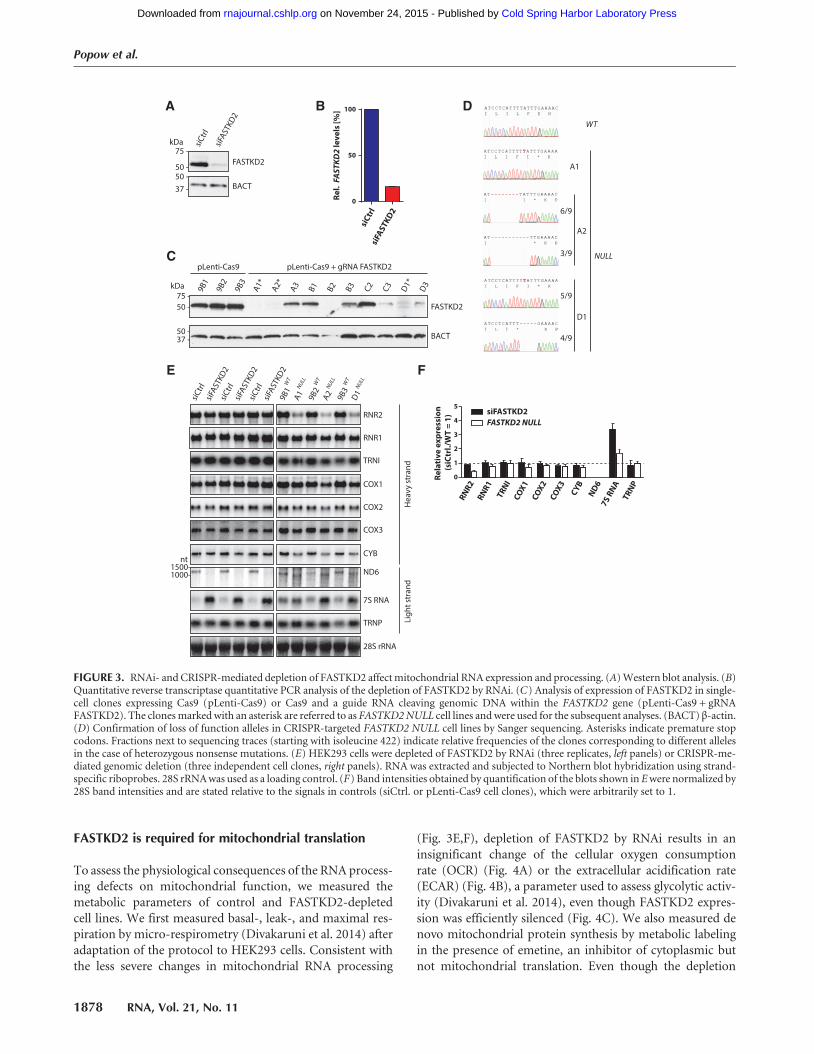
HEK293 cells both by RNA interference (RNAi) and the Cas9nuclease-based CRISPR system (Hsu et al. 2013). RNAi faith-fully reduced the expression of FASTKD2 protein (Fig. 3A)and mRNA (Fig. 3B) to ∼15% of control levels. Completedepletion for FASTKD2 was achieved via the CRISPR/Cas9nuclease system (Sanjana et al. 2014). Tomimic the mutationcausing the loss of function of FASTKD2 reported for two pa-tients suffering from early onset encephalomyopathy (Ghezziet al. 2008), we chose a guide RNA sequence that induces
cleavage of genomic DNA in the immediate vicinity of theR432 codon of FASTKD2, which is nonsense mutated in af-fected patients. We isolated several independent clonal celllines without detectable FASTKD2 expression, referred to asFASTKD2 NULL cells (Fig. 3C). Sequencing of the genomicDNA of these cell lines confirmed successful nonsense muta-genesis of all alleles encoding FASTKD2 (Fig. 3D).Based on the iCLIP data, we analyzed the processing and
steady-state levels of FASTKD2 target RNAs in cells depleted
250 -
150 -
kDa
100 -75 -
50 -
37 -
25 -
MLS
-GF
P
#1
#2
#3
FASTKD2-GFP
IP:GFP
Silver stain
-MLS-GFP
-FASTKD2-GFP
B
D
log2 FASTKD2-GFP
MLS-GFP-log10 (p-value)Target
D-Loop 5.51 2.24
ND6 5.46 2.24
7S RNA 5.37 2.15
TRNP 4.71 7.55
Lig
ht
stra
nd
TRNT 6.84 3.59
TRNI 6.39 3.10
COX1 5.67 2.42
TRNL2 5.28 2.05
TRNH 5.22 2.04
COX2 5.20 2.03
RNR2 7.62 4.56
TRNL1 6.33 3.04
RNR1 6.04 2.76
CYB 5.76 2.49
He
av
y st
ran
d
F
150 -100 -
75 -
50 -
37 -
25 -
250 -kDa
-FASTKD2
-FASTKD2-GFP
0 2.5
10 40 160
320
Tetracyclin (ng/mL)
WB:FASTKD2
LysateA
*
-pre-FASTKD2-GFP
2 kb
MLS
-GF
PFA
ST
KD
2-G
FP
#1
#2
#3
#1
#2
#3
7S RNA TRNQ TRNATRNN
TRNYTRNC
TRNS1ND6
TRNE TRNP
TRNF TRNVRNR1 RNR2
TRNL1ND1
TRNITRNM
ND2TRNW
COX1TRND
COX2TRNK
ATP8/6 COX3TRNG
ND3
ND4L/4TRNHTRNS2
ND5 CYB
TRNR TRNL2
TRNT
Heavy20 55 20Light
Heavy
Light
-12 -8 -4 0 4 8 12
log2 FASTKD2-GFP
MLS-GFP
-log1
0 (p-
valu
e)
4
8
12
14
10
6
2
E
C
0
2
4
6
8
10
MLS-GFP #1
#2
#3
FASTKD2-GFP #1
#2
#3
Chr 1-22
Ch
r M
Ch
r X
Ch
r Y
Cro
ssli
nk
sit
e d
en
sity
[n
t-1]
FIGURE 2. Identification of FASTKD2 interacting transcripts in HEK293 cells. (A) Titration of tetracycline concentrations required for equal expres-sion of endogenous FASTKD2 and FASTKD2-GFP. The asterisk denotes a lower molecular mass species of unknown identity. (B) Analysis ofFASTKD2-GFP immunoprecipitates by polyacrylamide gel electrophoresis and silver stain. (C) Crosslink site density plot for iCLIP cDNA librariesafter alignment to the human genome and random barcode evaluation. Crosslink site density was calculated using the sum of annotated gene lengthsfor each chromosome. (D) Heat map representing significant (false discovery rate <0.05) RNA crosslink sites in MLS-GFP and FASTKD2-GFP librar-ies. Enrichment of light-strand transcripts is indicated in blue, enrichment of heavy-strand transcripts is indicated in red. (E) Enrichment plots forheavy- and light-strand FASTKD2-GFP crosslink sites. Enrichment and P-values were calculated using DESeq2. (F) High-confidence FASTKD2 RNAtargets (log2-fold enrichment ≥4, −log10 P-value≥ 2). Note that for a given target only the parameters for the highest scoring crosslink site arespecified.
Popow et al.
1876 RNA, Vol. 21, No. 11
Cold Spring Harbor Laboratory Press on November 24, 2015 - Published by rnajournal.cshlp.orgDownloaded from

for FASTKD2 by RNAi or CRISPR. Depletion of FASTKD2by RNAi leaves the noncoding heavy-strand transcripts un-changed (Fig. 3E, left panels; Fig. 3F black bars). In contrast,CRISPR-mediated depletion of FASTKD2 causes a profoundand selective reduction of 16S mitochondrial rRNA (RNR2)(Fig. 3E, top panel; Fig. 3F). However, the levels of 12S rRNA(RNR1), which arises from the same polycistronic heavy-strand precursor transcripts, are largely unaffected (Fig. 3E,F). Fully consistent with the results reported here, depletionof FASTKD2 by RNAi was just found in to cause misassemb-ly of the mitochondrial ribosome and impaired mitochon-drial protein synthesis in human immortalized fibroblasts(Antonicka and Shoubridge 2015). Our observations forFASTKD2 also complement a recent report implicatingRAP, a protein of Arabidopsis thaliana that shares the RAPdomain with FASTKD2, in precursor processing of the smallribosomal subunit RNA in chloroplasts (Kleinknecht et al.2014). Despite this surprising similarity, our data suggestthat FASTKD2 may function somewhat differently, as wedid not observe the accumulation of ribosomal precursortranscripts upon depletion of FASTKD2. Moreover, the un-changed levels of RNR1 suggest that the biogenesis of thecommon precursor of RNR1 and RNR2 is unaffected in theabsence of FASTKD2. Thus, selective depletion of RNR2may occur due to its aberrant processing or due to ribosomemisassembly. Indeed, recent studies suggest that failure to as-semble RNR2 with mitochondrial ribosomal proteins leadsto its selective depletion, which has been proposed to be me-diated by the mitochondrial degradosome (Antonicka andShoubridge 2015; Tu and Barrientos 2015).The levels of isoleucyl tRNA (TRNI), one of the heavy-
strand-encoded tRNAs highly enriched in FASTKD2-GFPlibraries, are unaffected by depletion of FASTKD2 by RNAiand CRISPR/Cas9-mediated mutagenesis (Fig. 3E,F). Wenext analyzed the expression of the heavy-strand protein-encoding transcripts bound by FASTKD2. The mRNA levelsof the complex IV components COX1 and COX2 are un-affected or mildly reduced by either form of depletion ofFASTKD2 (Fig. 3E,F). Complex IV was found to be most se-
verely affected in patients with mutated FASTKD2 (Ghezziet al. 2008). We therefore also examined the expression ofthe mitochondrial mRNA encoding the complex IV subunit,COX3, even though this transcript did not classify as a directFASTKD2 target (Fig. 2F). Comparable to the remaining mi-tochondrial complex IV subunit RNAs, the levels of COX3mRNA appear to be mildly affected by both RNAi- andCRISPR-mediated loss of FASTKD2 (Fig. 3E,F). Similarly,both modes of FASTKD2 depletion only mildly reduce thelevels of the remaining FASTKD2 protein-coding heavy-strand target transcript, the complex III subunit mRNACYB (Fig. 3E,F).Depletion of FASTKD2 by RNAi or CRISPR affects ND6
RNA processing and levels (Fig. 3E,F). As suggested byJourdain et al. (2015), depletion of FASTK has a similar ef-fect on expression of the ND6 mRNA as loss of FASTKD2.This study concludes that a mitochondrial form of FASTKregulates the exonucleolytic processing of the 3′-end of theND6 transcript in cooperation with the mitochondrial de-gradosome (Jourdain et al. 2015). However, our low FDRFASTKD2 crosslink sites cluster within the coding regionof the ND6 transcript, suggesting that FASTKD2 may deter-mine the fate of the mature forms of ND6, possibly in addi-tion to a role in precursor processing.Expression of 7S RNA is strongly induced by both modes
of depletion of FASTKD2 (Fig. 3E,F). This observation isconsistent with an induction of mitochondrial transcrip-tion, a possible consequence of perturbed mitochondrialhomeostasis (Metodiev et al. 2009), as 7S RNA levels havepreviously been proposed to correlate with de novo tran-scriptional activity in mitochondria (Terzioglu et al. 2013).Interfering with FASTKD2 expression by RNAi or CRISPRleaves TRNP steady-state levels unchanged (Fig. 3E,F).Taken together, both depletion of FASTKD2 by RNAi andCRISPR mutagenesis affect the levels of several mitochondri-al transcripts. CRISPR-mediated loss of FASTKD2 causestranscript-specific mitochondrial RNA expression defectscharacterized by a drastic reduction of 16S ribosomal RNA,loss of mature ND6 mRNA and the induction of 7S RNA.
TABLE 1. Summary statistics for genomic mapping and random barcode evaluation of FASTKD2-GFP and MLS-GFP cDNA libraries
Cell line MLS-GFP FASTKD2-GFP
Replicate number 1 2 3 1 2 3Initial sequencing reads 131,934,543
100%a
After experiment separation 22,240,321 21,165,609 22,869,088 20,835,753 17,587,180 22,740,08817%a 16%a 17%a 16%a 13%a 17%a
After mapping to human genome (unique hits) 12,854,926 13,189,145 15,283,924 13,116,022 13,545,405 17,925,85458%b 62%b 67%b 63%b 77%b 79%b
After mapping and random barcode evaluation 447,504 1,069,127 2,823,635 3,814,187 2,274,754 2,789,9843%c 8%c 18%c 29%c 17%c 16%c
aWith respect to initial sequencing reads.bWith respect to total reads after experiment separation.cWith respect to mapped reads.
FASTKD2 functions in mitochondrial RNA processing
www.rnajournal.org 1877
Cold Spring Harbor Laboratory Press on November 24, 2015 - Published by rnajournal.cshlp.orgDownloaded from
FASTKD2 is required for mitochondrial translation
To assess the physiological consequences of the RNA process-ing defects on mitochondrial function, we measured themetabolic parameters of control and FASTKD2-depletedcell lines. We first measured basal-, leak-, and maximal res-piration by micro-respirometry (Divakaruni et al. 2014) afteradaptation of the protocol to HEK293 cells. Consistent withthe less severe changes in mitochondrial RNA processing
(Fig. 3E,F), depletion of FASTKD2 by RNAi results in aninsignificant change of the cellular oxygen consumptionrate (OCR) (Fig. 4A) or the extracellular acidification rate(ECAR) (Fig. 4B), a parameter used to assess glycolytic activ-ity (Divakaruni et al. 2014), even though FASTKD2 expres-sion was efficiently silenced (Fig. 4C). We also measured denovo mitochondrial protein synthesis by metabolic labelingin the presence of emetine, an inhibitor of cytoplasmic butnot mitochondrial translation. Even though the depletion
75 -
50 -
kDa
50 -
37 -si
Ctr
l
siFA
STKD
2
FASTKD2
BACT
A
siCtr
lsiF
ASTKD2
Rel.
FAS
TKD
2 le
vels
[%]
0
50
100B
75 -
50 -
kDa
50 -37 -
FASTKD2
BACT
9B1
9B2
9B3
A1*
A2*
B1A3
B2 C2
B3 C3
D1*
D3
pLenti-Cas9 pLenti-Cas9 + gRNA FASTKD2C
E
WT
A1
A2
6/9
3/9
5/9
4/9
D1
NULL
D
nt1500-1000-
siC
trl
siFA
STKD
2
9B1
COX1
RNR1
ND6
siC
trl
siFA
STKD
2si
Ctr
lsi
FAST
KD2
9B2
9B3
A1
A2
D1
WT
NULL WT
WT
NULL
NULL
CYB
TRNP
7S RNA
RNR2
COX3
TRNI
28S rRNA
COX2 He
avy
stra
nd
Lig
ht
stra
nd
0
1
2
3
4
5
RNR2
RNR1
TRNI
COX1
COX2
COX3
CYB
ND67S
RNA
TRNP
Rela
tive
exp
ress
ion
(siC
trl./
WT
= 1)
F
siFASTKD2FASTKD2 NULL
FIGURE 3. RNAi- and CRISPR-mediated depletion of FASTKD2 affectmitochondrial RNA expression and processing. (A)Western blot analysis. (B)Quantitative reverse transcriptase quantitative PCR analysis of the depletion of FASTKD2 by RNAi. (C) Analysis of expression of FASTKD2 in single-cell clones expressing Cas9 (pLenti-Cas9) or Cas9 and a guide RNA cleaving genomic DNA within the FASTKD2 gene (pLenti-Cas9 + gRNAFASTKD2). The clonesmarkedwith an asterisk are referred to as FASTKD2NULL cell lines andwere used for the subsequent analyses. (BACT) β-actin.(D) Confirmation of loss of function alleles in CRISPR-targeted FASTKD2 NULL cell lines by Sanger sequencing. Asterisks indicate premature stopcodons. Fractions next to sequencing traces (starting with isoleucine 422) indicate relative frequencies of the clones corresponding to different allelesin the case of heterozygous nonsense mutations. (E) HEK293 cells were depleted of FASTKD2 by RNAi (three replicates, left panels) or CRISPR-me-diated genomic deletion (three independent cell clones, right panels). RNA was extracted and subjected to Northern blot hybridization using strand-specific riboprobes. 28S rRNAwas used as a loading control. (F) Band intensities obtained by quantification of the blots shown in Ewere normalized by28S band intensities and are stated relative to the signals in controls (siCtrl. or pLenti-Cas9 cell clones), which were arbitrarily set to 1.
Popow et al.
1878 RNA, Vol. 21, No. 11
Cold Spring Harbor Laboratory Press on November 24, 2015 - Published by rnajournal.cshlp.orgDownloaded from

0.00
0.05
0.10
0.15
0.20
0.25
0.00
0.05
0.10
0.15
75 -
50 -
kDa
50 -
37 -
siC
trl
siFA
STKD
2
FASTKD2
BACT
C
CO1/
BACT
[-]
J
0
200
400
WT
NULL
Rel.
grow
th (G
lc =
100
%)G
0
10
20
30
40
50
WT
NULL
B
siCtr
lsiF
ASTKD2
0
20
40
60
80
ECA
R (O
ligo
= 10
0 %
)F
0
20
40
60
80
100
HEK29
39B
19B
29B
3 A1 A2 D1
WT NULL
WT
NULL
ECA
R (O
ligo
= 10
0 %
)
AO
CR [p
mol
/min
]
0
50
100
150
200
t (min)
siCtrl
siFASTKD2
0 20 40 60 80 100
Basal Oligo FCCP1 FCCP2 A+R
E
0
100
200
300HEK293
9B1
9B2
9B3
A1
A2
D1
WT
NULL
t (min)0 20 40 60 80 100
OCR
[pm
ol/m
in]
Basal Oligo FCCP1 FCCP2 A+R
kDa
75 -
50 -
9B3
A1
FASTKD2
Crude mitochondriaH
A2
D1
9B1
9B2
25- MRPP2
WT NULL
I
C-I/C
S
C-II/
CS
C-II-
III/C
S
C-IV
/CS
C-V
/CS
0.00
0.05
0.10
0.15
0.20
0.25
0.00
0.01
0.02
0.03
0.04
WT
NULL WT
NULL WT
NULL WT
NULLWT
NULL
0.00
0.02
0.04
0.06
0.08
siC
trl
siFA
STKD
2
Au
tora
dio
gra
ph
yC
BB
D
COI
CYBND2COIIICOIIATP6
ND6ND3
ND4L
ND5
ND4
ATP8
9B1
9B2
9B3
A1
A2
D1
WT NULL
ND5COIND4CYBND2COIIICOIIATP6
ND6ND3ND4LATP8
Au
tora
dio
gra
ph
yC
BB
FIGURE 4. Metabolic analysis of cells depleted of FASTKD2. (A) Oxygen consumption rate (OCR) of HEK293 cells depleted of FASTKD2 by RNAi([siCtrl] control siRNA; [siFASTKD2] FASTKD2 targeting siRNA pool; [Basal] basal respiration; [Oligo] leak respiration in the presence of oligomy-cin; [FCCP1] uncoupled respiration in the presence of 200 nM FCCP (carbonyl cyanide-4-(trifluoromethoxy)phenylhydrazone); [FCCP2] uncoupledrespiration in the presence of 250 nM FCCP; [A + R] residual respiration in the presence of antimycin a and rotenone). (B) Extracellular acidificationrate (ECAR) of cells depleted of FASTKD2 by RNAi. Maximum ECAR measured in the presence of oligomycin (Oligo) was set to 100%. (C)Confirmation of RNAi-mediated depletion of FASTKD2 by Western blot. (D) Metabolic labeling of de novo synthesis of mitochondrial proteinsin cells depleted of FASTKD2 by RNAi (left panel) or CRISPR-mediated mutagenesis (right panel) in the presence of emetine. (CBB) Coomassie bril-liant blue stain. (E) OCR of HEK293 cells depleted of FASTKD2 by CRISPR. Abbreviations are as in A. (F) ECAR of cells depleted of FASTKD2 byCRISPR. (G) Relative growth of cells depleted of FASTKD2 by CRISPR in galactose media. (H) Western blot analysis of expression of FASTKD2 incrude mitochondrial preparations isolated from cells depleted of FASTKD2 by CRISPR. (I) Activity of ETC complexes in cells depleted of FASTKD2by CRISPR. Values were normalized by the activity of citrate synthase and are stated for both cells depleted of FASTKD2 by CRISPR-mediatedmutagenesis and Cas9-expressing control cells. (J) mtDNA levels of cells depleted of FASTKD2 by CRISPR. Values are stated as relative ratios ofCOXI (mitochondrial) to β-actin (nuclear) amplicons as determined by quantitative reverse transcriptase PCR.
FASTKD2 functions in mitochondrial RNA processing
www.rnajournal.org 1879
Cold Spring Harbor Laboratory Press on November 24, 2015 - Published by rnajournal.cshlp.orgDownloaded from

of FASTKD2 by RNAi does not appreciably affect the steady-state levels or processing pattern of RNR2 on Northernblots (Fig. 3E, top panel on the left), it appreciably reducesglobal mitochondrial translation (Fig. 4D left panel). Thefact that the metabolic activity of siRNA-treated cells appearslargely unaltered despite a generalized reduction of mito-chondrial translation suggests a remarkable capacity of thecells to buffer perturbations of mitochondrial protein syn-thesis for limited periods of time. Genomic deletion ofFASTKD2 by CRISPR-mediated mutagenesis impairs globalmitochondrial translation to an even greater extent thanRNAi-mediated depletion, thus confirming the importanceof the protein for mitochondrial protein synthesis (Fig. 4Dright panel).
Lack of FASTKD2 compromises cellular respiration
Against the background of the apparently high functionalstability of mitochondrial functions to RNAi-mediatedFASTKD2 depletion, we also analyzed the metabolic param-eters of FASTKD2 NULL cells. Of note, CRISPR-mediatedloss of FASTKD2 causes a pronounced decrease of both basalandmaximal respiration (Fig. 4E, 0–20 or 40–80min, respec-tively). The OCR is diminished for all three tested clonalFASTKD2 NULL cell lines, compared with the rates mea-sured for the Cas9 control and parental cell lines. In additionto reduced oxygen consumption, each of the FASTKD2-defi-cient cell lines also displays an increased ECAR (Fig. 4F), sug-gesting that the energy deficit arising from dysfunctionaloxidative phosphorylation may be compensated by an in-creased rate of glycolysis. As cells with defects in oxidativephosphorylation frequently exhibit diminished prolifera-tion in media containing galactose as the main carbohydrate(Robinson et al. 1992), we compared the growth rates ofFASTKD2 WT (wild type) and NULL cells in glucose- versusgalactose-containing media. Whereas both FASTKD2 WTand NULL cell lines show lower proliferation rates in gal-actose compared to glucose media, the FASTKD2 NULLcells are far more severely affected (Fig. 4G). Therefore,FASTKD2-deficient cells display multiple signs of a markeddefect in cellular respiration.
We alsomeasured the activity of the complexes of the respi-ratory chain in crude mitochondria isolated from FASTKD2NULL or the control WT cell lines (Fig. 4H). All activitieswere normalized to citrate synthase activity measured inthe respective mitochondrial preparations. Consistent withthe generalized defect in mitochondrial translation ofFASTKD2-depleted cells, all mitochondrially encoded ETCcomplexes display diminished activities (30%–60% ofWT levels, Fig. 4I). Of all ETC complexes, it appears as ifNADH:ubiquinone oxidoreductase (complex I) is the mostseverely affected (∼30%ofWT levels, Fig. 4I, first panel), con-sistent with one of its subunit transcripts, ND6, being stronglyaffected by both modes of depletion of FASTKD2. Of note,succinate dehydrogenase (complex II) activity was unaffected
(Fig. 4I, second panel), consistent with its subunits and as-sembly factors being encoded by the nuclear genome.Note that 7S RNA is a polyadenylated mitochondrial tran-
script of ∼200 nt length with poorly defined functions (Ojalaet al. 1981a; Clayton 1984; Mercer et al. 2011). Accordingto the current model of mitochondrial DNA replication, anonpolyadenylated fragment of 7S RNA, produced by earlytermination, serves as an RNA primer for the replication ofmitochondrial heavy-strand DNA (Agaronyan et al. 2015).To assess a possible functional consequence of altered 7SRNA expression upon depletion of FASTKD2, we deter-mined the mitochondrial DNA content of FASTKD2 WTand NULL cell lines. mtDNA levels are slightly increasedin FASTKD2 NULL cells compared to WT controls (Fig.4J). Importantly, this excludes mtDNA depletion as a causeof the respiration defect of FASTKD2 NULL cells. Furthercontributions of altered 7S RNA expression to the pheno-type of FASTKD2-deficient cells are difficult to assign, asthis transcript still awaits further functional characterization(Ojala et al. 1981a; Clayton 1984). Taken together, CRISPR-mediated deletion of FASTKD2 has drastic effects onboth mitochondrial gene expression and physiology. Boththe broadly compromised cellular respiration and inducedglycolysis are consistent with the drastic depletion of thelarge mitochondrial ribosomal subunit RNA (RNR2) associ-ated with the complete loss of FASTKD2 expression. Never-theless, the activity of ETC complexes remains detectable inFASTKD2 NULL cells, which suggests residual respiratorychain protein synthesis in the absence of FASTKD2.
CONCLUSIONS
The FASTK family represents an emerging family of RNA-binding proteins. Despite early evidence that FASTK familyproteins localize to mitochondria and, as demonstrated forFASTKD3, affect cellular respiration (Simarro et al. 2010),mechanistic and functional information is only now begin-ning to emerge. A recent high-throughput screen for factorsinvolved in mitochondrial RNA processing implicatedFASTKD4 in the regulation of the turnover of a select spec-trum of mitochondrial transcripts (Wolf and Mootha2014). With the aim to identify the molecular underpinningsof a rare form of encephalomyopathy associated with mutat-ed FASTKD2 (Ghezzi et al. 2008), we here propose a mech-anistic model for its function. Our identification of themolecular targets of FASTKD2 by iCLIP yields a high-resolu-tion map normalized to exclude PCR amplification-inducedartifacts and controlled for specificity. The ensuing charac-terization of the expression of FASTKD2-bound transcriptsupon depletion of FASTKD2 by RNAi or the CRISPR/Cas9system provides insights into FASTKD2 function in mito-chondrial biology and pathophysiology. In combinationwith the metabolic analyses we thus establish the physiolog-ical consequences of a genomic deletion of FASTKD2 in cul-tured human cells.
Popow et al.
1880 RNA, Vol. 21, No. 11
Cold Spring Harbor Laboratory Press on November 24, 2015 - Published by rnajournal.cshlp.orgDownloaded from

The effects of FASTKD2 deficiency appear to be cell type-and tissue-specific, as the originally reported complex IVdeficiency was only detectable in muscle biopsies but notin fibroblasts of the affected patients (Ghezzi et al. 2008).The variable capability of different cell types to compensatefor the consequences of impaired mitochondrial translation,as suggested by a recent study (Richman et al. 2015), mayat least to some degree explain the apparently contrastingconsequences of the loss of FASTKD2 in different tissuesor cell types. Alternatively, the lack of FASTKD2 might becompensated for by further, tissue-specific factors such asother mitochondrial granule components or FASTK familyproteins. It therefore remains to be determined to whichdegree FASTKD- and other mitochondrial RNA granule pro-teins cooperate or are functionally interdependent, as sug-gested by the comparable effects of FASTK (Jourdain et al.2015) and FASTKD2 (this study) depletion on ND6 expres-sion. Future work on the other members of the FASTK familyof proteins will help complete the picture of their mode offunction.
MATERIALS AND METHODS
Expression and detection of GFP-fusion proteins
All constructs were inserted into pcDNA5/FRT/TO-GFP, a deriva-tive of pcDNA5/FRT/TO (Invitrogen) with a Gly/Ser-linker EGFPconstruct inserted between the NotI and ApaI restriction sites.The open reading frame of FASTKD2 comprising amino acids me-thionine 17, the conserved start codon, to glutamine 710 (Ghezziet al. 2008) was amplified by PCR using the primers FASTKD2_F(5′-AGCTGGTACCATGAATAACAAAGCG-3′, start codon under-lined) and FASTKD2_R (5′-ACGTCTCGAGTTGTGTGCTTTG-CAC-3′). The mitochondrial localization signal of FASTKD2 (ami-no acids 16–68) was amplified using the primers FASTKD2_F andMLS_R (5′-ACGTCTCGAGCATTCTGTTATGAAAGTTATTTAA-AATG-3′). The resulting amplicons were inserted into pcDNA5/FRT/TO-GFP using the XhoI and KpnI restriction sites, thus gen-erating C-terminal GFP-fusion proteins. The constructs were in-troduced into HEK293 Flip-In TRex cell lines according to themanufacturer’s recommendations (Life Technologies) using Effec-tene transfection reagents (Qiagen). For fluorescence microscopy,pcDNA5/FRT/TO-based constructs expressing FASTKD2-GFP orGFP only were transiently transfected into MCF-7 or U2OS cells.Forty-eight hours after transfection, the cells were incubated for30 min with 250 nM Mitotracker deep red CMX ros (Life Technol-ogies), fixed with formaldehyde and visualized using a Leica SP2confocal microscope.
UV-cross-linking, immunoprecipitation, and labelingof FASTKD2-associated transcripts
Induced HEK293 Flp-In TRex cell lines expressing FASTKD2-GFP(10 ng/mL tetracycline, 16 h) were washed twice with PBS and irra-diated with UV light at 254 nm (150 mJ/cm2) on ice. Immediatelyafter irradiation, cells were lysed in three cell pellet volumes of lysisbuffer (100 mM KCI, 5 mM MgCl2, 10 mM Tris–HCI pH 7.5,
0.5% NP-40, 4 mMDTT, 100 U/mL RNasin [Promega], 200 µM ri-bonucleoside vanadyl complex [NEB], EDTA-free protease in-hibitor cocktail [Roche]). Lysates were incubated with 50 U/mLof DNase I (Takara) and indicated concentrations of RNaseA(Sigma) for 10 min at 37°C. Subsequently the lysates were chilledon ice and cleared by centrifugation at 16,000g for 20 min at 4°C.Fusion proteins were captured by incubation with 10 µL of pre-equilibrated magnetic particle suspension (GFP-Trap_M, Chromo-tek) per mL of lysate with rotation for 2 h at 4°C. Beads were recov-ered and washed four times with high salt buffer (500 mMNaCl, 20mM Tris–HCl pH 7.5, 1 mM MgCl2, 0.05% NP40, 0.1% sodiumdodecyl sulfate, EDTA-free protease inhibitor cocktail). Subse-quently, beads were washed two times with PNK buffer (50 mMTris–HCl pH 7.5, 50 mM NaCl, 10 mM MgCl2, 0.5% NP-40, 5mMDTT). Beads were resuspended in the original bead suspensionvolume and incubated with 1 U/μL T4 polynucleotide kinase (NEB)and 0.1 μCi/μL [γ-32P]ATP (Hartmann Analytics) for 30 min at 37°C and 800 rpm to label the 5′-end of cross-linked RNAs. The mag-netic beads were then washed five times with PNK buffer free ofDTT. Immunoprecipitates were resolved on 4%–12% gradientgels (BioRad) and blotted on PVDF membranes. Cross-linkedRNA–protein complexes were visualized by autoradiography usinga phosphorimager (Fuji).
Identification of FASTKD2-associatedtranscripts by iCLIP
Cell lines expressing FASTKD2-GFP or MLS-GFP were subjected toUV cross-linking and lysis as described above. The cleared lysates(typically 2 mL) were then mixed 1:4 with 5× High Salt Buffer(1.25 MNaCl, 100 mMTris–HCl pH 7.5, 0.5% sodium dodecyl sul-fate). The lysates were precleared by incubation with 60 µL of equil-ibrated blocked magnetic particle suspension (Chromotek) per mLof lysate (1 h, 4°C, gentle rotation). GFP-fusion proteins were thencaptured from precleared lysates by incubation with 60 µL of GFP-Trap magnetic particle suspension (GFP-Trap_M, Chromotek) permL of lysate (2 h, 4°C, gentle rotation). Beads were collected using amagnet and washed twice with high salt buffer. The beads were thenwashed twice with medium salt buffer (250 mMNaCl, 20 mMTris–HCl pH 7.5, 1 mM MgCl2, 0.05% NP-40, 100 U/mL RNasin, 200µM ribonucleoside vanadyl complex, EDTA-free protease inhibitorcocktail) and once with low salt buffer (150 mMNaCl, 20 mMTris–HCl pH 7.5, 1 mMMgCl2, 0.01% NP-40, 50 U/mL RNasin, EDTA-free protease inhibitor cocktail). Beads were resuspended in 1 mLlow salt buffer without RNasin and incubated (3 min, 37°C, 1 100rpm) with 4 U of RNase I (Life Technologies) and 4 U of TurboDNase (Life Technologies). Beads were washed once with highsalt buffer, twice with low salt buffer, resuspended in 20 µL ofPNK mix (70 mM Tris–HCl pH 6.5, 10 mM MgCl2, 5 mM DTT,250 U/mL T4 polynucleotide kinase [NEB], 500 U/mL RNasin)and incubated with agitation (1 100 rpm) for 20 min at 37°C.Beads were then washed twice with high salt buffer, twice withlow salt buffer and incubated for 16 h (1 100 rpm, 16°C) in 20 µLligation mix (50 mM Tris–HCl pH 7.5, 10 mM MgCl2, 10 mMDTT, 500 U/mL T4 RNA ligase 1, 500 U/mL RNasin, 1.5 μM pre-adenylated linker L3 [5′-rApp-AGATCGGAAGAGCGGTTCAG-ddC-3′], 20% PEG-400 [Sigma]). The beads were washed threetimes with high salt buffer, twice with low salt buffer, proteins di-gested with proteinase K and samples processed for all subsequent
FASTKD2 functions in mitochondrial RNA processing
www.rnajournal.org 1881
Cold Spring Harbor Laboratory Press on November 24, 2015 - Published by rnajournal.cshlp.orgDownloaded from

steps as described previously (Konig et al. 2010). cDNA libraries ob-tained after PCR amplification with universal Solexa primers (25 cy-cles) were multiplexed and sequenced using an Illumina HiSeq2000platform. Mapping of the retrieved reads to the hg19 assembly ofthe human genome (considering only single hits in the genome),random barcode evaluation, and evaluation of the significance ofcrosslink sites (Fig. 2D) were carried out as previously described(Konig et al. 2010). Low FDR (<0.05) crosslink site counts of thethree replicates sequenced for each MLS-GFP and FASTKD2-GFPwere then used to compute log2(enrichment) and log10(P-value) us-ing DESeq2 (Anders and Huber 2010).
Cell culture, depletion of FASTKD2 by RNAi,and CRISPR-induced mutagenesis
Cells were cultured at 37°C, 95% humidity and 5% CO2 in 1× high-glucose DMEM supplemented with 10% fetal bovine serum, 2 mML-glutamine, 100 U/mL penicillin, and 100 μg/mL streptomycin sul-fate. For galactose cultivation experiments, cells were cultured inDMEM without glucose and glutamine (Life Technologies) plus 2mM L-glutamine, 0.9 g/L D-galactose or 4.5 g/L D-glucose, 10%dialyzed fetal bovine serum, and 1 mM pyruvate. Depletion ofFASTKD2 by RNAi was achieved by transfection of Dharmaconsmartpool siRNAs targeting FASTKD2 using the MISSION siRNAuniversal negative control (Sigma) for control experiments andLipofectamine RNAiMAX reagent (Life Technologies) as suggestedby the manufacturer. Typically, cells were harvested and analyzed 72h after transfection. To achieve the genomic deletion of FASTKD2by CRISPR/Cas9-mediated mutagenesis, the annealed oligonucleo-tides FASTKD2_1269F (5′-CACCGCAAGGTTTTCAAATAAAAT-G-3′) and FASTKD2_1269R (5′-AAACCATTTTATTTGAAAACC-TTGC-3′) were inserted into pXPR_001 (obtained from Addgene)via the BsmBI restriction sites. The resulting plasmid was introducedinto HEK293 Flp-In TRex cells by lentiviral infection using standardprocedures. Infected cell pools were selected with 100 µg/mL zeocin,5 µg/mL blasticidin, and 1 µg/mL puromycin and antibiotic-resis-tant single-cell clones isolated by limiting dilution. Upon clonal ex-pansion, loss of FASTKD2 expression was assessed by Western blot(Protein Tech No. 17464-1-AP). To directly identify the mutationsintroduced by CRISPR/Cas9-mediated mutagenesis, the genomicDNA sequence surrounding the protospacer adjacent motif wasamplified using the primers FASTKD2_gSEQ_F (5′-cggtacccgggg-atcGGATATATTCTGTTGATCAGGACAGTTGAATTGCTGC-3′,lower case indicates homology sequences for In-Fusion cloning) andFASTKD2_gSEQ_R (5′-cgactctagaggatcAGTATGAAGAATAGATA-GAATGTTTTTCATGTTTAGGGATTCAGG-3′) using PhusionDNA polymerase (NEB). PCR amplicons were either directly se-quenced or inserted into linearized pUC19 by In-Fusion cloning(Clontech) following the manufacturer’s recommendations. Aftertransformation, plasmids obtained from individual colonies weresubjected to Sanger sequencing.
Analysis of mitochondrial RNA processingby Northern blotting
Northern blot analysis of mitochondrial transcripts was carried outessentially as described previously (Jourdain et al. 2013). Briefly, 10µg of total RNA were resolved by electrophoresis in 1.2% agarose–formaldehyde gels and blotted by capillary transfer on nylon mem-
branes (Genescreen PlusR, PerkinElmer). Cross-linked membraneswere probed in 50% formamide, 7% SDS, 0.2 M NaCl, 80 mMsodium phosphate (pH 7.4), 0.1 mg/mL salmon sperm DNA at60°C or 45°C (for tRNA probes) with antisense riboprobes preparedby transcription from PCR products. Blots were briefly washed with1× SSC, 0.1% SDS and afterwards for 30 min with 0.5× SSC, 0.1%SDS. Bands on blots were detected by phosphorimaging.
Metabolic characterization of cell lines
OCR and ECAR were determined using a Seahorse XFe96 analyzer(Seahorse Bioscience) as suggested by the manufacturer. Typically,25,000 HEK293 cells were seeded into poly-L-lysine (PLL) coated96-well analyzer plates (Seahorse Bioscience) and grown for 16 hto allow for attachment. OCR and ECAR were recorded in XF assaymedium (base medium plus 25 mM D-glucose, 5 mM pyruvate, 4mM L-glutamine). Leak respiration was measured after injectionof 2 µM oligomycin, uncoupled respiration after injection of 200and 250 nM FCCP and residual oxygen consumption in the pres-ence of 0.5 µM rotenone and 0.5 µM antimycin A. To assess growthof cells in galactose-containing media, 2500 cells were seeded intoPLL-coated 96-well plates and grown for 16 h in standard mediumto allow for attachment. Cells were washed thrice and furthergrown in parallel in either galactose or glucose containing media.After 48 h, cell numbers were determined using the CyQUANTDirect cell proliferation assay (Life Technologies) as specified bythe manufacturer.
Pulse labeling of mitochondrial translation products
Labeling of mitochondrial translation products was carried out asdescribed previously (Sasarman and Shoubridge 2012). Briefly,500,000 cells were seeded into PLL-coated wells of a six-well plateon the day before labeling. Cells were washed twice with DMEMwithout cysteine and methionine and incubated for 30 min (37°C,5% CO2, 95% relative humidity) with equilibrated labeling medium(DMEM without cysteine and methionine, 2 mM glutamine, 10%dialyzed serum and 1 mM pyruvate). Cytoplasmic translation wasinhibited by addition of 100 µg/mL emetine (Sigma) and plates in-cubated for further 5 min. Finally, translation products were labeledby addition of 200 µCi/mL of L-[35S]Methionine (HartmannAnalytics) for 1 h. Cells were then incubated for 10 min with stan-dard culturing medium, harvested by scraping in ice-cold PBS, lysedwith RIPA buffer (25 mM Tris–HCl pH 7.5, 150 mMNaCl, 1% NP-40, 1% sodium deoxycholate, 0.1% sodium dodecyl sulfate, 1 mMDTT, EDTA-free protease inhibitor cocktail), cleared by centrifuga-tion (16,000g, 10 min) and 30 µg aliquots (Bradford assay, BSA stan-dard) analyzed by SDS gel electrophoresis (15% gels). Gels werestained with Coomassie brilliant blue, dried, and visualized byphosphorimaging.
Respiratory chain assays
Cells were resuspended in mitochondrial isolation buffer (250 mMsucrose, 50 mM KCl, 5 mM MgCl2, 20 mM Tris–HCl pH 7.5) andlysed by 12 passages through a 27 2/3 G needle. The lysate was cen-trifuged for 10 min at 600g at 4°C, the supernatant recovered andcrude mitochondria recovered by centrifugation at 14,000g for 15
Popow et al.
1882 RNA, Vol. 21, No. 11
Cold Spring Harbor Laboratory Press on November 24, 2015 - Published by rnajournal.cshlp.orgDownloaded from

min. Mitochondrial pellets were resuspended in a minimal volumeof mitochondrial isolation buffer. The protein concentration of mi-tochondrial preparations was determined by Bradford assay usingBSA as a standard. Mitochondrial enzyme activities were recordedusing a Tecan infinite M1000pro plate reader equipped with a ther-mostat essentially as described previously (Sauer et al. 2005). Thefollowing extinction coefficients were used to calculate relativeenzyme activities from absorption values: NADH: ε340–400 nm = 6.1mM−1 cm−1, cytochrome c: ε550–540 nm = 19.0 mM−1 cm−1, 2,6-dichlorphenol-indophenol (DCPIP): ε610–750 nm = 22.0 mM−1
cm−1, 5,5′-dithiobis-(2-nitrobenzoic acid) (DTNB): ε412 nm = 14.2mM−1 cm−1. To assay complex I activity, mitochondria were resus-pended in 25 mM potassium phosphate pH 7.5 plus 5 mM MgCl2and disrupted by sonication in a BioRuptor device (Diagenode, 20cycles, 30-sec duty, 30-sec pause at 4°C). Complex I activity (absorp-tion at 340 nm against absorption at 400 nm as a background) wasrecorded as the rotenone-sensitive (20 µM) decylubiquinone reduc-tase activity (50 mM potassium phosphate pH 7.2, 3 mg/mL BSA,0.3 mM KCN, 200 µM NADH, 4 µM antimycin A, 60 µM decylubi-quinone). Complex II activity (absorption at 610 nm against absorp-tion at 750 nm as a background) was recorded in 20 mM Tris–HClpH 7.5, 250 mM sucrose, 50 mM KCl, 5 mM MgCl2, 0.05% TritonX-100, 20 mM sodium succinate, 40 µM decylubiquinone, 60 µMDCPIP, 2 mM sodium azide. Prior to measurements, mitochondriawere preincubated for 10 min at 25°C in mitochondrial isolationbuffer containing 2 mM sodium azide and 20 mM succinate.Complex II–III activity was recorded as succinate:cytochrome c re-ductase activity (50 mM potassium phosphate pH 7.5, 20 mMsodium succinate, 1 mMKCN, 10 µM rotenone, 50 µM oxidized cy-tochrome c) at 550 nm using absorption at 540 nm as a background.Complex IV activity was recorded as cytochrome c oxidase activity(250 mM sucrose, 120 mM potassium phosphate pH 7.5, 5 mMMgCl2, 0.05% laurylmaltoside, 200 µM reduced cytochrome c) at550 nm using absorption at 540 nm as a background. Complex Vactivity (absorption at 340 nm using absorption at 400 nm as a back-ground) wasmeasured by a coupled assay in assay mixtures contain-ing 250 mM sucrose, 50 mM KCl, 5 mM MgCl2, 20 mM Tris–HClpH 7.5, 250 µM NADH, 1 mM phosphoenolpyruvate, 2 U/mL py-ruvate kinase, 2.5 U/mL lactate dehydrogenase and 2 mM ATP.Citrate synthase activity was measured at 412 nm in assay mixturescontaining 200mMTris–HCl, 0.25% Triton X-100, 100 µMDTNB,300 µM acetyl-coenzyme A, and 0.5 mM oxaloacetate.
Real-time PCR-based quantification of gene expressionand mitochondrial DNA content
RNA was isolated using TRI reagent (Sigma) according to themanufacturer’s protocol. cDNA was synthesized using SuperscriptII reverse transcriptase (Life Technologies) and quantitative PCR re-actions were set up using a SYBR green PCR master mix (AppliedBiosystems) both as specified by the manufacturer. FASTKD2 ex-pression was detected using the primers FASTKD2_QF (5′-TCCT-GAATCCCTAAACATGAAAA-3′) and FASTKD2_QR (5′-GCCA-TAACTTCCACGAAC TG-3′) using the β-actin transcript BACTF(5′-TCACCGGAGTCCATCACGAT-3′) and BACTR (5′-CGCGA-GAAGATGACCCAGA-3′) for normalization. To determine the mi-tochondrial DNA content, genomic DNA was isolated as previouslydescribed (Guo et al. 2009). Mitochondrial DNA was quantifiedusing the primers CO1F (5′-ATACTACTAACAGACCGCAACCT-
C-3′) and CO1R (5′-GAGATTATTCCGAAGCCTGGT-3′), whereasnuclear DNAwas quantified with the primers BACTGF (5′-GAAGG-ATTCCTATGTGGGCGA-3′) and BACTGR (5′-CAGGGTGAGG-ATGCCTCTCTT-3′).
ACKNOWLEDGMENTS
We thank E. Zielonka for help with the generation of CRISPR celllines, B. Klaus for help with analysis of the data using DESeq2,C. Asencio, R. Horos, and J. Perez-Perri for helpful commentson the manuscript, and A. Castello, A. Jourdain, J.C. Martinou,W. Rossmanith, and all members of the Hentze laboratory for fruit-ful discussions. This work was supported by a European MolecularBiology Organization (EMBO) long-term fellowship (1371–2013)and a Marie Curie intra-European fellowship (MoMeFAST) to J.P., and a European Research Council advanced grant to M.W.H.
Received April 27, 2015; accepted August 13, 2015.
REFERENCES
Agaronyan K, Morozov YI, Anikin M, Temiakov D. 2015.Mitochondrial biology. Replication-transcription switch in humanmitochondria. Science 347: 548–551.
Anders S, Huber W. 2010. Differential expression analysis for sequencecount data. Genome Biol 11: R106.
Antonicka H, Shoubridge EA. 2015. Mitochondrial RNA granules arecenters for posttranscriptional RNA processing and ribosome bio-genesis. Cell Rep 10: 920–932.
Baltz AG, Munschauer M, Schwanhäusser B, Vasile A, Murakawa Y,Schueler M, Youngs N, Penfold-Brown D, Drew K, Milek M, et al.2012. The mRNA-bound proteome and its global occupancy profileon protein-coding transcripts. Mol Cell 46: 674–690.
Brzezniak LK, Bijata M, Szczesny RJ, Stepien PP. 2011. Involvement ofhuman ELAC2 gene product in 3′ end processing of mitochondrialtRNAs. RNA Biol 8: 616–626.
Calvo SE, Mootha VK. 2010. The mitochondrial proteome and humandisease. Annu Rev Genomics Hum Genet 11: 25–44.
Calvo S, Jain M, Xie X, Sheth SA, Chang B, Goldberger OA,Spinazzola A, Zeviani M, Carr SA, Mootha VK. 2006. Systematicidentification of human mitochondrial disease genes through inte-grative genomics. Nat Genet 38: 576–582.
Castello A, Fischer B, Eichelbaum K, Horos R, Beckmann BM, Strein C,Davey NE, Humphreys DT, Preiss T, Steinmetz LM, et al. 2012.Insights into RNA biology from an atlas of mammalian mRNA-binding proteins. Cell 149: 1393–1406.
Chang JH, Tong L. 2012. Mitochondrial poly(A) polymerase and poly-adenylation. Biochim Biophys Acta 1819: 992–997.
Clayton DA. 1984. Transcription of the mammalian mitochondrialgenome. Annu Rev Biochem 53: 573–594.
Divakaruni AS, Paradyse A, Ferrick DA, Murphy AN, Jastroch M. 2014.Analysis and interpretation of microplate-based oxygen consump-tion and pH data. Methods Enzymol 547: 309–354.
Fernández-Vizarra E, Tiranti V, Zeviani M. 2009. Assembly of the oxi-dative phosphorylation system in humans: what we have learned bystudying its defects. Biochim Biophys Acta 1793: 200–211.
Ghezzi D, Saada A, D’Adamo P, Fernandez-Vizarra E, Gasparini P,Tiranti V, Elpeleg O, Zeviani M. 2008. FASTKD2 nonsensemutation in an infantile mitochondrial encephalomyopathy associ-ated with cytochrome c oxidase deficiency. Am J Hum Genet 83:415–423.
Guo W, Jiang L, Bhasin S, Khan SM, Swerdlow RH. 2009. DNA extrac-tion procedures meaningfully influence qPCR-based mtDNA copynumber determination. Mitochondrion 9: 261–265.
FASTKD2 functions in mitochondrial RNA processing
www.rnajournal.org 1883
Cold Spring Harbor Laboratory Press on November 24, 2015 - Published by rnajournal.cshlp.orgDownloaded from

Hsu PD, Scott DA, Weinstein JA, Ran FA, Konermann S, Agarwala V,Li Y, Fine EJ, Wu X, Shalem O, et al. 2013. DNA targeting specificityof RNA-guided Cas9 nucleases. Nat Biotechnol 31: 827–832.
Jourdain AA, Koppen M, Wydro M, Rodley CD, Lightowlers RN,Chrzanowska-Lightowlers ZM, Martinou JC. 2013. GRSF1 regulatesRNA processing in mitochondrial RNA granules. Cell Metab 17:399–410.
Jourdain AA, Koppen M, Rodley CD, Maundrell K, Gueguen N,Reynier P, Guaras AM, Enriquez JA, Anderson P, Simarro M,et al. 2015. A mitochondria-specific isoform of FASTK is presentin mitochondrial RNA granules and regulates gene expression andfunction. Cell Rep 10: 1110–1121.
Kleinknecht L, Wang F, Stübe R, Philippar K, Nickelsen J, Bohne AV.2014. RAP, the sole octotricopeptide repeat protein in Arabidopsis,is required for chloroplast 16S rRNA maturation. Plant Cell 26:777–787.
Konig J, Zarnack K, Rot G, Curk T, Kayikci M, Zupan B, Turner DJ,Luscombe NM, Ule J. 2010. iCLIP reveals the function of hnRNPparticles in splicing at individual nucleotide resolution. Nat StructMol Biol 17: 909–915.
KoopmanWJ, Distelmaier F, Smeitink JA, Willems PH. 2013. OXPHOSmutations and neurodegeneration. EMBO J 32: 9–29.
Kwon SC, Yi H, Eichelbaum K, Föhr S, Fischer B, You KT, Castello A,Krijgsveld J, Hentze MW, Kim VN. 2013. The RNA-binding proteinrepertoire of embryonic stem cells. Nat Struct Mol Biol 20: 1122–1130.
Lee I, Hong W. 2004. RAP—a putative RNA-binding domain. TrendsBiochem Sci 29: 567–570.
Mercer TR, Neph S, DingerME, Crawford J, SmithMA, Shearwood AM,Haugen E, Bracken CP, Rackham O, Stamatoyannopoulos JA, et al.2011. The human mitochondrial transcriptome. Cell 146: 645–658.
Metodiev MD, Lesko N, Park CB, Cámara Y, Shi Y, Wibom R,Hultenby K, Gustafsson CM, Larsson NG. 2009. Methylation of12S rRNA is necessary for in vivo stability of the small subunit ofthe mammalian mitochondrial ribosome. Cell Metab 9: 386–397.
Nagao A, Hino-Shigi N, Suzuki T. 2008. Measuring mRNA decay in hu-man mitochondria. Methods Enzymol 447: 489–499.
Ojala D, Crews S, Montoya J, Gelfand R, Attardi G. 1981a. A small poly-adenylated RNA (7 S RNA), containing a putative ribosome attach-ment site, maps near the origin of human mitochondrial DNAreplication. J Mol Biol 150: 303–314.
Ojala D, Montoya J, Attardi G. 1981b. tRNA punctuationmodel of RNAprocessing in human mitochondria. Nature 290: 470–474.
Pagliarini DJ, Calvo SE, Chang B, Sheth SA, Vafai SB, Ong SE,Walford GA, Sugiana C, Boneh A, Chen WK, et al. 2008. A mito-chondrial protein compendium elucidates complex I disease biol-ogy. Cell 134: 112–123.
Piechota J, Tomecki R, Gewartowski K, Szczesny R, Dmochowska A,Kudła M, Dybczynska L, Stepien PP, Bartnik E. 2006. Differential
stability of mitochondrial mRNA in HeLa cells. Acta Biochim Pol53: 157–168.
Richman TR, Ermer JA, Davies SM, Perks KL, Viola HM,Shearwood AM, Hool LC, Rackham O, Filipovska A. 2015.Mutation in MRPS34 compromises protein synthesis and causesmitochondrial dysfunction. PLoS Genet 11: e1005089.
Robinson BH, Petrova-Benedict R, Buncic JR, Wallace DC. 1992.Nonviability of cells with oxidative defects in galactose medium: ascreening test for affected patient fibroblasts. Biochem Med MetabBiol 48: 122–126.
Rothbauer U, Zolghadr K, Muyldermans S, Schepers A, Cardoso MC,Leonhardt H. 2008. A versatile nanotrap for biochemical and func-tional studies with fluorescent fusion proteins. Mol Cell Proteomics7: 282–289.
Sanchez MI, Mercer TR, Davies SM, Shearwood AM, Nygård KK,Richman TR, Mattick JS, Rackham O, Filipovska A. 2011. RNA pro-cessing in human mitochondria. Cell Cycle 10: 2904–2916.
Sanjana NE, Shalem O, Zhang F. 2014. Improved vectors andgenome-wide libraries for CRISPR screening. Nat Methods 11:783–784.
Sasarman F, Shoubridge EA. 2012. Radioactive labeling of mitochondri-al translation products in cultured cells.Methods Mol Biol 837: 207–217.
Sauer SW, Okun JG, Schwab MA, Crnic LR, Hoffmann GF,Goodman SI, Koeller DM, Kölker S. 2005. Bioenergetics in glu-taryl-coenzyme A dehydrogenase deficiency: a role for glutaryl-coenzyme A. J Biol Chem 280: 21830–21836.
Simarro M, Gimenez-Cassina A, Kedersha N, Lazaro JB, Adelmant GO,Marto JA, Rhee K, Tisdale S, Danial N, Benarafa C, et al. 2010. Fastkinase domain-containing protein 3 is a mitochondrial proteinessential for cellular respiration. Biochem Biophys Res Commun401: 440–446.
Terzioglu M, Ruzzenente B, Harmel J, Mourier A, Jemt E, López MD,Kukat C, Stewart JB, Wibom R, Meharg C, et al. 2013. MTERF1binds mtDNA to prevent transcriptional interference at the light-strand promoter but is dispensable for rRNA gene transcription reg-ulation. Cell Metab 17: 618–626.
Tian Q, Taupin J, Elledge S, Robertson M, Anderson P. 1995. Fas-acti-vated serine/threonine kinase (FAST) phosphorylates TIA-1 duringFas-mediated apoptosis. J Exp Med 182: 865–874.
Tu YT, Barrientos A. 2015. The human mitochondrial DEAD-Box pro-tein DDX28 resides in RNA granules and functions in mitoribosomeassembly. Cell Rep 10: 854–864.
Urlaub H, Hartmuth K, Lührmann R. 2002. A two-tracked approach toanalyze RNA-protein crosslinking sites in native, nonlabeled smallnuclear ribonucleoprotein particles. Methods 26: 170–181.
Wolf AR, Mootha VK. 2014. Functional genomic analysis of humanmitochondrial RNA processing. Cell Rep 7: 918–931.
Popow et al.
1884 RNA, Vol. 21, No. 11
Cold Spring Harbor Laboratory Press on November 24, 2015 - Published by rnajournal.cshlp.orgDownloaded from

10.1261/rna.052365.115Access the most recent version at doi: 2015 21: 1873-1884 originally published online September 14, 2015RNA
Johannes Popow, Anne-Marie Alleaume, Tomaz Curk, et al. processing and translationFASTKD2 is an RNA-binding protein required for mitochondrial RNA
References
http://rnajournal.cshlp.org/content/21/11/1873.full.html#ref-list-1
This article cites 42 articles, 6 of which can be accessed free at:
Open Access
Open Access option.RNAFreely available online through the
License
Commons Creative
.http://creativecommons.org/licenses/by-nc/4.0/(Attribution-NonCommercial 4.0 International), as described at
, is available under a Creative Commons LicenseRNAThis article, published in
ServiceEmail Alerting
click here.right corner of the article or
Receive free email alerts when new articles cite this article - sign up in the box at the top
http://rnajournal.cshlp.org/subscriptions go to: RNATo subscribe to
© 2015 Popow et al.; Published by Cold Spring Harbor Laboratory Press for the RNA Society
Cold Spring Harbor Laboratory Press on November 24, 2015 - Published by rnajournal.cshlp.orgDownloaded from