fda briefing document · fda briefing document peripheral and central nervous system drugs....
TRANSCRIPT

FDA Briefing Document
Peripheral and Central Nervous System Drugs Advisory Committee Meeting
January 22, 2016
NDA 206488 Eteplirsen

Disclaimer Statement
The attached package contains background information prepared by the Food and Drug Administration (FDA) for the panel members of the advisory committee. The FDA background package often contains assessments and/or conclusions and recommendations written by individual FDA reviewers. Such conclusions and recommendations do not necessarily represent the final position of the individual reviewers, nor do they necessarily represent the final position of the Review Division or Office. We have brought these issues to this Advisory Committee in order to gain the Committee’s insights and opinions, and the background package may not include all issues relevant to the final regulatory recommendation and instead is intended to focus on issues identified by the Agency for discussion by the advisory committee. The FDA will not issue a final determination on the issues at hand until input from the advisory committee process has been considered and all reviews have been finalized. The final determination may be affected by issues not discussed at the advisory committee meeting.
Table of Contents
I. Briefing Memorandum to the Committee
II. Draft Points to Consider
III. Clinical Team Leader Memorandum to the Committee
IV. Statistical Review
V. Summary of Clinical Pharmacology Findings

I. Briefing Memorandum to the Committee

MEMORANDUM
DATE: December 15, 2015
FROM: Division of Neurology Products and Office of Drug Evaluation-I Office of New Drugs Center for Drug Evaluation and Research, FDA
TO: Members and Invited Guests of the Peripheral and Central Nervous Systems Drugs Advisory Committee (PCNS AC)
SUBJECT: Briefing Memo for New Drug Application (NDA) 206488, for the use of eteplirsen for the treatment of Duchenne muscular dystrophy in patients with mutations amenable to exon 51 skipping
Introduction:
The Peripheral and Central Nervous Systems Drugs Advisory Committee will be meeting on January 22, 2016, to discuss the NDA for eteplirsen, submitted by Sarepta Therapeutics, Inc., for the treatment of certain patients with Duchenne muscular dystrophy (DMD). The Committee includes experts on DMD, general neurology, clinical trial design, and biostatistics, as well as representatives from the DMD patient community. Sarepta is seeking accelerated approval for eteplirsen for patients with DMD who have a confirmed mutation of the dystrophin gene amenable to exon 51 skipping (≈13% of patients with DMD). In such patients, skipping of exon 51 might restore the reading frame of dystrophin, increase the production of dystrophin, and lead to a clinical benefit for patients.
To support the efficacy of eteplirsen, the applicant undertook two small exploratory studies (Study 28 and Study 33) to assess eteplirsen’s potential to increase dystrophin expression, and a single 12-patient controlled clinical study (Study 201/202) to assess whether eteplirsen increased expression of dystrophin protein, leading to clinical benefit. The design and results of these studies have been reviewed in detail by a multidisciplinary review team led by Dr. Ronald Farkas (Cross-Disciplinary Team Leader), who provides an integrated summary review of the eteplirsen data. Also included in this briefing package are the statistical review of Study 201/202 by Dr. Xiang Ling, and a summary of clinical pharmacology findings by Dr. Atul Bhattaram, Dr. Ta-Chen Wu, and Dr. Bart Rogers.
As explained by the applicant, eteplirsen’s intended mechanism of action is removal of exon 51 of the pre-messenger RNA, thereby restoring the mRNA “reading frame.” This shift would enable the production of a truncated dystrophin protein. By increasing the quantity of an
1
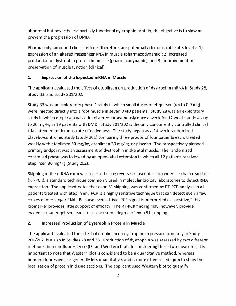
abnormal but nevertheless partially functional dystrophin protein, the objective is to slow or prevent the progression of DMD.
Pharmacodynamic and clinical effects, therefore, are potentially demonstrable at 3 levels: 1) expression of an altered messenger RNA in muscle (pharmacodynamic); 2) increased production of dystrophin protein in muscle (pharmacodynamic); and 3) improvement or preservation of muscle function (clinical).
1. Expression of the Expected mRNA in Muscle
The applicant evaluated the effect of eteplirsen on production of dystrophin mRNA in Study 28, Study 33, and Study 201/202.
Study 33 was an exploratory phase 1 study in which small doses of eteplirsen (up to 0.9 mg) were injected directly into a foot muscle in seven DMD patients. Study 28 was an exploratory study in which eteplirsen was administered intravenously once a week for 12 weeks at doses up to 20 mg/kg in 19 patients with DMD. Study 201/202 is the only concurrently controlled clinical trial intended to demonstrate effectiveness. The study began as a 24-week randomized placebo-controlled study (Study 201) comparing three groups of four patients each, treated weekly with eteplirsen 50 mg/kg, eteplirsen 30 mg/kg, or placebo. The prospectively planned primary endpoint was an assessment of dystrophin in skeletal muscle. The randomized controlled phase was followed by an open-label extension in which all 12 patients received eteplirsen 30 mg/kg (Study 202).
Skipping of the mRNA exon was assessed using reverse transcriptase polymerase chain reaction (RT-PCR), a standard technique commonly used in molecular biology laboratories to detect RNA expression. The applicant notes that exon 51 skipping was confirmed by RT-PCR analysis in all patients treated with eteplirsen. PCR is a highly sensitive technique that can detect even a few copies of messenger RNA. Because even a trivial PCR signal is interpreted as “positive,” this biomarker provides little support of efficacy. The RT-PCR finding may, however, provide evidence that eteplirsen leads to at least some degree of exon 51 skipping.
2. Increased Production of Dystrophin Protein in Muscle
The applicant evaluated the effect of eteplirsen on dystrophin expression primarily in Study 201/202, but also in Studies 28 and 33. Production of dystrophin was assessed by two different methods: immunofluorescence (IF) and Western blot. In considering these two measures, it is important to note that Western blot is considered to be a quantitative method, whereas immunofluorescence is generally less quantitative, and is more often relied upon to show the localization of protein in tissue sections. The applicant used Western blot to quantify
2
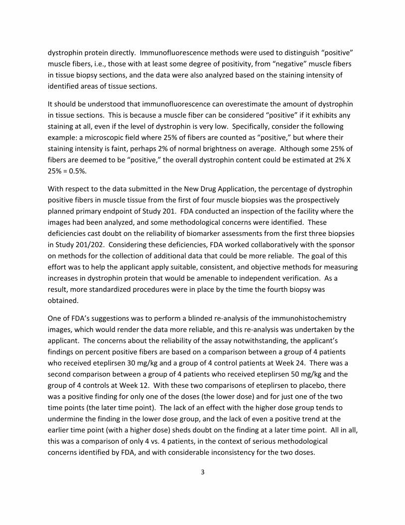
dystrophin protein directly. Immunofluorescence methods were used to distinguish “positive” muscle fibers, i.e., those with at least some degree of positivity, from “negative” muscle fibers in tissue biopsy sections, and the data were also analyzed based on the staining intensity of identified areas of tissue sections.
It should be understood that immunofluorescence can overestimate the amount of dystrophin in tissue sections. This is because a muscle fiber can be considered “positive” if it exhibits any staining at all, even if the level of dystrophin is very low. Specifically, consider the following example: a microscopic field where 25% of fibers are counted as “positive,” but where their staining intensity is faint, perhaps 2% of normal brightness on average. Although some 25% of fibers are deemed to be “positive,” the overall dystrophin content could be estimated at 2% X 25% = 0.5%.
With respect to the data submitted in the New Drug Application, the percentage of dystrophin positive fibers in muscle tissue from the first of four muscle biopsies was the prospectively planned primary endpoint of Study 201. FDA conducted an inspection of the facility where the images had been analyzed, and some methodological concerns were identified. These deficiencies cast doubt on the reliability of biomarker assessments from the first three biopsies in Study 201/202. Considering these deficiencies, FDA worked collaboratively with the sponsor on methods for the collection of additional data that could be more reliable. The goal of this effort was to help the applicant apply suitable, consistent, and objective methods for measuring increases in dystrophin protein that would be amenable to independent verification. As a result, more standardized procedures were in place by the time the fourth biopsy was obtained.
One of FDA’s suggestions was to perform a blinded re-analysis of the immunohistochemistry images, which would render the data more reliable, and this re-analysis was undertaken by the applicant. The concerns about the reliability of the assay notwithstanding, the applicant’s findings on percent positive fibers are based on a comparison between a group of 4 patients who received eteplirsen 30 mg/kg and a group of 4 control patients at Week 24. There was a second comparison between a group of 4 patients who received eteplirsen 50 mg/kg and the group of 4 controls at Week 12. With these two comparisons of eteplirsen to placebo, there was a positive finding for only one of the doses (the lower dose) and for just one of the two time points (the later time point). The lack of an effect with the higher dose group tends to undermine the finding in the lower dose group, and the lack of even a positive trend at the earlier time point (with a higher dose) sheds doubt on the finding at a later time point. All in all, this was a comparison of only 4 vs. 4 patients, in the context of serious methodological concerns identified by FDA, and with considerable inconsistency for the two doses.
3
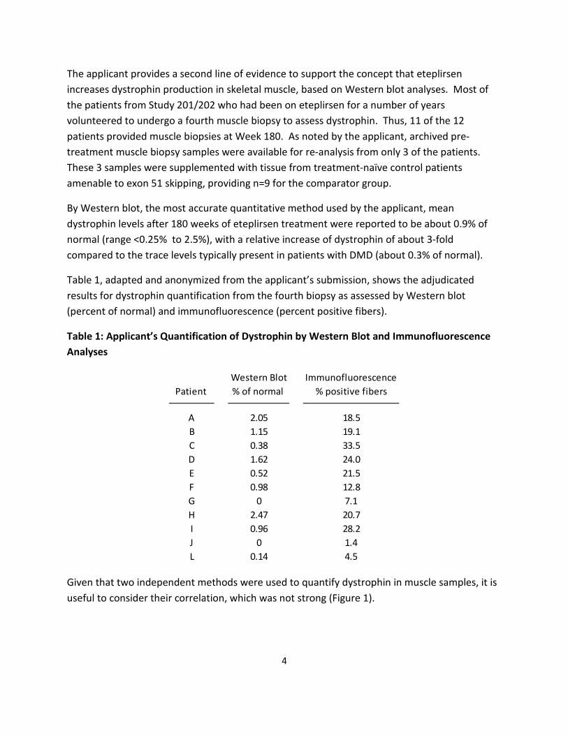
The applicant provides a second line of evidence to support the concept that eteplirsen increases dystrophin production in skeletal muscle, based on Western blot analyses. Most of the patients from Study 201/202 who had been on eteplirsen for a number of years volunteered to undergo a fourth muscle biopsy to assess dystrophin. Thus, 11 of the 12 patients provided muscle biopsies at Week 180. As noted by the applicant, archived pretreatment muscle biopsy samples were available for re-analysis from only 3 of the patients. These 3 samples were supplemented with tissue from treatment-naïve control patients amenable to exon 51 skipping, providing n=9 for the comparator group.
By Western blot, the most accurate quantitative method used by the applicant, mean dystrophin levels after 180 weeks of eteplirsen treatment were reported to be about 0.9% of normal (range <0.25% to 2.5%), with a relative increase of dystrophin of about 3-fold compared to the trace levels typically present in patients with DMD (about 0.3% of normal).
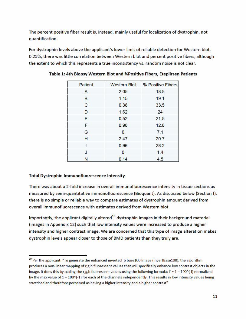
Table 1, adapted and anonymized from the applicant’s submission, shows the adjudicated results for dystrophin quantification from the fourth biopsy as assessed by Western blot (percent of normal) and immunofluorescence (percent positive fibers).
Table 1: Applicant’s Quantification of Dystrophin by Western Blot and Immunofluorescence Analyses
Patient Western Blot % of normal
Immunofluorescence % positive fibers
A B C D E F G H I J L
2.05 1.15 0.38 1.62 0.52 0.98
0 2.47 0.96
0 0.14
18.5 19.1 33.5 24.0 21.5 12.8 7.1 20.7 28.2 1.4 4.5
Given that two independent methods were used to quantify dystrophin in muscle samples, it is useful to consider their correlation, which was not strong (Figure 1).
4
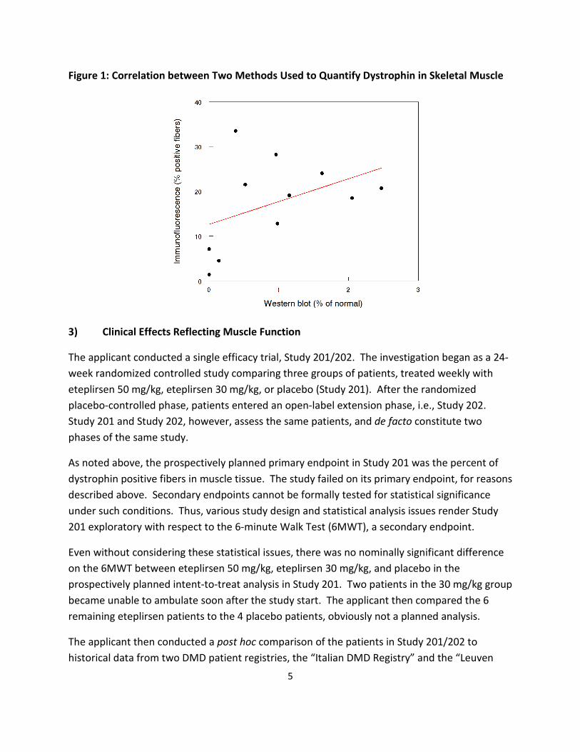
Figure 1: Correlation between Two Methods Used to Quantify Dystrophin in Skeletal Muscle
3) Clinical Effects Reflecting Muscle Function
The applicant conducted a single efficacy trial, Study 201/202. The investigation began as a 24week randomized controlled study comparing three groups of patients, treated weekly with eteplirsen 50 mg/kg, eteplirsen 30 mg/kg, or placebo (Study 201). After the randomized placebo-controlled phase, patients entered an open-label extension phase, i.e., Study 202. Study 201 and Study 202, however, assess the same patients, and de facto constitute two phases of the same study.
As noted above, the prospectively planned primary endpoint in Study 201 was the percent of dystrophin positive fibers in muscle tissue. The study failed on its primary endpoint, for reasons described above. Secondary endpoints cannot be formally tested for statistical significance under such conditions. Thus, various study design and statistical analysis issues render Study 201 exploratory with respect to the 6-minute Walk Test (6MWT), a secondary endpoint.
Even without considering these statistical issues, there was no nominally significant difference on the 6MWT between eteplirsen 50 mg/kg, eteplirsen 30 mg/kg, and placebo in the prospectively planned intent-to-treat analysis in Study 201. Two patients in the 30 mg/kg group became unable to ambulate soon after the study start. The applicant then compared the 6 remaining eteplirsen patients to the 4 placebo patients, obviously not a planned analysis.
The applicant then conducted a post hoc comparison of the patients in Study 201/202 to historical data from two DMD patient registries, the “Italian DMD Registry” and the “Leuven
5

Neuromuscular Reference Center” registry. The applicant attempted to match patients in Study 202 with patients in the historical control cohort, based on five factors: 1) corticosteroid use at baseline (use/non-use); 2) sufficient longitudinal data for 6MWT available (Y/N); 3) age ≥ 7 years (Y/N); 4) genotype amenable to any exon skipping therapy (Y/N); and 5) genotype amenable to exon 51 skipping therapy (Y/N). (Of course there is no way to assure that criteria for selecting patients from a historical database are made by individuals who are blinded to individual results.) The applicant describes “statistically significant” results in favor of eteplirsen 30 mg/kg” in Study 202, with a difference of 141 meters over the historical control.
The problems of historically-controlled studies are well-recognized, but FDA regulations (21 CFR 314.126) and accepted international guidelines (International Conference on Harmonization Guideline, Choice of Control Group and Related Issues in Clinical Trials – E10 [2000]) recognize that historical control studies can be considered adequate and well-controlled studies under the proper circumstances. FDA agreed, therefore, to consider historically-controlled data for demonstration of efficacy, but identified several issues that needed to be addressed prior to submission of the NDA.
First, the intent-to-treat (ITT) analysis, including all randomized patients, was negative for the comparison between the eteplirsen and placebo groups. In that setting, all subsequent analyses should ordinarily be considered exploratory and hypothesis-generating.
Second, patients in Study 202 appeared to be receiving optimal care, including intensive physical therapy and intensive steroid regimens. FDA asked the applicant to establish that treatment modalities in the historically-controlled population were similar, such that the historical group would be an appropriate control for the Study 202 patients.
Third, FDA noted that for most of its duration, Study 202 was open-label with all patients receiving eteplirsen, and that performance on the 6-minute walk test could be influenced by motivation and coaching. FDA expressed concern that open-label trials are susceptible to bias on the part of investigators, patients, and parents.
Although this is generally unavoidable, except in some cases where a historically controlled study is planned, historical control groups are selected with data in hand, in this case data on the 6-minute walk test.
The applicant considers the results from the historical control comparison to constitute a result on an “intermediate clinical endpoint” – a clinical endpoint that can be measured earlier than irreversible morbidity or mortality (IMM), that is reasonably likely to predict an effect on IMM or other clinical benefit, and that could suffice as a basis for accelerated approval. It should be noted that accelerated approval is based on the endpoints selected (surrogates; intermediate),
6
7
not on the adequacy of the studies supporting an effect on these endpoints. Thus, the evidence of an effect on an intermediate endpoint, if it is to serve as the basis for accelerated approval, must meet the evidentiary standard for an adequate and well-controlled study. In this case, the historically-controlled study would need to be considered adequate and well-controlled to support full or accelerated approval.
FDA has concerns regarding the comparability of treatment groups and the overall persuasiveness of the historical control comparison, as described in the FDA briefing materials. In fact, the clinical course of the 12 patients participating in Study 201/202 appears to be within the expected natural history of DMD. As discussed in the background materials, the natural history in patients amenable to exon 51 skipping indicates that the age range at loss of ambulation is wide, ranging from 8 to 16 years for most patients. Progression in DMD occurs in a generally predictable stepwise fashion, with loss of ability to stand from the floor preceding loss of ability to walk independently, which itself precedes a decline in pulmonary function. Eteplirsen-treated patients experienced a sequential worsening of functional abilities and muscle weakness, as demonstrated by changes in rise time from the floor and the evolution of North Star Ambulatory Assessment (NSAA) scores. These two outcome measures are particularly important to the interpretation of the study results. The NSAA has been specifically designed to measure functional ability in ambulatory patients with Duchenne muscular dystrophy,1 and is considered a reliable test that can be used across a range of settings. Among other functions, the NSAA measures activities of standing, walking, standing up from a chair, standing on one leg, climbing onto and descending from a box step, getting from lying to sitting, rising from the floor, jumping, hopping, and running. The NSAA is a comprehensive outcome measure, and arguably more fully reflects function in DMD than the 6MWT. In addition, the loss of the ability to rise has been described as a strong predictor of the loss of ambulation over the following 48 weeks.
The three figures below illustrate the progression of functional deficits in eteplirsen-treated patients in Study 201/202.
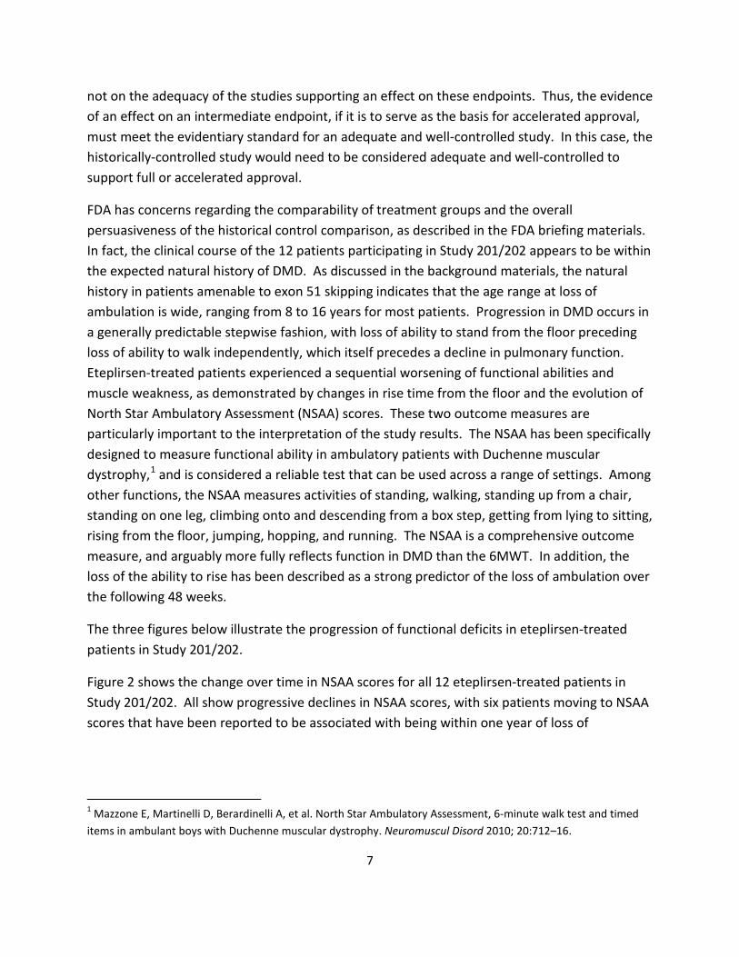
Figure 2 shows the change over time in NSAA scores for all 12 eteplirsen-treated patients in Study 201/202. All show progressive declines in NSAA scores, with six patients moving to NSAA scores that have been reported to be associated with being within one year of loss of
1 Mazzone E, Martinelli D, Berardinelli A, et al. North Star Ambulatory Assessment, 6-minute walk test and timed items in ambulant boys with Duchenne muscular dystrophy. Neuromuscul Disord 2010; 20:712–16.
ambulation, and an additional four patients moving to scores associated with being within 2 years from loss of ambulation.2
Figure 2: North Star Ambulatory Assessment (NSAA) scores in eteplirsen-treated patients in Study 201/202. The two horizontal lines indicate NSAA scores of 9 and 13, which have been reported to be associated with being either 1 or 2 years, respectively, from loss of ambulation.
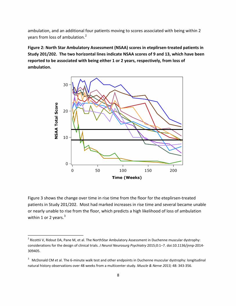
Figure 3 shows the change over time in rise time from the floor for the eteplirsen-treated patients in Study 201/202. Most had marked increases in rise time and several became unable or nearly unable to rise from the floor, which predicts a high likelihood of loss of ambulation within 1 or 2 years.3
2 Ricottii V, Ridout DA, Pane M, et al. The NorthStar Ambulatory Assessment in Duchenne muscular dystrophy: considerations for the design of clinical trials. J Neurol Neurosurg Psychiatry 2015;0:1–7. doi:10.1136/jnnp-2014309405.
McDonald CM et al. The 6-minute walk test and other endpoints in Duchenne muscular dystrophy: longitudinal natural history observations over 48 weeks from a multicenter study. Muscle & Nerve 2013; 48: 343-356.
8
3
(Figure 4).
Figure 3: Rise time in eteplirsen-treated patients in Study 201/202
In addition, the eteplirsen 6MWT open-label data were compared to publicly available data from patients treated with placebo in a recent large randomized controlled study in DMD patients with mutations amenable to exon 51 skipping4 The declines in 6MWT over time in eteplirsen-treated patients (colored lines) appear to lie within the range of changes observed in the cohort of patients treated with placebo (grey lines). Arguably, placebo-treated patients who were blinded to treatment assignment from other controlled trials are more appropriate as matched controls than registry patients, as they may receive special care and attention as trial participants, and may be more highly motivated.
4http://www.fda.gov/downloads/AdvisoryCommittees/CommitteesMeetingMaterials/Drugs/PeripheralandCentral NervousSystemDrugsAdvisoryCommittee/UCM475956.pdf
9
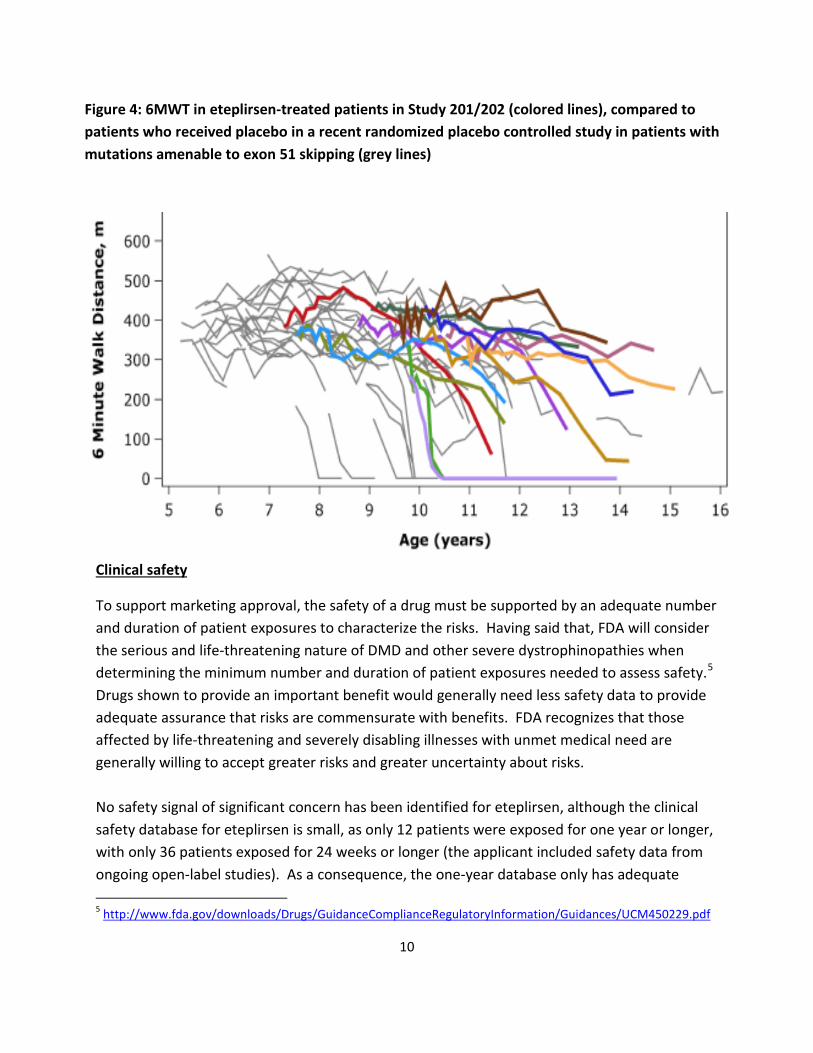
Figure 4: 6MWT in eteplirsen-treated patients in Study 201/202 (colored lines), compared to patients who received placebo in a recent randomized placebo controlled study in patients with mutations amenable to exon 51 skipping (grey lines)
Clinical safety
To support marketing approval, the safety of a drug must be supported by an adequate number and duration of patient exposures to characterize the risks. Having said that, FDA will consider the serious and life-threatening nature of DMD and other severe dystrophinopathies when determining the minimum number and duration of patient exposures needed to assess safety.5
Drugs shown to provide an important benefit would generally need less safety data to provide adequate assurance that risks are commensurate with benefits. FDA recognizes that those affected by life-threatening and severely disabling illnesses with unmet medical need are generally willing to accept greater risks and greater uncertainty about risks.
No safety signal of significant concern has been identified for eteplirsen, although the clinical safety database for eteplirsen is small, as only 12 patients were exposed for one year or longer, with only 36 patients exposed for 24 weeks or longer (the applicant included safety data from ongoing open-label studies). As a consequence, the one-year database only has adequate
5 http://www.fda.gov/downloads/Drugs/GuidanceComplianceRegulatoryInformation/Guidances/UCM450229.pdf
10
power to assess the frequencies of the more common adverse events. Less frequent events, possibly serious, may have been missed because of the small database.
Regulatory Requirements for Approval
Although approvability of a drug reflects a benefit-risk assessment, the decision about approvability is necessarily stepwise, requiring first that the drug be found effective, prior to consideration of benefit-risk.
The effectiveness requirement for a drug was added to the Food Drug and Cosmetic Act (FD&CA, the Act) in 1962. The 1962 amendments included a provision requiring manufacturers of drug products to establish a drug’s effectiveness by “substantial evidence.” Substantial evidence was defined in section 505(d) of the Act as:
“…evidence consisting of adequate and well-controlled investigations, by experts qualified by scientific training and experience to evaluate the effectiveness of the drug involved, on the basis of which it could be fairly and responsibly concluded by such experts that the drug will have the effect it purports or is represented to have under the conditions of use prescribed, recommended, or suggested in the labeling or proposed labeling thereof.”
It has been FDA’s position, based on the language of the statute and the legislative history of the 1962 amendments, that Congress generally intended to require at least two adequate and well-controlled trials, each convincing on its own, to establish effectiveness.
In 1997 under the FDA Modernization Act (FDAMA) section 505(d) of the Act was amended to make it clear that the Agency may consider “data from one adequate and well-controlled clinical investigation and confirmatory evidence” to constitute substantial evidence if FDA determines that such data and evidence are sufficient to establish effectiveness.
Thus, a single highly persuasive positive trial combined with independent findings that substantiate efficacy might support approval, but it is critical that the possibility of an incorrect outcome be considered and that all the available data be examined for their potential to either support or undercut reliance on a single trial. FDA described in a guidance document6 the characteristics of a single adequate and well-controlled study that could support an effectiveness claim. These include: 1) large multicenter study; 2) consistency across study subsets; 3) multiple studies within a study (e.g., properly designed factorial study analyzed as a
6 US Food and Drug Administration. Guidance for Industry. Providing Clinical Evidence of Effectiveness for Human Drug and Biological Products. http://www.fda.gov/downloads/Drugs/.../Guidances/ucm078749.pdf. May 1998. Accessed December 17, 2015.
11
series of pairwise comparisons); 4) multiple endpoints involving different events; and 5) statistically very persuasive findings. These factors should be considered in assessing whether Study 201/202 as a single study could be sufficient to support approval.
DMD is a rare and serious disease without approved treatments, and FDA has long stressed that it is appropriate to exercise the broadest flexibility in applying the statutory standards to drugs for such diseases, while preserving appropriate guarantees for effectiveness and safety.7
Accelerated Approval
The applicant is seeking accelerated approval for eteplirsen. Accelerated approval is a particular type of approval that FDA may grant for a product for a serious or life-threatening disease or condition upon a determination that the product has an effect on a surrogate endpoint that is reasonably likely to predict clinical benefit, or on a clinical endpoint that can be measured earlier than irreversible morbidity or mortality and that is reasonably likely to predict an effect on irreversible morbidity or mortality or other clinical benefit, taking into account the severity, rarity, or prevalence of the condition and the availability or lack of alternative treatments.8
Two potential pathways to accelerated approval were discussed with the applicant during the eteplirsen development program:
1. Using clinical data from Study 201/202 on 6-minute walk distance as an intermediate clinical endpoint that could have the potential to support accelerated approval.
Under that approach, the basis for accelerated approval would be a conclusion that eteplirsen reduced the rate of decline of walking performance to an extent that is reasonably likely to predict a long-term beneficial effect on irreversible morbidity or mortality. Study 201 clearly failed to show an advantage of eteplirsen over placebo on 6-minute walk distance in the placebo-controlled trial. The specific finding proposed by the applicant as supporting accelerated approval is the comparison of 6-minute walk distance between the 12 patients in Study 201/202 and historical controls, where the control patients were selected post hoc. There are significant concerns regarding the ability to draw valid conclusions from this historically controlled comparison. Moreover, comparisons between patients in Study 201/202 and patients in a related development program who had received placebo suggest that the
7 21 CFR 312.80, subpart E
8 http://www.fda.gov/downloads/drugs/guidancecomplianceregulatoryinformation/guidances/ucm358301.pdf
12
change in 6-minute walk distance with eteplirsen was consistent with the natural history of the disease.
2. Using dystrophin data as a surrogate endpoint to support accelerated approval.
FDA indicated in the DMD guidance1 that biomarkers that reliably reflect the health and amount of skeletal muscle may, if supported by sufficient scientific evidence and acceptable analytical methods, be used as surrogate endpoints to support accelerated approval of a new DMD drug. For eteplirsen, the quantification of dystrophin present in the fourth muscle biopsy was assessed by Western Blot, and compared with treatment-naïve controls that were selected by the applicant. The apparent treatment effect could be expressed as a 3-fold increase over the trace amount present at baseline, but relative changes can be difficult to interpret. The mean dystrophin level in patients who had been treated with eteplirsen for some 180 weeks was on average 0.9% of normal, far below levels observed in a milder form of muscular dystrophy known as Becker-type muscular dystrophy (BMD). The minimum level of dystrophin that might be reasonably likely to predict clinical benefit in patients with BMD remains unknown, but experts in DMD9,10 have stated that levels less than 3% of that of normal healthy muscle are generally associated with the typical DMD phenotype, and have proposed that “induction of approximately 10% of normal dystrophin levels sets a minimum level to confer measurable clinical benefit.”11 In addition, so called “exon 51-model” BMD patients, who have the same truncated form of dystrophin that would be produced by eteplirsen in DMD patients, and experience a mild disease, express truncated dystrophin at levels reported to range from 50% to 100% of normal.
Importantly, the evidentiary standards for effectiveness are not lower for biomarker endpoints used to support accelerated approval, nor should accelerated approval be used to compensate for weak or inconsistent clinical findings.
Although FDA is prepared to be flexible with respect to a devastating illness with no treatment options, we cannot approve drugs for which substantial evidence of effectiveness has not been established. Thus, as you digest the background materials, we hope you will carefully consider
9 Flanigan KM. Duchenne and Becker muscular dystrophies. Neurol Clin 2014; 32: 671-688
10 Lu QL, Cirak S, Partridge T. What can we learn from clinical trials of exon skipping for DMD? Mol Ther Nucleic acids. 2014; 3: e152
11 Wilton SD, Veedu RN, Fletcher S. The emperor’s new dystrophin: finding sense in the noise. Trends in Molecular Medicine 2015; 21: 417-426.
13

the strengths and weaknesses of all of the data, and be prepared to consider and discuss whether or not you believe that efficacy has been established.
It is important to recognize that no final conclusions have been reached on the approvability of this application, and we look forward to a fruitful discussion of these issues at the Advisory Committee Meeting on January 22, 2016.
14

II. Drafts Points To Consider
FOOD AND DRUG ADMINISTRATION (FDA)
Center for Drug Evaluation and Research (CDER)
Peripheral and Central Nervous System Drugs Advisory Committee Meeting
DRAFT POINTS TO CONSIDER
January 22, 2016
1. Consider the data for dystrophin expression, including the following a. Experimental methods, including consideration of accuracy, reliability,
reproducibility, etc. b. Potential clinical meaning, including consideration of amount of dystrophin relative
to patients with Becker muscular dystrophy, functionality of the truncated dystrophin, and percent of muscle fibers with detectable dystrophin.
2. Consider the data for clinical measures, including the following a. Design and potential interpretability of Study 201/202, including consideration of a)
the placebo-controlled period, and b) comparison of the open-label experience to natural history.
b. Results of Study 201/202 in the context of the study design.
3. Consider the possible design of any future efficacy and safety studies that might be necessary.
III. Clinical Team Leader Memorandum to the Committee
MEMORANDUM
DATE: December 8, 2015
FROM: Ronald Farkas, M.D., Ph.D. Clinical Team Leader Division of Neurology Products, CDER, FDA
TO: Members and Invited Guests of the Peripheral and Central Nervous Systems Drugs Advisory Committee (PCNS AC)
SUBJECT: Clinical Team Leader Memo for New Drug Application (NDA) 206488, for the use of Exondys 51 (eteplirsen) for the treatment of Duchenne muscular dystrophy in patients with mutations amenable to exon 51 skipping
1

Table of Contents
Table of Figures ............................................................................................................................................. 3
1. Disease Background .............................................................................................................................. 3
2. Eteplirsen Drug Development Rationale............................................................................................... 3
3. Dystrophin Evidence ............................................................................................................................. 4
a. Study 33 ............................................................................................................................................ 4
b. Study 28 ............................................................................................................................................ 4
c. Study 201/202, First 3 Biopsies ......................................................................................................... 5
d. Study 201/202, 4th Biopsy ................................................................................................................. 9
e. Dystrophin in BMD .......................................................................................................................... 12
f. Reviewer Discussion, Dystrophin Quantification Methods ............................................................ 13
g. FDA Review Team Preliminary Conclusions on Dystrophin Findings .............................................. 15
4. Clinical Efficacy Evidence .................................................................................................................... 16
a. Design and analysis of Study 201/202 ............................................................................................ 16
Increases in rise time in eteplirsen-treated patients predict a high likelihood of sequential loss of
Issues with comparison of eteplirsen-treated patients with applicant’s proposed historical
ambulation within 1 or 2 years ........................................................................................................... 23
controls ............................................................................................................................................... 25
b. FDA Review Team Preliminary Conclusions, Clinical Endpoints ..................................................... 29
6. Clinical Safety ...................................................................................................................................... 30
2
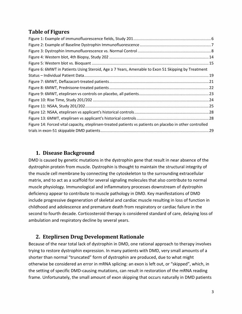
Table of Figures Figure 1: Example of immunofluorescence fields, Study 201 ....................................................................... 6
Figure 2: Example of Baseline Dystrophin Immunofluorescence ................................................................. 7
Figure 3: Dystrophin Immunofluorescence vs. Normal Control ................................................................... 8 Figure 4: Western blot, 4th Biopsy, Study 202 ........................................................................................... 14
Figure 5: Western blot vs. Bioquant ........................................................................................................... 15
Figure 6: 6MWT in Patients Using Steroid, Age ≥ 7 Years, Amenable to Exon 51 Skipping by Treatment Status – Individual Patient Data .................................................................................................................. 19
Figure 7: 6MWT, Deflazacort-treated patients ........................................................................................... 21
Figure 8: 6MWT, Prednisone-treated patients ........................................................................................... 22 Figure 9: 6MWT, eteplirsen vs controls on placebo, all patients................................................................ 23
Figure 10: Rise Time, Study 201/202 .......................................................................................................... 24
Figure 11: NSAA, Study 201/202 ................................................................................................................. 25 Figure 12: NSAA, eteplirsen vs applicant's historical controls .................................................................... 28
Figure 13: 6MWT, eteplirsen vs applicant's historical controls .................................................................. 28
Figure 14: Forced vital capacity, eteplirsen-treated patients vs patients on placebo in other controlled
trials in exon-51 skippable DMD patients ................................................................................................... 29
1. Disease Background DMD is caused by genetic mutations in the dystrophin gene that result in near absence of the dystrophin protein from muscle. Dystrophin is thought to maintain the structural integrity of the muscle cell membrane by connecting the cytoskeleton to the surrounding extracellular matrix, and to act as a scaffold for several signaling molecules that also contribute to normal muscle physiology. Immunological and inflammatory processes downstream of dystrophin deficiency appear to contribute to muscle pathology in DMD. Key manifestations of DMD include progressive degeneration of skeletal and cardiac muscle resulting in loss of function in childhood and adolescence and premature death from respiratory or cardiac failure in the second to fourth decade. Corticosteroid therapy is considered standard of care, delaying loss of ambulation and respiratory decline by several years.
2. Eteplirsen Drug Development Rationale Because of the near total lack of dystrophin in DMD, one rational approach to therapy involves trying to restore dystrophin expression. In many patients with DMD, very small amounts of a shorter than normal “truncated” form of dystrophin are produced, due to what might otherwise be considered an error in mRNA splicing: an exon is left out, or “skipped”, which, in the setting of specific DMD-causing mutations, can result in restoration of the mRNA reading frame. Unfortunately, the small amount of exon skipping that occurs naturally in DMD patients
3
does not appear to appreciably slow muscle degeneration. It was reasoned, however, that if exon skipping could be augmented by drug therapy, levels of the truncated dystrophin could be increased to a level high enough to confer clinical benefit. Eteplirsen was designed to bind to dystrophin mRNA at a specific site to cause the splicing machinery to skip exon 51, thus restoring the dystrophin reading frame in certain amenable patients, and increasing production of the truncated dystrophin. How much of the truncated dystrophin would be necessary to confer clinical benefit remains an open question, but a related form of muscular dystrophy, called Becker muscular dystrophy (BMD), provides a natural model of what exon skipping in DMD might achieve. In so-called “exon 51-model” BMD patients, the same truncated form of dystrophin that would be produced by eteplirsen in DMD patients occurs naturally. These BMD patients experience a mild, or in some cases asymptomatic, muscle disease. Importantly, however, the truncated dystrophin in these BMD patients is expressed at high levels, roughly 50- to 100%1 of what would be expected for normal dystrophin.
3. Dystrophin Evidence Dr. Ashutosh Rao, from the Office of Biotechnology Products, reviewed dystrophin methodologies and supporting assays. The effect of eteplirsen on dystrophin expression was examined in 3 clinical studies: Study 33, Study 28, and Study 201/202, as follows:
a. Study 33: In this exploratory phase 1 study, small doses of eteplirsen (up to 0.9 mg total) were injected directly into a foot muscle in 7 patients with DMD. An increase in dystrophin expression was reported adjacent to the needle track, but it is not clear if, or to what degree, this might reflect the activity of eteplirsen when given by the intravenous (IV) route, which does not produce similar high local concentrations or mechanical effects.
b. Study 28: In this exploratory study, eteplirsen was administered intravenously once a week for 12 weeks at doses ranging from 0.5 to 20 mg/kg, with up to 4 patients per dose level. The methods for dystrophin quantification were not reviewed by FDA prior to the conduct of the study, and FDA has concerns about the reliability of the methods and procedures. In one response from the applicant to an information request from FDA about quality control methods, the applicant responded that “Study 28 was an exploratory phase 1b study which was only intended to generate proof of concept data to guide future studies. For this reason, quality controls for the dystrophin data in Study 28 were not properly optimized.” In addition, Study 28 examined dystrophin levels after 12 weeks of dosing, but it is necessary to understand dystrophin levels that are present with longer
1 Anthony K et al (2011) Dystrophin quantification and clinical correlations in Becker muscular dystrophy: implications for clinical trials. Brain. 134,3544-3556.
4
term, more clinically relevant durations of therapy. Thus, as mentioned above, FDA considers the 4th biopsy from patients in Study 201/202, which was taken after 180-weeks of treatment with eteplirsen, to be of greater potential clinical relevance.
c. Study 201/202, First 3 Biopsies: Study 201/202 was a 3-arm, 12-patient study comparing the effects of 30 mg/kg or 50 mg/kg IV eteplirsen to placebo. Biopsies were taken at baseline, week 12 (for half the patients), week 24 (for the other half), and week 48 for all patients. During the development of eteplirsen FDA communicated to the applicant concerns about the biomarker studies on the first 3 biopsies.2 With additional review following submission of the NDA, it is not clear that any of the dystrophin biomarker data from the first 3 biopsies are reliable or interpretable.
Immunofluorescence images
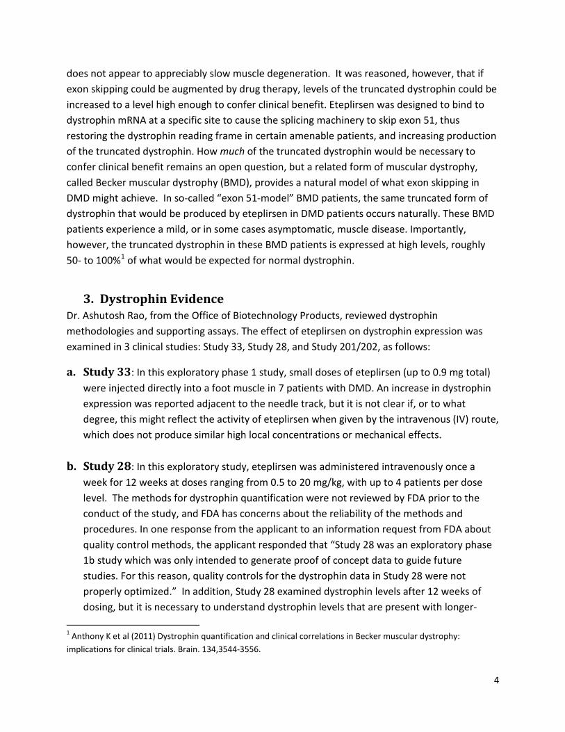
The measurement of total dystrophin immunofluorescence by Bioquant was first carried out on blinded baseline, Week 12, and Week 24 images, captured at 20x magnification. The results showed essentially no change in intensity for any patient. Negative results were obtained both when the study was conducted with MANDYS106 antibody or with Dys2 antibody. However, investigators attributed the negative results to the image magnification, and captured new images at 40x magnification after the blind was broken, with personnel reporting to FDA site inspectors that positive fields were uniquely selected for further quantitation. The images selected at 40x magnification showed roughly a doubling of immunofluorescence intensity for all patients between baseline and Week 12 (50 mg/kg patients) or week 24 (30 mg/kg patients). Because the analyses were intentionally targeted to fibers whose staining intensity exceeded a particular threshold, it is not clear whether these results are representative or interpretable.
The 20x immunofluorescence images on samples obtained through Week 24 were selected by an individual blinded to treatment group, but the microscopic fields to be photographed were selected manually by the operator, as opposed to a more automated method introduced for studies of the 4th biopsy. Bias in field selection may have resulted in preferential capture of bright fibers that appear similar to revertant fibers.
2 e.g. at a meeting on March 13, 2013, FDA stated “while we do not believe that you have adequately characterized the quantity of truncated dystrophin produced by eteplirsen treatment (Western blot data is not available), the immunofluorescence data you presented suggest that a much lower quantity of truncated dystrophin is produced by eteplirsen treatment than is present in BMD.” In the April 15, 2014, advice letter in which potential pathways for approval were discussed, FDA stated “After examining the source data and images you provided in support of dystrophin protein expression from eteplirsen treatment, we remain skeptical about the persuasiveness of the data, and concerned about serious methodological problems explained previously.”
5
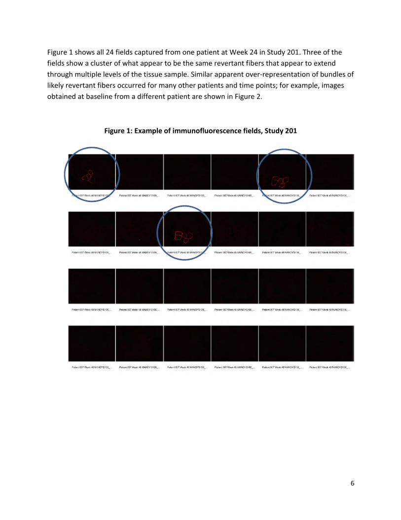
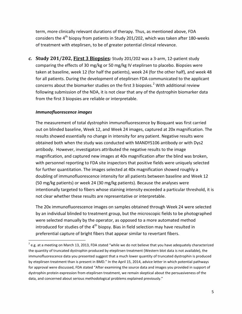
Figure 1 shows all 24 fields captured from one patient at Week 24 in Study 201. Three of the fields show a cluster of what appear to be the same revertant fibers that appear to extend through multiple levels of the tissue sample. Similar apparent over-representation of bundles of likely revertant fibers occurred for many other patients and time points; for example, images obtained at baseline from a different patient are shown in Figure 2.
Figure 1: Example of immunofluorescence fields, Study 201
6
Figure 2: Example of Baseline Dystrophin Immunofluorescence
Week 48 samples were processed separately for dystrophin immunofluorescence from earlier samples, and had higher background staining. As a consequence, valid comparison is not possible with earlier time points for percent positive fibers or total immunofluorescence because the higher background staining, and not necessarily an effect of drug, could be responsible for any differences observed.
Importantly, the Week 48 immunofluorescence was still very low, and much less intense than normal controls, as shown in Figure 3. The top two images show the intensity as originally captured, and the bottom two images show the intensity converted to “heatmap” images that represent the observed (unmodified) pixel intensity as color, from low intensity blue to high intensity red and white.
7
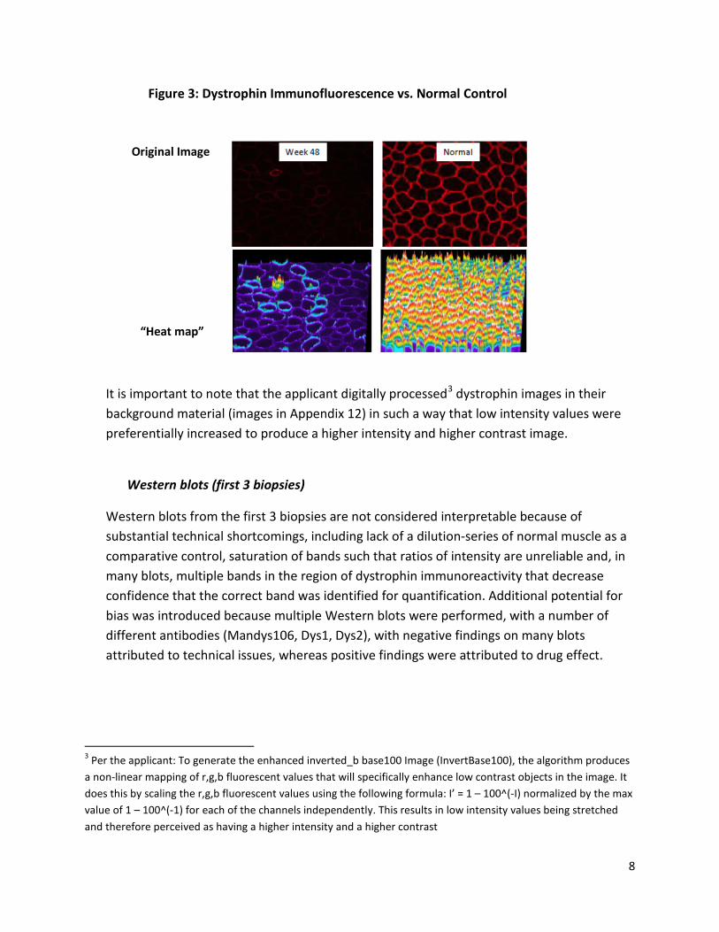
Figure 3: Dystrophin Immunofluorescence vs. Normal Control
Original Image
“Heat map”
It is important to note that the applicant digitally processed3 dystrophin images in their background material (images in Appendix 12) in such a way that low intensity values were preferentially increased to produce a higher intensity and higher contrast image.
Western blots (first 3 biopsies)
Western blots from the first 3 biopsies are not considered interpretable because of substantial technical shortcomings, including lack of a dilution-series of normal muscle as a comparative control, saturation of bands such that ratios of intensity are unreliable and, in many blots, multiple bands in the region of dystrophin immunoreactivity that decrease confidence that the correct band was identified for quantification. Additional potential for bias was introduced because multiple Western blots were performed, with a number of different antibodies (Mandys106, Dys1, Dys2), with negative findings on many blots attributed to technical issues, whereas positive findings were attributed to drug effect.
3 Per the applicant: To generate the enhanced inverted_b base100 Image (InvertBase100), the algorithm produces a non-linear mapping of r,g,b fluorescent values that will specifically enhance low contrast objects in the image. It does this by scaling the r,g,b fluorescent values using the following formula: I’ = 1 – 100^(-I) normalized by the max value of 1 – 100^(-1) for each of the channels independently. This results in low intensity values being stretched and therefore perceived as having a higher intensity and a higher contrast
8

d. Study 201/202, 4th Biopsy Biomarker studies on the 4th biopsy obtained at Week 180 were conducted by the applicant with technical advice from FDA. However, the reliability of results remains questionable for a number of reasons, including the following:
• Controls were not matched by muscle group: Biopsies at Week 180 were taken from the deltoid, one of the few muscle groups that, along with the calf muscle, can be hypertrophied in DMD.4 In contrast, both the baseline samples available from eteplirsen-treated patients, and most of the new external controls from untreated patients, were obtained from the biceps (except for one, which was obtained from deltoid). There is little human data on differences in dystrophin levels between muscle groups but, in nonclinical models of DMD, there is evidence that dystrophin levels vary between muscles,5 with may affect the readout of experiments in which the effectiveness of the treatment is not particularly high.
• Controls were not matched by patient: There appears to be considerable inter-patient variability in dystrophin levels present in exon-51 skippable DMD. In Western blots from biopsies of extensor digitorum brevis (EDB),6 dystrophin levels averaged about 0.3% of normal, but ranged from undetectable to ≈ 1% of normal or somewhat higher. The applicant obtained data from biopsies of 9 untreated patients, and reported an average dystrophin level of 0.08%. 7 However, such a small sample size may not provide a reliable estimate of baseline levels that were present in the eteplirsen-treated patients. The dystrophin level estimated in these biceps controls is lower than the estimate from the EDB biopsies, perhaps because dystrophin levels truly differ between these muscle groups, or perhaps only secondary to chance when a small number of observations with high variability are compared.
4 Pradhan S (2002) Valley sign in Duchenne muscular dystrophy: importance in patients with inconspicuous calves. Neurol India. 50,184-186.
5 Pigozzo S et al (2013) Revertant fibers in the mdx murine model of Duchenne muscular dystrophy: an age- and muscle-related reappraisal. PLOS ONE. 8,e72147
6 FDA Advisory Committee presentation for drisapersen, slide 43.
7 Noting, however, that values <0.25% were rounded to zero. Including those lower values leads to an average level about twice as high, but still half as much as in EDB.
9

• Lack of independent confirmation: The applicant has not obtained independent confirmation of dystrophin findings.8
Exon Skipping The applicant reported positive findings for all patients on detection of exon 51-skipped mRNA, as measured by RT-PCR. However, RT-PCR is highly sensitive to the presence of even a few molecules of mRNA, and does not indicate how much, or even if any, dystrophin protein might have been produced.
Western Blot, 4th biopsy
Western blot results for eteplirsen-treated patients is shown in Table 1. Dystrophin levels in treated patients were, on average, about 0.9% of normal9 (range <0.25% -2.5%) as measured by Western blot, the most quantitative method used by the applicant.
At the low dystrophin levels present in the Week 180 biopsies, random measurement error can be large in comparison to the estimated amount of dystrophin. Consequently, little confidence can be placed on any individual patient value, and the data should not be considered as reliable evidence that some patients failed to produce any dystrophin from eteplirsen whereas others were more responsive.
Percent Positive Fibers
Table 1 shows the percent positive fibers in eteplirsen patients. On average, the percentage of fibers with any detectable staining was about 17%, versus about 1% in the controls selected by the applicant. It is important to stress, however, that the applicant’s definition of a positive fiber was not based on a threshold amount of dystrophin or staining brightness, but rather only on “a majority of the fiber perimeter stain at an intensity judged by eye to be above background of the image.”[emphasis added] Consequently, “17% positive fibers” does not correspond to 17% of normal dystrophin levels, or to 17% of fibers being as bright as in BMD.
8 For example, in the April 15, 2014, letter discussing data that would be filed with the NDA, FDA stated “We expect that the initial biomarker data from these [newly exposed patients] exposures will start becoming available at about the time of NDA submission and shortly thereafter.” Also, as early as the July 23, 2013 meeting FDA expressed concern that “all muscle biopsies were obtained and processed by a single technician at a single study center” and that in part because of concern about bias, “we also ask that you confirm, [biomarker results] by an independent laboratory.”
9 The applicant notes that Week 180 samples were measured relative to a single normal individual’s deltoid muscle biopsy, which introduces additional uncertainty into the interpretation of fold increase vs. normal because dystrophin appears to vary about 2-fold among different normal individuals.
10

e. Dystrophin in BMD
Quantity: The minimum level of Becker-type dystrophin that might be reasonably likely to predict clinical benefit remains unknown, but experts in DMD,11 including those directly involved in the development of eteplirsen,12 have stated that levels less than 3% of that of normal healthy muscle, as identified by Western blotting, are generally associated with the typical DMD phenotype, and have proposed, based on a wide range of scientific observations, that “induction of approximately 10% of normal dystrophin levels sets a minimum level to confer measurable clinical benefit.” 13
Dystrophin levels in exon-51 model BMD patients have been observed to be much higher than these estimates, roughly 80% of normal on average.14 The clinical phenotype in these patients is, however, generally much milder than DMD, and this should not be taken to suggest that such high levels would be necessary for any benefit.
Timing: Experts have cautioned that dystrophin is present in BMD from birth, and that “we should not conclude that dystrophin restitution in DMD patients with established dystrophic pathology will confer comparable benefits to the dystrophins in BMD patients”15 for reasons including the pro-inflammatory environment that develops in DMD.16
Functionality: The exact dystrophin mutation affects the clinical phenotype in BMD, and likely also in DMD, confounding interpretation of any possible clinical impact of small differences in dystrophin levels among DMD patients, with experts stressing that “it will be essential to
11 Flanigan KM (2014) Duchenne and Becker muscular dystrophies. Neurol Clin. 32,l 671-688.
12 Lu QL, Cirak S, Partridge T (2014) What can we learn from clinical trials of exon skipping for DMD? Mol Ther Nucleic acids. 3, e152.
13 Wilton SD, Veedu RN, Fletcher S (2015) The emporer’s new dystrophin: finding sense in the noise. Trends in Molecular Medicine. 21, 417-426.
14 Anthony K et al (2011) Dystrophin quantification and clinical correlations in Becker muscular dystrophy: implications for clinical trials. Brain. 134, 3544-3556.
15 Wilton SD, Fletcher S, Flanigan KM(2014) Dystrophin as a therapeutic biomarker: Are we ignoring data from the
pase? Neuromuscular Disorder. 24, 463-466.
16 Rosenberg et al (2015) Immune-mediated pathology in Duchenne muscular dystrophy. Sci Transl Med 7,299rv4.
12
account for different mutations when looking at other possible contributing factors to disease severity.”17
Localization: In BMD, dystrophin is typically present in all or most fibers18,19 and, in addition to the total amount, this is thought to be important for function of the dystrophin. In contrast, in DMD many patients have no detectable dystrophin staining, while others have bright staining in a small percentage (1- to 5%) of “revertant” fibers in which exon skipping is thought to occur spontaneously. Some DMD patients can also show faint dystrophin staining in up to about 25% of fibers,20 with the percentage of positive fibers appearing to depend in part on technical factors that affect assay sensitivity.
Unusual BMD Patients: Rarely, patients with BMD are encountered who have dystrophin levels that are less than 1% of normal, which is as low as typical DMD patients. Importantly, however, rather than suggesting that very low levels of drug-induced dystrophin are likely to be beneficial, such patients highlight the complexity of the relationship between dystrophin levels and phenotype. The fact that such patients can have mild disease appears to be unrelated to, not necessarily the result of, low levels of dystrophin. In this context, the applicant selected three BMD patients as comparators for the Week 180 dystrophin studies, one of whom had low dystrophin level of about 2% of normal. However, the BMD patients selected by the applicant do not appear representative, and this patient may correspond to one of the rare BMD patients with very low dystrophin levels.
f. Reviewer Discussion, Dystrophin Quantification Methods Considerable confusion can be created by the fact that a number of different methods have been used to quantify dystrophin expression, some more quantitative than others, and some producing higher absolute numbers than others. As discussed above, immunofluorescence is mainly informative of dystrophin localization, but is not a reliable measure of dystrophin amount (beyond perhaps the binary distinction between “undetectable” and “detectable”). For
17 Van den Bergen JC et al (2014) Dystrophin levels and clinical severity in Becker muscular dystrophy patients. Neurol Neurosurg Psychiatry. 85, 747-753.
18 Arahata et al (1989) Dystrophin diagnosis: comparison of dystrophin abnormalities by immunofluorescence and immunoblot analysis. Proc Natl Acad Sci. 86,7154-7158.
19 Morandi et al (1995) Dystrophin characterization in BMD patients: correlation of abnormal protein with clinical phenotype. Journal of Neurological Sciences 132,146-155.
20 Arechavala-Gomeza et al (2010) Revertant fibres and dystrophin traces in Duchenne muscular dystrophy: implications for clinical trials. Neuromuscul Disord. 20,295-301.
13
example, in many patients with typical DMD, only trace levels of dystrophin are present, yet this results in 25% or more of fibers being faintly dystrophin-positive.
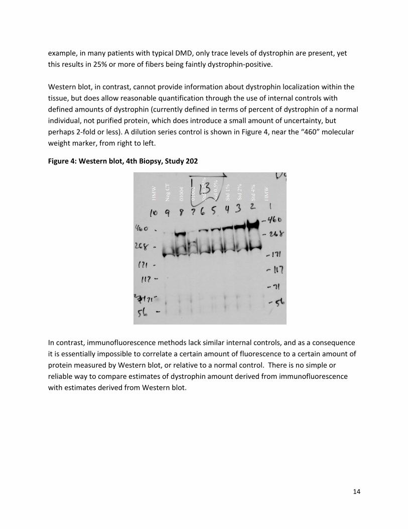
Western blot, in contrast, cannot provide information about dystrophin localization within the tissue, but does allow reasonable quantification through the use of internal controls with defined amounts of dystrophin (currently defined in terms of percent of dystrophin of a normal individual, not purified protein, which does introduce a small amount of uncertainty, but perhaps 2-fold or less). A dilution series control is shown in Figure 4, near the “460” molecular weight marker, from right to left.
Figure 4: Western blot, 4th Biopsy, Study 202
In contrast, immunofluorescence methods lack similar internal controls, and as a consequence it is essentially impossible to correlate a certain amount of fluorescence to a certain amount of protein measured by Western blot, or relative to a normal control. There is no simple or reliable way to compare estimates of dystrophin amount derived from immunofluorescence with estimates derived from Western blot.
14

Figure 5 shows that at low levels of dystrophin (<5% by Western blot), immunofluorescence appears to overestimate the amount of dystrophin; for example, immunofluorescence shows about 25% intensity for samples with roughly 1- or 2% of normal dystrophin by Western blot, and shows about 10% of normal intensity for samples with <1% of normal dystrophin levels.
Figure 5: Western blot vs. Bioquant
Finally, a representation of the change in dystrophin levels in terms of percent change from baseline is problematic in this situation, because the trace baseline dystrophin levels in many patients are too low to be measured accurately, resulting in ratios that are imprecise, and that are greatly affected by small amounts of random variability in denominators that are close to zero.
g. FDA Review Team Preliminary Conclusions on Dystrophin Findings
Adequate scientific methods appear to be available to measure dystrophin expression in DMD. As discussed in the recent FDA draft Guidance on DMD,21 there is justifiable interest in dystrophin as a potential surrogate endpoint for accelerated approval in DMD. However, the Guidance also states that the potential for a biomarker to predict clinical benefit in DMD is inseparable from such factors as the magnitude of change of the biomarker. Regarding
21 Duchenne muscular dystrophy and related dystrophinopathies: developing drugs for treatment. http://www.fda.gov/Drugs/GuidanceComplianceRegulatoryInformation/Guidances/default.htm
15
methodology, the Guidance stresses the importance of the performance characteristics of the biomarker assays, including quality-control measures.
Based on the data submitted by the applicant, considerable doubt remains about how much, or perhaps even whether, dystrophin levels were increased by eteplirsen. The degree of uncertainty about the dystrophin data hinders discussion of its use as surrogate endpoint for eteplirsen. However, to the degree that the dystrophin data may be interpretable, the amount and distribution of dystrophin in treated patients appears to be within the range typically associated with DMD, not BMD. Data suggesting that higher levels of dystrophin were produced by eteplirsen appear unreliable.
4. Clinical Efficacy Evidence
The only study that evaluated clinical efficacy is Study 201/202. Dr. Xiang Ling, from the Office of Biometrics, provided a statistical review of that study. As described below, and in Dr. Ling’s review, Study 201/202 was not designed in a way that allows reliable use of statistical hypothesis testing (i.e., “p-values”), and is only capable of providing interpretable evidence of efficacy if the beneficial effect of eteplirsen is so large that it is essentially self-evident, without the use of statistics.
a. Design and analysis of Study 201/202
Clinical efficacy was examined in one single-center, 24-week, 3-arm controlled trial (Study 201) in 12 patients assigned 1:1:1 to 30 mg/kg eteplirsen, 50 mg/kg eteplirsen, or placebo. Study 201 was continued as an open-label extension, called Study 202, which has been ongoing for more than 3 years. Multiple functional endpoints were assessed both in the placebo-controlled and open-label extension periods, including 6 minute walk test (6MWT), North Star Ambulatory Assessment (NSAA), and a number of measures of pulmonary function. Analysis of clinical endpoints was not controlled for multiplicity, but in Study 201 the clinical endpoints were essentially uniformly negative, without trends supportive of efficacy.
Shortly after Study 202 passed 1 year duration, the applicant proposed a post-hoc analysis with a number of changes from the original analysis: a) data for 2 out of 8 patients treated with eteplirsen (patients who quickly lost ambulation) were dropped, b) the prespecified comparison of each dose arm to placebo was changed to comparison of the 6 remaining treated patients to the 4 placebo-treated patients, and c) the endpoint was taken to be Week 36, instead of Week 24. FDA explained in detail to the applicant in March of 2013 why the proposed analysis was unreasonable even for hypothesis generation, and why Study 201 did not provide evidence of efficacy.
16
As the duration of exposure in Study 202 increased, the applicant proposed comparing the clinical course of treated patients to historical controls. FDA expressed strong reservations regarding the potential interpretability of the applicant’s proposed comparison to historical controls and the use of 6MWT as the primary endpoint in such a historical comparison. Because of these concerns, FDA noted that a dramatic effect size would be necessary for any such analysis to be potentially interpretable. Well-designed historically-controlled trials can, in certain circumstances, be considered adequate and well-controlled designs that can support FDA approval. However, Study 201/202 is not a well-designed historically-controlled trial. It is well established, as detailed in guidelines developed by U.S. and international regulatory bodies,22 that “inability to control bias is the major and well-recognized limitation of externally-controlled trials, and it is always difficult, and in many cases impossible, to establish comparability of the treatment and control groups.” Furthermore “a consequence of the recognized inability to control bias is that the potential persuasiveness of findings from externally controlled trials depends on obtaining much more extreme levels of statistical significance and much larger estimated differences between treatments than would be considered necessary in concurrently controlled trials.”
FDA encouraged the sponsor at the March 2013 meeting to conduct an adequately powered placebo-controlled trial of eteplirsen, stating “if it is true that eteplirsen leads to remarkable clinical benefit in even some patients, there is no doubt that a feasible placebo controlled study can be designed to demonstrate that benefit.” FDA also stated that “there is considerable variation among individual patients with regard to clinical measures and important milestones” and that data from an open-label study “may only be interpretable if a relevant objective endpoint obviously insulated from bias demonstrated compelling data that are clearly outside the know variability range for DMD.” FDA further stated that, at that time, comparison of data from Study 202 did not provide interpretable evidence of benefit “given the limitations of the open-label design for protecting against bias on effort-dependent endpoints like 6MWT.” At a July 2013 meeting with the applicant, at which the possibility of NDA filing based on dystrophin production was discussed, FDA similarly expressed reservations about natural history controls “due to the usual difficulty in showing comparability between the study populations in natural history studies,” and reiterated that 6MWT was susceptible to bias in the proposed natural history comparison.
Discussions about comparison of Study 202 patients to natural history continued with the April 15, 2014, communication from FDA to the applicant which stated that, with additional data to support the efficacy and safety of eteplirsen, an NDA should be filable. FDA noted that patients
22 Choice of control group and related issues in clinical trials, E10. International Conference on Harmonisation of Technical Requirements for Registration of Pharmaceuticals for Human Use, 2000.
17
in Study 202 appeared to be receiving optimal care, including intensive physical therapy and intensive steroid regimens, and again stated that “performance on the 6-minute walk test is strongly influenced by motivation and coaching, and open-label trials are susceptible to bias on the part of investigators, patients, and parents.” In a September 2014 communication, FDA explained its concern that, as noted by DMD experts, “preservation of ambulation and other skills is affected by the value that families and caregivers put on maintaining those skills, with such factors as risk of falls and injury from continued ambulation weighed against the safety and speed of allowing patients to use a wheelchair.” FDA further advised the applicant that while it was not clear that such biases could be adequately controlled, the applicant should present data from measures of muscle strength in the NDA to assist in determining if measures of ambulation had been affected by these types of bias. As discussed below, results from rise time measures and the NSAA appear to be reasonable measures of muscle strength in this context, and thus important for interpreting the 6MWT results.
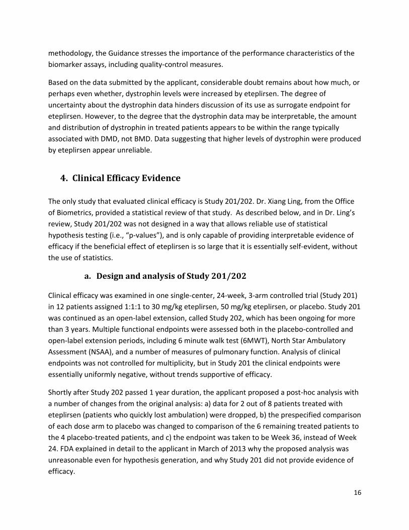
To interpret the applicant’s comparison of 6MWT results for eteplirsen patients to historical controls, it is also important to understand the progression of 6MWT as DMD patients near the time of loss of ambulation. At younger ages, during the period of relative stability or slow decline of 6MWT, a difference between two patients in 6MWT of 100 m is likely to predict a difference of several years in time to loss of ambulation, particularly if one patient is below about 300 meters and the other above. Differences between patients of 150- or 200 m on 6MWT have even larger prognostic implications, with patients who can walk in the range of 400- to 500 m on 6MWT unlikely to lose ambulation for many years. In contrast, however, large differences in 6MWT between patients near the time of loss of ambulation occur even when patients have generally similar prognoses.
Figure 6, taken from the applicant’s NDA, shows patient-level data for eteplirsen and historical controls. Consider two patients in their final year or two of ambulation: the historical control patient with a baseline of about 200 m (arrow), and the eteplirsen patient with a baseline of about 260 (star). At Month 12, the eteplirsen patient has lost ambulation, whereas the 6MWT for the historical control patient remains at about 200 m, such that the difference in 6MWT has increased from 60 m at baseline to about 200 m. By Month 24, the historical control patient has also lost ambulation, such that the difference between patients has become zero. Thus, in contrast to younger patients, the 200 m difference near the time of loss of ambulation corresponded to about 1 year difference in age at loss of ambulation. The general pattern and size of this effect is typical, with many DMD patients decreasing from about 300 m on 6MWT to loss of ambulation over 1- to 2 years, leading to brief but very large differences in 6MWT between patients whose disease course is otherwise generally similar. This does not imply that a difference of 150- or 200 m on 6MWT would not be clinically meaningful, but does suggest that even modest differences between study arms in poorly controlled studies such as Study
18
202 can exaggerate differences in certain functional measures near the time that patients lose ambulation.
Figure 6: 6MWT in Patients Using Steroid, Age ≥ 7 Years, Amenable to Exon 51 Skipping by Treatment Status – Individual Patient Data
Rate of progression of 6MWT in eteplirsen-treated patients is consistent with expected natural history
Patients from the placebo arm of randomized double-blind trials are likely to be better matched to patients in eteplirsen trials for factors that are difficult to measure, such as motivation and compliance with supportive therapy, compared to patients from registries. Placebo-controlled trials have recently been conducted with patients with DMD amenable to exon-51 skipping. Data from patients from the placebo group from some of these studies are publically available, and were used for a comparison with eteplirsen-treated patients.23 The figures below show the clinical course on 6MWT of eteplirsen-treated patients from Study 201/202 (colored lines) compared to patients treated with placebo in other controlled studies in exon-51 skippable patients with DMD (grey lines). Patients are divided by baseline rise from floor time (an
http://www.fda.gov/downloads/AdvisoryCommittees/CommitteesMeetingMaterials/Drugs/PeripheralandCentral NervousSystemDrugsAdvisoryCommittee/UCM475956.pdf
19
23
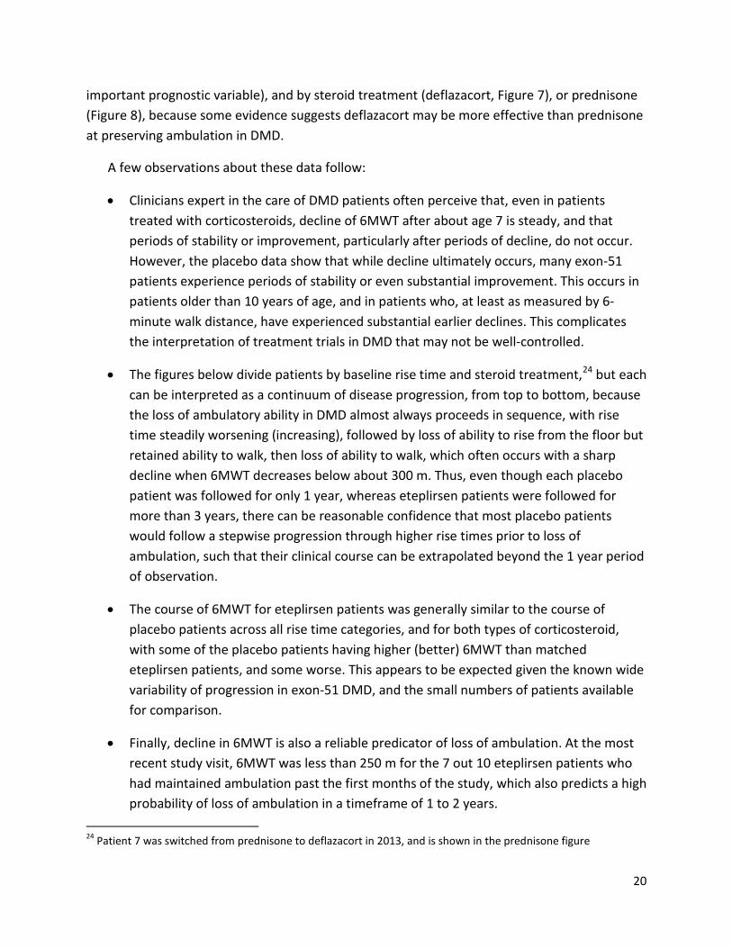
important prognostic variable), and by steroid treatment (deflazacort, Figure 7), or prednisone (Figure 8), because some evidence suggests deflazacort may be more effective than prednisone at preserving ambulation in DMD.
A few observations about these data follow:
• Clinicians expert in the care of DMD patients often perceive that, even in patients treated with corticosteroids, decline of 6MWT after about age 7 is steady, and that periods of stability or improvement, particularly after periods of decline, do not occur. However, the placebo data show that while decline ultimately occurs, many exon-51 patients experience periods of stability or even substantial improvement. This occurs in patients older than 10 years of age, and in patients who, at least as measured by 6minute walk distance, have experienced substantial earlier declines. This complicates the interpretation of treatment trials in DMD that may not be well-controlled.
• The figures below divide patients by baseline rise time and steroid treatment,24 but each can be interpreted as a continuum of disease progression, from top to bottom, because the loss of ambulatory ability in DMD almost always proceeds in sequence, with rise time steadily worsening (increasing), followed by loss of ability to rise from the floor but retained ability to walk, then loss of ability to walk, which often occurs with a sharp decline when 6MWT decreases below about 300 m. Thus, even though each placebo patient was followed for only 1 year, whereas eteplirsen patients were followed for more than 3 years, there can be reasonable confidence that most placebo patients would follow a stepwise progression through higher rise times prior to loss of ambulation, such that their clinical course can be extrapolated beyond the 1 year period of observation.
• The course of 6MWT for eteplirsen patients was generally similar to the course of placebo patients across all rise time categories, and for both types of corticosteroid, with some of the placebo patients having higher (better) 6MWT than matched eteplirsen patients, and some worse. This appears to be expected given the known wide variability of progression in exon-51 DMD, and the small numbers of patients available for comparison.
• Finally, decline in 6MWT is also a reliable predicator of loss of ambulation. At the most recent study visit, 6MWT was less than 250 m for the 7 out 10 eteplirsen patients who had maintained ambulation past the first months of the study, which also predicts a high probability of loss of ambulation in a timeframe of 1 to 2 years.
24 Patient 7 was switched from prednisone to deflazacort in 2013, and is shown in the prednisone figure
20

In the figures below, many of the eteplirsen patients appear to have few or no matches to the placebo patients in the most recent year of treatment, but this is a result of the division of the figures into categories based on baseline rise time. Most eteplirsen patients are currently in the >15 s rise time category (10 of the 12 eteplirsen patients, including at least 5 who lost ability to rise), and can be compared to the >15 s rise time group of control patients. In general, the course of eteplirsen-treated patients in Study 201/202 is similar to the course in these control patients, as shown in Figure 9, which combines all eteplirsen and control patients.
Figure 7: 6MWT, Deflazacort-treated patients
21
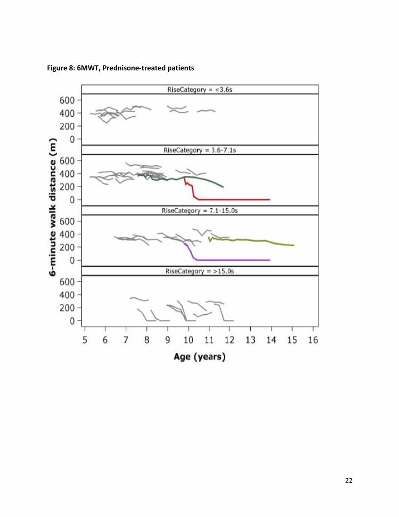
Figure 8: 6MWT, Prednisone-treated patients
22

Figure 9: 6MWT, eteplirsen vs controls on placebo, all patients
Because evidence that even a few eteplirsen patients might have progressed markedly differently than expected by natural history would be of interest, a few additional observations about these data are important. Assignment of eteplirsen patients to rise time category is affected by random noise in the baseline measure. Specific patients may appear to progress faster or slower than “matched” controls, but the noise inherent in matching needs to be considered. For example, the patient indicated by the bright green line in
Figure 7 was placed in the 7.1- to 15-second rise time category, but had large variability for rise time values, and a more accurate estimate of rise time for this patient might be closer to 5 seconds, suggesting that matching to a less advanced group of historical controls might have been as, or more, appropriate. In addition, a number of other factors can confound efforts to match treated with historical patients. For example, the sponsor has argued that loss of muscle, as measured by MRI, was more severe at baseline in two patients than suggested by functional tests, decreasing the interpretability of the rapid loss of ambulation experienced by these patients after starting eteplirsen.
Increases in rise time in eteplirsen-treated patients predict a high likelihood of sequential loss of ambulation within 1 or 2 years
Figure 10 shows rise time from floor for the eteplirsen patients. Three eteplirsen patients lost the ability to rise from the floor in the first year of Study 201. The applicant has, at times,
23
proposed that after an initial time period in which dystrophin levels from eteplirsen accumulated, disease progression largely stabilized in treated patients. All patients in Study 202 have continued to progress steadily while taking eteplirsen, as indicated by rise time from floor, without any discernible stabilization or slowing. Most have now become unable, or nearly unable, to rise from the floor, which predicts a high likelihood of sequential loss of ambulation within 1 or 2 years.
Figure 10: Rise Time, Study 201/202
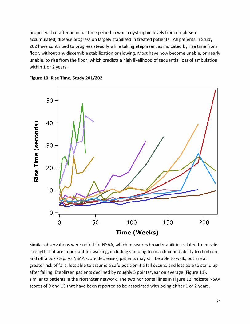
Similar observations were noted for NSAA, which measures broader abilities related to muscle strength that are important for walking, including standing from a chair and ability to climb on and off a box step. As NSAA score decreases, patients may still be able to walk, but are at greater risk of falls, less able to assume a safe position if a fall occurs, and less able to stand up after falling. Eteplirsen patients declined by roughly 5 points/year on average (Figure 11), similar to patients in the NorthStar network. The two horizontal lines in Figure 12 indicate NSAA scores of 9 and 13 that have been reported to be associated with being either 1 or 2 years,
24
respectively, from loss of ambulation.25 Combined with loss of ability to rise from the floor, the NSAA scores suggest that the eteplirsen patients, who are currently 11 to 14 years or age, are at, or close to, a level of muscle strength often associated with use of a wheelchair.
Figure 11: NSAA, Study 201/202
Issues with comparison of eteplirsen-treated patients with applicant’s proposed historical controls
Untreated historical control groups tend to have worse outcomes than apparently similar control groups in randomized studies. Patients in randomized studies need to meet certain criteria to be entered that generally select a less sick population than is typical of external
25 Ricottii et al (2015) The NorthStar Ambulatory Assessment in Duchenne muscular dystrophy: considerations for the design of clinical trials. J. Neurol Neurosurg Psychiatry. 0,1-7.
25

control groups. Such concerns appear to apply to muscular dystrophy, although the magnitude of this effect is difficult to quantify. In patients with fascioscapulhumoral muscular dystrophy Statland et al.26 observed that “whereas natural history data showed a decrease in strength over 1 year, there was an apparent increase in strength at 6 months in 2 of the 3 clinical trials in both the placebo and treatment groups.” [emphasis added] The authors concluded that this type of bias should be taken as a reminder of the importance of placebo groups when measuring strength in muscular dystrophy.
Supportive care can prolong ambulation in DMD by several years, but its effectiveness is dependent on both type and intensity of care, which is likely to differ substantially between patients enrolled in observational studies or registries versus interventional treatment studies. DMD care guidelines specify that corticosteroid efficacy needs to be balanced with side effects in the context of the individual patient’s goals. Patients enrolled in efficacy trials would likely be more interested in maximizing steroid efficacy compared to patients enrolled in observational natural history studies. This appears to have been the case for the eteplirsen patients compared to the controls selected by the applicant. A higher proportion, 69% vs. 8%, of the natural history controls vs. eteplirsen patients were on regimens other than daily dosing that are often selected to decrease side effects but that are thought to be associated with lessefficacy. Doses of corticosteroids also appear to have been lower in the applicant’s natural history patients, which included those “in whom the dose had not been always completely adjusted to the current weight.”27 Adherence to treatment guidelines is difficult to measure, but adherence in the eteplirsen study was reported to be exceptional, while there is evidence that care received in the regions of origin of many of the sponsor’s historical control patients was likely of lower intensity.28 Finally, as the sponsor’s natural history study proceeded, some patients left to enter interventional clinical trials, further decreasing the similarity of the natural history cohort to the eteplirsen patients.
Recent evidence from the Cooperative International Neuromuscular Research Group (CINRG) reinforces the observation that seemingly small differences in steroid treatment and clinical care may have relatively large effects, up to several years, on age at loss of ambulation.29 The
26 Statland JM et al (2013) Reevaluating measures of disease progression in facioscapulohumeral muscular dystrophy. Neuromuscul Disord. 23,306-12.
27 Mazzone E et al (2010) North Star ambulatory assessment, 6-minute walk test and timed items in ambulant boys with Duchenne muscular dystrophy. Neuromuscular Disorders. 20,712-716.
28 Landfeldt E et al (2015) Compliance to care guidelines for Duchenne muscular dystrophy. Journal of Neuromuscular Diseases. 2,63-72.
29 Bello L et al (2015) Prednisone/prednisolone and deflazacort regimens in the CINRG Duchenne natural history study. Neurology. 85, 1048-1055.
26
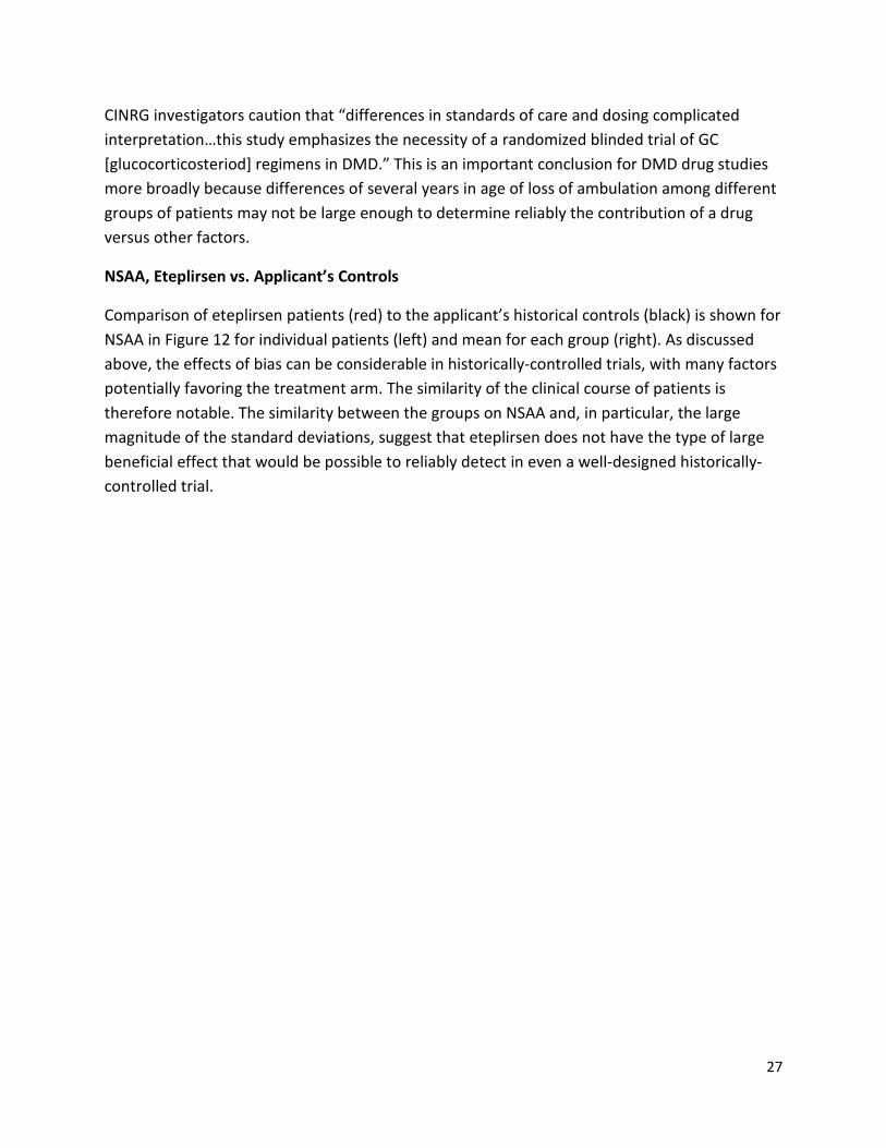
CINRG investigators caution that “differences in standards of care and dosing complicated interpretation…this study emphasizes the necessity of a randomized blinded trial of GC [glucocorticosteriod] regimens in DMD.” This is an important conclusion for DMD drug studies more broadly because differences of several years in age of loss of ambulation among different groups of patients may not be large enough to determine reliably the contribution of a drug versus other factors.
NSAA, Eteplirsen vs. Applicant’s Controls
Comparison of eteplirsen patients (red) to the applicant’s historical controls (black) is shown for NSAA in Figure 12 for individual patients (left) and mean for each group (right). As discussed above, the effects of bias can be considerable in historically-controlled trials, with many factors potentially favoring the treatment arm. The similarity of the clinical course of patients is therefore notable. The similarity between the groups on NSAA and, in particular, the large magnitude of the standard deviations, suggest that eteplirsen does not have the type of large beneficial effect that would be possible to reliably detect in even a well-designed historically-controlled trial.
27
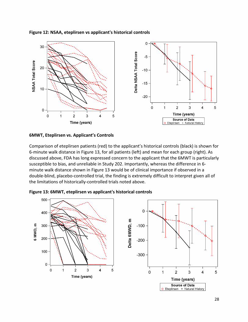
Figure 12: NSAA, eteplirsen vs applicant's historical controls
6MWT, Eteplirsen vs. Applicant’s Controls
Comparison of eteplirsen patients (red) to the applicant’s historical controls (black) is shown for 6-minute walk distance in Figure 13, for all patients (left) and mean for each group (right). As discussed above, FDA has long expressed concern to the applicant that the 6MWT is particularly susceptible to bias, and unreliable in Study 202. Importantly, whereas the difference in 6minute walk distance shown in Figure 13 would be of clinical importance if observed in a double-blind, placebo-controlled trial, the finding is extremely difficult to interpret given all of the limitations of historically-controlled trials noted above.
Figure 13: 6MWT, eteplirsen vs applicant's historical controls
28
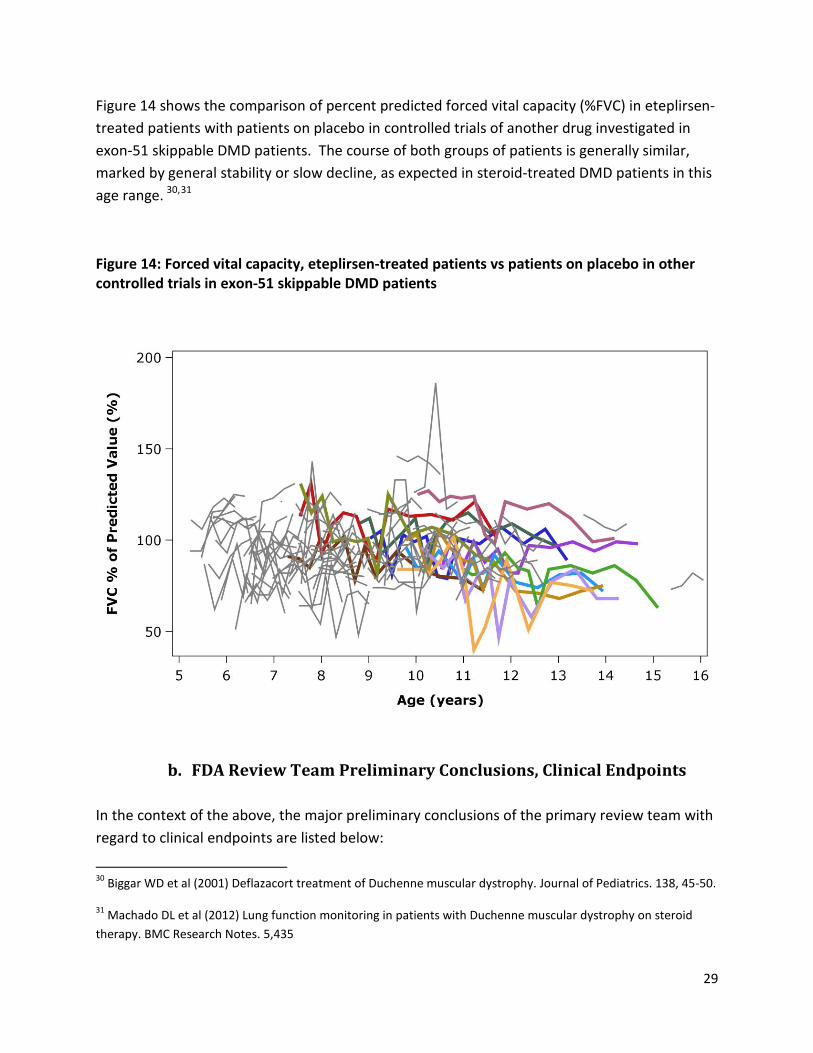
Figure 14 shows the comparison of percent predicted forced vital capacity (%FVC) in eteplirsentreated patients with patients on placebo in controlled trials of another drug investigated in exon-51 skippable DMD patients. The course of both groups of patients is generally similar, marked by general stability or slow decline, as expected in steroid-treated DMD patients in this
30, 31 age range.
Figure 14: Forced vital capacity, eteplirsen-treated patients vs patients on placebo in other controlled trials in exon-51 skippable DMD patients
b. FDA Review Team Preliminary Conclusions, Clinical Endpoints
In the context of the above, the major preliminary conclusions of the primary review team with regard to clinical endpoints are listed below:
30 Biggar WD et al (2001) Deflazacort treatment of Duchenne muscular dystrophy. Journal of Pediatrics. 138, 45-50.
31 Machado DL et al (2012) Lung function monitoring in patients with Duchenne muscular dystrophy on steroid therapy. BMC Research Notes. 5,435
29
1. The natural history of DMD in patients amenable to exon 51 skipping has been characterized in a number of observational natural history studies and controlled trials, and the range of age at loss of ambulation is very wide, currently between about 8 and 16 years for most patients. Eteplirsen patients have experienced a sequential loss of ambulatory abilities and increasing muscle weakness, as measured by rise time from floor, NSAA, 6MWT, and other tests. In the context of this considerable variability among patients, the clinical course of eteplirsen patients over more than 3 ½ years of treatment with eteplirsen has been generally similar to expected natural history of patients provided with intensive supportive care.
2. There are important differences between patients enrolled in observational natural history studies and patients enrolled in interventional drug efficacy studies, some of which are quantifiable, and some of which are not. Corticosteroid therapy appears to have been more intensive in eteplirsen patients compared to the natural history patients selected by the applicant, and this, itself, may have been capable of affecting performance. Near the time when patients lose ambulation, decisions are made by patients and caregivers about whether weakness has progressed to the point that it is in the patient’s best interest to use a wheelchair to avoid the risk of falls and injuries and to decrease the effort and time required for mobility. Differences in individual care decisions, therefore, seemingly could produce large differences in 6MWT and time to loss of ambulation between eteplirsen patients and natural history controls. NSAA results, potentially representing a more direct measure of strength, suggest that differences in DMD progression between eteplirsen patients and the applicant’s natural history controls were too small and variable, in the context of a poorly-controlled trial, to be reliably attributed to drug treatment.
3. With regard to future efficacy studies, any beneficial effects of eteplirsen are unlikely to be large enough to be detectable outside of a placebo-controlled trial.
6. Clinical Safety The clinical safety database for eteplirsen is small: 114 total patients exposed, with only 36 exposed for ≥24 weeks and 12 exposed for ≥1 year. Most of these exposures were outside of placebo-controlled studies, limiting ability to determine if adverse events were the result of drug effect or chance. However, the serious and severe adverse events that occurred were generally consistent with events expected in DMD. The 12 patients in Study 202 were exposed for >3 years, which provides some reassurance against delayed toxicity.
It is important to note that dose-limiting toxicity was not observed, such that higher doses, with potentially greater likelihood of efficacy, could be studied in the future.
30

In animal studies, the primary target organ was the kidney, with dose-dependent renal tubular cytoplasmic basophilia and/or vacuolation and, at the high dose, tubular degeneration/necrosis. In a mouse study, dilatation of the lateral ventricles of the brain was observed at the mid and high doses. The mechanism of this effect, and its relevance to humans, is unknown. Mean eteplirsen plasma exposures (AUC) at the NOAELs for monkey and juvenile rat were 20-fold and 6-fold, respectively, greater than that in patients dosed once weekly with 30 mg/kg IV eteplirsen.
31

IV. Statistical Review
U.S. Department of Health and Human Services Food and Drug Administration Center for Drug Evaluation and Research Office of Translational Sciences Office of Biostatistics
S T A T I S T I C A L R E V I E W A N D E V A L U A T I O N
NDA/BLA #: Drug Name: Indication(s): Applicant: Date(s):
Review Priority:
Biometrics Division: Statistical Reviewer: Concurring Reviewers:
Medical Division: Clinical Team:
Project Manager:
CL IN IC A L ST UDI E S
206488
EXONDYS 51™ (eteplirsen)
Duchenne muscular dystrophy (DMD)
Sarepta
Submission date: 6/26/2015
PDUFA Date: 2/26/2016
Priority Review
Division I, Office of Biometrics (HFD -710)
Xiang Ling, Ph.D.
Kun Jin, Ph.D., Team Leader
Jim Hung, Ph.D., Director
Division of Neuropharm (HFD -120)
Christopher Breder, M.D., Ph.D.
Ronald Farkas, M.D., Ph.D., Team Leader
Fannie Choy
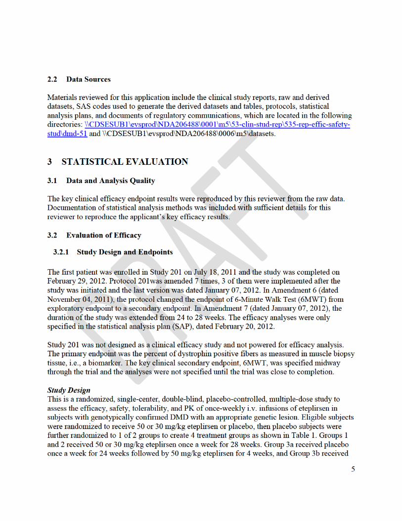
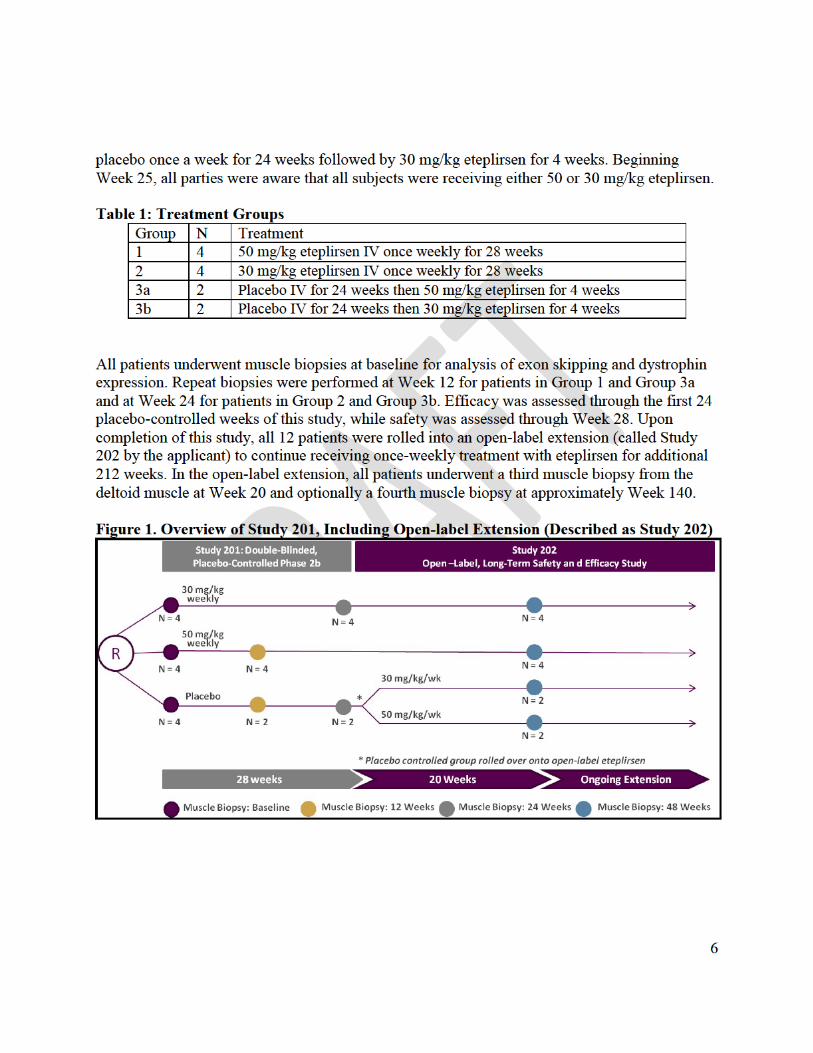

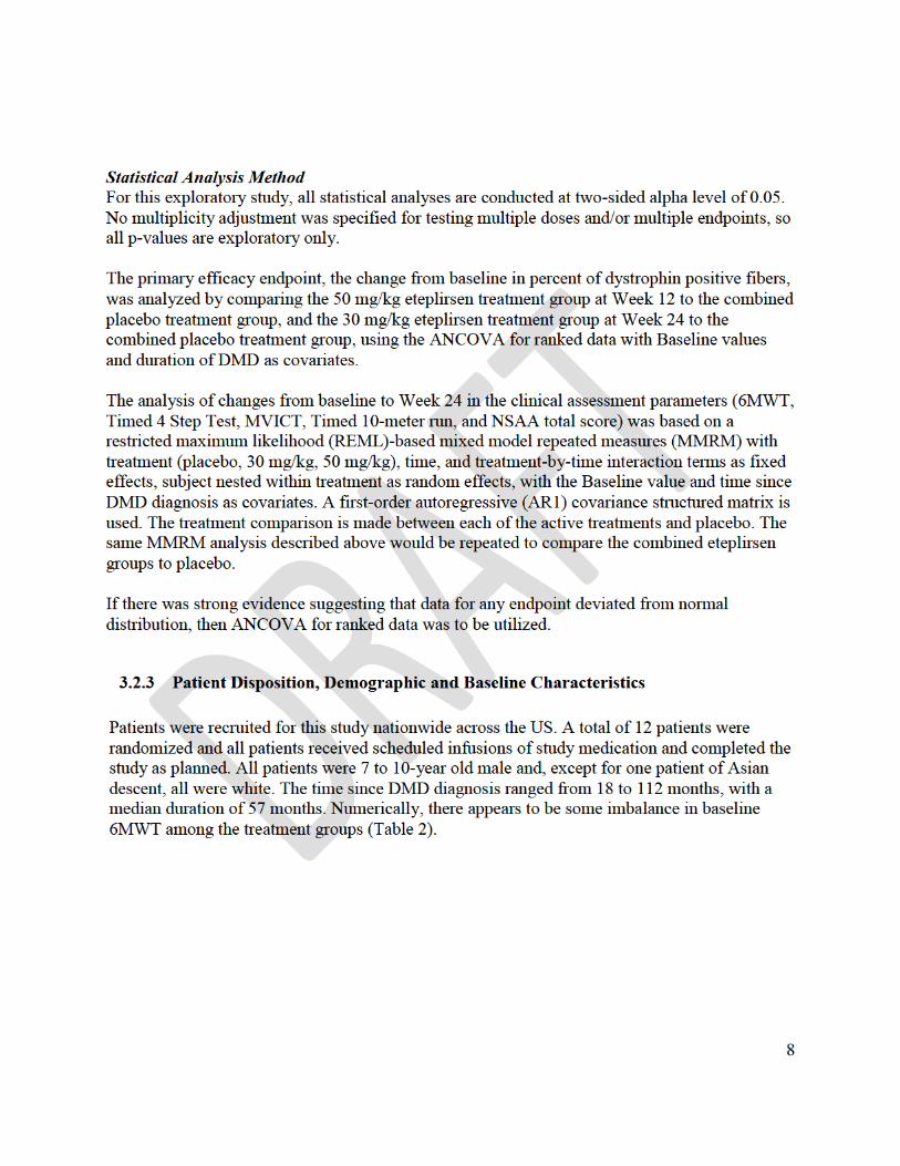
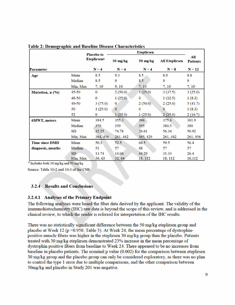
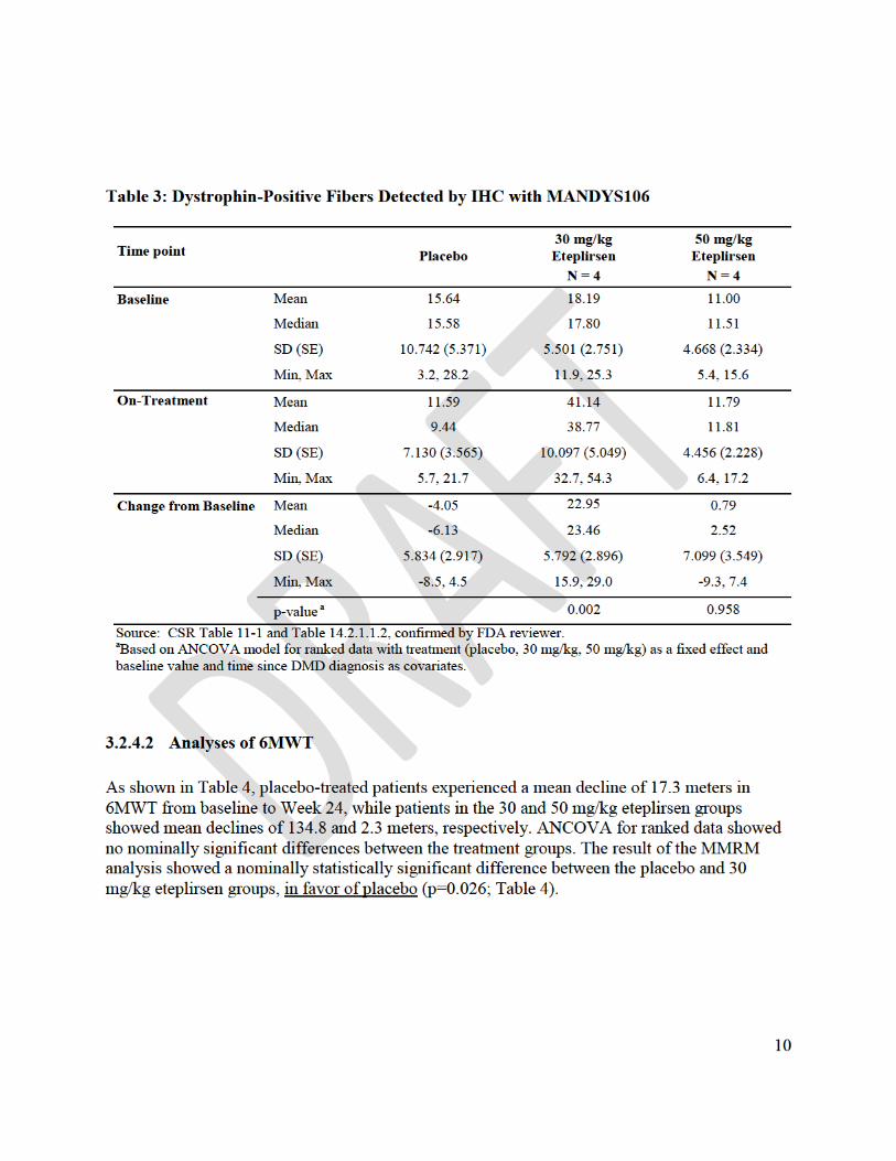
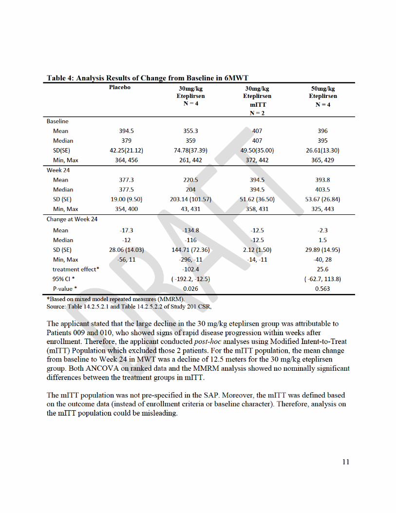

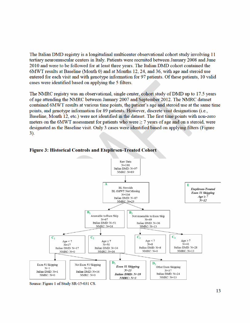

V. Summary of Clinical Pharmacology Findings
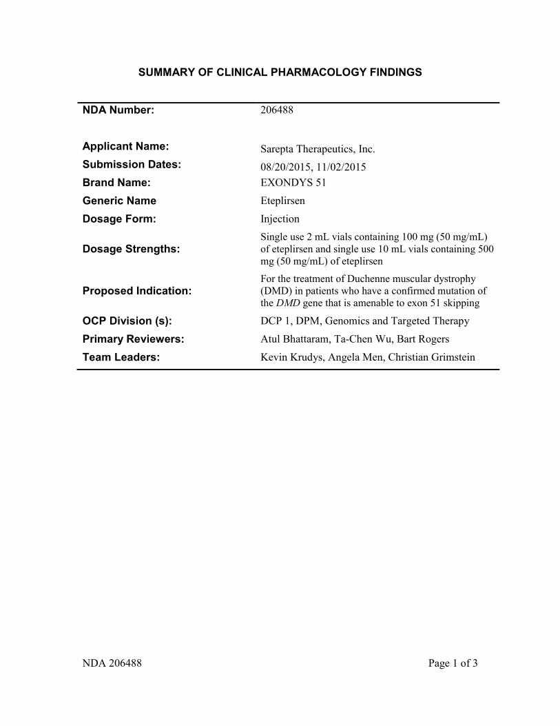
SUMMARY OF CLINICAL PHARMACOLOGY FINDINGS
NDA Number: 206488
Applicant Name: Sarepta Therapeutics, Inc. Submission Dates: 08/20/2015, 11/02/2015 Brand Name: EXONDYS 51
Generic Name Eteplirsen
Dosage Form: Injection Single use 2 mL vials containing 100 mg (50 mg/mL)
Dosage Strengths: of eteplirsen and single use 10 mL vials containing 500 mg (50 mg/mL) of eteplirsen
For the treatment of Duchenne muscular dystrophy Proposed Indication: (DMD) in patients who have a confirmed mutation of
the DMD gene that is amenable to exon 51 skipping
OCP Division (s): DCP 1, DPM, Genomics and Targeted Therapy
Primary Reviewers: Atul Bhattaram, Ta-Chen Wu, Bart Rogers
Team Leaders: Kevin Krudys, Angela Men, Christian Grimstein
NDA 206488 Page 1 of 3
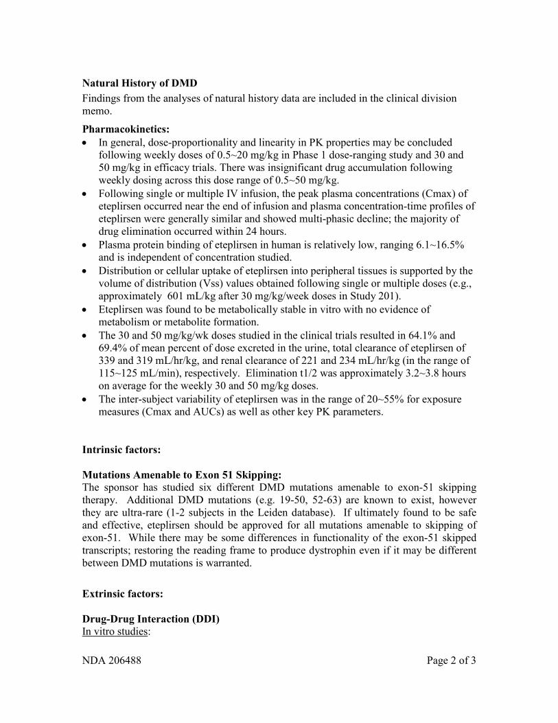
Mutations Amenable to Exon 51 Skipping:
Natural History of DMD Findings from the analyses of natural history data are included in the clinical division memo.
Pharmacokinetics: • In general, dose-proportionality and linearity in PK properties may be concluded
following weekly doses of 0.5~20 mg/kg in Phase 1 dose-ranging study and 30 and 50 mg/kg in efficacy trials. There was insignificant drug accumulation following weekly dosing across this dose range of 0.5~50 mg/kg.
• Following single or multiple IV infusion, the peak plasma concentrations (Cmax) of eteplirsen occurred near the end of infusion and plasma concentration-time profiles of eteplirsen were generally similar and showed multi-phasic decline; the majority of drug elimination occurred within 24 hours.
• Plasma protein binding of eteplirsen in human is relatively low, ranging 6.1~16.5% and is independent of concentration studied.
• Distribution or cellular uptake of eteplirsen into peripheral tissues is supported by the volume of distribution (Vss) values obtained following single or multiple doses (e.g., approximately 601 mL/kg after 30 mg/kg/week doses in Study 201).
• Eteplirsen was found to be metabolically stable in vitro with no evidence of metabolism or metabolite formation.
• The 30 and 50 mg/kg/wk doses studied in the clinical trials resulted in 64.1% and 69.4% of mean percent of dose excreted in the urine, total clearance of eteplirsen of 339 and 319 mL/hr/kg, and renal clearance of 221 and 234 mL/hr/kg (in the range of 115~125 mL/min), respectively. Elimination t1/2 was approximately 3.2~3.8 hours on average for the weekly 30 and 50 mg/kg doses.
• The inter-subject variability of eteplirsen was in the range of 20~55% for exposure measures (Cmax and AUCs) as well as other key PK parameters.
Intrinsic factors:
The sponsor has studied six different DMD mutations amenable to exon-51 skipping therapy. Additional DMD mutations (e.g. 19-50, 52-63) are known to exist, however they are ultra-rare (1-2 subjects in the Leiden database). If ultimately found to be safe and effective, eteplirsen should be approved for all mutations amenable to skipping of exon-51. While there may be some differences in functionality of the exon-51 skipped transcripts; restoring the reading frame to produce dystrophin even if it may be different between DMD mutations is warranted.
Extrinsic factors:
Drug-Drug Interaction (DDI) In vitro studies:
NDA 206488 Page 2 of 3
Eteplirsen is expected to have a low potential for DDI in humans based on results of in vitro investigation on microsomal metabolism, plasma protein binding, inhibition or induction of major CYP isozymes or major drug transporters at the concentration range studied for clinical dosing regimen: o Eteplirsen had insignificant inhibitory effects for CYP2B6, CYP2C8, CYP2D6,
CYP3A4/5, CYP1A2, CYP2C9, or CYP2C19 in human liver microsomes. There was no metabolism-dependent inhibition observed with any of the CYPs tested.
o Eteplirsen at the concentration range studied did not show significant enzyme inducing capability for CYP1A2, CYP2B6, and CYP3A4 in human hepatocytes.
o Eteplirsen is not a substrate and/or an inhibitor of major human drug transporters OAT1, OAT3, OCT1, OCT2, OATP1B1, OATP1B3, P-gp, BCRP, MRP2, and BSEP in transfected CHO cells, Caco-2 monolayers, or inside-out human membranes.
NDA 206488 Page 3 of 3