fisb mediates membrane fission during sporulation in...
TRANSCRIPT

FisB mediates membrane fission duringsporulation in Bacillus subtilis
Thierry Doan,1,2 Jeff Coleman,3 Kathleen A. Marquis,1 Alex J. Meeske,1 Briana M. Burton,4,7
Erdem Karatekin,5,6,8 and David Z. Rudner1,8
1Department of Microbiology and Immunobiology, Harvard Medical School, Boston, Massachusetts 02115, USA; 2Laboratoire deChimie Bacterienne, UMR7283, Aix-Marseille Universite-CNRS, Institut de Microbiologie de la Mediterranee, 13009Marseilles, France; 3Department of Cell Biology, School of Medicine, Yale University, New Haven, Connecticut 06520, USA;4Department of Cell Biology, Harvard Medical School, Boston, Massachusetts 02115, USA; 5Department of Cellularand Molecular Physiology, School of Medicine, Yale University, New Haven, Connecticut 06520, USA; 6NanobiologyInstitute, Yale University, Orange, Connecticut 06477, USA
How bacteria catalyze membrane fission during growth and differentiation is an outstanding question inprokaryotic cell biology. Here, we describe a protein (FisB, for fission protein B) that mediates membrane fissionduring the morphological process of spore formation in Bacillus subtilis. Sporulating cells divide asymmetrically,generating a large mother cell and smaller forespore. After division, the mother cell membranes migrate aroundthe forespore in a phagocytic-like process called engulfment. Membrane fission releases the forespore into themother cell cytoplasm. Cells lacking FisB are severely and specifically impaired in the fission reaction. Moreover,GFP-FisB forms dynamic foci that become immobilized at the site of fission. Purified FisB catalyzes lipid mixing invitro and is only required in one of the fusing membranes, suggesting that FisB–lipid interactions drive membraneremodeling. Consistent with this idea, the extracytoplasmic domain of FisB binds with remarkable specificity tocardiolipin, a lipid enriched in the engulfing membranes and regions of negative curvature. We propose thatmembrane topology at the final stage of engulfment and FisB–cardiolipin interactions ensure that the mother cellmembranes are severed at the right time and place. The unique properties of FisB set it apart from the knownfission machineries in eukaryotes, suggesting that it represents a new class of fission proteins.
[Keywords: sporulation; engulfment; fission; morphogenesis; fusion; dynamin; ESCRT-III]
Supplemental material is available for this article.
Received October 31, 2012; revised version accepted January 2, 2013.
The remodeling of lipid bilayers is essential to all organ-isms. These remodeling events include membrane fusion,wherein two membranes merge to become one, andmembrane fission, where a single lipid bilayer dividesinto two. Our understanding of the molecular mecha-nisms underlying these processes comes from extensivestudies in eukaryotes and the enveloped viruses thatinfect them. In eukaryotes, fusion and fission play centralroles in a broad array of cell biological processes, in-cluding exocytosis, endocytosis, cytokinesis, and intra-cellular membrane trafficking (Chernomordik andKozlov 2003; Sudhof and Rothman 2009; Kozlov et al.2010). They are also critical for the entry and exit ofenveloped viruses into (and out of) their host (Kielian andRey 2006; Harrison 2008). In contrast, virtually nothing isknown about how bacteria remodel their membranes
during growth and differentiation despite the fact thatthey undergo membrane fission in every cell divisioncycle. Here, we describe the identification and character-ization of a newly identified protein (FisB, for fissionprotein B) that functions in membrane fission during thedevelopmental process of spore formation in the bacte-rium Bacillus subtilis.
From a topological perspective, fusion and fission canbe seen as opposed processes. However, at the molecularlevel, both involve local disruption, bending, and bridgingof lipid bilayers. Achieving lipid mixing during eitherfusion or fission requires forcing bilayers to within acritical distance of a few nanometers (Rand and Parsegian1986; Wong et al. 1999; Kozlovsky and Kozlov 2003;Bashkirov et al. 2008; Kozlov et al. 2010). For bothprocesses, hydration forces are thought to provide themajor obstacle against lipid merger, which is alleviated bybending coupled to lipid tilting that allows hydrophobicattraction between the hydrocarbon chains of the lipids(Chernomordik and Kozlov 2003; Kozlov et al. 2010).Studies in eukaryotes and the viruses that infect themhave revealed that specialized proteins generate the work
7Present address: Department of Molecular and Cellular Biology, Har-vard University, Cambridge, MA 02138, USA8Corresponding authorsE-mail [email protected] [email protected] is online at http://www.genesdev.org/cgi/doi/10.1101/gad.209049.112.
322 GENES & DEVELOPMENT 27:322–334 � 2013 by Cold Spring Harbor Laboratory Press ISSN 0890-9369/13; www.genesdev.org
Cold Spring Harbor Laboratory Press on January 19, 2014 - Published by genesdev.cshlp.orgDownloaded from

required for these lipid rearrangements. In the case ofeukaryotic membrane fusion, SNARE proteins anchoredin apposed membranes form trans-complexes that over-come repulsive forces to bring bilayers into intimatecontact. In enveloped viruses, membrane fusion proteinsthat are anchored in the viral membrane insert into hostcell membranes and, through a series of conformationalchanges, bring the lipid bilayers into close proximity,leading to fusion. As for fission, the only factors describedto date that participate in it are dynamin (Ferguson andDe Camilli 2012) and the endosomal sorting complexrequired for transport (ESCRT-III complex) (Hurley 2010;Wollert and Hurley 2010; Henne et al. 2011). Dynaminand dynamin-like proteins are involved in endocytosis,multivesicular membrane budding, cytokinesis in chlo-roplasts, and mitochondrial genesis. These proteins arethought to function as ‘‘pinching machines’’: They poly-merize around the outside of the neck that connects twoparts of a contiguous membrane, constricting the neck toa critical radius. GTP hydrolysis then produces confor-mational changes that result in fission (Ferguson and DeCamilli 2012). In contrast, the ESCRT-III complex appearsto assemble on the inside or at one of the openings of theneck to induce membrane constriction and, ultimately,fission (Hurley 2010; Henne et al. 2011). The ESCRT-IIIcomplex has been implicated in the biogenesis of multi-vesicular bodies (Gruenberg and Stenmark 2004; Hurley2008; Wollert et al. 2009; Wollert and Hurley 2010), mem-brane abscission during cytokinesis (Elia et al. 2011), andbudding of some enveloped viruses from the host mem-brane (Strack et al. 2003; Morita and Sundquist 2004;Bieniasz 2006). Interestingly, a homologous set of proteinsappears to function in cytokinesis in the CrenarchaeaSulfolobus (Lindas et al. 2008; Samson et al. 2008).
The molecular mechanism underlying membrane fis-sion during cytokinesis in bacteria remains enigmatic. Ithas been proposed that protein components of the celldivision machinery could catalyze fission (Sharp andPogliano 1999; Liu et al. 2006; de Boer 2010; Fleminget al. 2010). Alternatively, it has been suggested that cellwall synthesis on the outside of the cell could force theconstricting membranes into close proximity, ultimatelyleading to fission (Weiss 2004; Judd et al. 2005; Meyeret al. 2010). To date, no protein has been directlyimplicated in this fission reaction.
Here, we investigated a specialized membrane fissionevent that occurs during sporulation in B. subtilis (Fig. 1).In response to starvation, B. subtilis differentiates intoa dormant spore (Stragier and Losick 1996; Errington2003). Upon initiation of this developmental process,the cell divides asymmetrically, generating two cells ofunequal size and dissimilar fate. The smaller cell is theprospective spore and is referred to as the forespore. Thelarger cell is called the mother cell. Initially, these twocells lie side by side, separated by a double-membraneseptum. However, shortly after polar division, the mothercell membranes migrate around the forespore in a phago-cytic-like process called engulfment. In the last stage ofthis process, the leading edges of the migrating mem-branes meet at the cell pole and, upon membrane fission,
the forespore is released into the mother cell cytoplasm(Fig. 1). The forespore is thus surrounded by two mem-branes: an inner membrane that contains the foresporecytoplasm and an outer membrane derived from themother cell membrane. The molecular mechanism un-derlying this membrane remodeling event is the focus ofour work.
We describe the identification and characterization ofa membrane protein, FisB, that is required for membranefission at the last stage of engulfment. FisB is a bitopicmembrane protein that is produced in the mother cellafter polar division. In its absence, the migration of themother cell membranes around the forespore proceedsnormally, but engulfment stalls when the membranesreach the cell pole. In support of the idea that FisBfunctions in membrane remodeling, a functional GFP-FisB fusion localizes as a focus at the cell pole at the timeof membrane fission. Consistent with a direct role inmembrane remodeling, we show that recombinant FisBcatalyzes lipid mixing in vitro. Interestingly, lipid-mixingactivity required FisB in only one of the two interactingmembranes, suggesting that FisB–lipid interactions drivemembrane remodeling. In support of this idea, we showthat the extracellular domain of FisB directly interactswith liposomes. Moreover, FisB–liposome interactionrequired the presence of the anionic phospholipid cardi-olipin (CL). Altogether, our data support the idea that FisB
Figure 1. Membrane fission during spore development. Sche-matic representation of the morphological stages of the engulf-ment process. After polar division, the mother cell membranesmigrate around the forespore. When they meet at (or near) the cellpole, membrane fission releases the forespore into the mother cellcytoplasm. Enlarged schematics highlight the engulfing mem-branes pre- and post-fission. (Middle panel) At the final stage,the membranes form a tube that is severed in the fission re-action. Fluorescent images show sporulating cells (strainBKM015) before and after membrane fission. Prior to fission,the lipophylic dye FM4-64 labels the peripheral membranes ofthe mother cell and the double membrane surrounding theforespore. After fission, the dye cannot access the membranessurrounding the forespore. The presence of a forespore withinthe mother cell is assessed by a fluorescent forespore reporter(PspoIIQ-cfp).
FisB mediates engulfment fission
GENES & DEVELOPMENT 323
Cold Spring Harbor Laboratory Press on January 19, 2014 - Published by genesdev.cshlp.orgDownloaded from

directly catalyzes the membrane fission event that marksthe end of engulfment. Given the topology of FisB andthat of the membrane tube to be severed, we suggest thatFisB promotes fission from within or near one of theopenings of the tube by interacting with CL that isenriched in the engulfment membranes and in regionsof high negative curvature.
Results
FisB is required for membrane fission at the last stageof engulfment
Previous studies on membrane fission during sporulationimplicated the polytopic membrane protein SpoIIIE(Sharp and Pogliano 1999, 2003). SpoIIIE is a DNA trans-porter that is required to translocate the forespore chro-mosome into the forespore compartment (Wu andErrington 1994). In the absence of SpoIIIE, sporulatingcells are unable to pump the forespore chromosome andfail to make viable spores. Using an innovative mem-brane fission assay (described below), Sharp and Pogliano(1999) discovered that cells lacking SpoIIIE are also de-fective in membrane fission at a late stage of engulfment.In support of the idea that SpoIIIE functions directly incatalyzing fission, a SpoIIIE-GFP fusion protein localizedto the forespore pole around the time of membrane fission(Sharp and Pogliano 1999). In the course of our studies onchromosome organization and segregation during sporu-lation (Burton et al. 2007; Marquis et al. 2008; Sullivanet al. 2009), we observed that sporulating cells lackingSpoIIIE exhibited defects in engulfment at stages priorto membrane fission. Under our assay conditions, 80%–90% of the SpoIIIE mutant cells had bulged septal mem-branes and/or invaginations that appeared to preventcomplete membrane migration around the forespore(Supplemental Fig. S1). These observations led us to con-sider the possibility that SpoIIIE might only be indirectlyinvolved in membrane fission and that other proteinscatalyze this reaction.
We began our studies by investigating two candidatefactors for a role in catalyzing fission: the B. subtilisdynamin homolog DynA (Burmann et al. 2011) and thepeptidoglycan synthetic machinery (Meyer et al. 2010).B. subtilis DynA has been shown to catalyze membranefusion in vitro; however, cells lacking DynA have vir-tually no phenotype during vegetative growth (Burmannet al. 2011). To investigate whether the DynA mutantwas impaired in membrane fission during sporulation,we took advantage of the fission assay developed bySharp and Pogliano (1999). In this assay, cells are inducedto sporulate synchronously by nutrient downshift. Attime points after polar division, the cells are stainedwith the membrane dye FM4-64 and visualized byfluorescence microscopy. Since this dye is unable tocross the lipid bilayer, the membranes surroundingthe forespore are only labeled in cells that have notyet undergone fission (Fig. 1). Once fission occurs andthe forespore is released into the mother cell cytosol,the membrane-impermeant dye only has access to the
peripheral mother cell membranes. To assess whetherthese cells have an engulfed forespore inside them, weused a forespore fluorescent reporter in which the geneencoding the cyan fluorescent protein (CFP) was placedunder the control of the forespore-specific transcriptionfactor sF (PspoIIQ-cfp), as described previously (Fig. 1;Meyer et al. 2010). Using this assay, we found thatengulfment and membrane fission were indistinguishablein wild-type cells and cells lacking DynA (Fig. 2A). Thus,dynamin does not appear to function in membrane fissionduring sporulation.
Next, we investigated whether cell wall synthesisbetween the forespore and mother cell membranes isinvolved in membrane fission. Dworkin and colleagues(Meyer et al. 2010) recently proposed that the action ofcell wall synthetic complexes anchored in the mother cellmembrane could promote membrane migration duringengulfment and membrane fission. Using a membranefission assay similar to the one described above, theseresearchers reported that the addition of antibiotics thatinhibit cell wall synthesis during the engulfment processresulted in a fourfold reduction in fission. To investigatewhether the force generated from cell wall synthesis wasthe principal driver of membrane fission, we performedsimilar antibiotic addition experiments (see the Materialsand Methods). Under our assay conditions, we observeda defect in membrane fission, although the effect wasmore modest than observed previously. Membrane fis-sion was reduced from 86% to 51% when the antibioticfosfomycin was added at hour 1.5 of sporulation (Fig. 2A).The reduction in fission was even less pronounced whenthe drug was added at later time points (data not shown).These results suggest that peptidoglycan synthesis andthe force generated by it could help bring the membranesinto close proximity but indicate that the protein (orproteins) that catalyze membrane fission remain to bediscovered.
DynA was not required for membrane fission, and wewere unable to identify homologs of the ESCRT-III fissionmachinery in the B. subtilis genome. Accordingly, weturned to the hypothesis that fission is mediated bya fusion-like mechanism. We reasoned that if sucha fission protein existed, it would likely be synthesizedin the mother cell at an early stage of sporulation prior tothe fission reaction and would have at least one trans-membrane segment, and cells lacking this protein wouldhave reduced sporulation efficiency. The first mothercell-specific transcription factor that is induced duringsporulation is the alternative s factor sE (Stragier andLosick 1996; Piggot and Losick 2002). The sE regulon hasbeen defined by transcriptional profiling (Eichenbergeret al. 2003; Feucht et al. 2003; Steil et al. 2005), andstrains harboring deletions in most of the genes in theregulon have been generated (Eichenberger et al. 2003).Using the criteria described above, we examined a subsetof this ordered library of mutants using the fluorescentmembrane fission assay of Sharp and Pogliano (1999). Ouranalysis identified one mutant (in the yunB gene) thatwas severely impaired in membrane fission (Fig. 2A).Based on its mutant phenotype and the characterization
Doan et al.
324 GENES & DEVELOPMENT
Cold Spring Harbor Laboratory Press on January 19, 2014 - Published by genesdev.cshlp.orgDownloaded from

of the yunB gene product described below, we renamedthis gene fisB for fission protein B.
To quantitatively assess the requirement for FisB inmembrane fission, we compared wild type and the FisBmutant in a sporulation time course (Fig. 2B; Supplemental
Fig. S2). Two hours after the initiation of sporulation,13% of wild-type cells (n > 1000) had completed mem-brane fission, releasing the forespore into the mother cellcytoplasm. At this time point, there was no detectablefission in the mutant. Thirty minutes later, at hour 2.5,51% of wild-type sporulating cells (n > 1000) had un-dergone membrane fission compared with <1% in theFisB mutant (n > 1000). By hour 3, 79% of wild-typesporulating cells had completed membrane fission, butonly 2% had completed membrane fission in the mutant.Importantly, in cells lacking FisB, the migration of themother cell membranes around the forespore appearedindistinguishable from wild type (Fig. 2B; SupplementalFig. S2). Moreover, similar to wild type, membranemigration appeared complete in most cells lacking FisBby hour 2.5. These results indicate that the absence ofFisB does not impact the early steps of engulfment andthus suggest that FisB plays a specific role in the mem-brane fission process.
Interestingly, membrane fission was not completelyabolished in cells lacking FisB. By hour 4.5, ;21% of theFisB mutant sporulating cells appeared to have undergonefission. This percentage is likely to be an overestimatebecause sporulating cells lacking FisB begin to lyse at thistime point, reducing the total number of sporulating cellsin the culture (Supplemental Fig. S2). FisB mutants havea sporulation efficiency of 12%–15%, suggesting thatmost of the cells that successfully undergo membranefission are able to produce mature spores.
Altogether, these results suggest that FisB plays a spe-cific role in membrane fission. In its absence, membranemigration around the forespore proceeds normally, butmost sporulating cells fail to undergo membrane remod-eling and release of the forespore into the mother cellcytoplasm. Cells that stall at the fission step ultimatelylyse. Those that do complete fission do so after an ;2-hdelay.
FisB forms dynamic foci that localize to the poleat the time of membrane fission
FisB is predicted to be a bitopic membrane protein witha small N-terminal domain that resides in the mother cellcytoplasm and a larger (23-kDa) extracellular domain. Todetermine the subcellular localization of FisB, we gener-ated an N-terminal monomeric GFP fusion to FisBexpressed under its native promoter- and ribosome-bind-ing site. The fusion protein complemented the FisB-nullmutant for membrane fission and sporulation (data notshown). We visualized the GFP fusion by epifluorescencemicroscopy in a sporulation time course. For this exper-iment, we visualized engulfment and membrane fissionusing the membrane dye TMA-DPH. TMA-DPH ineffi-ciently crosses the lipid bilayer and thus weakly labelsthe mother cell membrane that surrounds the foresporeafter fission is complete. Accordingly, using TMA-DPH,it is possible to distinguish pre- and post-fission stageswithout a forespore reporter. Supplemental Figure S3shows a direct comparison of the two membrane dyesFM4-64 and TMA-DPH.
Figure 2. Cells lacking FisB are impaired in membrane fission.(A) Histogram shows fission efficiency (the percentage of spor-ulating cells that have undergone fission compared with allsporulating cells; n > 1000), 3 h after the onset of sporulationfrom a representative experiment. Wild type (BKM015), a DynAmutant (BAM027), wild type after the addition of 5 mMfosfomycin (Fos) that inhibits peptidoglycan synthesis at hour1.5, and a FisB mutant (BTD3200) are shown. (B) Fissionefficiency as a function of time from a representative experimentin a wild-type strain (wt, BKM15) and a FisB mutant (BTD3200).(C) Membrane fission was assessed by fluorescent microscopyduring a sporulation time course in wild type (BKM015) anda FisB mutant (BTD3200). Both strains contained a fluorescentforespore reporter (PspoIIQ-cfp) to label all forespores. Themembranes (false-colored red) were visualized with the fluores-cent dye FM4-64 and merged with forespore CFP (false-coloredgreen). Examples of sporulating cells in which fission hasoccurred are highlighted (yellow arrowheads). Time (in hours)after the initiation of sporulation is indicated. Bar, 1 mm.
FisB mediates engulfment fission
GENES & DEVELOPMENT 325
Cold Spring Harbor Laboratory Press on January 19, 2014 - Published by genesdev.cshlp.orgDownloaded from

During engulfment at hour 2.5, GFP-FisB localized asdiscrete foci in both the peripheral membranes as well asthe mother cell membranes that were engulfing theforespore (Fig. 3A). We were unable to quantify thenumber of FisB proteins in the foci, but the fluorescenceintensity suggests that each focus is not a single proteinand likely represents an oligomeric complex. Consistentwith this idea, we found that both full-length FisB andthe extracellular domain of FisB when purified fromEscherichia coli can form oligomeric complexes in vitro(Supplemental Fig. S4). Time-lapse imaging revealed thatthe GFP-FisB foci were highly mobile in the membrane(Supplemental Movie M1). Importantly, at the time ofmembrane fission or just after it, a GFP-FisB focus waspresent near the cell pole adjacent to the forespore ina significant proportion of the cells. Strikingly, time-lapseimaging indicates that these polar foci remained immo-bile (Supplemental Movie M2). Figure 3B shows repre-sentative stills from a typical movie and an image inwhich each pixel is averaged over the entire time lapse.Dynamic foci are lost upon averaging, while immobileones at the cell pole are retained.
In some cases, lowering the level of a fluorescent fusionwill reveal or highlight specific subcellular localization,perhaps by reducing nonspecific or low-affinity binding(Rudner et al. 2002; Gregory et al. 2008). Accordingly, weconstructed a YFP-FisB fusion expressed at levels approxi-mately fivefold lower than the native protein (see theMaterials and Methods; data not shown). The fusion proteincomplemented the fisB-null, restoring sporulation effi-ciency to wild-type levels. Moreover, YFP-FisB supportedefficient membrane fission, albeit with a 20- to 30-mindelay compared with wild type (Fig. 3C; data not shown).
During early engulfment stages, YFP-FisB localized infoci in the mother cell membranes, as observed with the
GFP-FisB fusion expressed at wild-type levels. These fociwere more often found in the mother cell membranesthat surrounded the forespore (Fig. 3C; Supplemental Fig.S5). Importantly, at hour 3, when sporulating cells wereabout to undergo fission or had just completed it, ;65%of the sporulating cells (n > 1000) exhibited a YFP-FisBfocus at or near the cell pole adjacent to the forespore (Fig.3C, yellow arrowheads; Supplemental Fig. S5, yellowarrowheads). In most cases, the polar focus was brighterthan the foci in the peripheral and spore membranes,consistent with the idea that FisB assembles into a largeroligomeric complex at the pole. Because of the lowerexpression levels, we were unable to assess FisB dynamicsdue to rapid photobleaching of the fusion protein.
These results suggest that FisB forms mobile oligo-meric complexes in the membranes of the mother cell.When the engulfing membranes reach the cell pole,a subset of these complexes localize to this site, wherethey participate in the fission reaction.
FisB mediates membrane remodeling in vitro
Altogether, our results show that FisB localizes to the siteof fission and is required for membrane remodeling invivo. To investigate whether FisB plays a direct role in thefission reaction and gain insight into the mechanism, weturned to in vitro experiments. Based on tomographiccryoelectron microscopy of Caulobacter crescentus cellsundergoing cytokinesis (Judd et al. 2005), the connectionbetween daughter cells prior to fission can be viewed asa tube 20–30 nm in length. We imagine a similarly sizedmembrane tube exists between the peripheral membraneof the mother cell and that surrounding the forespore.Since most of the FisB protein (amino acids 39–254) ispresent on the extracytoplasmic side of the membrane, if
Figure 3. GFP-FisB forms dynamic foci that becomeimmobilized at or near the cell pole at the time ofmembrane fission. (A) Images show GFP-FisB (strainBAM003) at hour 3 of sporulation. The membraneswere visualized with the fluorescent dye TMA-DPH.Examples of sporulating cells with a discrete GFP-FisBfocus at the cell pole are highlighted (yellow arrow-heads). (B) Representative images from a time-lapsemovie showing the dynamic behavior of GFP-FisB fociin the mother cell membranes at hour 2.5 of sporula-tion. Images were acquired every 2 min. The bottompanel (AVG) shows an image in which each pixel isaveraged over the entire time lapse. Yellow arrowheadshighlight immobile GFP-FisB foci at the cell pole. (C)Low levels of YFP-FisB (strain BTD3124) reveal specificlocalization at the cell pole at the time of fission.Fluorescent images are from hour 2 (T2) and hour 3(T3) of sporulation. Punctate YFP-FisB localization priorto fission is highlighted with white arrowheads. Yellowarrowheads show discrete YFP-FisB foci at the cell poleat the time of membrane fission. A schematic repre-sentation of FisB localization is shown below themicrographs. Bar, 1 mm.
Doan et al.
326 GENES & DEVELOPMENT
Cold Spring Harbor Laboratory Press on January 19, 2014 - Published by genesdev.cshlp.orgDownloaded from

FisB indeed mediates membrane fission, it is unlikely todo so via a dynamin-like constriction from outside of thetube. Instead, FisB must do work on the membrane tubefrom within or at one of its openings, interacting withproteins and/or lipids across the tube. This mechanism isreminiscent of those employed by eukaryotic SNAREsand viral fusion proteins to force apposing bilayers intoclose proximity. We therefore reasoned that if FisBcatalyzes fission in vivo, purified full-length FisB insertedinto artificial liposomes should be able to induce lipidmixing and membrane remodeling in vitro, as has beenshown for SNAREs (Weber et al. 1998). We used a standardfluorescence dequenching assay for lipid mixing (Strucket al. 1981) in which a population of liposomes bearingthe fluorescent lipid 1,2-dioleoyl-sn-glycero-3-phospho-ethanolamine-N-(7-nitro-2-1,3-benzoxadiazol-4-yl) (NBD-PE) and its lipid-linked quencher, 1,2-dioleoyl-sn-glyc-ero-3-phosphoethanolamine-N-(lissamine rhodamine Bsulfonyl) (LR-PE), are mixed with unlabeled liposomes.Fusion of labeled and unlabeled liposomes increases themean distance between the NBD-PE fluorophores andtheir quenchers, resulting in an increase in fluorescence(Fig. 4).
Full-length his-tagged FisB was expressed in E. coli andpurified from detergent-solubilized membranes using the
lipid-like detergent Fos-choline-12 (see the Materialsand Methods). The purified protein was then reconsti-tuted into labeled or unlabeled liposomes composed ofa mixture of phospholipids (phosphatidylcholine [PC],phosphatidylglycerol [PG], phosphatidylethanolamine[PE], and CL) found in sporulating B. subtilis cells(Griffiths and Setlow 2009) or a mixture of the minimallipids (1-palmitoyl-2-oleoyl-sn-glycero-3-phosphocho-line [POPC] and dioleyl phosphatidyl serine [DOPS])commonly used for SNARE-mediated fusion (Weberet al. 1998; Karatekin et al. 2010). To control for contam-inating membrane proteins in the His-FisB-purified frac-tion, we performed a mock purification from E. coli cellsharboring the empty expression plasmid (SupplementalFig. S6). Labeled liposomes reconstituted with FisB orwith mock-purified proteins (mock) or that were protein-free were mixed with unlabeled protein-free, FisB, ormock liposomes, and lipid mixing was monitored as anincrease in NBD fluorescence as a function of time.
Using liposomes composed of lipids found in sporulat-ing B. subtilis, efficient lipid mixing was observed whenFisB liposomes were mixed with either FisB or protein-free liposomes (Fig. 4). Labeled FisB liposomes and un-labeled FisB liposomes resulted in slightly less efficientfusion than between FisB and protein-free liposomes,perhaps due to surface coverage by FisB reducing theavailable area of target membranes to interact with FisBin trans. Importantly, all combinations involving protein-free liposomes and liposomes containing mock-purifiedproteins resulted in negligible lipid mixing (Fig. 4; datanot shown). Interestingly, using liposomes composed ofPOPC/DOPS (the minimal SNARE lipid mixture), nofusion was observed between FisB–FisB or FisB–protein-free liposomes (data not shown). In contrast, neuronalSNAREs incorporated into liposomes containing POPC/DOPS resulted in efficient lipid mixing comparable withprevious reports (Supplemental Fig. S7). These resultsshow that FisB possesses an intrinsic capacity to remodellipid membranes in vitro and strongly suggest it can alsodo so in vivo. The data further imply that the lipidremodeling activity of FisB critically relies on specificprotein–lipid interactions.
FisB binds to CL
The results of the membrane fusion assay with the twodifferent lipid mixtures suggest that an interaction be-tween a particular lipid in the B. subtilis lipid composi-tion was essential for FisB’s membrane remodeling activ-ity. The simplest model is that this interaction occurs intrans and involves the extracellular domain of FisB(ECD). To test this idea and identify the lipids to whichFisB binds, we used a coflotation assay (Fig. 5A; Bigayet al. 2005). The extracellular domain of FisB lacking itstransmembrane segment was fused to a His tag, and thefusion protein was expressed in E. coli and purified fromthe soluble fraction (see the Materials and Methods). Thesoluble protein was then mixed with protein-free lipo-somes consisting of either the B. subtilis lipid mix (PG/PE/CL/PC) or the standard minimal SNARE composition
Figure 4. FisB catalyzes membrane remodeling in vitro. Thelipid-mixing assay based on fluorescence dequenching is shownschematically above the graph. Fusion of labeled and unlabeledliposomes increases the mean distance between the NBD-PEfluorophores and their quenchers, resulting in an increase influorescence. See the Materials and Methods for details. Lipidmixing was monitored as an increase in NBD fluorescence asa function of time after incubation of labeled and unlabelledliposomes. Liposomes contained FisB or mock-purified proteins(mock) or were protein-free (pf). Fluorescence is expressed as thefraction of the maximum dequenching obtained after addition ofdetergent at the end of the experiment. The graph shows theaverage of four independent fusion experiments from threedifferent reconstitutions (error bars correspond to the standarderror).
FisB mediates engulfment fission
GENES & DEVELOPMENT 327
Cold Spring Harbor Laboratory Press on January 19, 2014 - Published by genesdev.cshlp.orgDownloaded from
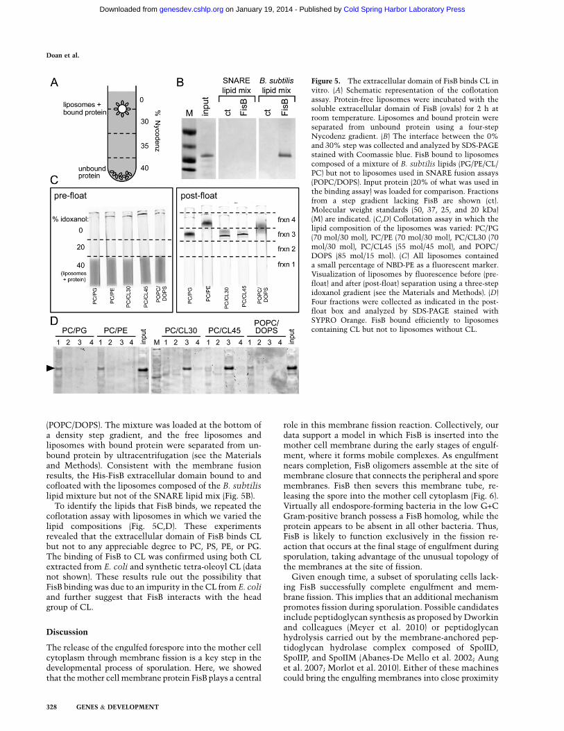
(POPC/DOPS). The mixture was loaded at the bottom ofa density step gradient, and the free liposomes andliposomes with bound protein were separated from un-bound protein by ultracentrifugation (see the Materialsand Methods). Consistent with the membrane fusionresults, the His-FisB extracellular domain bound to andcofloated with the liposomes composed of the B. subtilislipid mixture but not of the SNARE lipid mix (Fig. 5B).
To identify the lipids that FisB binds, we repeated thecoflotation assay with liposomes in which we varied thelipid compositions (Fig. 5C,D). These experimentsrevealed that the extracellular domain of FisB binds CLbut not to any appreciable degree to PC, PS, PE, or PG.The binding of FisB to CL was confirmed using both CLextracted from E. coli and synthetic tetra-oleoyl CL (datanot shown). These results rule out the possibility thatFisB binding was due to an impurity in the CL from E. coliand further suggest that FisB interacts with the headgroup of CL.
Discussion
The release of the engulfed forespore into the mother cellcytoplasm through membrane fission is a key step in thedevelopmental process of sporulation. Here, we showedthat the mother cell membrane protein FisB plays a central
role in this membrane fission reaction. Collectively, ourdata support a model in which FisB is inserted into themother cell membrane during the early stages of engulf-ment, where it forms mobile complexes. As engulfmentnears completion, FisB oligomers assemble at the site ofmembrane closure that connects the peripheral and sporemembranes. FisB then severs this membrane tube, re-leasing the spore into the mother cell cytoplasm (Fig. 6).Virtually all endospore-forming bacteria in the low G+CGram-positive branch possess a FisB homolog, while theprotein appears to be absent in all other bacteria. Thus,FisB is likely to function exclusively in the fission re-action that occurs at the final stage of engulfment duringsporulation, taking advantage of the unusual topology ofthe membranes at the site of fission.
Given enough time, a subset of sporulating cells lack-ing FisB successfully complete engulfment and mem-brane fission. This implies that an additional mechanismpromotes fission during sporulation. Possible candidatesinclude peptidoglycan synthesis as proposed by Dworkinand colleagues (Meyer et al. 2010) or peptidoglycanhydrolysis carried out by the membrane-anchored pep-tidoglycan hydrolase complex composed of SpoIID,SpoIIP, and SpoIIM (Abanes-De Mello et al. 2002; Aunget al. 2007; Morlot et al. 2010). Either of these machinescould bring the engulfing membranes into close proximity
Figure 5. The extracellular domain of FisB binds CL invitro. (A) Schematic representation of the coflotationassay. Protein-free liposomes were incubated with thesoluble extracellular domain of FisB (ovals) for 2 h atroom temperature. Liposomes and bound protein wereseparated from unbound protein using a four-stepNycodenz gradient. (B) The interface between the 0%and 30% step was collected and analyzed by SDS-PAGEstained with Coomassie blue. FisB bound to liposomescomposed of a mixture of B. subtilis lipids (PG/PE/CL/PC) but not to liposomes used in SNARE fusion assays(POPC/DOPS). Input protein (20% of what was used inthe binding assay) was loaded for comparison. Fractionsfrom a step gradient lacking FisB are shown (ct).Molecular weight standards (50, 37, 25, and 20 kDa)(M) are indicated. (C,D) Coflotation assay in which thelipid composition of the liposomes was varied: PC/PG(70 mol/30 mol), PC/PE (70 mol/30 mol), PC/CL30 (70mol/30 mol), PC/CL45 (55 mol/45 mol), and POPC/DOPS (85 mol/15 mol). (C) All liposomes containeda small percentage of NBD-PE as a fluorescent marker.Visualization of liposomes by fluorescence before (pre-float) and after (post-float) separation using a three-stepidoxanol gradient (see the Materials and Methods). (D)Four fractions were collected as indicated in the post-float box and analyzed by SDS-PAGE stained withSYPRO Orange. FisB bound efficiently to liposomescontaining CL but not to liposomes without CL.
Doan et al.
328 GENES & DEVELOPMENT
Cold Spring Harbor Laboratory Press on January 19, 2014 - Published by genesdev.cshlp.orgDownloaded from

and promote fission (Judd et al. 2005), although at a slowerrate than FisB. Alternatively, SpoIIIE or an as yet un-identified factor could function in an independent fissionpathway.
SpoIIIE and membrane fission
Our analysis of engulfment in cells lacking the SpoIIIEDNA translocase suggests that its activity is critical formembrane migration around the forespore prior tofission. These results do not exclude the possibility thatit could also function in the fission reaction. However,our analysis of the FisB protein in SpoIIIE mutantsprovides a simpler explanation for the original observa-tions of Sharp and Pogliano (1999). Immunoblot analysisof sporulating cells revealed that the levels of FisB weresignificantly reduced in the SpoIIIE-null mutant com-pared with wild type (Supplemental Fig. S8A). Moreover,in a SpoIIIE point mutant (SpoIIIE36) that binds DNA butis impaired in DNA transport (Wu and Errington 1994;Wu et al. 1995), FisB protein levels were unaffected, butYFP-FisB localization was disrupted (Supplemental Fig.S8B). In accordance with this finding, Sharp and Pogliano(1999) reported that the SpoIIIE36 mutant was impairedin membrane fission but had a less severe defect thanthe null. Based on these observations, we favor the ideathat SpoIIIE is only indirectly involved in the fissionreaction, impacting membrane migration and FisB abun-dance and/or localization. Interestingly, it has also been
proposed that SpoIIIE participates in fission duringasymmetric division in sporulating cells (Liu et al.2006; Fleming et al. 2010). This membrane remodelingevent, wherein cytosol fills the gap inside the closingmembrane annulus, has the opposite topology to thefission at the end of engulfment, where the extracellularmedium fills the gap to be closed by the engulfingmembrane (Supplemental Fig. S9). Intriguingly, SpoIIIE’sarchitecture (large cytoplasmic domain anchored to themembrane) is opposite to that of FisB (membrane-an-chored extracytoplasmic domain). Establishing a directrole for SpoIIIE in membrane remodeling awaits in vitroreconstitution.
Interactions between the extracellular domain of FisBand CL
We found that the extracellular domain of FisB interactswith CL, and our data suggest that this interaction isimportant for membrane remodeling in vitro. The spec-ificity of this interaction is remarkable for a solubledomain. Although CL has high affinity for proteins withtransmembrane segments involved in oxidative phos-phorylation, such as the F0F1 ATPase (Haines 2009),many soluble proteins that bind CL, such as cytochrome c(Kagan et al. 2009) and tBid (Epand et al. 2002) ineukaryotes and MinD (Mileykovskaya et al. 2003) inbacteria, also interact with other, negatively chargedlipids such as PS and PG to varying extents. In contrast,we could not detect significant interaction between theextracellular domain of FisB and the acidic lipids PG andPS or with the zwitterionic lipids PE and PC (or withdigalactosyldiacylglycerol) (data not shown). Thus, weexpect the dynamics of FisB and CL to be intimatelycoupled (see below). In this regard, it is noteworthy thatduring sporulation, the levels of CL increase, and CL isenriched in the engulfing membranes (Kawai et al. 2004).B. subtilis possesses three genes that encode proteinshomologous to CL synthases: clsA (ywnE), which en-codes the major CL synthase; clsB (ywjE); and ywiE(Kunst et al. 1997; Kawai et al. 2004). The expression ofboth clsA and clsB increases during sporulation, consis-tent with the idea that CL plays an important role duringthis process. Cells lacking all three genes are viable andhave undetectable levels of CL during vegetative growth(Kawai et al. 2004). However, CL is readily detectable inthe triple mutant during the early stages of sporulation,indicating that an additional sporulation-specific CLsynthase exists or that CL is produced as a side reactionby a lipid synthesis/modification enzyme (Kawai et al.2004, 2006; Tan et al. 2012). In accordance with thepresence of CL during engulfment in cells lacking ClsA,ClsB, and YwiE, we found that the triple mutant wasable to undergo fission at a frequency comparable withwild type (data not shown). This result suggests thatif FisB requires CL to catalyze fission in vivo, then thereduced amount of CL made in the absence of theknown synthases must be sufficient. Alternatively,FisB could use another phospholipid in addition to CLin vivo.
Figure 6. Model for FisB-mediated fission. Schematic repre-sentation of the membrane tube formed at the cell pole by theengulfing mother cell membranes. Due to its high negativecurvature, CL (not shown) is thought to become enrichedwithin the tube. FisB (green) becomes immobilized at this sitethrough its interaction with CL. Oligomerization of FisB orFisB–lipid interactions are proposed to sever the tube, releasingthe spore into the mother cell cytoplasm. See the Discussionfor details.
FisB mediates engulfment fission
GENES & DEVELOPMENT 329
Cold Spring Harbor Laboratory Press on January 19, 2014 - Published by genesdev.cshlp.orgDownloaded from

Late forespore gene expression does not requirethe completion of engulfment
Previous work has shown that the developmental pro-grams of gene expression in the mother cell and foresporeare not free-running clocks but are linked to each other bycell–cell signaling pathways (Stragier and Losick 1996;Rudner and Losick 2001; Rudner and Doan 2008). Fur-thermore, it has been hypothesized that these cell type-specific transcriptional programs are coupled to morpho-genesis. Specifically, it was proposed that the activationof the forespore transcription factor sG is coupled to thecompletion of engulfment and membrane fission (Stragierand Losick 1996; Rudner and Losick 2001; Rudner andDoan 2008; Regan et al. 2012). One compelling model wasthat the isolation of the forespore from the externalenvironment as a result of fission served as the triggerfor sG. Recently, we reported evidence that arguedagainst the coupling of sG activation to membrane fission(Doan et al. 2009). Specifically, we found that a subset(13%) of sporulating cells that was blocked at early stagesof engulfment successfully activated sG in the forespore.However, the severe defect in morphogenesis in thesemutants and the small number of cells that turned on sG
raised concern that the activation might not be biologi-cally relevant. Furthermore, recent genetic experimentsusing large insertions into the B. subtilis chromosomethat delay DNA transport into the forespore suggest thatsG activation could indeed be coupled to completion ofengulfment (Regan et al. 2012). The FisB mutant allowedus to revisit the link between sG activity and morpho-genesis in a strain in which engulfment proceeds nor-mally and only fission is impaired.
Analysis of sG activity in the FisB mutant revealed that47% (n > 1000) of the sporulating cells that were stalled atthe membrane fission step successfully activated sG inthe forespore compartment (Supplemental Fig. S10). Thisobservation lends further support to the idea that sG
activation and the fission reaction are not coupled events.Intriguingly, we also found that sporulating FisB mutantslyse at an elevated frequency, and this lysis was sup-pressed in cells lacking sG (Supplemental Fig. S11). Thisobservation suggests that activation of sG prior to thecompletion of membrane fission is deleterious to thesporulating cell and leads to death. Thus, althougha mechanism that holds sG inactive until fission iscomplete would appear to be beneficial, our data suggestthat B. subtilis does not employ one. We suspect thatunder normal circumstances (in wild-type cells), sG
activation and fission are temporally distinct enough thata coupling device is not required.
How might FisB mediate membrane scission?
Although membrane fission is a fundamental and ubiq-uitous biological process used by species ranging from thesimplest bacteria and enveloped viruses to the mostsophisticated neuronal cells, only two general fissionmachineries have been described. The first and bestunderstood involves constriction of a membrane neckor tube from the outside by dynamin and its homologs
(Ferguson and De Camilli 2012). This constriction pro-cess relies on oligomerization of dynamin around themembrane neck and GTP hydrolysis to sever it. Thesecond fission machinery is the ESCRT-III system (Hurley2010; Henne et al. 2011), in which all components aresoluble proteins that are recruited to the membrane viaprotein–protein and protein–lipid interactions. The onlyESCRT-III component that seems to be absolutely re-quired for fission is Snf7, whose oligomerization initiatesthe fission process, at least in vitro (Wollert and Hurley2010). Other subunits likely terminate Snf7 filamentassembly and recruit Vps4, an ATPase that disassemblesthe ESCRT-III complex and perhaps contributes to thefission reaction. The mechanism by which ESCRT-IIImediates fission is still intensely debated (Hurley 2010;Henne et al. 2011); however, it is clear that the machineryworks at the opening of and/or inside the membrane neckto achieve fission, rather than using constriction fromoutside the tube. Based on the topology of FisB with itslarge extracytoplasmic domain and very short (15-amino-acid) cytoplasmic domain, we suspect that FisB alsocatalyzes fission by doing work at one (or both) of theopenings of the tube or from within it.
The activity of eukaryotic fusion and fission machinessuch as SNAREs and dynamin are exquisitely regulated intime and space by other proteins and intracellular signals.Potential regulators of FisB other than CL have not beenidentified. However, given the topology of the mem-branes during engulfment of the forespore, it is possible,in principle, that the dynamics of CL during sporulation,its propensity to associate with negatively curved mem-branes, FisB binding to CL, and homo-oligomerization ofFisB could provide sufficient control over when andwhere fission occurs.
At the final stages of engulfment, the tube connectingthe membrane engulfing the forespore to the rest of themother cell membrane must shrink in diameter (Fig. 6).This will increase the negative curvature of the tube’sinner leaflet, making it increasingly favorable for CLlocalization. We hypothesize that FisB becomes immobi-lized at the fission site as a result of its interaction withCL within the membrane tube. We found that FisB formsoligomers in vitro, and one possibility is that oligomeri-zation leads to a filling of the tube with FisB and overflowto the tube’s openings. The positive curvature of theseopenings would be unfavorable for CL localization. If FisBrecruits CL to these positively curved regions, this couldlead to the destabilization and membrane scission. Ina variation of this model, FisB would localize to thehighly positively curved regions at the openings of thetube by virtue of its own curvature preference and, byrecruiting CL to such regions, would accumulate stresses,which could be relieved by severing the tube.
Other alternatives exist that are not mutually exclu-sive. For example, FisB oligomerization with concomi-tant conformational changes could be coupled to thedestabilization of the tube, perhaps via twisting or com-pression. Alternatively, FisB could induce lateral segre-gation of CL (de Kruijff and Cullis 1980), with theresulting line tension and/or the discontinuity in elastic
Doan et al.
330 GENES & DEVELOPMENT
Cold Spring Harbor Laboratory Press on January 19, 2014 - Published by genesdev.cshlp.orgDownloaded from

properties between the CL-rich and CL-poor phasesleading spontaneously to fission. Such fission occurs atdomain boundaries between cholesterol-rich and choles-terol-poor tubules under certain conditions (Allain et al.2004; Roux et al. 2005). Finally, FisB might act througha mechanism reminiscent of viral fusion proteins. Al-though fusion and fission are topologically opposite pro-cesses, microscopically, both require cutting, bending,and rejoining of membranes. From this point of view, it isperhaps not surprising that some homologs of dynamin-1have been described to mediate fusion, not fission (Kozlovet al. 2010; Ferguson and De Camilli 2012). In this model,FisB anchored inside the membrane tube would interactwith CL across the tube via its extracytoplasmic domain.CL binding could trigger a conformational change inFisB, as in the case of cytochrome c (Kagan et al. 2009),ultimately leading to membrane fission.
Further biochemical analysis of FisB in vitro, alongwith the identification of other proteins that functionwith or regulate FisB and the missing CL synthase, willhelp distinguish among these models for FisB function.However, our characterization of FisB raises the intrigu-ing possibility that it is part of a novel fission mechanism.
Materials and methods
General methods
All B. subtilis strains were derived from the prototrophic strainPY79 (Youngman et al. 1983). Sporulation was induced byresuspension at 37°C according to the method of Sterlini-Mandelstam (Harwood and Cutting 1990) or by exhaustion insupplemented DS medium (Schaeffer et al. 1965). Sporulationefficiency was determined in 36-h cultures as the total number ofheat-resistant (20 min at 80°C) colony-forming units (CFUs)compared with wild-type heat-resistant CFUs. Analysis of mem-brane fission after inhibition of cell wall synthesis was per-formed as described previously (Meyer et al. 2010) except cellswere sporulated at 37°C and fosfomycin (5 mM final) was addedat hour 1.5. Membrane fission was assessed at hours 2, 2.5, and 3.Insertion–deletion mutations were generated by isothermalassembly (Gibson et al. 2009) of PCR products followed by directtransformation into B. subtilis. Tables of strains, plasmids, andoligonucleotide primers and a description of plasmid construc-tion can be found online in the Supplemental Material (Supple-mental Tables S1–S3; Supplemental Material).
Protein purification and antibody production
His6-FisBFL and His6-FisBECD fusion proteins were expressed inE. coli BL21 DE3 pLysS at 30°C and 16°C, respectively, andpurified by affinity chromatography on Ni2+-NTA agarose (Qiagen).MBP-FisBECD fusion protein was expressed in E. coli NB42 andpurified by affinity chromatography on amylose resin (NewEngland Biolabs). A complete description of the purificationscan be found in the Supplemental Material. MBP-FisBECD peakfractions were pooled and used to generate rabbit polyclonalantibodies (Covance). Crude serum was affinity-purified as de-scribed (Campo and Rudner 2006) using His6-FisBECD.
Immunoblot analysis
Whole-cell lysates from sporulating cells induced by resuspen-sion were prepared as described (Doan and Rudner 2007).
Samples were heated for 10 min at 50°C prior to loading.Equivalent loading was based on OD600 at the time of harvest.Proteins were separated by SDS-PAGE on 12.5% polyacrylamidegels, electroblotted onto Immobilon-P membranes (Millipore),and blocked in 5% nonfat milk in phosphate-buffered saline(PBS) and 0.5% Tween-20. The blocked membranes were probedwith affinity-purified anti-His (1:1000; Santa Cruz Biotechnol-ogy), anti-FisB (1:10,000), and anti-sF (15,000) (Carniol et al.2004) diluted into 3% BSA in 13 PBS-0.05% Tween-20. Primaryantibodies were detected using horseradish peroxidase-conju-gated goat anti-rabbit G (Bio-Rad) and the Western Lightningreagent kit as described by the manufacturer (PerkinElmer).
In vitro lipid mixing assay
The protocol described in Weber et al. (1998) was followed withminor modifications. All lipids were purchased from AvantiPolar Lipids. For fluorescent donor liposomes, 100 mL of 3 mMlipids (L-a-PG [E. coli; sodium salt], CL [E. coli; sodium salt], L-a-phosphatidylethanolamine [E. coli, PE], L-a-PC [egg, chicken],NBD-PE [ammonium salt; 18:1], LR-PE [ammonium salt; 18:1])in a molar ratio of 30:45:13:10:1:1 dissolved in chloroform weredried under nitrogen gas, placed under a vacuum dessicator for1 h, and then dissolved in reconstitution buffer (RB; 25 mMHepes at pH 7.4, 100 mM KCl, 10% glycerol, 1 mM DTT)supplemented with 1% (w/v) octyl-b-D-glucopyranoside (OG)and either FisB protein, the mock purification, or nothing toobtain protein-free liposomes. Unlabeled acceptor liposomeswere prepared using 100 mL of 15 mM lipids as above in a molarratio of 30:45:15:10. FisB protein was reconstituted into thelipids at a 1:1000 (protein:lipid) molar ratio, and then the OG wasremoved by overnight dialysis (6000- to 8000-Da molecularweight cutoff). Unincorporated protein was removed by flotationof the liposomes through a Nycodenz (Accurate Chemicals) stepgradient. Labeled liposomes were recovered in a 150-mL volume(2 mM lipid). Unlabeled liposomes were collected in 400 mL (3.75mM lipid).
The lipid-mixing assay was performed in white 96-wellMaxisorp plates (Nunc). Liposomes (10 mL of labeled and 45 mLof unlabeled) were mixed on ice and then placed in a roomtemperature SpectraMax M5 Microplate Reader (Molecular De-vices), and NBD fluorescence was recorded with filters set at 460nm (excitation) and 538 nm (emission) at 30-sec intervals for 45min. To determine the maximum NBD fluorescence, 10 mL of2.5% (w/v) n-dodecyl-b-D-maltoside (DM) (Thermo Scientific)was added at the end of the experiment, and fluorescences weremonitered for 15 min.
Flotation assay
Natural (PG, PE, and CL, all from E. coli, or egg PC) lipids orsynthetic lipids (POPC), 1,2-dioleoyl-sn-glycero-3-phospho-L-serine (sodium salt) , 19,39-bis(1,2-dioleoyl-sn-glycero-3-phospho)-sn-glycerol (sodium salt; 18:1 CL), and 1,2-dioleoyl-sn-glycero-3-phospho-(19-rac-glycerol) (sodium salt) ) from Avanti Polar Lipidswere used. All liposomes included 0.2 mole percent NBD-PE(ammonium salt; 18:1) as a fluorescent marker. The lipids weredissolved in a 2:1 (v/v) mixture of chloroform and methanol.Lipids (1 mmol total) were mixed at desired ratios, and the mix-tures were dried under nitrogen flow. Residual organic solventwas removed under vacuum for at least 2 h in a vacuumdessicator. Lipids were hydrated in 1 mL of RB-EDTA buffer[25 mM Hepes at pH 7.4, 140 mM KCl, 1 mM EDTA, 0.2 mMtris(2-carboxyethyl)phosphine] by shaking for >30 min. The lipidsuspension was then immersed in liquid nitrogen and thenallowed to thaw in a water bath at 37°C. This freeze–thaw cycle
FisB mediates engulfment fission
GENES & DEVELOPMENT 331
Cold Spring Harbor Laboratory Press on January 19, 2014 - Published by genesdev.cshlp.orgDownloaded from
was repeated five additional times to form large unilamellarvesicles (LUVs) (Pick 1981). The LUVs were either flash-frozen inliquid nitrogen and stored at �80°C or used fresh for extrusion.The LUV stock was extruded 19 times through 50-nm pore sizepolycarbonate filters (Avanti) at 40°C using a miniextruder.
The extruded liposomes (40 nmol total lipid) were incubatedwith 40 pmol of FisB extracytoplasmic domain in a total volumeof 100 mL for 1 h at room temperature. Two-hundred milliliters of60% iodoxanol density gradient medium (Optiprep, Sigma-Aldrich) was added to make a 40% iodoxanol solution (density,1.215 g/mL) that was layered at the bottom of a 5-mm 3 41-mmBeckman Ultra-Clear ultracentrifuge tube (Beckman Coulter,Inc.). This was overlaid with 200 mL of 20% iodoxanol solution(diluted in RB-EDTA; density, 1.110 g/mL), followed by a toplayer of 200 mL of RB-EDTA. To assess liposome redistributionafter floatation, NBD-PE fluorescence was measured before andafter centrifugation as follows: Step gradients were prepared inparallel, and the tubes were placed in a rack in a gel-imaginginstrument (ImageQuant LAS 4000, GE Healtcare). A mirror wasplaced at a 45° angle next to the rack, reflecting the image of thetubes to the camera placed above (Fig. 5C, pre-float; Bigay andAntonny 2005; Bigay et al. 2005). Blue epi-illumination was used,and fluorescence was collected through a green long-pass filter(Y515 Di). After imaging, the tubes were spun at 48,000 rpm(269,000g) for 1.5 h at 20°C. After floatation, the tubes wereimaged again to assess the position of the liposomes in thegradient, and then fractions were collected from the bottom ofeach tube as indicated in Figure 5C, post-float. Because FisBcould have varying affinities for the different lipids, we quanti-fied both lipid and protein in all fractions. The amount of lipid ineach fraction was quantified using NBD-PE fluorescence, and theamount of protein was quantified by PAGE (Novex mini Nu-PAGE Bis-tris, 12%, 1 mm thick; Invitrogen), stained withSYPRO Orange (Invitrogen). The sum of the protein from eachfraction was usually less than the total initially mixed withliposomes (Fig. 5D). This is probably because some of the proteinsticks to the walls of the ultracentrifuge tube. However, this wasnot an issue when comparing relative amounts of protein indifferent fractions (Bigay and Antonny 2005; Bigay et al. 2005).
Fluorescence microscopy
Fluorescence microscopy was performed on an Olympus BX61microscope as previously described (Doan et al. 2005). Fluores-cent signals were visualized with a phase-contrast objectiveUplanF1 1003 and captured with a monochrome CoolSnapHQdigital camera (Photometrics) using Metamorph software (Mo-lecular Device). Exposure times were typically 500 msec for CFPand GFP and 2000 msec for YFP-FisB. Membranes were stainedwith either TMA-DPH or FM4-64 (Molecular Probes) at a finalconcentration of 0.01 mM and 3 mg/mL, respectively. Exposuretimes were typically 200 msec. Images were analyzed, adjusted,and cropped using Metamorph software. Cells were concentratedby centrifugation (8000 rpm for 30 sec) prior to visualization.This step had no impact on the localization of the fusionproteins.
Acknowledgments
We thank members of the Rudner laboratory (past and present)for advice and encouragement, Tom Rapoport and JochenZimmer for advice on membrane protein purification, TomBernhardt and Tsuyoshi Uehara for help with gel filtration,Mike Strauss and Chris Rodrigues for stimulating discussions,Annick Turbe-Doan for help with figures, Bruno Antonny fordiscussions and advice on lipid–protein interactions, and Rich
Losick for generously providing the library of sE regulon mu-tants. E.K. thanks Jim Rothman for a home to do the fusion andflotation experiments. T.D. thanks Anne Galinier for her pa-tience and generosity. Support for this work comes from theNational Institutes of Health Grant GM086466 (to D.Z.R.) andCNRS and Marie-Curie International Reintegration GrantPIRG08-GA-2010-276750 (to T.D.).
References
Abanes-De Mello A, Sun YL, Aung S, Pogliano K. 2002. Acytoskeleton-like role for the bacterial cell wall duringengulfment of the Bacillus subtilis forespore. Genes Dev16: 3253–3264.
Allain JM, Storm C, Roux A, Ben Amar M, Joanny JF. 2004.Fission of a multiphase membrane tube. Phys Rev Lett 93:158104.
Aung S, Shum J, Abanes-De Mello A, Broder DH, Fredlund-Gutierrez J, Chiba S, Pogliano K. 2007. Dual localizationpathways for the engulfment proteins during Bacillus sub-tilis sporulation. Mol Microbiol 65: 1534–1546.
Bashkirov PV, Akimov SA, Evseev AI, Schmid SL, Zimmerberg J,Frolov VA. 2008. GTPase cycle of dynamin is coupled tomembrane squeeze and release, leading to spontaneousfission. Cell 135: 1276–1286.
Bieniasz PD. 2006. Late budding domains and host proteins inenveloped virus release. Virology 344: 55–63.
Bigay J, Antonny B. 2005. Real-time assays for the assembly-disassembly cycle of COP coats on liposomes of defined size.Methods Enzymol 404: 95–107.
Bigay J, Casella JF, Drin G, Mesmin B, Antonny B. 2005.ArfGAP1 responds to membrane curvature through thefolding of a lipid packing sensor motif. EMBO J 24: 2244–2253.
Burton BM, Marquis KA, Sullivan NL, Rapoport TA, RudnerDZ. 2007. The ATPase SpoIIIE transports DNA across fusedseptal membranes during sporulation in Bacillus subtilis.Cell 131: 1301–1312.
Burmann F, Ebert N, van Baarle S, Bramkamp M. 2011. A bacterialdynamin-like protein mediating nucleotide-independent mem-brane fusion. Mol Microbiol 79: 1294–1304.
Campo N, Rudner DZ. 2006. A branched pathway governing theactivation of a developmental transcription factor by regu-lated intramembrane proteolysis. Mol Cell 23: 25–35.
Carniol K, Eichenberger P, Losick R. 2004. A threshold mech-anism governing activation of the developmental regulatoryprotein sF in Bacillus subtilis. J Biol Chem 279: 14860–14870.
Chernomordik LV, Kozlov MM. 2003. Protein–lipid interplay infusion and fission of biological membranes. Annu RevBiochem 72: 175–207.
de Boer PA. 2010. Advances in understanding E. coli cell fission.Curr Opin Microbiol 13: 730–737.
de Kruijff B, Cullis PR. 1980. Cytochrome c specifically inducesnon-bilayer structures in cardiolipin-containing model mem-branes. Biochim Biophys Acta 602: 477–490.
Doan T, Rudner DZ. 2007. Perturbations to engulfment triggera degradative response that prevents cell-cell signallingduring sporulation in Bacillus subtilis. Mol Microbiol 64:500–511.
Doan T, Marquis KA, Rudner DZ. 2005. Subcellular localizationof a sporulation membrane protein is achieved througha network of interactions along and across the septum. Mol
Microbiol 55: 1767–1781.Doan T, Morlot C, Meisner J, Serrano M, Henriques AO, Moran
CP, Rudner DZ. 2009. Novel secretion apparatus maintains
Doan et al.
332 GENES & DEVELOPMENT
Cold Spring Harbor Laboratory Press on January 19, 2014 - Published by genesdev.cshlp.orgDownloaded from

spore integrity and developmental gene expression in Bacil-
lus subtilis. PLoS Genet 5: e1000566.Eichenberger P, Jensen ST, Conlon EM, van Ooij C, Silvaggi J,
Gonzalez-Pastor JE, Fujita M, Ben-Yehuda S, Stragier P, LiuJS, et al. 2003. The sE regulon and the identification ofadditional sporulation genes in Bacillus subtilis. J Mol Biol
327: 945–972.Elia N, Sougrat R, Spurlin TA, Hurley JH, Lippincott-Schwartz J.
2011. Dynamics of endosomal sorting complex required fortransport (ESCRT) machinery during cytokinesis and its rolein abscission. Proc Natl Acad Sci 108: 4846–4851.
Epand RF, Martinou JC, Fornallaz-Mulhauser M, Hughes DW,Epand RM. 2002. The apoptotic protein tBid promotesleakage by altering membrane curvature. J Biol Chem 277:32632–32639.
Errington J. 2003. Regulation of endospore formation in Bacillussubtilis. Nat Rev Microbiol 1: 117–126.
Ferguson SM, De Camilli P. 2012. Dynamin, a membrane-remodelling GTPase. Nat Rev Mol Cell Biol 13: 75–88.
Feucht A, Evans L, Errington J. 2003. Identification of sporula-tion genes by genome-wide analysis of the sE regulon ofBacillus subtilis. Microbiology 149: 3023–3034.
Fleming TC, Shin JY, Lee SH, Becker E, Huang KC, BustamanteC, Pogliano K. 2010. Dynamic SpoIIIE assembly mediatesseptal membrane fission during Bacillus subtilis sporulation.Genes Dev 24: 1160–1172.
Gibson DG, Young L, Chuang RY, Venter JC, Hutchison CA,Smith HO. 2009. Enzymatic assembly of DNA molecules upto several hundred kilobases. Nat Methods 6: 343–345.
Gregory JA, Becker EC, Pogliano K. 2008. Bacillus subtilis MinCdestabilizes FtsZ-rings at new cell poles and contributes tothe timing of cell division. Genes Dev 22: 3475–3488.
Griffiths KK, Setlow P. 2009. Effects of modification of mem-brane lipid composition on Bacillus subtilis sporulation andspore properties. J Appl Microbiol 106: 2064–2078.
Gruenberg J, Stenmark H. 2004. The biogenesis of multivesic-ular endosomes. Nat Rev Mol Cell Biol 5: 317–323.
Haines TH. 2009. A new look at cardiolipin. Biochim Biophys
Acta 1788: 1997–2002.Harrison SC. 2008. Viral membrane fusion. Nat Struct Mol Biol
15: 690–698.Harwood C, Cutting S. 1990. Molecular biological methods for
Bacillus. Wiley, New York.Henne WM, Buchkovich NJ, Emr SD. 2011. The ESCRT pathway.
Dev Cell 21: 77–91.Hurley JH. 2008. ESCRT complexes and the biogenesis of
multivesicular bodies. Curr Opin Cell Biol 20: 4–11.Hurley JH. 2010. The ESCRT complexes. Crit Rev Biochem Mol
Biol 45: 463–487.Judd EM, Comolli LR, Chen JC, Downing KH, Moerner WE,
McAdams HH. 2005. Distinct constrictive processes, sepa-rated in time and space, divide caulobacter inner and outermembranes. J Bacteriol 187: 6874–6882.
Kagan VE, Bayir HA, Belikova NA, Kapralov O, Tyurina YY,Tyurin VA, Jiang J, Stoyanovsky DA, Wipf P, Kochanek PM,et al. 2009. Cytochrome c/cardiolipin relations in mitochon-dria: A kiss of death. Free Radic Biol Med 46: 1439–1453.
Karatekin E, Di Giovanni J, Iborra C, Coleman J, O’ShaughnessyB, Seagar M, Rothman JE. 2010. A fast, single-vesicle fusionassay mimics physiological SNARE requirements. Proc Natl
Acad Sci 107: 3517–3521.Kawai F, Shoda M, Harashima R, Sadaie Y, Hara H, Matsumoto
K. 2004. Cardiolipin domains in Bacillus subtilis marburgmembranes. J Bacteriol 186: 1475–1483.
Kawai F, Hara H, Takamatsu H, Watabe K, Matsumoto K.2006. Cardiolipin enrichment in spore membranes and its
involvement in germination of Bacillus subtilis Marburg.Genes Genet Syst 81: 69–76.
Kielian M, Rey FA. 2006. Virus membrane-fusion proteins: Morethan one way to make a hairpin. Nat Rev Microbiol 4: 67–76.
Kozlov MM, McMahon HT, Chernomordik LV. 2010. Protein-driven membrane stresses in fusion and fission. Trends
Biochem Sci 35: 699–706.Kozlovsky Y, Kozlov MM. 2003. Membrane fission: Model for
intermediate structures. Biophys J 85: 85–96.Kunst F, Ogasawara N, Moszer I, Albertini AM, Alloni G,
Azevedo V, Bertero MG, Bessieres P, Bolotin A, Borchert S,et al. 1997. The complete genome sequence of the gram-positive bacterium Bacillus subtilis. Nature 390: 249–256.
Lindas AC, Karlsson EA, Lindgren MT, Ettema TJ, Bernander R.2008. A unique cell division machinery in the Archaea. Proc
Natl Acad Sci 105: 18942–18946.Liu NJ, Dutton RJ, Pogliano K. 2006. Evidence that the SpoIIIE
DNA translocase participates in membrane fusion duringcytokinesis and engulfment. Mol Microbiol 59: 1097–1113.
Marquis KA, Burton BM, Nollmann M, Ptacin JL, BustamanteC, Ben-Yehuda S, Rudner DZ. 2008. SpoIIIE strips proteins offthe DNA during chromosome translocation. Genes Dev 22:1786–1795.
Meyer P, Gutierrez J, Pogliano K, Dworkin J. 2010. Cell wallsynthesis is necessary for membrane dynamics during spor-ulation of Bacillus subtilis. Mol Microbiol 76: 956–970.
Mileykovskaya E, Fishov I, Fu X, Corbin BD, Margolin W,Dowhan W. 2003. Effects of phospholipid composition onMinD–membrane interactions in vitro and in vivo. J Biol
Chem 278: 22193–22198.Morita E, Sundquist WI. 2004. Retrovirus budding. Annu Rev
Cell Dev Biol 20: 395–425.Morlot C, Uehara T, Marquis KA, Bernhardt TG, Rudner DZ.
2010. A highly coordinated cell wall degradation machinegoverns spore morphogenesis in Bacillus subtilis. Genes Dev24: 411–422.
Pick U. 1981. Liposomes with a large trapping capacity preparedby freezing and thawing of sonicated phospholipid mixtures.Arch Biochem Biophys 212: 186–194.
Piggot P, Losick R. 2002. Sporulation genes and intercompart-mental regulation. In Bacillus subtilis and its closest rela-
tives: From genes to cells (ed. A Sonenshein et al.), pp. 483–517. ASM Press, Washington, D.C.
Rand RP, Parsegian VA. 1986. Mimicry and mechanism inphospholipid models of membrane fusion. Annu Rev Physiol
48: 201–212.Regan G, Itaya M, Piggot PJ. 2012. Coupling of sG activation to
completion of engulfment during sporulation of Bacillus
subtilis survives large perturbations to DNA translocationand replication. J Bacteriol 194: 6264–6271.
Roux A, Cuvelier D, Nassoy P, Prost J, Bassereau P, Goud B.2005. Role of curvature and phase transition in lipid sortingand fission of membrane tubules. EMBO J 24: 1537–1545.
Rudner D, Doan T. 2008. Intercompartmental signal transduc-tion during sporulation in Bacillus subtilis. In Chemical
communication among bacteria (ed. S Winans, B Bassler),pp. 3–12. ASM Press, Washington, D.C.
Rudner DZ, Losick R. 2001. Morphological coupling in de-velopment: Lessons from prokaryotes. Dev Cell 1: 733–742.
Rudner DZ, Pan Q, Losick RM. 2002. Evidence that sub-cellular localization of a bacterial membrane protein isachieved by diffusion and capture. Proc Natl Acad Sci 99:8701–8706.
Samson RY, Obita T, Freund SM, Williams RL, Bell SD. 2008. Arole for the ESCRT system in cell division in archaea.Science 322: 1710–1713.
FisB mediates engulfment fission
GENES & DEVELOPMENT 333
Cold Spring Harbor Laboratory Press on January 19, 2014 - Published by genesdev.cshlp.orgDownloaded from

Schaeffer P, Millet J, Aubert JP. 1965. Catabolic repression ofbacterial sporulation. Proc Natl Acad Sci 54: 704–711.
Sharp MD, Pogliano K. 1999. An in vivo membrane fusion assayimplicates SpoIIIE in the final stages of engulfment duringBacillus subtilis sporulation. Proc Natl Acad Sci 96: 14553–14558.
Sharp MD, Pogliano K. 2003. The membrane domain of SpoIIIEis required for membrane fusion during Bacillus subtilis
sporulation. J Bacteriol 185: 2005–2008.Steil L, Serrano M, Henriques AO, Volker U. 2005. Genome-
wide analysis of temporally regulated and compartment-specific gene expression in sporulating cells of Bacillus
subtilis. Microbiology 151: 399–420.Strack B, Calistri A, Craig S, Popova E, Gottlinger HG. 2003.
AIP1/ALIX is a binding partner for HIV-1 p6 and EIAV p9functioning in virus budding. Cell 114: 689–699.
Stragier P, Losick R. 1996. Molecular genetics of sporulation inBacillus subtilis. Annu Rev Genet 30: 297–341.
Struck DK, Hoekstra D, Pagano RE. 1981. Use of resonanceenergy transfer to monitor membrane fusion. Biochemistry20: 4093–4099.
Sudhof TC, Rothman JE. 2009. Membrane fusion: Grapplingwith SNARE and SM proteins. Science 323: 474–477.
Sullivan NL, Marquis KA, Rudner DZ. 2009. Recruitment ofSMC by ParB–parS organizes the origin region and promotesefficient chromosome segregation. Cell 137: 697–707.
Tan BK, Bogdanov M, Zhao J, Dowhan W, Raetz CR, Guan Z.2012. Discovery of a cardiolipin synthase utilizing phospha-tidylethanolamine and phosphatidylglycerol as substrates.Proc Natl Acad Sci 109: 16504–16509.
Weber T, Zemelman BV, McNew JA, Westermann B, Gmachl M,Parlati F, Sollner TH, Rothman JE. 1998. SNAREpins: Min-imal machinery for membrane fusion. Cell 92: 759–772.
Weiss DS. 2004. Bacterial cell division and the septal ring. Mol
Microbiol 54: 588–597.Wollert T, Hurley JH. 2010. Molecular mechanism of multi-
vesicular body biogenesis by ESCRT complexes. Nature 464:864–869.
Wollert T, Wunder C, Lippincott-Schwartz J, Hurley JH. 2009.Membrane scission by the ESCRT-III complex. Nature 458:172–177.
Wong JY, Park CK, Seitz M, Israelachvili J. 1999. Polymer-cushioned bilayers. II. An investigation of interaction forcesand fusion using the surface forces apparatus. Biophys J 77:1458–1468.
Wu LJ, Errington J. 1994. Bacillus subtilis SpoIIIE proteinrequired for DNA segregation during asymmetric cell di-vision. Science 264: 572–575.
Wu LJ, Lewis PJ, Allmansberger R, Hauser PM, Errington J.1995. A conjugation-like mechanism for prespore chromo-some partitioning during sporulation in Bacillus subtilis.Genes Dev 9: 1316–1326.
Youngman PJ, Perkins JB, Losick R. 1983. Genetic transpositionand insertional mutagenesis in Bacillus subtilis with Strep-
tococcus faecalis transposon Tn917. Proc Natl Acad Sci 80:2305–2309.
Doan et al.
334 GENES & DEVELOPMENT
Cold Spring Harbor Laboratory Press on January 19, 2014 - Published by genesdev.cshlp.orgDownloaded from