fish health sample collection procedures us/lab... · if a parasite infection is suspected fix fish...
TRANSCRIPT
Fish Health Sample Collection Fish Health Sample Collection ProceduresProcedures
Wyoming Game and Fish Laboratory

How to do a general examination of your How to do a general examination of your fish at the hatchery.fish at the hatchery.
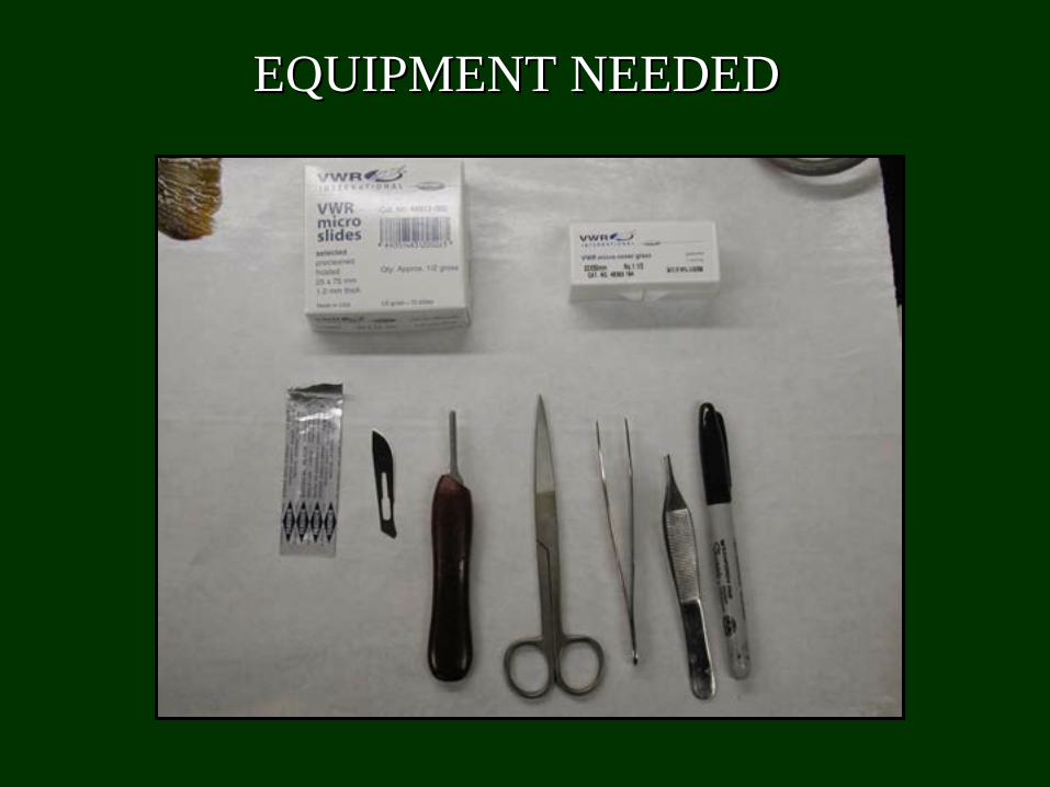
EQUIPMENT NEEDEDEQUIPMENT NEEDED

USE FRESHLY DEAD FISHUSE FRESHLY DEAD FISHExamine ASAP after deathExamine ASAP after death
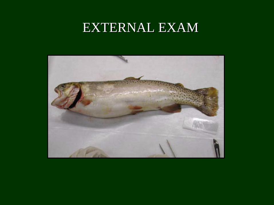
EXTERNAL EXAMEXTERNAL EXAM
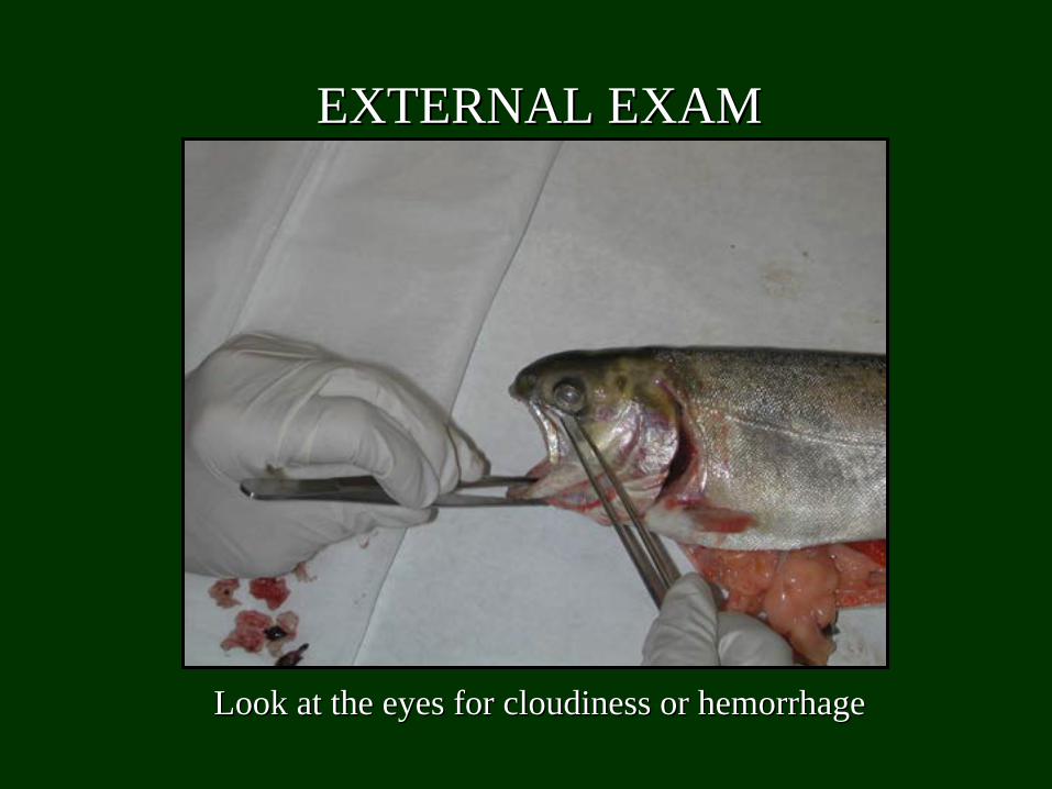
Look at the eyes for cloudiness or hemorrhageLook at the eyes for cloudiness or hemorrhage
EXTERNAL EXAMEXTERNAL EXAM
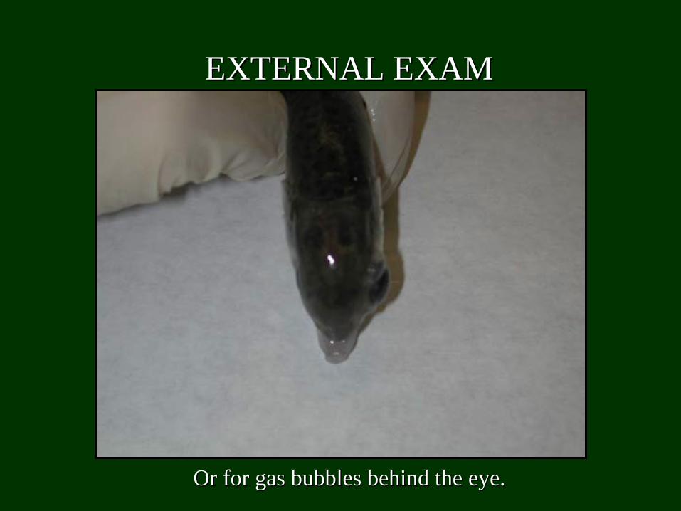
Or for gas bubbles behind the eye.Or for gas bubbles behind the eye.
EXTERNAL EXAMEXTERNAL EXAM
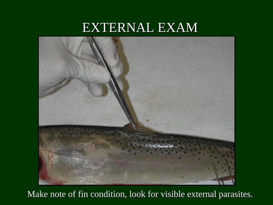
Make note of fin condition, look for visible external parasites.Make note of fin condition, look for visible external parasites.
EXTERNAL EXAMEXTERNAL EXAM
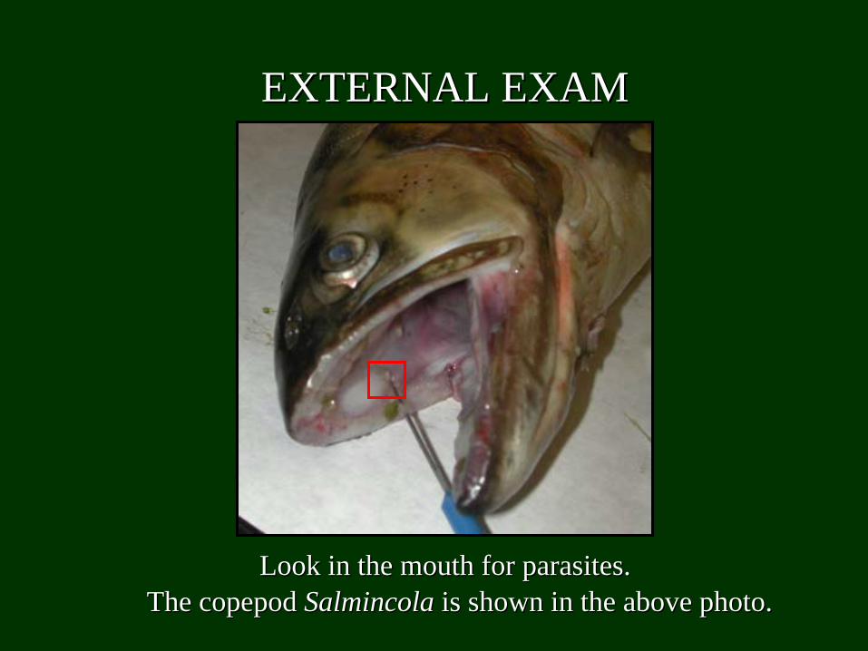
Look in the mouth for parasites.Look in the mouth for parasites.The copepod The copepod Salmincola Salmincola is shown in the above photo.is shown in the above photo.
EXTERNAL EXAMEXTERNAL EXAM
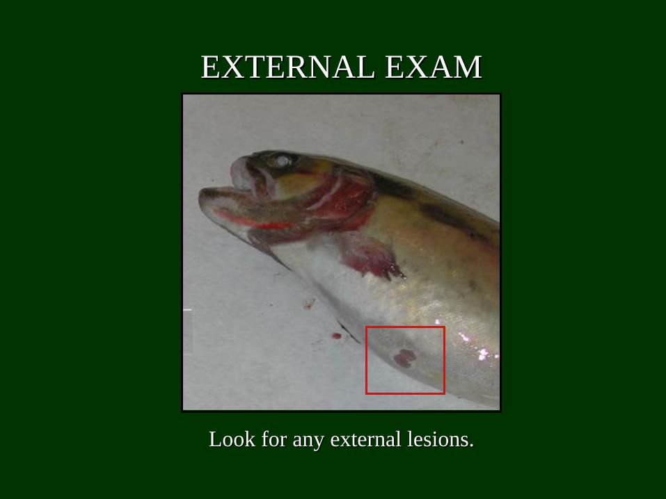
Look for any external lesions.Look for any external lesions.
EXTERNAL EXAMEXTERNAL EXAM
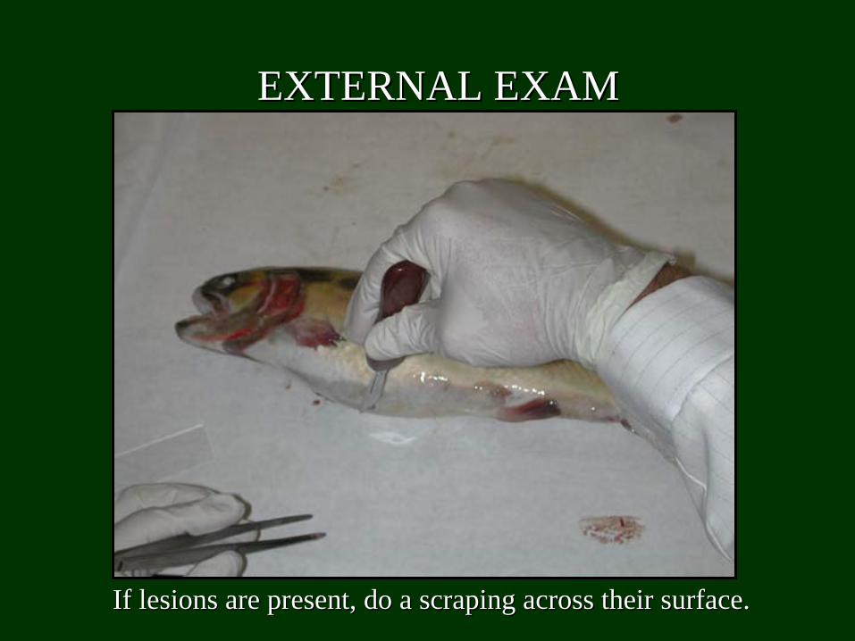
If lesions are present, do a scraping across their surface.If lesions are present, do a scraping across their surface.
EXTERNAL EXAMEXTERNAL EXAM
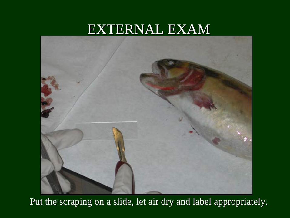
Put the scraping on a slide, let air dry and label appropriatelyPut the scraping on a slide, let air dry and label appropriately..
EXTERNAL EXAMEXTERNAL EXAM
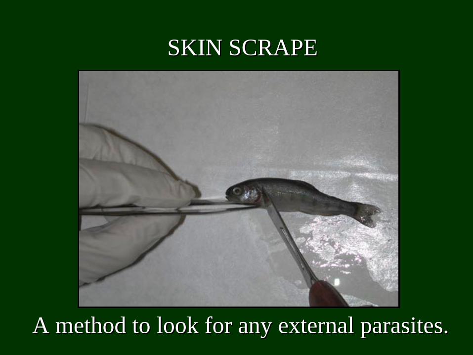
SKIN SCRAPESKIN SCRAPE
A method to look for any external parasites.A method to look for any external parasites.
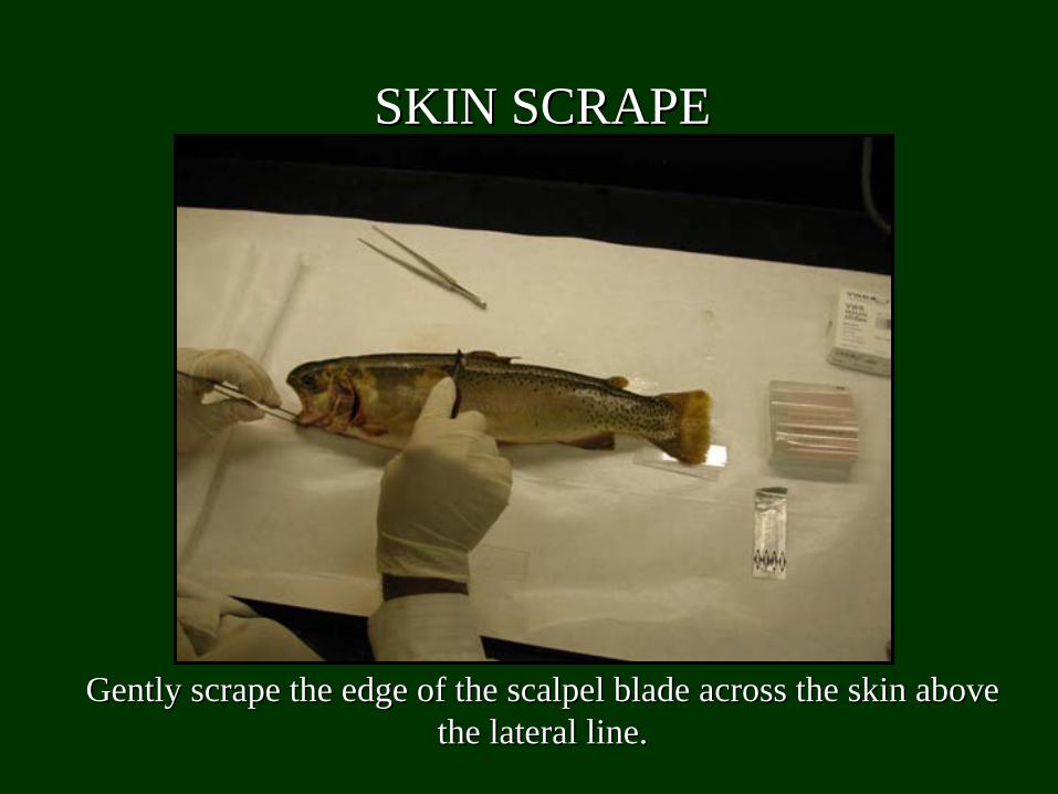
Gently scrape the edge of the scalpel blade across the skin abovGently scrape the edge of the scalpel blade across the skin above e the lateral line.the lateral line.
SKIN SCRAPESKIN SCRAPE
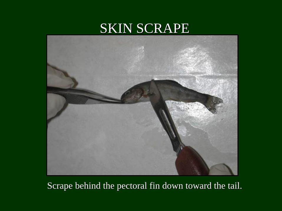
Scrape behind the pectoral fin down toward the tail.Scrape behind the pectoral fin down toward the tail.
SKIN SCRAPESKIN SCRAPE
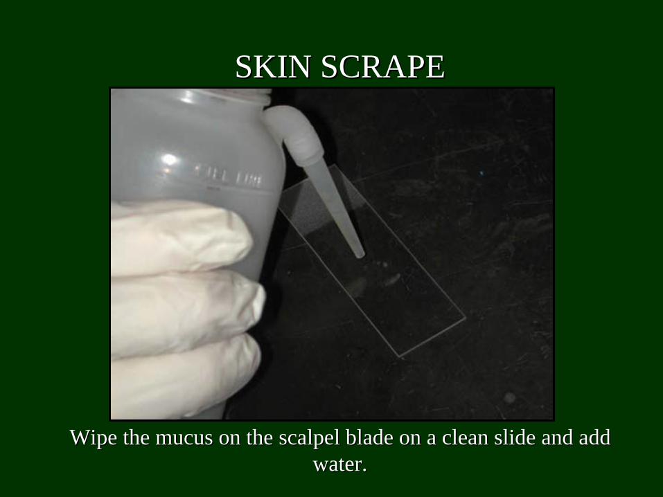
Wipe the mucus on the scalpel blade on a clean slide and add Wipe the mucus on the scalpel blade on a clean slide and add water.water.
SKIN SCRAPESKIN SCRAPE
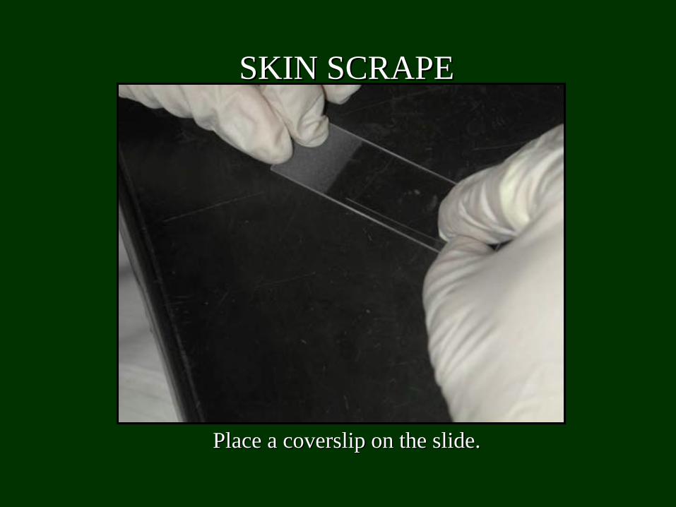
Place a coverslip on the slide.Place a coverslip on the slide.
SKIN SCRAPESKIN SCRAPE
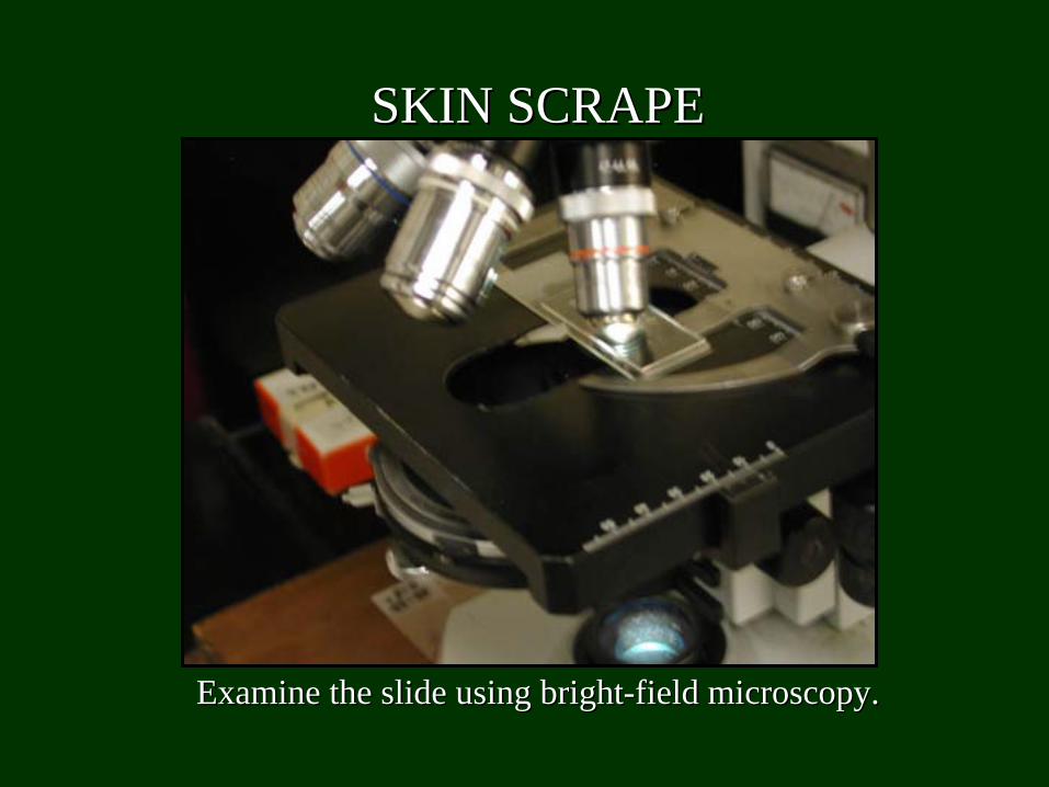
Examine the slide using brightExamine the slide using bright--field microscopy.field microscopy.
SKIN SCRAPESKIN SCRAPE
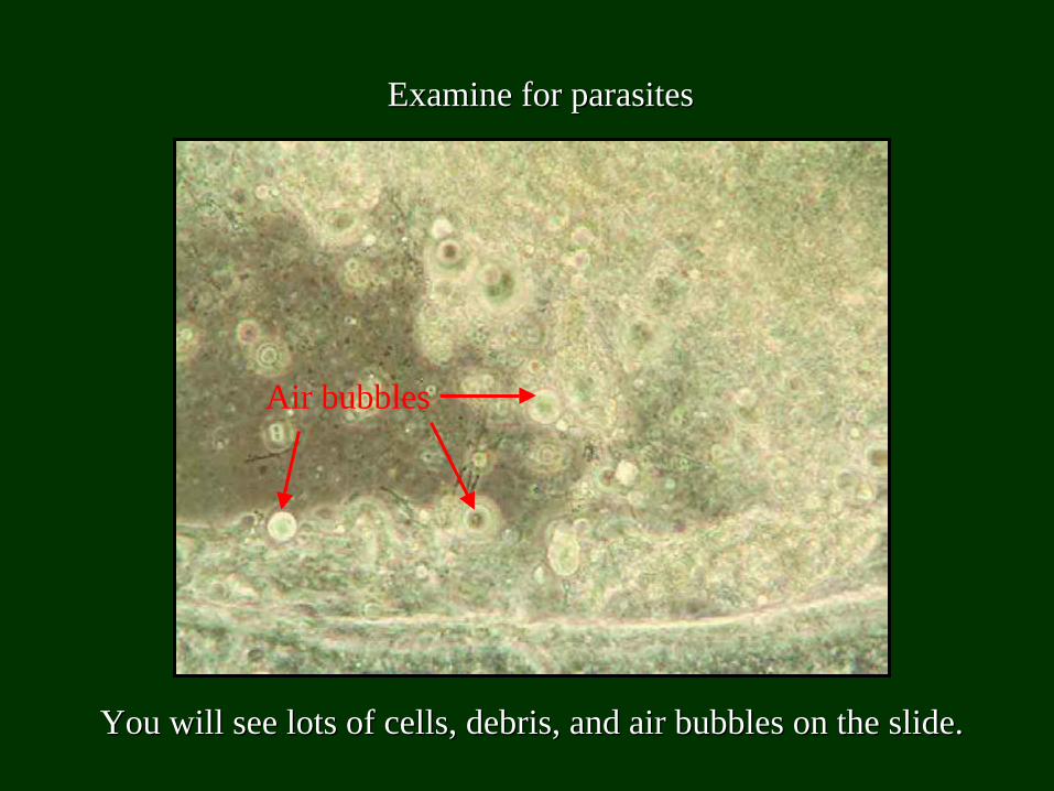
Examine for parasitesExamine for parasites
You will see lots of cells, debris, and air bubbles on the slideYou will see lots of cells, debris, and air bubbles on the slide..
Air bubbles
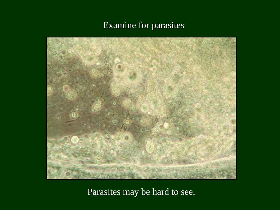
Examine for parasitesExamine for parasites
Parasites may be hard to see.Parasites may be hard to see.
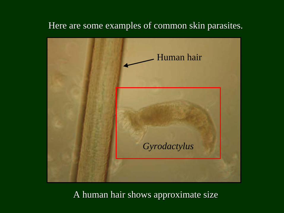
Here are some examples of common skin parasites.Here are some examples of common skin parasites.
A human hair shows approximate sizeA human hair shows approximate size
Human hair
Gyrodactylus
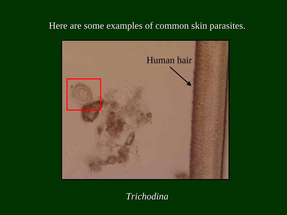
Here are some examples of common skin parasites.Here are some examples of common skin parasites.
TrichodinaTrichodina
Human hair
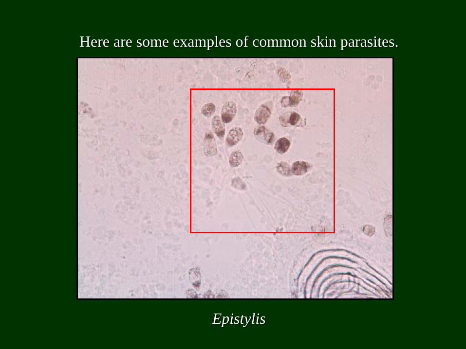
Here are some examples of common skin parasites.Here are some examples of common skin parasites.
EpistylisEpistylis

If fish is not fresh, parasites can degrade and will not be as If fish is not fresh, parasites can degrade and will not be as noticeable.noticeable.
Gyrodactylus Gyrodactylus haptors (mouthparts)haptors (mouthparts)
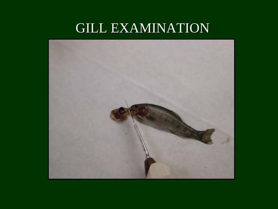
GILL EXAMINATIONGILL EXAMINATION
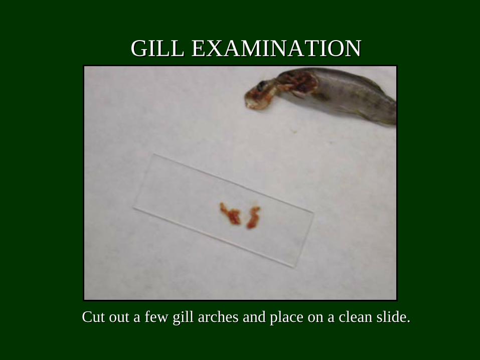
Cut out a few gill arches and place on a clean slide.Cut out a few gill arches and place on a clean slide.
GILL EXAMINATIONGILL EXAMINATION
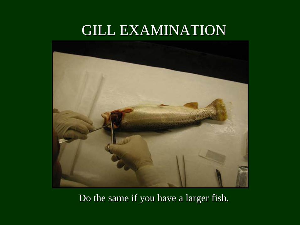
Do the same if you have a larger fish.Do the same if you have a larger fish.
GILL EXAMINATIONGILL EXAMINATION
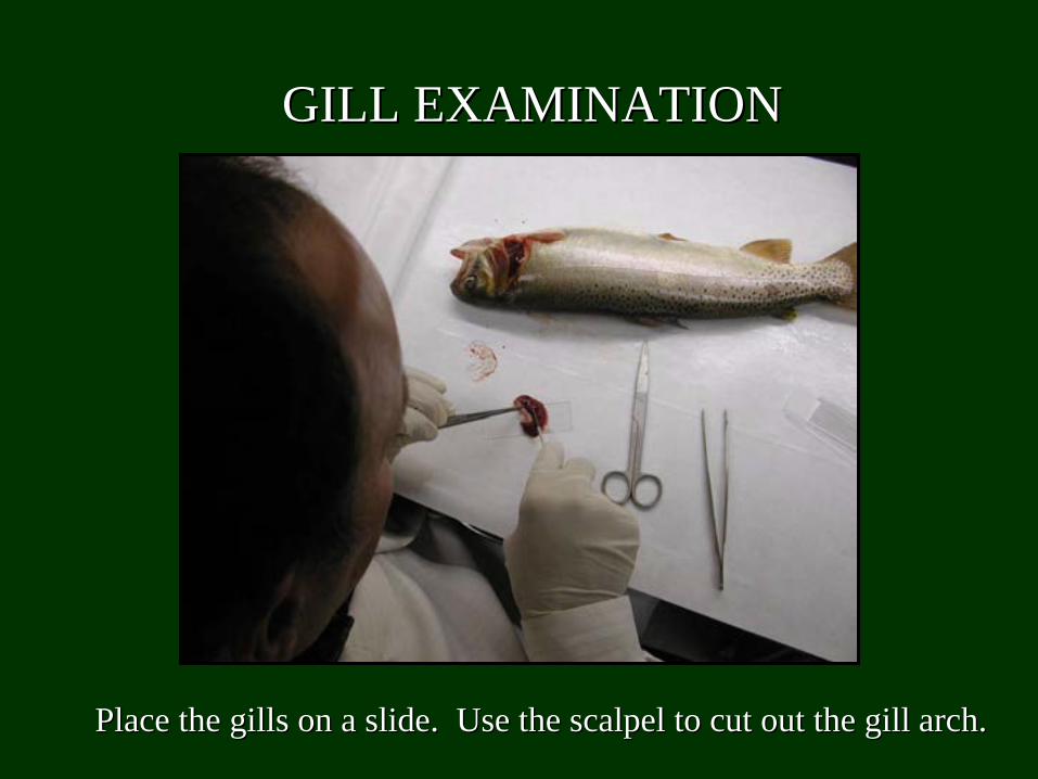
Place the gills on a slide. Use the scalpel to cut out the gillPlace the gills on a slide. Use the scalpel to cut out the gill arch.arch.
GILL EXAMINATIONGILL EXAMINATION

Add enough water to cover the sample.Add enough water to cover the sample.
GILL EXAMINATIONGILL EXAMINATION

GILL EXAMINATIONGILL EXAMINATION
Add a coverslip.Add a coverslip.
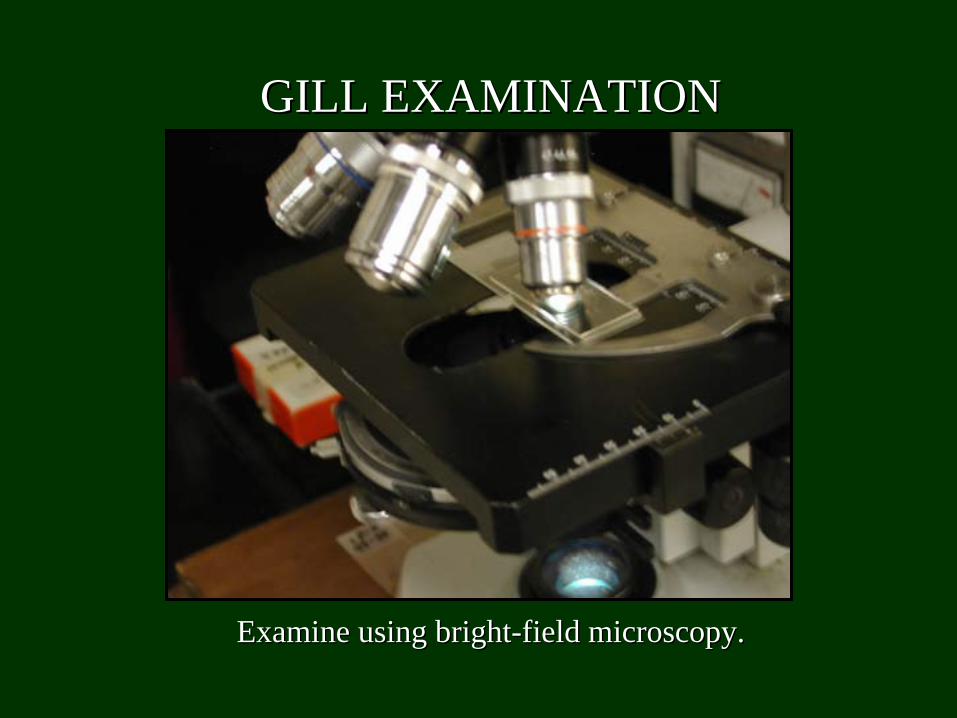
Examine using brightExamine using bright--field microscopy.field microscopy.
GILL EXAMINATIONGILL EXAMINATION
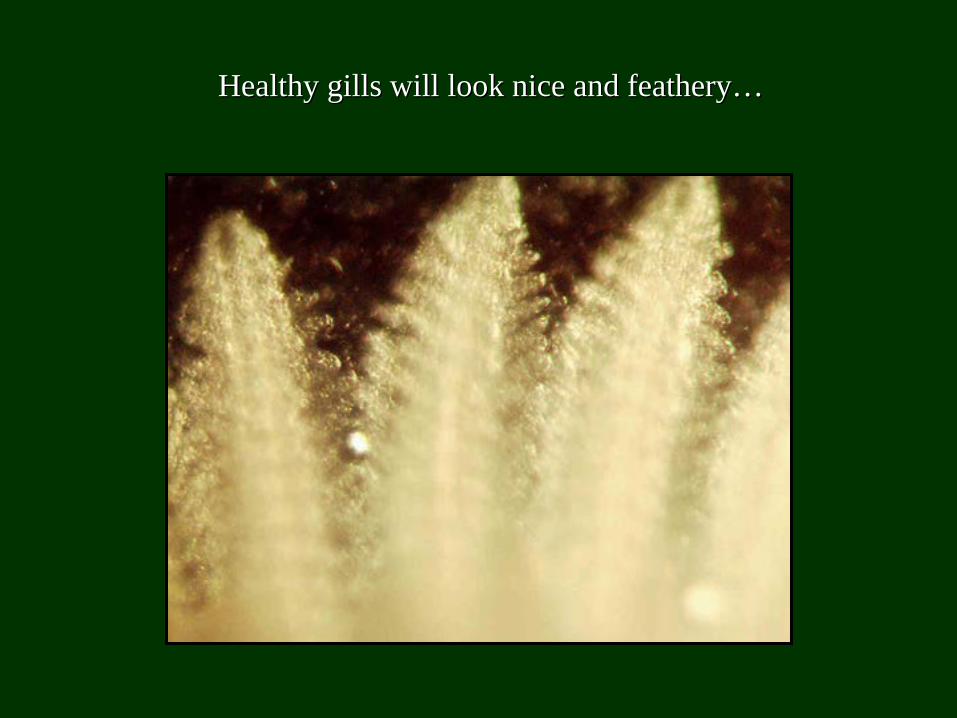
Healthy gills will look nice and feathery…Healthy gills will look nice and feathery…
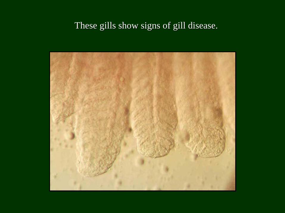
These gills show signs of gill disease.These gills show signs of gill disease.
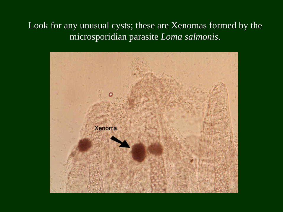
Look for any unusual cysts; these are Xenomas formed by the Look for any unusual cysts; these are Xenomas formed by the microsporidian parasite microsporidian parasite Loma salmonisLoma salmonis..
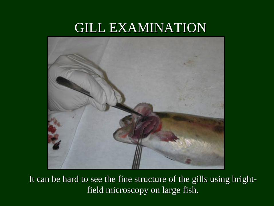
It can be hard to see the fine structure of the gills using brigIt can be hard to see the fine structure of the gills using brightht-- field microscopy on large fish.field microscopy on large fish.
GILL EXAMINATIONGILL EXAMINATION
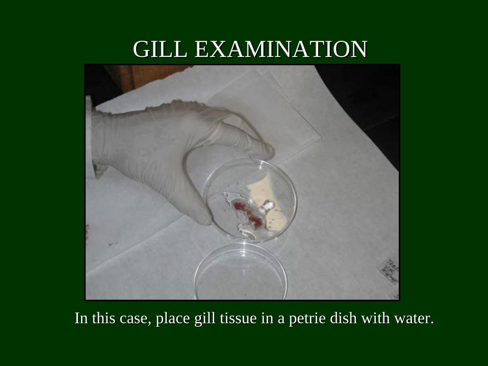
In this case, place gill tissue in a In this case, place gill tissue in a petrie petrie dish with water.dish with water.
GILL EXAMINATIONGILL EXAMINATION

Examine under the dissecting microscope.Examine under the dissecting microscope.
GILL EXAMINATIONGILL EXAMINATION
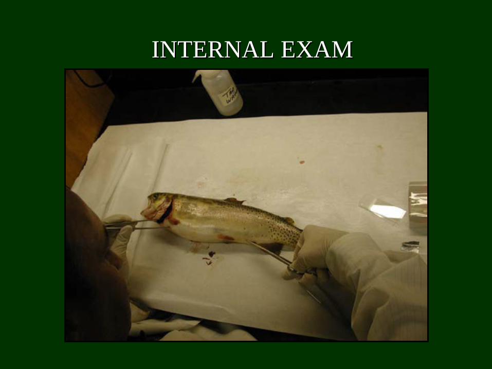
INTERNAL EXAMINTERNAL EXAM

Cut from the vent up to the pectoral fin, in a halfCut from the vent up to the pectoral fin, in a half--moon shape.moon shape.
INTERNAL EXAMINTERNAL EXAM
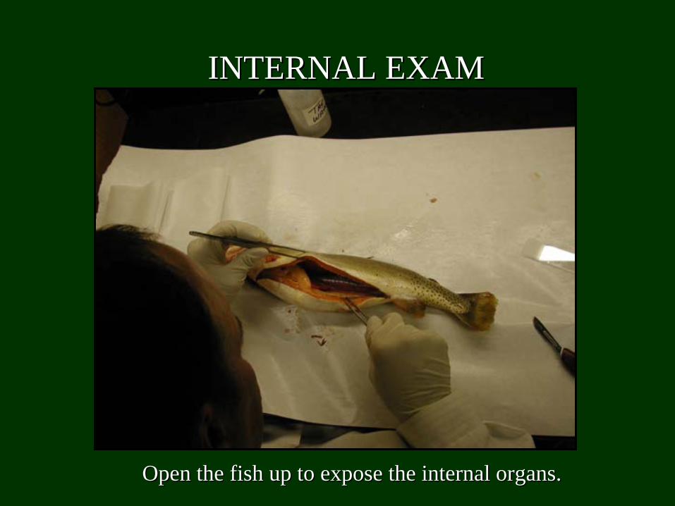
Open the fish up to expose the internal organs.Open the fish up to expose the internal organs.
INTERNAL EXAMINTERNAL EXAM
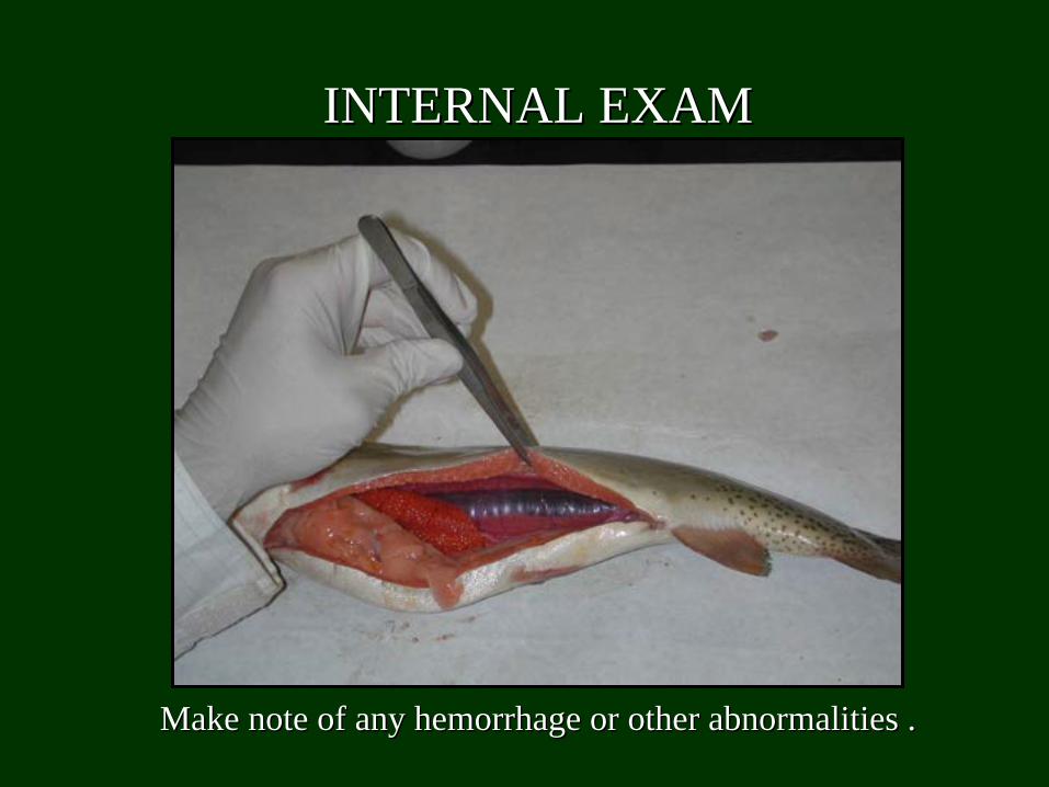
Make note of any hemorrhage or other abnormalities .Make note of any hemorrhage or other abnormalities .
INTERNAL EXAMINTERNAL EXAM
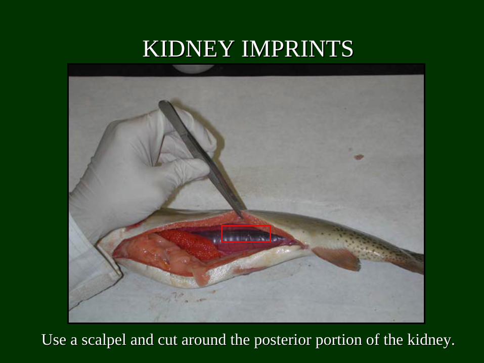
Use a scalpel and cut around the posterior portion of the kidneyUse a scalpel and cut around the posterior portion of the kidney..
KIDNEY IMPRINTSKIDNEY IMPRINTS
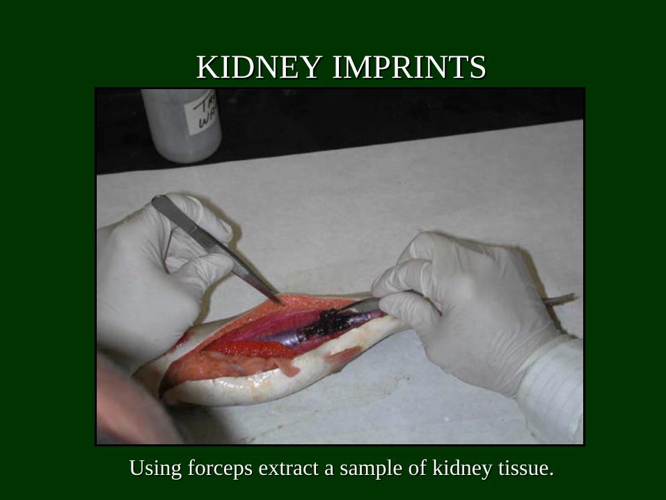
Using forceps extract a sample of kidney tissue.Using forceps extract a sample of kidney tissue.
KIDNEY IMPRINTSKIDNEY IMPRINTS
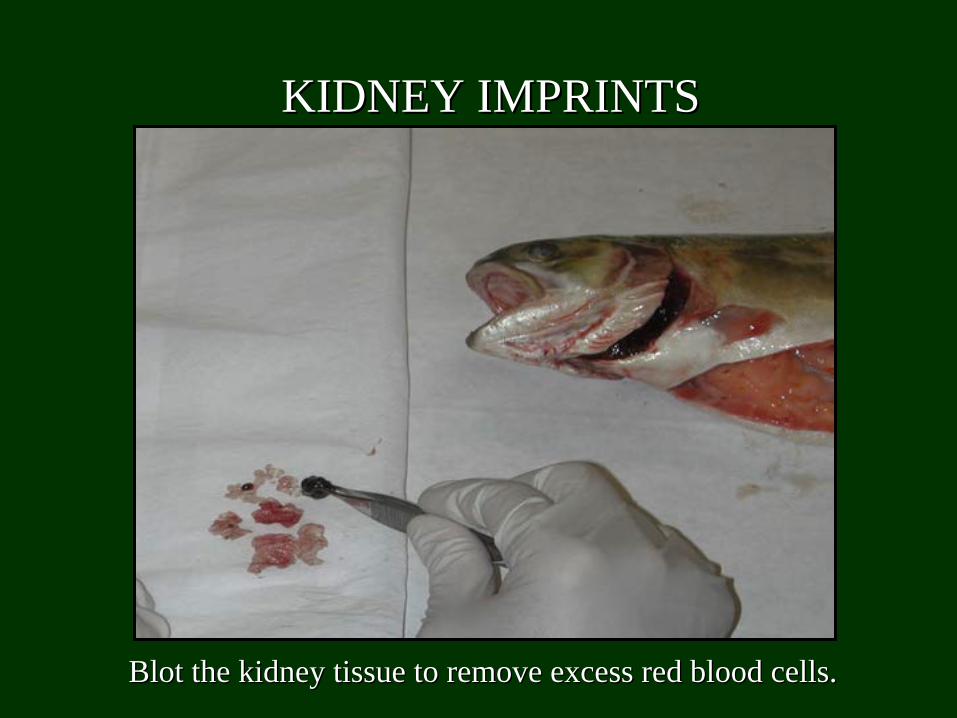
Blot the kidney tissue to remove excess red blood cells.Blot the kidney tissue to remove excess red blood cells.
KIDNEY IMPRINTSKIDNEY IMPRINTS
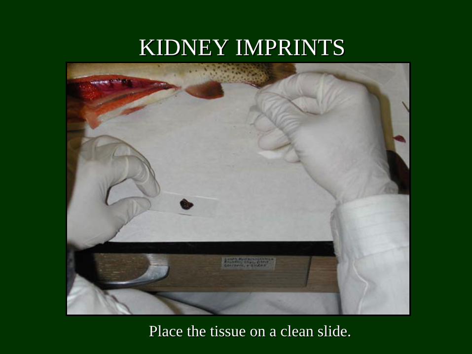
Place the tissue on a clean slide.Place the tissue on a clean slide.
KIDNEY IMPRINTSKIDNEY IMPRINTS
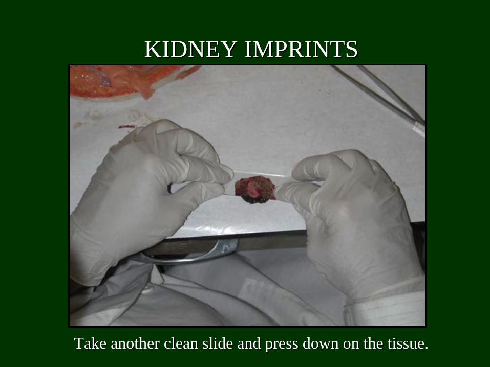
Take another clean slide and press down on the tissue.Take another clean slide and press down on the tissue.
KIDNEY IMPRINTSKIDNEY IMPRINTS
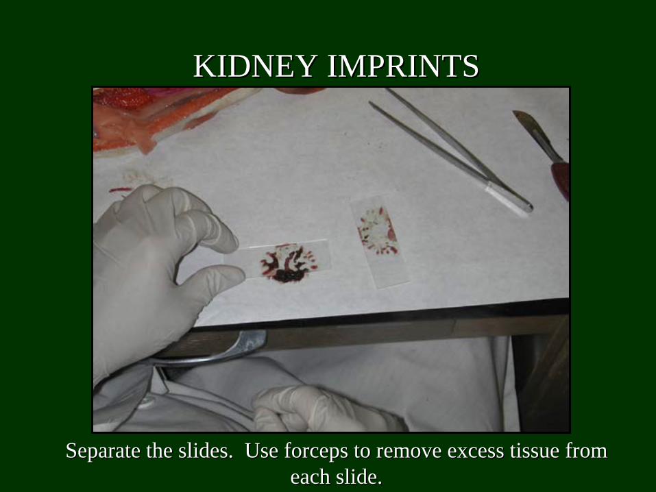
Separate the slides. Use forceps to remove excess tissue from Separate the slides. Use forceps to remove excess tissue from each slide.each slide.
KIDNEY IMPRINTSKIDNEY IMPRINTS
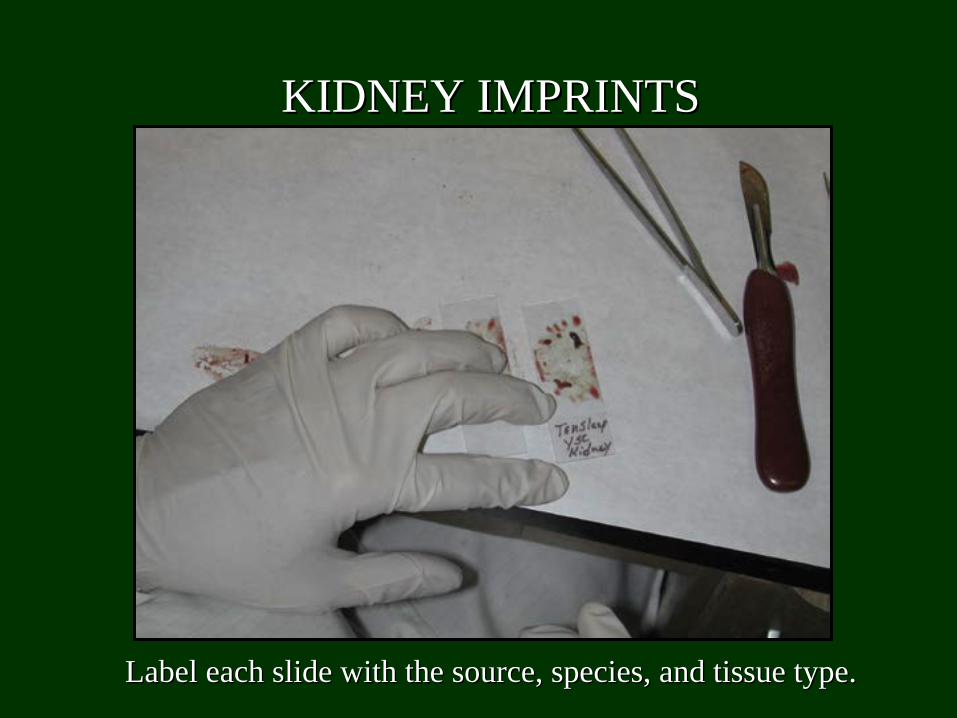
Label each slide with the source, species, and tissue type.Label each slide with the source, species, and tissue type.
KIDNEY IMPRINTSKIDNEY IMPRINTS
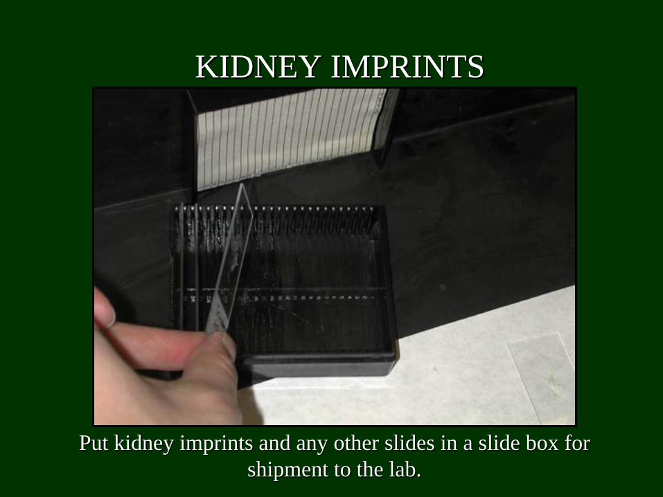
Put kidney imprints and any other slides in a slide box for Put kidney imprints and any other slides in a slide box for shipment to the lab.shipment to the lab.
KIDNEY IMPRINTSKIDNEY IMPRINTS
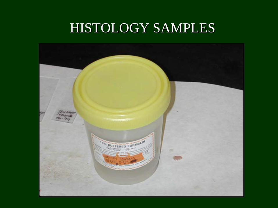
HISTOLOGY SAMPLESHISTOLOGY SAMPLES
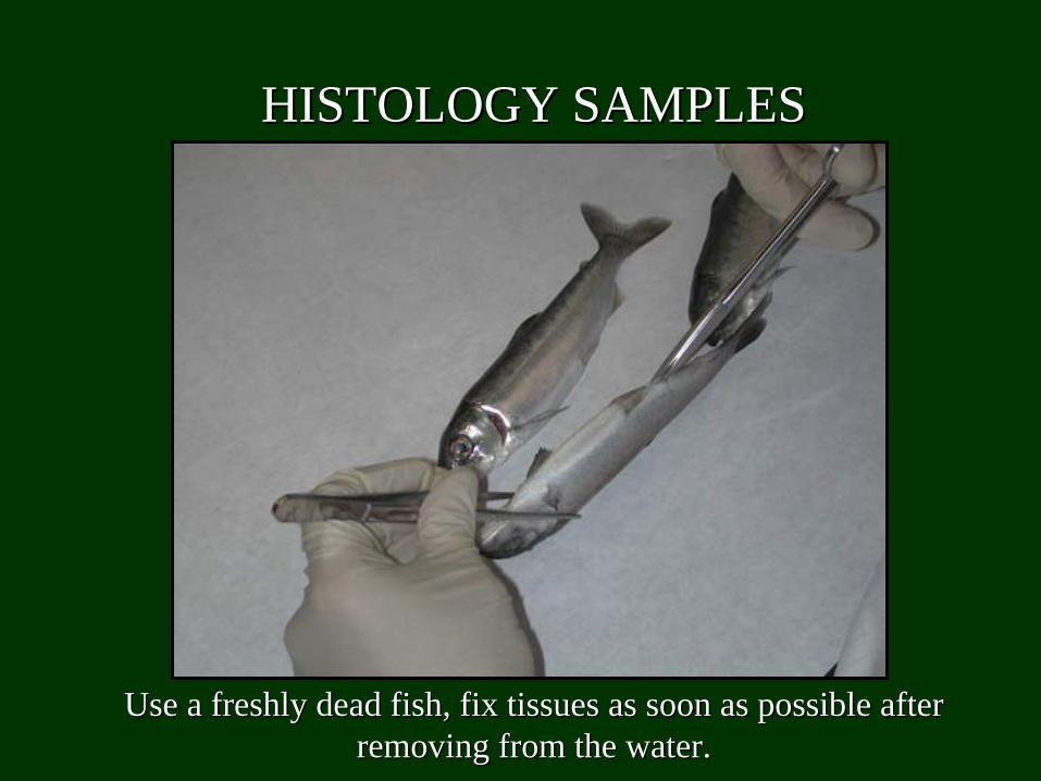
Use a freshly dead fish, fix tissues as soon as possible after Use a freshly dead fish, fix tissues as soon as possible after removing from the water.removing from the water.
HISTOLOGY SAMPLESHISTOLOGY SAMPLES
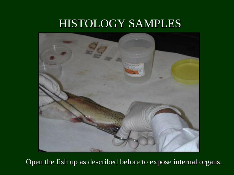
Open the fish up as described before to expose internal organs.Open the fish up as described before to expose internal organs.
HISTOLOGY SAMPLESHISTOLOGY SAMPLES
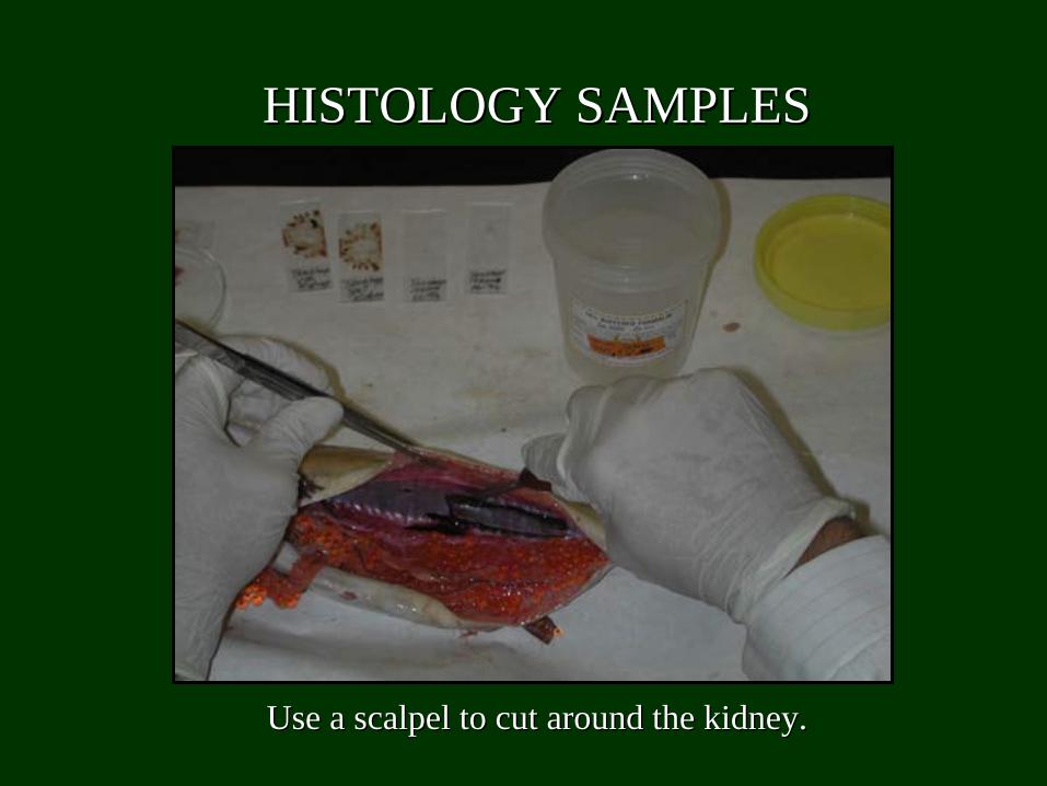
Use a scalpel to cut around the kidney.Use a scalpel to cut around the kidney.
HISTOLOGY SAMPLESHISTOLOGY SAMPLES
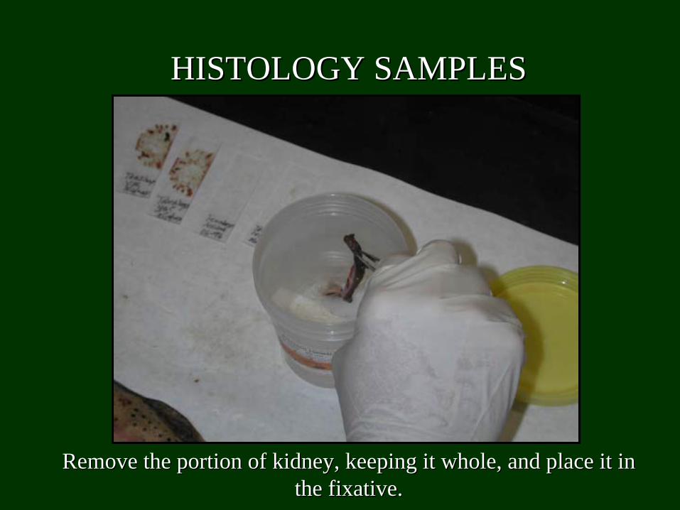
Remove the portion of kidney, keeping it whole, and place it in Remove the portion of kidney, keeping it whole, and place it in the fixative.the fixative.
HISTOLOGY SAMPLESHISTOLOGY SAMPLES
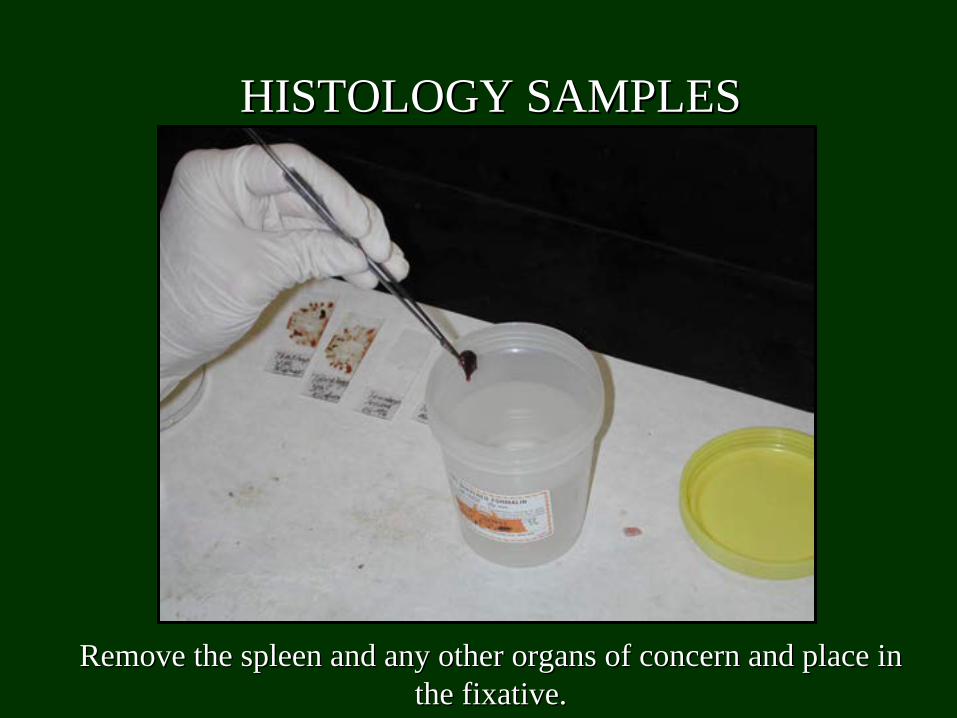
Remove the spleen and any other organs of concern and place in Remove the spleen and any other organs of concern and place in the fixative.the fixative.
HISTOLOGY SAMPLESHISTOLOGY SAMPLES
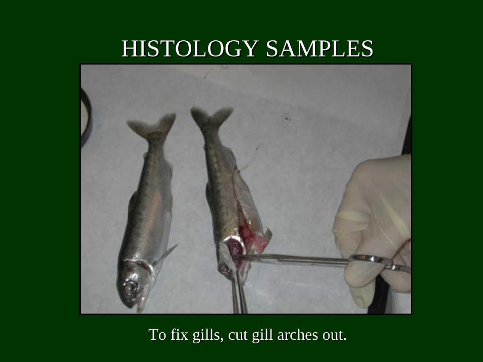
To fix gills, cut gill arches out.To fix gills, cut gill arches out.
HISTOLOGY SAMPLESHISTOLOGY SAMPLES
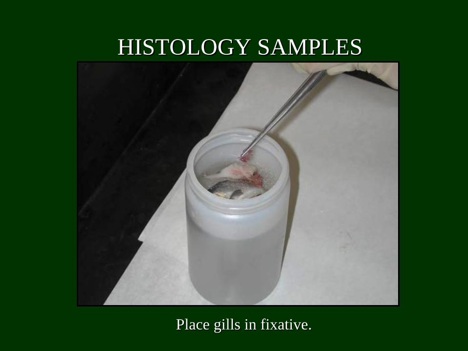
Place gills in fixative.Place gills in fixative.
HISTOLOGY SAMPLESHISTOLOGY SAMPLES
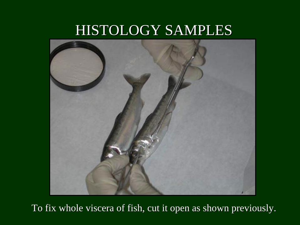
To fix whole viscera of fish, cut it open as shown previously.To fix whole viscera of fish, cut it open as shown previously.
HISTOLOGY SAMPLESHISTOLOGY SAMPLES
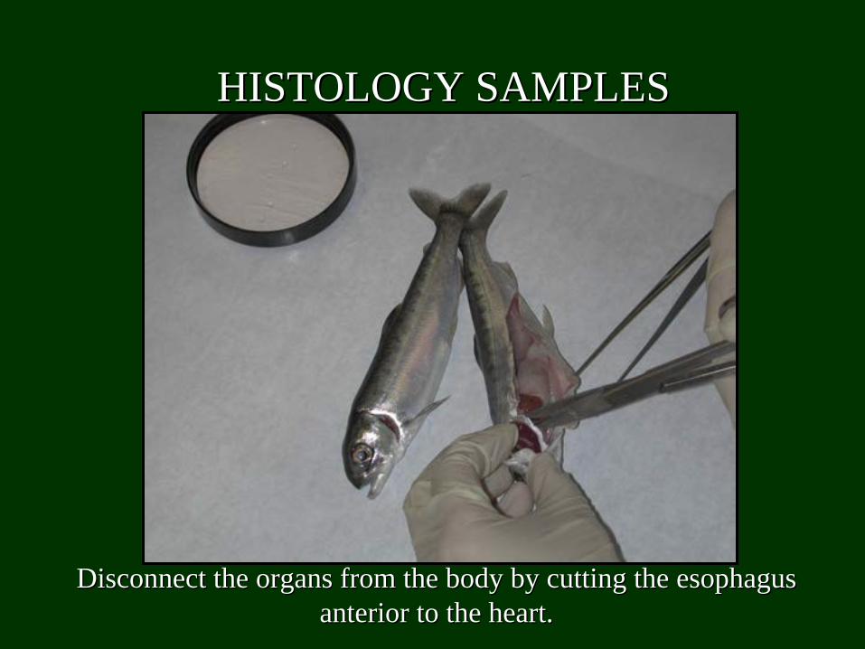
Disconnect the organs from the body by cutting the esophagus Disconnect the organs from the body by cutting the esophagus anterior to the heart.anterior to the heart.
HISTOLOGY SAMPLESHISTOLOGY SAMPLES
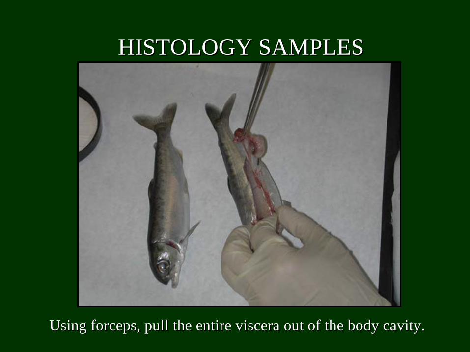
Using forceps, pull the entire viscera out of the body cavity.Using forceps, pull the entire viscera out of the body cavity.
HISTOLOGY SAMPLESHISTOLOGY SAMPLES

Cut the descending intestine at the vent.Cut the descending intestine at the vent.
HISTOLOGY SAMPLESHISTOLOGY SAMPLES
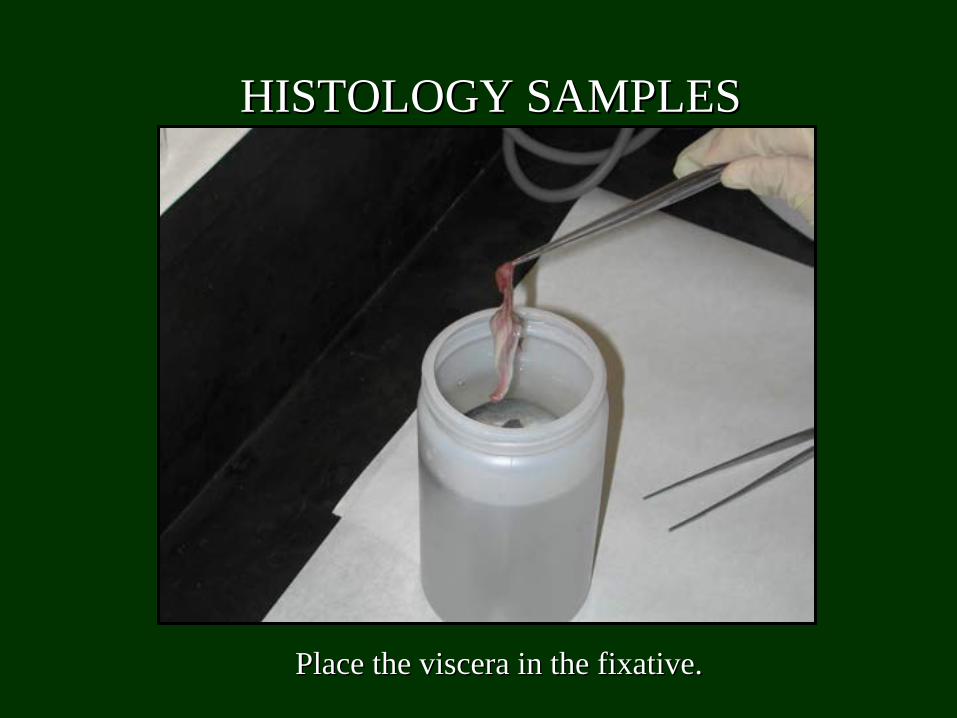
Place the viscera in the fixative.Place the viscera in the fixative.
HISTOLOGY SAMPLESHISTOLOGY SAMPLES
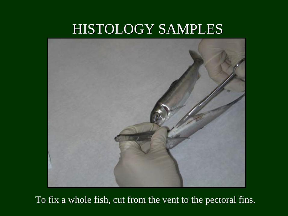
To fix a whole fish, cut from the vent to the pectoral fins.To fix a whole fish, cut from the vent to the pectoral fins.
HISTOLOGY SAMPLESHISTOLOGY SAMPLES

Make another cut into the muscle tissue above the lateral line. Make another cut into the muscle tissue above the lateral line. These cuts allow the fixative to penetrate all of the fish tissuThese cuts allow the fixative to penetrate all of the fish tissues.es.
HISTOLOGY SAMPLESHISTOLOGY SAMPLES
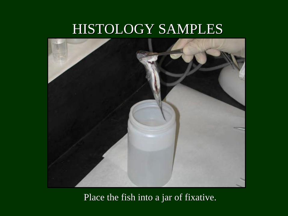
Place the fish into a jar of fixative.Place the fish into a jar of fixative.
HISTOLOGY SAMPLESHISTOLOGY SAMPLES
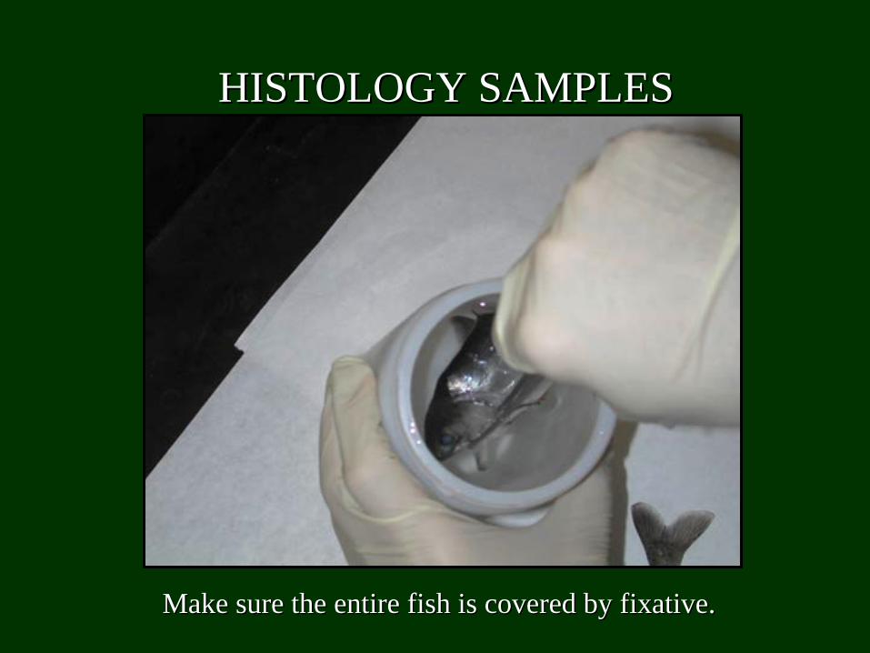
Make sure the entire fish is covered by fixative.Make sure the entire fish is covered by fixative.
HISTOLOGY SAMPLESHISTOLOGY SAMPLES
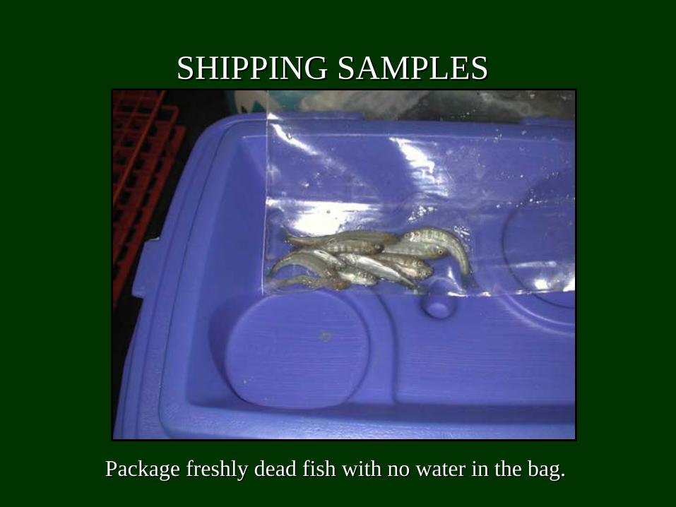
SHIPPING SAMPLESSHIPPING SAMPLES
Package freshly dead fish with no water in the bag.Package freshly dead fish with no water in the bag.
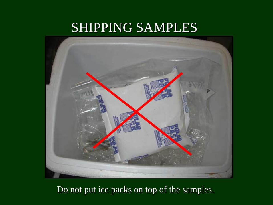
Do not put ice packs on top of the samples.Do not put ice packs on top of the samples.
SHIPPING SAMPLESSHIPPING SAMPLES

Do put samples on top of ice preferably with newspaper or other Do put samples on top of ice preferably with newspaper or other insulation between the samples and ice so they don’t freeze insulation between the samples and ice so they don’t freeze
during shipment.during shipment.
SHIPPING SAMPLESSHIPPING SAMPLES
SHIPPING SAMPLESSHIPPING SAMPLES
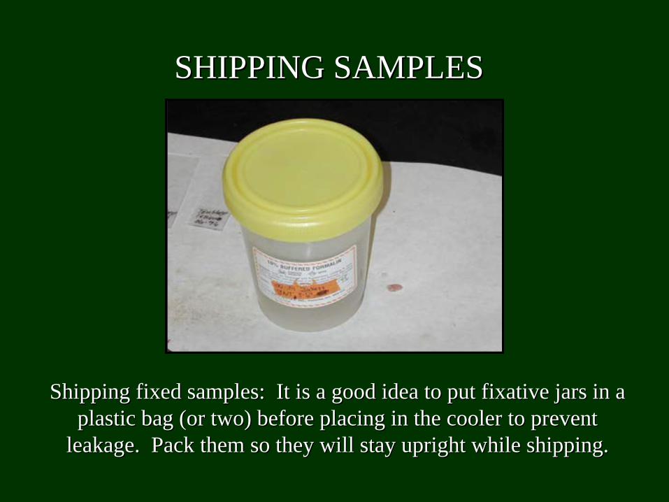
Shipping fixed samples: It is a good idea to put fixative jars Shipping fixed samples: It is a good idea to put fixative jars in a in a plastic bag (or two) before placing in the cooler to prevent plastic bag (or two) before placing in the cooler to prevent
leakage. Pack them so they will stay upright while shipping.leakage. Pack them so they will stay upright while shipping.
SHIPPING SAMPLESSHIPPING SAMPLES
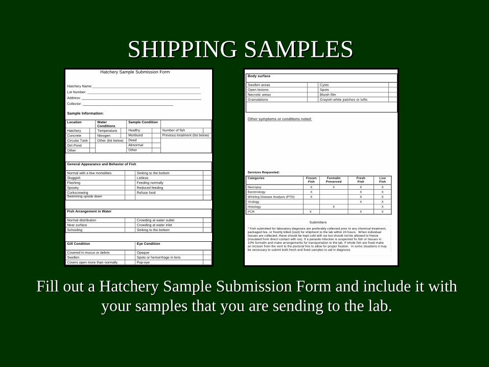
Fill out a Hatchery Sample Submission Form and include it with Fill out a Hatchery Sample Submission Form and include it with your samples that you are sending to the lab.your samples that you are sending to the lab.
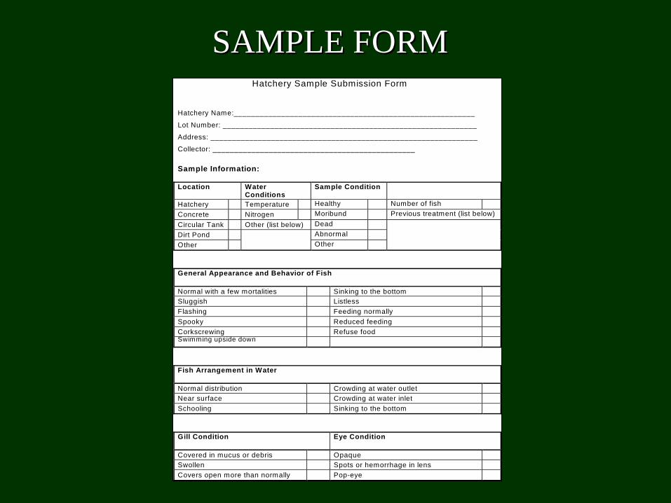
Hatchery Sample Submission Form Hatchery Name:________________________________________________________
Lot Number: ___________________________________________________________
Address: ______________________________________________________________
Collector: _______________________________________________ Sample Information: Location Water
Conditions Sample Condition
Hatchery Temperature Healthy Number of fish Concrete Nitrogen Moribund Previous treatment (list below) Circular Tank Other (list below) Dead Dirt Pond Abnormal Other
Other
General Appearance and Behavior of Fish
Normal with a few mortalities Sinking to the bottom Sluggish Listless Flashing Feeding normally Spooky Reduced feeding Corkscrewing Refuse food Swimming upside down Fish Arrangement in Water
Normal distribution Crowding at water outlet Near surface Crowding at water inlet Schooling Sinking to the bottom Gill Condition Eye Condition
Covered in mucus or debris Opaque Swollen Spots or hemorrhage in lens Covers open more than normally Pop-eye
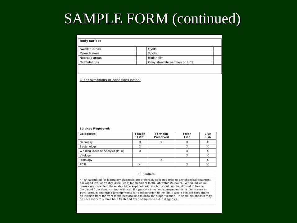
Body surface
Swollen areas Cysts Open lesions Spots Necrotic areas Bluish film Granulations Grayish-white patches or tufts
Other symptoms or conditions noted: Services Requested:
Categories Frozen Fish
Formalin Preserved
Fresh Fish
Live Fish
Necropsy X X X X Bacteriology X X X Whirling Disease Analysis (PTD) X X X Virology X X Histology X X PCR X X X
Submitters
* Fish submitted for laboratory diagnosis are preferably collected prior to any chemical treatment, packaged live, or freshly killed (iced) for shipment to the lab within 24 hours. When individual tissues are collected, these should be kept cold with ice but should not be allowed to freeze (insulated from direct contact with ice). If a parasite infection is suspected fix fish or tissues in 10% formalin and make arrangements for transportation to the lab. If whole fish are fixed make an incision from the vent to the pectoral fins to allow for proper fixation. In some situations it may be necessary to submit both fresh and fixed samples to aid in diagnosis
Hatchery Sample Submission Form Hatchery Name:________________________________________________________
Lot Number: ___________________________________________________________
Address: ______________________________________________________________
Collector: _______________________________________________ Sample Information: Location Water
Conditions Sample Condition
Hatchery Temperature Healthy Number of fish Concrete Nitrogen Moribund Previous treatment (list below) Circular Tank Other (list below) Dead Dirt Pond Abnormal Other
Other
General Appearance and Behavior of Fish
Normal with a few mortalities Sinking to the bottom Sluggish Listless Flashing Feeding normally Spooky Reduced feeding Corkscrewing Refuse food Swimming upside down Fish Arrangement in Water
Normal distribution Crowding at water outlet Near surface Crowding at water inlet Schooling Sinking to the bottom Gill Condition Eye Condition
Covered in mucus or debris Opaque Swollen Spots or hemorrhage in lens Covers open more than normally Pop-eye
SAMPLE FORMSAMPLE FORM
Body surface
Swollen areas Cysts Open lesions Spots Necrotic areas Bluish film Granulations Grayish-white patches or tufts
Other symptoms or conditions noted:
Services Requested:
Categories Frozen Fish
Formalin Preserved
Fresh Fish
Live Fish
Necropsy X X X X Bacteriology X X X W hirling Disease Analysis (PTD) X X X Virology X X Histology X X PCR X X X
Submitters
* Fish submitted for laboratory diagnosis are preferably collected prior to any chemical treatment, packaged live, or freshly killed (iced) for shipment to the lab within 24 hours. W hen individual tissues are collected, these should be kept cold with ice but should not be allowed to freeze (insulated from direct contact with ice). If a parasite infection is suspected fix fish or tissues in 10% formalin and make arrangements for transportation to the lab. If whole fish are fixed make an incision from the vent to the pectoral fins to allow for proper fixation. In some situations it may be necessary to submit both fresh and fixed samples to aid in diagnosis
SAMPLE FORM (continued)SAMPLE FORM (continued)
DISCLAIMERDISCLAIMER
Although this presentation was developed to assist Although this presentation was developed to assist hatchery personnel with the basic principals for parasite hatchery personnel with the basic principals for parasite
examinations and sample collection procedures, we examinations and sample collection procedures, we recognize that fish disease diagnosis is often difficult recognize that fish disease diagnosis is often difficult and requires an experienced fish health professional and requires an experienced fish health professional
and full service testing facilities.and full service testing facilities.

Contact us!Contact us!
We welcome any and all feedback on the usefulness of We welcome any and all feedback on the usefulness of this presentation. Please contact us and let us know this presentation. Please contact us and let us know
what you think.what you think.
[email protected]@uwyo.edu oror
[email protected]@uwyo.edu