flexible fiberoptic workshop: basic course - …entpa.org/resources/documents/flex scopes basic...
TRANSCRIPT

Flexible Fiberoptic Workshop: Basic Course Cheryl Parnell, PA-C
Updated 12/08/2015
March 31, 2016 Orlando, FL

Flexible Fiberoptic Workshop: Basic Course
Basic Instruction
Demonstration
Hands-On Practice
Learn by doing
Practice indirect laryngoscopy on mannequin
Practice indirect laryngoscopy on simulated patient
Perform flexible fiberoptic endoscopy on mannequin.
Perform flexible fiberoptic endoscopy on simulated patient
Demonstrate proper sterilization and handling technique.
Introduction
There are multiple methods and techniques available to successfully complete all the topics presented in this workshop. Some are based on
patient request, available equipment or supervising physician’s preference.
The goal of this workshop is to correctly
demonstrate the most common methods and give participants time for hands on training.
Flexible Fiberoptic Workshop: Basic Course
Learning objectives • Discuss normal anatomy visible via flexible
fiberoptic nasopharyngoscopy • Practice the use of the flexible fiberoptic
nasopharyngoscope on mannequins. • Practice the use of the flexible fiberoptic
nasopharyngoscope on simulated patient • Understand and practice proper endoscope use
and care. • Normal variants and abnormal findings will be
discussed in Advanced Course.
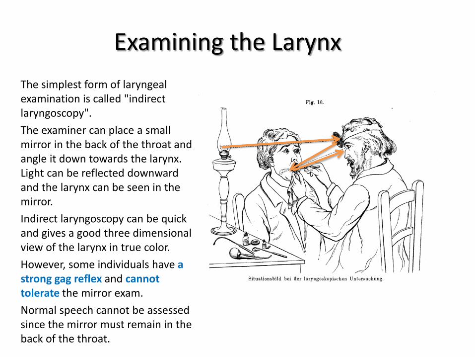
Examining the Larynx The simplest form of laryngeal examination is called "indirect laryngoscopy". The examiner can place a small mirror in the back of the throat and angle it down towards the larynx. Light can be reflected downward and the larynx can be seen in the mirror. Indirect laryngoscopy can be quick and gives a good three dimensional view of the larynx in true color. However, some individuals have a strong gag reflex and cannot tolerate the mirror exam. Normal speech cannot be assessed since the mirror must remain in the back of the throat.
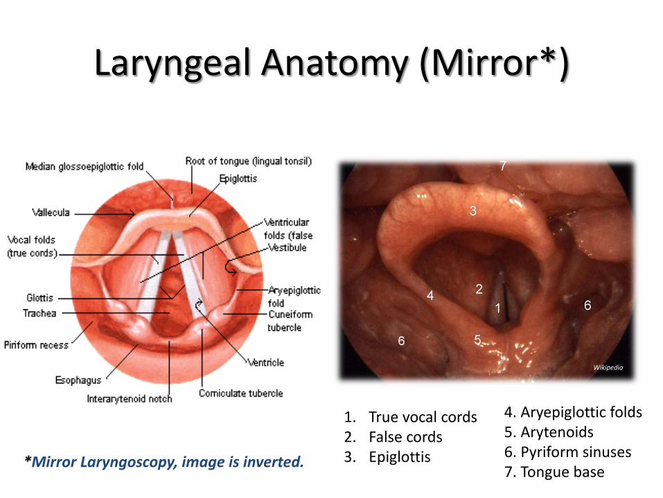
Laryngeal Anatomy (Mirror*)
1. True vocal cords 2. False cords 3. Epiglottis
4. Aryepiglottic folds 5. Arytenoids 6. Pyriform sinuses 7. Tongue base *Mirror Laryngoscopy, image is inverted.
Wikipedia
Indirect Laryngoscopy
Interactive, live demonstration of indirect laryngoscopy
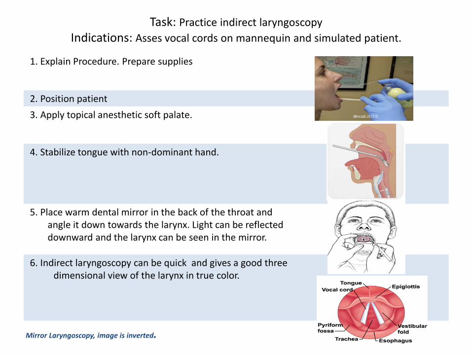
1. Explain Procedure. Prepare supplies
2. Position patient 3. Apply topical anesthetic soft palate.
4. Stabilize tongue with non-dominant hand.
5. Place warm dental mirror in the back of the throat and angle it down towards the larynx. Light can be reflected downward and the larynx can be seen in the mirror.
6. Indirect laryngoscopy can be quick and gives a good three dimensional view of the larynx in true color.
Task: Practice indirect laryngoscopy Indications: Asses vocal cords on mannequin and simulated patient.
Mirror Laryngoscopy, image is inverted.
Mercado 2013 ©
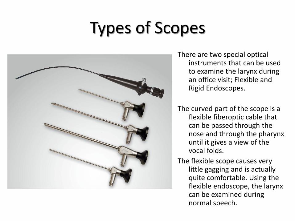
Types of Scopes There are two special optical
instruments that can be used to examine the larynx during an office visit; Flexible and Rigid Endoscopes.
The curved part of the scope is a
flexible fiberoptic cable that can be passed through the nose and through the pharynx until it gives a view of the vocal folds.
The flexible scope causes very little gagging and is actually quite comfortable. Using the flexible endoscope, the larynx can be examined during normal speech.
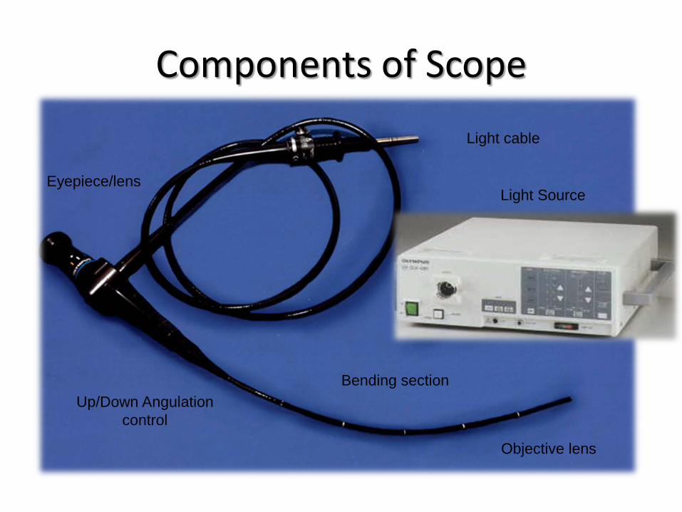
Components of Scope
Eyepiece/lens
Light cable
Up/Down Angulation control
Bending section
Objective lens
Light Source
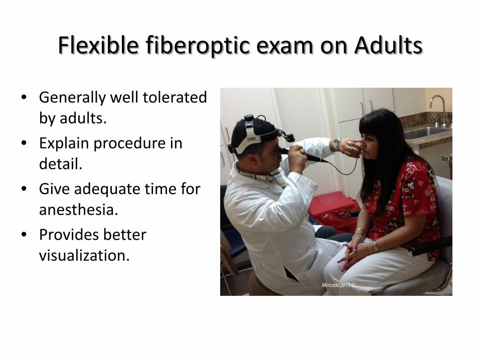
Flexible fiberoptic exam on Adults
• Generally well tolerated by adults.
• Explain procedure in detail.
• Give adequate time for anesthesia.
• Provides better visualization. Mercado 2013 ©
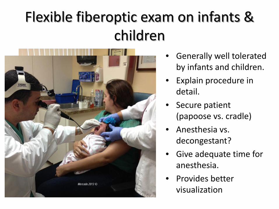
Flexible fiberoptic exam on infants & children
• Generally well tolerated by infants and children.
• Explain procedure in detail.
• Secure patient (papoose vs. cradle)
• Anesthesia vs. decongestant?
• Give adequate time for anesthesia.
• Provides better visualization Mercado 2013 ©
Mercado 2013 ©
Indications for Fiberoptic Endoscopy (FOE)
• Sinusitis • Epistaxis • Nasal obstruction • Foreign body • Strong gag reflex* • Failed mirror exam*
• Unilateral otitis media • Dysphonia • Dysphagia • Odynophagia • Symptoms of aspiration • Hemoptysis
*Documentation of a strong gag reflex and failed mirror exam should be included in note to justify procedure for billing purposes.
Contraindications for Fiberoptic Endoscopy (FOE)
• Epiglottitis (by inexperienced) • Relative:
– Coagulopathy – Craniofacial trauma
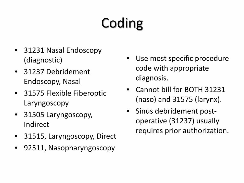
Coding
• 31231 Nasal Endoscopy (diagnostic)
• 31237 Debridement Endoscopy, Nasal
• 31575 Flexible Fiberoptic Laryngoscopy
• 31505 Laryngoscopy, Indirect
• 31515, Laryngoscopy, Direct • 92511, Nasopharyngoscopy
• Use most specific procedure code with appropriate diagnosis.
• Cannot bill for BOTH 31231 (naso) and 31575 (larynx).
• Sinus debridement post-operative (31237) usually requires prior authorization.
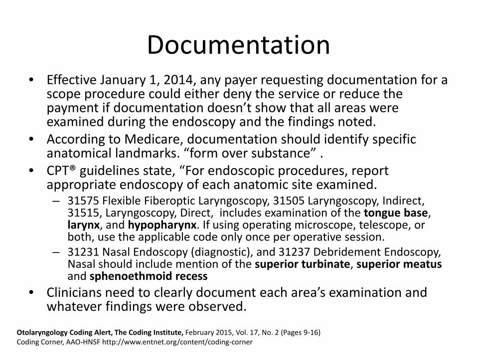
Documentation • Effective January 1, 2014, any payer requesting documentation for a
scope procedure could either deny the service or reduce the payment if documentation doesn’t show that all areas were examined during the endoscopy and the findings noted.
• According to Medicare, documentation should identify specific anatomical landmarks. “form over substance” .
• CPT® guidelines state, “For endoscopic procedures, report appropriate endoscopy of each anatomic site examined. – 31575 Flexible Fiberoptic Laryngoscopy, 31505 Laryngoscopy, Indirect,
31515, Laryngoscopy, Direct, includes examination of the tongue base, larynx, and hypopharynx. If using operating microscope, telescope, or both, use the applicable code only once per operative session.
– 31231 Nasal Endoscopy (diagnostic), and 31237 Debridement Endoscopy, Nasal should include mention of the superior turbinate, superior meatus and sphenoethmoid recess
• Clinicians need to clearly document each area’s examination and whatever findings were observed.
Otolaryngology Coding Alert, The Coding Institute, February 2015, Vol. 17, No. 2 (Pages 9-16) Coding Corner, AAO-HNSF http://www.entnet.org/content/coding-corner
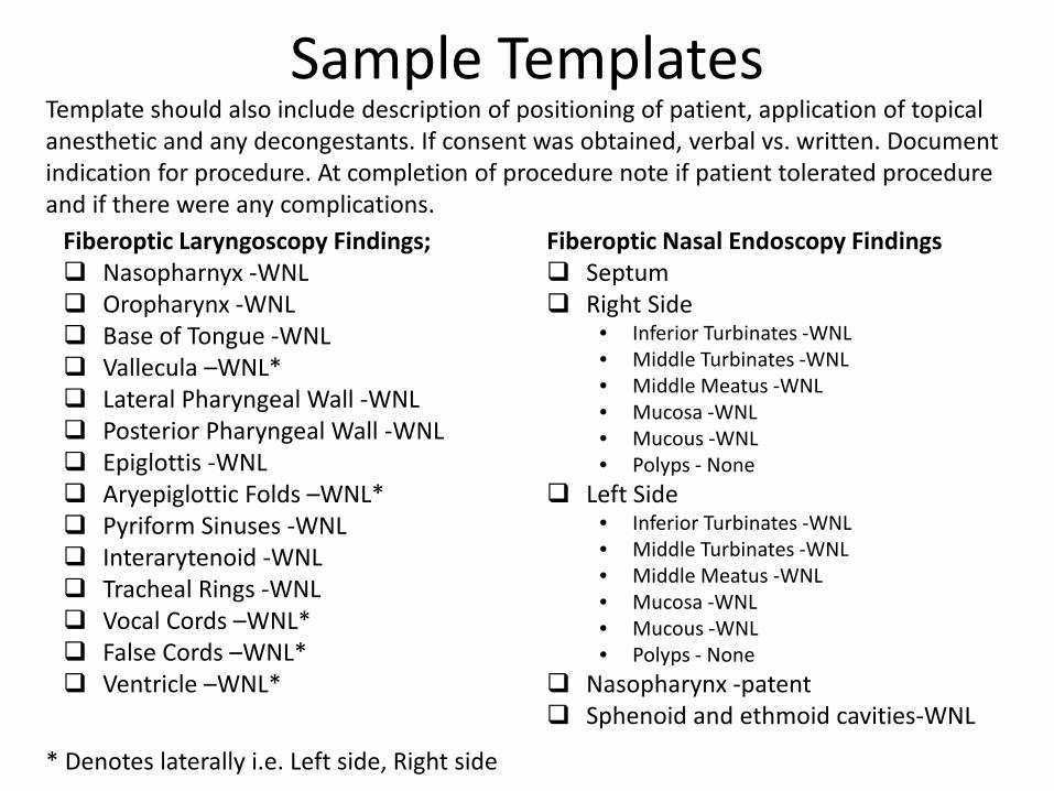
Sample Templates
Fiberoptic Nasal Endoscopy Findings Septum Right Side
• Inferior Turbinates -WNL • Middle Turbinates -WNL • Middle Meatus -WNL • Mucosa -WNL • Mucous -WNL • Polyps - None
Left Side • Inferior Turbinates -WNL • Middle Turbinates -WNL • Middle Meatus -WNL • Mucosa -WNL • Mucous -WNL • Polyps - None
Nasopharynx -patent Sphenoid and ethmoid cavities-WNL
Fiberoptic Laryngoscopy Findings; Nasopharnyx -WNL Oropharynx -WNL Base of Tongue -WNL Vallecula –WNL* Lateral Pharyngeal Wall -WNL Posterior Pharyngeal Wall -WNL Epiglottis -WNL Aryepiglottic Folds –WNL* Pyriform Sinuses -WNL Interarytenoid -WNL Tracheal Rings -WNL Vocal Cords –WNL* False Cords –WNL* Ventricle –WNL*
* Denotes laterally i.e. Left side, Right side
Template should also include description of positioning of patient, application of topical anesthetic and any decongestants. If consent was obtained, verbal vs. written. Document indication for procedure. At completion of procedure note if patient tolerated procedure and if there were any complications.
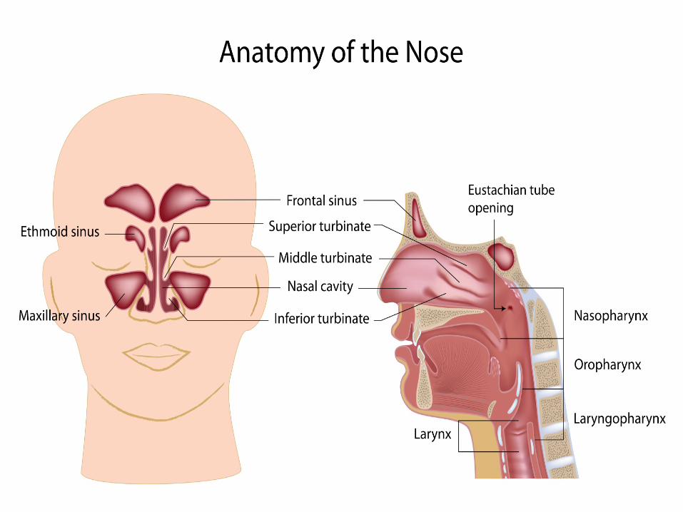
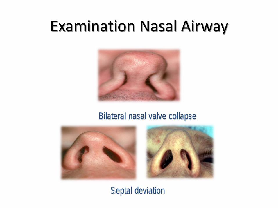
Examination Nasal Airway
Bilateral nasal valve collapse
Septal deviation
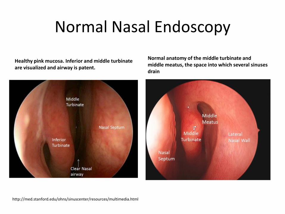
Healthy pink mucosa. Inferior and middle turbinate are visualized and airway is patent.
Normal anatomy of the middle turbinate and middle meatus, the space into which several sinuses drain
http://med.stanford.edu/ohns/sinuscenter/resources/multimedia.html
Normal Nasal Endoscopy
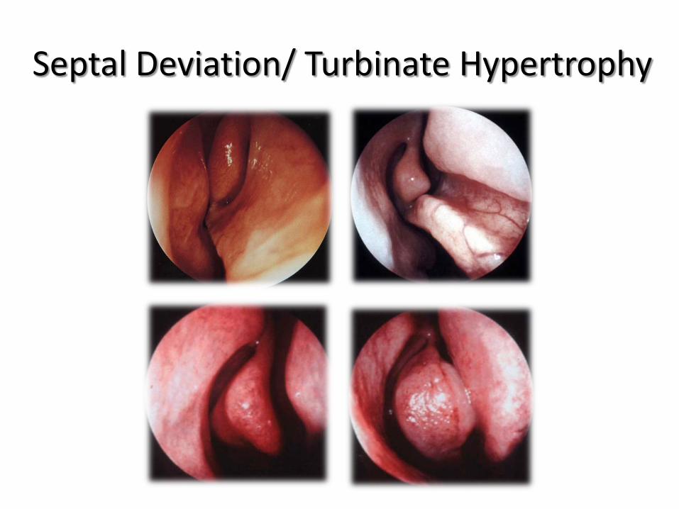
Septal Deviation/ Turbinate Hypertrophy
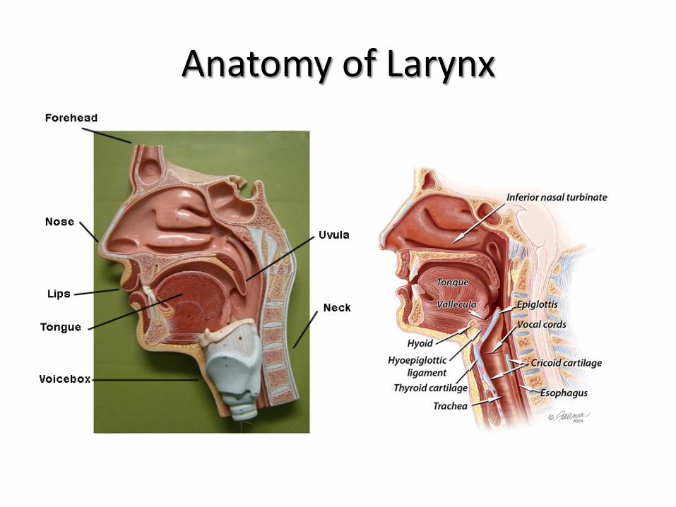
Anatomy of Larynx
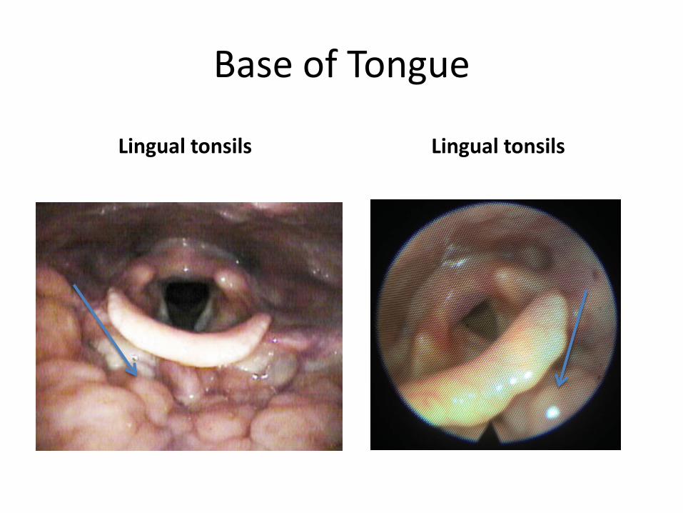
Base of Tongue
Lingual tonsils Lingual tonsils
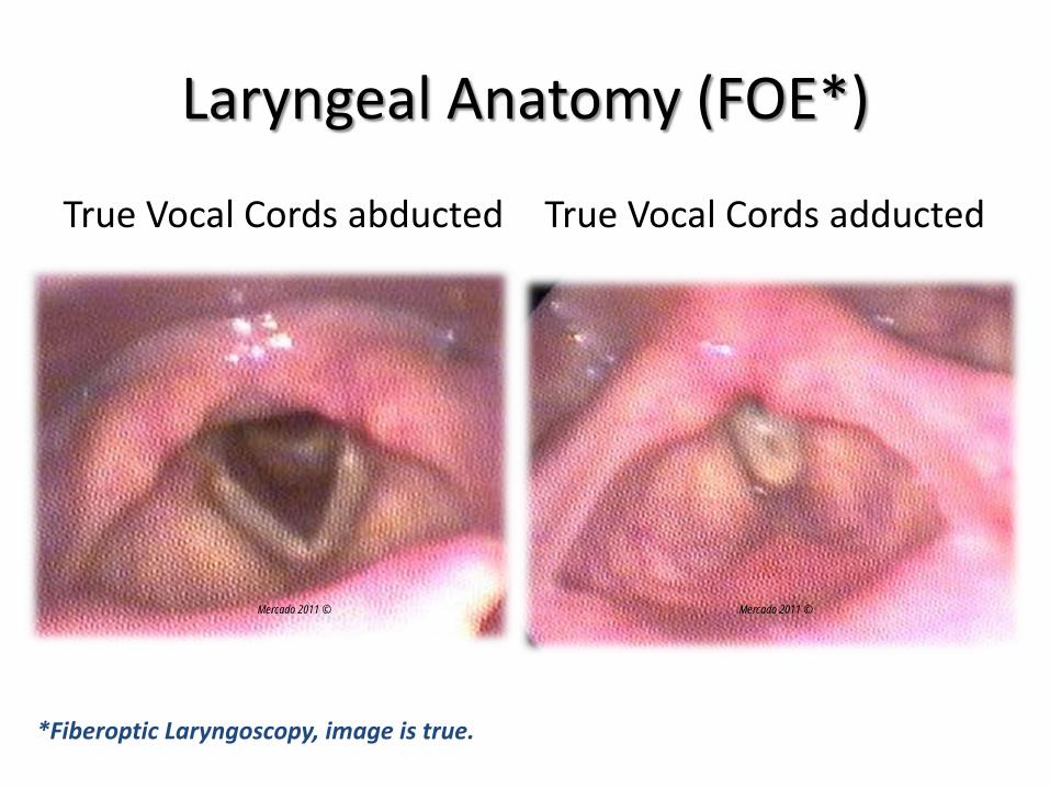
Laryngeal Anatomy (FOE*)
True Vocal Cords abducted True Vocal Cords adducted
*Fiberoptic Laryngoscopy, image is true.
Mercado 2011 © Mercado 2011 ©
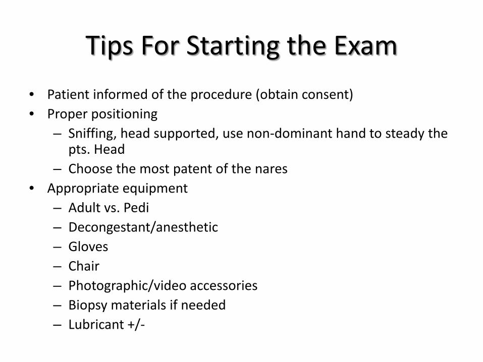
Tips For Starting the Exam • Patient informed of the procedure (obtain consent) • Proper positioning
– Sniffing, head supported, use non-dominant hand to steady the pts. Head
– Choose the most patent of the nares • Appropriate equipment
– Adult vs. Pedi – Decongestant/anesthetic – Gloves – Chair – Photographic/video accessories – Biopsy materials if needed – Lubricant +/-
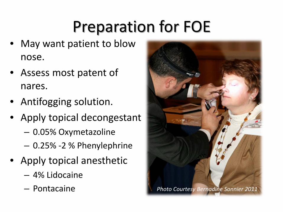
Preparation for FOE • May want patient to blow
nose. • Assess most patent of
nares. • Antifogging solution. • Apply topical decongestant
– 0.05% Oxymetazoline – 0.25% -2 % Phenylephrine
• Apply topical anesthetic – 4% Lidocaine – Pontacaine Photo Courtesy Bernadine Sonnier 2011
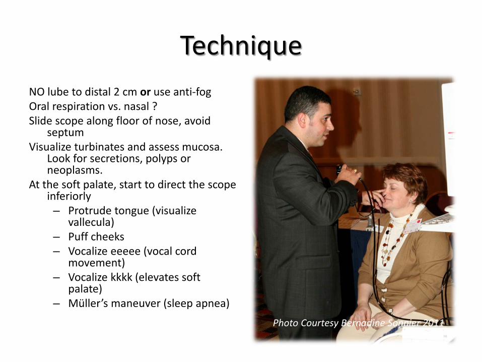
Technique NO lube to distal 2 cm or use anti-fog Oral respiration vs. nasal ? Slide scope along floor of nose, avoid
septum Visualize turbinates and assess mucosa.
Look for secretions, polyps or neoplasms.
At the soft palate, start to direct the scope inferiorly – Protrude tongue (visualize
vallecula) – Puff cheeks – Vocalize eeeee (vocal cord
movement) – Vocalize kkkk (elevates soft
palate) – Müller’s maneuver (sleep apnea) Photo Courtesy Bernadine Sonnier 2011
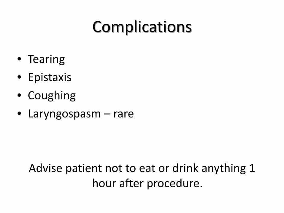
Complications
• Tearing • Epistaxis • Coughing • Laryngospasm – rare
Advise patient not to eat or drink anything 1 hour after procedure.
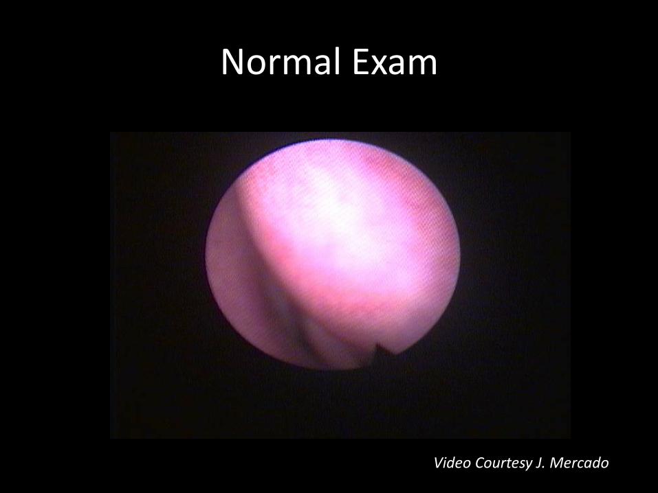
Normal Exam
Video Courtesy J. Mercado
Flexible Laryngoscopy
Interactive, live demonstration of flexible fiberoptic endoscopy
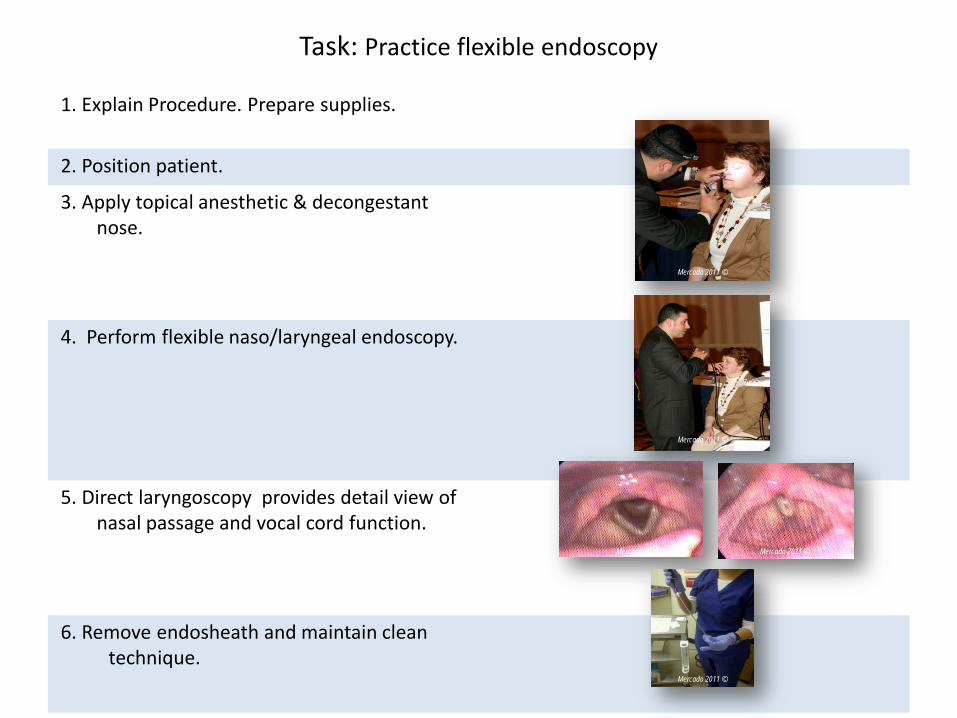
1. Explain Procedure. Prepare supplies.
2. Position patient.
3. Apply topical anesthetic & decongestant nose.
4. Perform flexible naso/laryngeal endoscopy. 5. Direct laryngoscopy provides detail view of
nasal passage and vocal cord function.
6. Remove endosheath and maintain clean
technique.
Task: Practice flexible endoscopy
Mercado 2011 ©
Mercado 2011 ©
Mercado 2011 ©
Mercado 2011 ©
Mercado 2011 ©
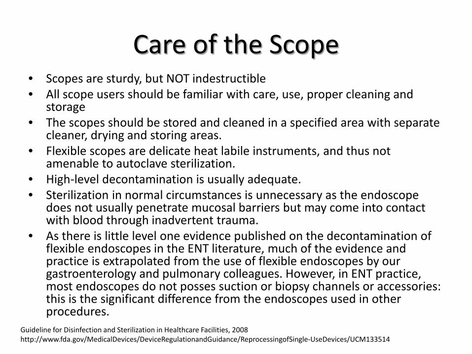
Care of the Scope • Scopes are sturdy, but NOT indestructible • All scope users should be familiar with care, use, proper cleaning and
storage • The scopes should be stored and cleaned in a specified area with separate
cleaner, drying and storing areas. • Flexible scopes are delicate heat labile instruments, and thus not
amenable to autoclave sterilization. • High-level decontamination is usually adequate. • Sterilization in normal circumstances is unnecessary as the endoscope
does not usually penetrate mucosal barriers but may come into contact with blood through inadvertent trauma.
• As there is little level one evidence published on the decontamination of flexible endoscopes in the ENT literature, much of the evidence and practice is extrapolated from the use of flexible endoscopes by our gastroenterology and pulmonary colleagues. However, in ENT practice, most endoscopes do not posses suction or biopsy channels or accessories: this is the significant difference from the endoscopes used in other procedures.
Guideline for Disinfection and Sterilization in Healthcare Facilities, 2008 http://www.fda.gov/MedicalDevices/DeviceRegulationandGuidance/ReprocessingofSingle-UseDevices/UCM133514
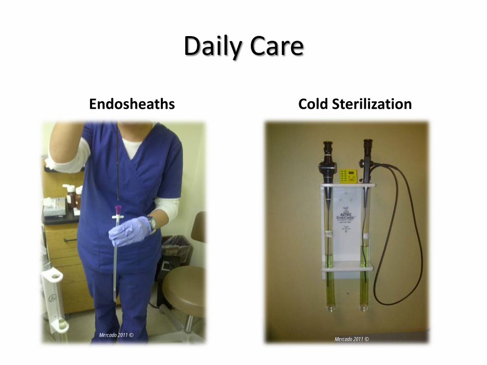
Daily Care
Endosheaths Cold Sterilization
Mercado 2011 © Mercado 2011 ©
Endosheaths vs. Cold Sterilization • Sterile disposable sheaths are
custom built for a variety of scopes and some models even come with a working channel.
• The tip of the sheath must be fully slid onto the scope so that the special optical element at the end of the sheath is flat against the tip of the scope.
• After using the sheath, it can be slid off and disposed of without the need to re-sterilize the scope.
• These sheaths should never be forcefully removed.
• Flexible scopes are non-autoclavable. • Clean length of flexible scope with an
enzymatic detergent solution like ENZOL® to remove debris and reduce bacterial burden before instruments are disinfected or sterilized.
• Soak flexible scope in a glutaraldehyde solution like Cidex ® which provides quick high-level disinfection.
• Noncorrosive solution reduces instrument damage and associated repair costs.
• Soaking times vary by product.
http://www.fda.gov/MedicalDevices/DeviceRegulationandGuidance/ReprocessingofSingle-UseDevices/UCM133514
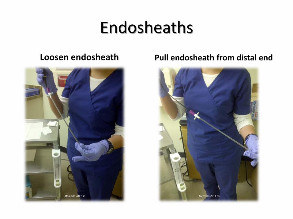
Endosheaths Loosen endosheath Pull endosheath from distal end
Mercado 2011 © Mercado 2011 ©
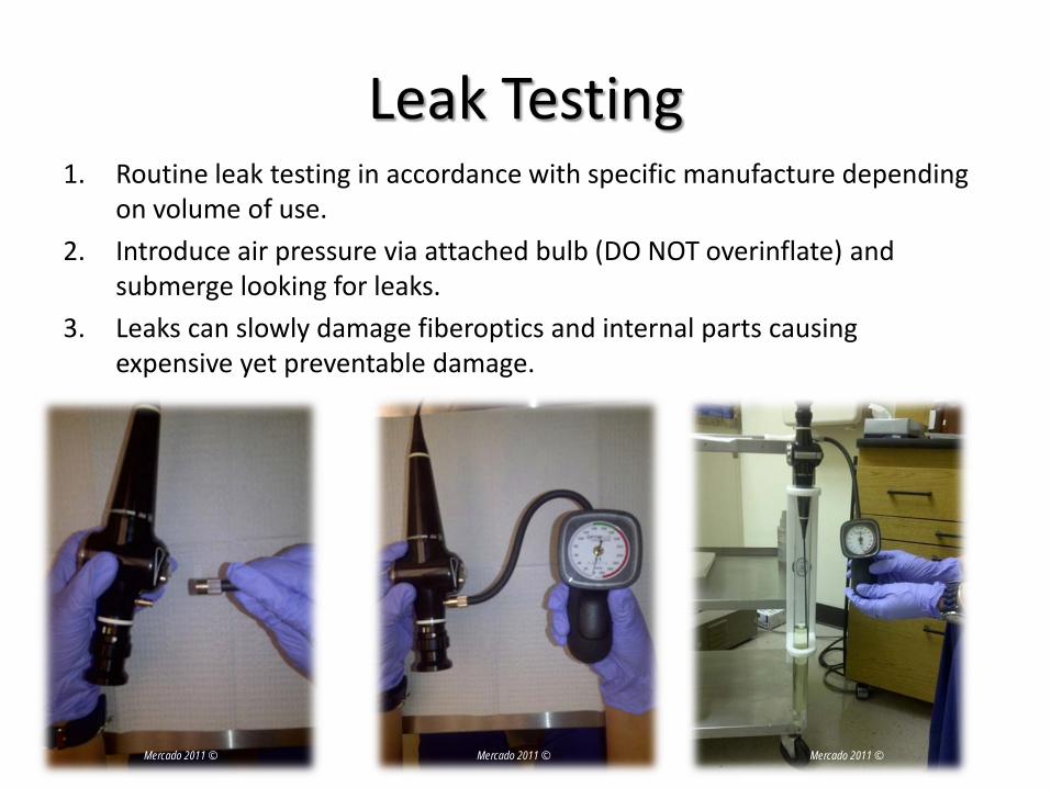
Leak Testing 1. Routine leak testing in accordance with specific manufacture depending
on volume of use. 2. Introduce air pressure via attached bulb (DO NOT overinflate) and
submerge looking for leaks. 3. Leaks can slowly damage fiberoptics and internal parts causing
expensive yet preventable damage.
Mercado 2011 © Mercado 2011 © Mercado 2011 ©
Helpful Care Tips
• Avoid bending scope in tight angles. • Clean lens with lens cleaner/paper. • Pre-clean with enzymatic cleaner. • Soak only for required period depending on
brand and manufacture. • Store in dry safe place. • Perform regular leak testing to avoid damage.
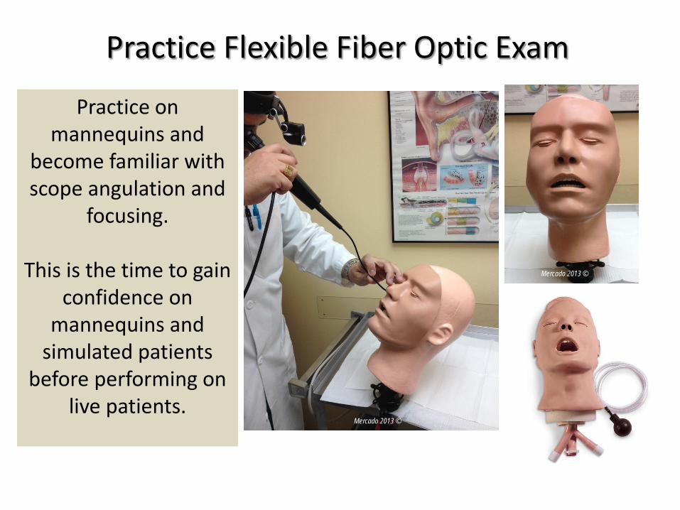
Practice on mannequins and
become familiar with scope angulation and
focusing.
This is the time to gain confidence on
mannequins and simulated patients
before performing on live patients.
Practice Flexible Fiber Optic Exam
Mercado 2013 ©
Mercado 2013 ©
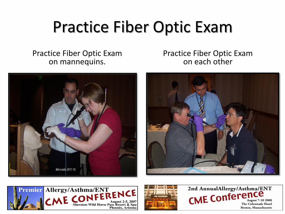
Practice Fiber Optic Exam Practice Fiber Optic Exam
on mannequins. Practice Fiber Optic Exam
on each other
Mercado 2011 © Mercado 2011 ©
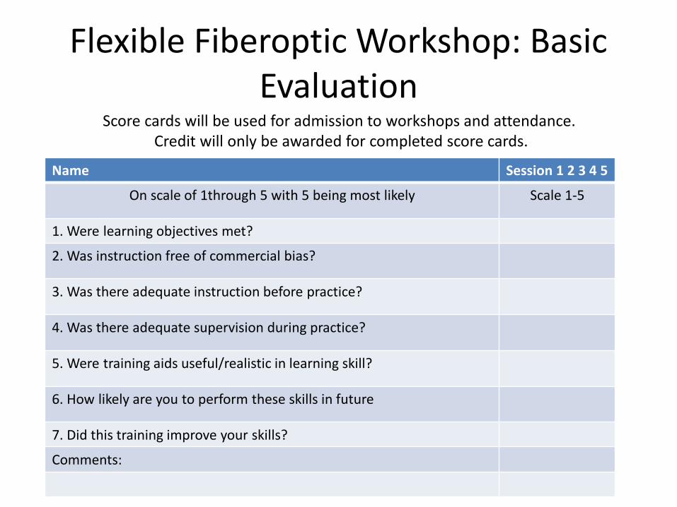
Flexible Fiberoptic Workshop: Basic Evaluation
Name Session 1 2 3 4 5
On scale of 1through 5 with 5 being most likely Scale 1-5
1. Were learning objectives met?
2. Was instruction free of commercial bias?
3. Was there adequate instruction before practice?
4. Was there adequate supervision during practice?
5. Were training aids useful/realistic in learning skill?
6. How likely are you to perform these skills in future
7. Did this training improve your skills?
Comments:
Score cards will be used for admission to workshops and attendance. Credit will only be awarded for completed score cards.
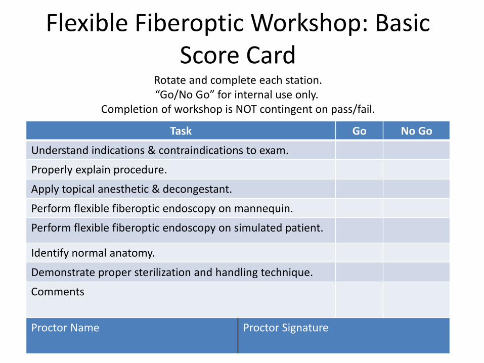
Flexible Fiberoptic Workshop: Basic Score Card
Task Go No Go
Understand indications & contraindications to exam.
Properly explain procedure.
Apply topical anesthetic & decongestant.
Perform flexible fiberoptic endoscopy on mannequin.
Perform flexible fiberoptic endoscopy on simulated patient.
Identify normal anatomy.
Demonstrate proper sterilization and handling technique.
Comments
Proctor Name Proctor Signature
Rotate and complete each station. “Go/No Go” for internal use only.
Completion of workshop is NOT contingent on pass/fail.
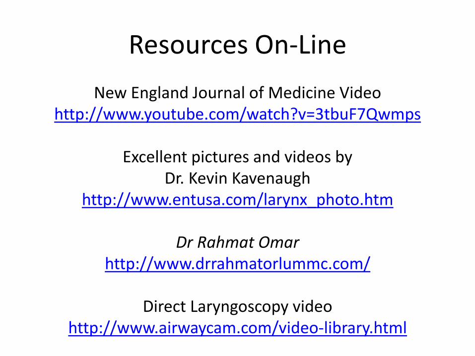
Resources On-Line New England Journal of Medicine Video
http://www.youtube.com/watch?v=3tbuF7Qwmps
Excellent pictures and videos by Dr. Kevin Kavenaugh
http://www.entusa.com/larynx_photo.htm
Dr Rahmat Omar http://www.drrahmatorlummc.com/
Direct Laryngoscopy video
http://www.airwaycam.com/video-library.html
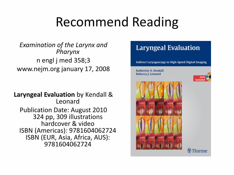
Recommend Reading Examination of the Larynx and
Pharynx n engl j med 358;3
www.nejm.org january 17, 2008
Laryngeal Evaluation by Kendall & Leonard
Publication Date: August 2010 324 pp, 309 illustrations
hardcover & video ISBN (Americas): 9781604062724
ISBN (EUR, Asia, Africa, AUS): 9781604062724
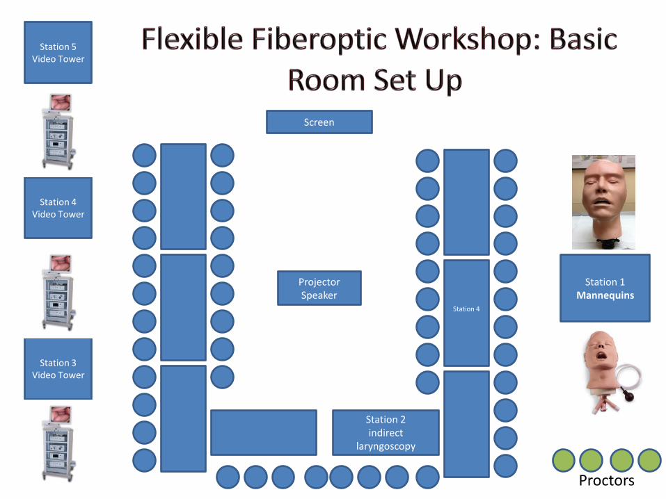
Station 5 Video Tower
Station 1 Mannequins
Station 4
Screen
Projector Speaker
Station 4 Video Tower
Station 3 Video Tower
Station 2 indirect
laryngoscopy
Proctors