fluoroscopy-assisted stress testing of the thumb metacarpophalangeal joint to assess the ulnar...
TRANSCRIPT

SURGERYARTICLES
Fluoroscopy-assisted stress testing of the thumbmetacarpophalangealjoint to assess the ulnar collateral ligament
Ashish Patel & Archit Patel & David Edelstein &
Jack Choueka
Published online: 13 February 2013# American Association for Hand Surgery 2013
AbstractBackground Diagnostic stress testing of ulnar collateral lig-ament (UCL) injuries of the thumb metacarpophalangeal(MCP) joint is pivotal to determining treatment. Compari-son to the uninjured extremity and fluoroscopy-assistedexamination are readily available modalities in the assessmentof these patients, with 5–10° differences impacting treatment.Comparative examination, however, assumes that bothextremities are normally equal, which has never been verifiedexperimentally. Comparison of clinical and fluoroscopicexamination has also never been scrutinized.Methods One hundred asymptomatic participants under-went both fluoroscopic and traditional stress examinationsto determine maximum passive radial deviation at neutralMCP flexion.Results Absolute clinical vs. fluoroscopic differences dem-onstrated a significant difference of 5.6° (SD 5.1°). Absolutevariability between left-to-right measurements was 4.5° (SD4.1°) and increased significantly as baseline stress deviationincreased (R = 0.43; p < 0.001). Left-to-right differenceexhibited no correlation to age, gender, or BMI.Conclusions The current investigation demonstrates right–left differences and differences between clinical and fluoro-scopic testing of which practitioners should be aware whenmaking treatment decisions for UCL injury of the thumbMCP joint.
Keywords Thumbmetacarpophalangeal joint . Thumbulnarcollateral ligament . Joint laxity . Stress testing .
Goniometry . Gamekeeper’s thumb . Skier’s thumb
Introduction
Ulnar collateral ligament (UCL) injury of the thumb meta-carpophalangeal (MCP) joint is a relatively common injuryprecipitated by acute trauma or repetitive MCP abduction.Joint laxity resulting from a significant UCL tear may con-tribute to hand weakness, radial deviation of the proximalphalanx, and range-of-motion instability [10]. Failure todiagnose and treat this injury can lead to chronic, painfulinstability, long-term weakness of pinch and grip strength,and premature, progressive arthritis of the joint [4, 10].
Injuries of the thumb UCL can be classified into threegeneral categories: grade I, which consists of an incompletetear without joint laxity; grade II, which comprises larger,but still incomplete tears with asymmetrical laxity in thejoint; and grade III, in which ligamentous disruption iscomplete [8, 9]. Ulnar MCP joint instability is evaluatedby securing the metacarpal and applying radial force to theproximal phalanx of the thumb until a firm endpoint isreached; the resultant metacarpal–phalanx angle at theMCP joint may then be measured with a goniometer. Previ-ous studies indicate that complete UCL rupture correlates toan absolute radial stress angulation of greater than 35° andthat differences in excess of 15° vs. the contralateral(uninjured) thumb suggest UCL tearing and that differencesof as little as 10° can indicate instability that may requiretreatment [1, 5, 7]. Accurate determination of metacarpal–phalanx angulation under stress is thus an important elementof proper diagnosis and subsequent treatment of UCL rup-ture. Furthermore, since goniometric testing relies partly ondifferentiating angular differences between the injuredand uninjured MCP, it is important to establish thevariability of contralateral MCP stress angulations inasymptomatic patients.
Though large variations in intersubject stress deviationhave been documented within the asymptomatic population,
A. Patel :A. Patel :D. Edelstein : J. Choueka (*)Department of Orthopaedic Surgery, Maimonides Medical Center,927 49th St., Brooklyn, NY 11219, USAe-mail: [email protected]
HAND (2013) 8:205–209DOI 10.1007/s11552-013-9500-2
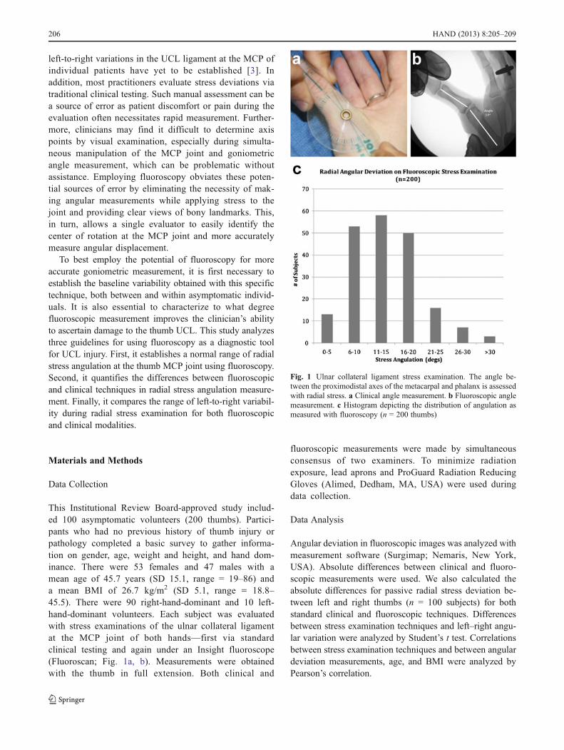
left-to-right variations in the UCL ligament at the MCP ofindividual patients have yet to be established [3]. Inaddition, most practitioners evaluate stress deviations viatraditional clinical testing. Such manual assessment can bea source of error as patient discomfort or pain during theevaluation often necessitates rapid measurement. Further-more, clinicians may find it difficult to determine axispoints by visual examination, especially during simulta-neous manipulation of the MCP joint and goniometricangle measurement, which can be problematic withoutassistance. Employing fluoroscopy obviates these poten-tial sources of error by eliminating the necessity of mak-ing angular measurements while applying stress to thejoint and providing clear views of bony landmarks. This,in turn, allows a single evaluator to easily identify thecenter of rotation at the MCP joint and more accuratelymeasure angular displacement.
To best employ the potential of fluoroscopy for moreaccurate goniometric measurement, it is first necessary toestablish the baseline variability obtained with this specifictechnique, both between and within asymptomatic individ-uals. It is also essential to characterize to what degreefluoroscopic measurement improves the clinician’s abilityto ascertain damage to the thumb UCL. This study analyzesthree guidelines for using fluoroscopy as a diagnostic toolfor UCL injury. First, it establishes a normal range of radialstress angulation at the thumb MCP joint using fluoroscopy.Second, it quantifies the differences between fluoroscopicand clinical techniques in radial stress angulation measure-ment. Finally, it compares the range of left-to-right variabil-ity during radial stress examination for both fluoroscopicand clinical modalities.
Materials and Methods
Data Collection
This Institutional Review Board-approved study includ-ed 100 asymptomatic volunteers (200 thumbs). Partici-pants who had no previous history of thumb injury orpathology completed a basic survey to gather informa-tion on gender, age, weight and height, and hand dom-inance. There were 53 females and 47 males with amean age of 45.7 years (SD 15.1, range = 19–86) anda mean BMI of 26.7 kg/m2 (SD 5.1, range = 18.8–45.5). There were 90 right-hand-dominant and 10 left-hand-dominant volunteers. Each subject was evaluatedwith stress examinations of the ulnar collateral ligamentat the MCP joint of both hands—first via standardclinical testing and again under an Insight fluoroscope(Fluoroscan; Fig. 1a, b). Measurements were obtainedwith the thumb in full extension. Both clinical and
fluoroscopic measurements were made by simultaneousconsensus of two examiners. To minimize radiationexposure, lead aprons and ProGuard Radiation ReducingGloves (Alimed, Dedham, MA, USA) were used duringdata collection.
Data Analysis
Angular deviation in fluoroscopic images was analyzed withmeasurement software (Surgimap; Nemaris, New York,USA). Absolute differences between clinical and fluoro-scopic measurements were used. We also calculated theabsolute differences for passive radial stress deviation be-tween left and right thumbs (n = 100 subjects) for bothstandard clinical and fluoroscopic techniques. Differencesbetween stress examination techniques and left–right angu-lar variation were analyzed by Student’s t test. Correlationsbetween stress examination techniques and between angulardeviation measurements, age, and BMI were analyzed byPearson’s correlation.
Fig. 1 Ulnar collateral ligament stress examination. The angle be-tween the proximodistal axes of the metacarpal and phalanx is assessedwith radial stress. a Clinical angle measurement. b Fluoroscopic anglemeasurement. c Histogram depicting the distribution of angulation asmeasured with fluoroscopy (n = 200 thumbs)
206 HAND (2013) 8:205–209

Results
Clinical vs. Fluoroscopic Stress Examination Techniques
Left and right thumbs from 100 subjects were analyzed todetermine the mean, standard deviation, and range for pas-sive radial angulation (Table 1). The absolute differencesdemonstrated a mean variation of 5.6° (SD 5.1°, range 0–26°). Angulation ranged from 5° to 31° for standard clinicalmeasurements and from 1° to 48° for fluoroscopic measure-ments (Fig. 1c). There was no difference between clinicaland fluoroscopic measurement with the thumb in neutralwhen analyzed by t test (p = 0.17) and Pearson’s correlationyielded an R value of 0.25 (p = 0.001).
Fluoroscopic Left-to-Right Difference
The absolute difference between right and left passive radialdeviation measurements was 4.5° (SD 4.1°, range 0–18°;Table 2, Fig. 2). For fluoroscopic measurements, unpairedt tests did not demonstrate a significant difference betweenleft and right angular deviation measurements (p = 0.69).Pearson correlation between left and right angular devia-tions yielded R = 0.59 (p < 0.001). The degree of correlationsubsided as baseline lateral deviation increased at neutral(R = 0.43, p < 0.001). Correlations between left-to-rightvariability and age, gender, BMI, dominant hand, andMCP flexion angle were not significant (p > 0.05).
Left-to-Right Differences with Clinical Examination
Absolute differences had a mean of 3.6° (SD 3.1°, range0–11°; Table 2). There were no subjects who exhibited agreater than 15° left–right differential by clinical mea-surement (Fig. 3). Unpaired t tests showed no significantdifference between left-to-right measurements at neutralflexion (p = 0.29). Pearson correlation tests for left-to-right angular deviation at neutral flexion produced an Rvalue of 0.64 (p < 0.001).
Discussion
Most practitioners use traditional physical examinations toevaluate and inform treatment of ulnar collateral ligamentinjuries. Visual examinations, even when combined withclinical goniometry techniques, have inherent flaws thatmay provide inaccurate measurements. This may at leastpartially account for the large variations in normative dataseen in the literature. One study used clinical goniometricstress examination on 1,000 thumbs at 15° of MCP flexionand reported a mean angulation of 10° (range 0–20°) [3].Another study reported means of 6° angulation at neutralflexion and 12° at 15° flexion in a clinical examination of750 thumbs [7]. Other work examining patients with UCLinjuries via clinical goniometry and making comparisonswith the contralateral (uninjured) thumb revealed a mean16.2° (SD 8.9°) at neutral [4]. The current investigationdemonstrated a mean angulation of 14.9° (SD 5.5°) at neu-tral flexion by clinical testing. These results are most con-sistent with the results obtained by Heyman et al.; however,there is still a lack of consensus in the literature and signif-icant potential for inter-examiner variability when using theclassical clinical technique [5].
Several key shortcomings of clinical examination thatmay contribute to decreased accuracy were noted duringthis investigation. In the clinic, angular assessment with agoniometer relies on using surface anatomy to approximatethe location and direction of the metacarpal and proximalphalanx; this complicates the determination of the rotationaxis. It was also difficult for a single examiner to simulta-neously apply radial stress and employ the goniometer.Holding the thumb in the appropriate level of MCP flexionto obtain comparative measurement further complicated thisprocess.
Assessing the thumb MCP under stress at 30° flexion vs.neutral is still a matter of debate. Proponents for testing inflexion believe that stressing the joint isolates the collateralligament, which is taut in flexion (unlike the accessoryligament, which tightens in extension). However, Posner
Table 1 Passive radial stressangulation for clinical andfluoroscopic examinations(n = 200) at neutral MCPflexion
Testing modality MCP position Hand Mean (°) Std deviation Min Max
Fluoroscopy Neutral Right 13.9 7.1 1 40
Left 14.3 6.1 2 39
Goniometer Neutral Right 14.5 5.6 5 31
Left 15.4 5.4 5 30
Table 2 Absolute left–rightangle differences for clinical andfluoroscopic techniques atneutral MCP flexion
Testing modality MCP position Mean (°) Std deviation Min Max
Goniometer Neutral 14.9 5.5 5 31
Fluoroscopy Neutral 14.1 6.6 1 40
HAND (2013) 8:205–209 207

did not observe any cases of MCP instability where thejoints were grossly unstable in flexion and stable in neutral.He thus recommended stress testing in neutral to minimizethe risk of an unseasoned examiner improperly diagnosing agrade I or II injury as grade III [9]. To limit the aforemen-tioned variability in our study, radial stress angulations weremeasured with the MCP joint at neutral flexion. The com-bination of these limitations may compromise the sensitivityof the clinical technique.
Fluoroscopy allows clear imaging of the metacarpal,proximal phalanx, and axis of rotation and permits theexaminer to freely manipulate the MCP joint. To date, thereare no data in the literature regarding the normal range of
fluoroscopic radial stress angulation in the MCP joint. Ourresults demonstrate that mean fluoroscopic stress angulationis consistent with both our clinical examination and aspreviously reported [5]. However, we observed a muchgreater range of angulation with fluoroscopic assessment.In addition, the distribution of angulation generated for theasymptomatic population using fluoroscopy produced a dis-tribution curve more closely resembling a normal distribu-tion than that derived from clinical measurements. Thenormal distribution of the fluoroscopic data reflects thevariability seen in the asymptomatic population; outliers inthe distribution illustrate that asymptomatic patients mayhave a stress angulations greater than 30° and left–rightdifferences greater than 15°. However, it is worth notingthat asymptomatic patients may not necessarily be unin-jured. Partial but painless tears in the UCL or acquiredligamentous laxity may potentially result in extreme left-to-right differences in angulation. In such cases, furthertests, such as direct imaging of the UCL to assess damage,may be indicated.
When comparing fluoroscopy to standard clinical techni-ques, the measurements differed by an absolute mean of5.5°. While this may not be a large difference, it maynonetheless prove significant for patients whose injury as-sessment falls on the border between grade II (partial thick-ness tears that can be treated nonsurgically) and grade III(full-thickness tears which require surgery) [9]. Additional-ly, the correlation coefficient of R = 0.30, though statisticallysignificant, demonstrates a weak association between thetwo modalities with a reduced correlation further from themean. So while the means between the two techniques werefairly similar, there is extensive variability between the twotechniques.
Other modalities for assessing UCL injuries, includingultrasound and MRI, have been explored in previous studies.Both these imaging methods provide excellent resolution ofligaments for devising treatment strategies, particularly whencomplete disruption of the ligament necessitates surgical re-pair [6, 11]. However, these modes were examined as ways todirectly visualize ligamentous trauma rather than as methodsto augment clinical assessments; they may thus not be practi-cal for initial screening. This is particularly true ofMRI, whichdespite its utility, is implemented at considerable expense [6].
Left-to-Right Intrasubject Stress Examination Variability
Using mini-fluoroscopy, left-to-right comparisons yielded amean absolute difference of 4.5° (SD 4.1°, range 0–18°) atneutral. Current surgical indications for UCL repair includea greater than 30° absolute stress angulation [3, 7, 9] orgreater than 15° angulation vs. the contralateral side [1, 5].Under fluoroscopic examination, our study found 1.5 % ofsubjects with joint laxity greater than 30° in neutral flexion
Fig. 2 Histogram illustrating absolute left–right variability on fluoro-scopic stress examination
Fig. 3 Absolute left–right variability on goniometric stress examination
208 HAND (2013) 8:205–209

and 3 % with left-to-right angular differentials greater than15°. There did not seem to be any relationship between theside with greatest joint laxity and subject hand dominance,although this may be due to the observed numbers beingvery small. These subjects represent presumably normalvariants with large baseline angulations in the absence ofclinical signs or symptoms of UCL injury. Additionally, theleft-to-right correlation was found to decrease with increas-ing baseline stress angulation. Since treatment decisions areoften based on 10° differences between left and right, theexaminer must be aware of potential left-to-right variabilityin the asymptomatic population, especially when the unin-jured extremity has a large angulation.
Assessing UCL injury through comparison to the contra-lateral side is a useful tool for the treating physician, al-though clinical signs and symptoms must be carefullyconsidered and incorporated into the evaluation of a patientwith large baseline angular deviations. In these patients,left–right comparisons may be misleading and other imag-ing modalities may be needed to confirm a UCL tear. It isalso important to consider that there is a qualitative aspect tothis type of clinical testing: specifically, the determination ofa “firm endpoint” under radial stress by the evaluator. Inorder to make comparisons based on differences in lateral–contralateral stress angulations, evaluators must acknowledgetheir own discretion in these decisions and be aware of anyvariability in their methods.
Though fluoroscopy results in lower radiation exposurethan standard X-rays and radiation-reducing gloves canattenuate exposure by 60–64 %, the amount of exposure toboth patient and surgeon must be considered [2]. We rec-ommend the use of fluoroscopy in lieu of conventionalradiographs both for the ease with which joint malrotationcan be corrected during imaging and to further reduce radi-ation exposure risks.
We endeavored to quantify the differences in standardclinical and fluoroscopic goniometric techniques and estab-lish the range of left-to-right intrasubject variability duringradial stress testing of the thumb MCP joint in an uninjuredpopulation. Clinical goniometry measurements demonstrat-ed a weak correlation to fluoroscopic measurements, whichsuggests inherent measurement errors in the absence offluoroscopic visualization. If a UCL tear is suspected, clin-ical stress testing may provide a practical screening test,
which, if positive, can be verified through fluoroscopicstress examination to obtain more accurate angular measure-ment. In addition, treatment decisions based on left-to-rightvariability should be made with care, especially in patientswith large baseline angular deviations.
Acknowledgments We would like to thank Maya Deza Culbertsonfor her helpful input during the writing of this manuscript.
Conflict of Interest The authors declare that they have no conflict ofinterest.
References
1. Bowers WH, Hurst LC. Gamekeeper’s thumb. Evaluation byarthrography and stress roentgenography. J Bone Joint Surg Am.1977;59:519–24.
2. Calder PR, Tennent TD, Allen PW. Assessment of the efficacy ofProguard RR-2 radio-protective gloves during forearm manipula-tion. Injury. 2003;34(2):159–61.
3. Coonrad RW, Goldner JL. A study of the pathological findings andtreatment in soft-tissue injury of the thumb metacarpophalangealjoint. With a clinical study of the normal range of motion in onethousand thumbs and a study of post mortem findings of ligamen-tous structures in relation to function. J Bone Joint Surg Am.1968;50:439–51.
4. Demirel M, Turhan E, Dereboy F, et al. Surgical treatment ofskier’s thumb injuries: case report and review of the literature.Mt Sinai J Med. 2006;73:818–21.
5. Heyman P, Gelberman RH, Duncan K, et al. Injuries of the ulnarcollateral ligament of the thumb metacarpophalangeal joint. Bio-mechanical and prospective clinical studies on the usefulness ofvalgus stress testing. Clin Orthop Relat Res. 1993;292:165–71.
6. Louis DS, Buckwalter KA. Magnetic resonance imaging of thecollateral ligaments of the thumb. J Hand Surg Am. 1989;14(4):739–41.
7. Palmer AK, Louis DS. Assessing ulnar instability of the metacar-pophalangeal joint of the thumb. J Hand Surg Am. 1978;3:542–6.
8. Patel S, Potty A, Taylor EJ, Sorene ED. Collateral ligament inju-ries of the metacarpophalangeal joint of the thumb: a treatmentalgorithm. Strat Trauma Limb Reconstr. 2010;5(1):1–10.
9. Posner MA, Retaillaud JL. Metacarpophalangeal joint injuries ofthe thumb. Hand Clin. 1992;8:713–32.
10. Sakellarides HT, DeWeese JW. Instability of the metacarpophalan-geal joint of the thumb. Reconstruction of the collateral ligamentsusing the extensor pollicis brevis tendon. J Bone Joint Surg Am.1976;58:106–12.
11. Schnur DP, DeLone FX, McClellan RM, Bonavita J, Witham RS.Ultrasound: a powerful tool in the diagnosis of ulnar collateralligament injuries of the thumb. Ann Plast Surg. 2002;49(1):19–23.
HAND (2013) 8:205–209 209