for the life sciences - biopac sysbiopac-sys.jp/support/images/biopac_mri_catalog_web.pdffor the...
TRANSCRIPT

For the Life Sciences
MRI Solutions for Human and Animal Studies• Physiological Data Acquisition Systems• AcqKnowledge Software—automation & specialized MRI tools• New! Remote Monitor• New! MRI Smart Amplifiers• Transducers, Electrodes, and Accessories• Human-safe Isolated RF Cable/Filter Systems

BIOPAC provides physiological data acquisition and analysis systems specifically for human and small animalMRI life science research applications.BIOPAC offers data acquisition systems, MRI Smart Amplifiers, transducers, stimulusoptions, electrodes, and leads with advancedsoftware tools for safe data collection, subjectmonitoring, and clean physiological signals inthe MRI environment.
MP Systems and amplifiers are placed in theMRI Control Room, and specialized cable systems optimize data quality with isolated and RF filtered interfacing between the subject/MRI Chamber and the Control Room.Filter leakage currents and dielectric isolationsatisfy IEC60601-1.
Systems include AcqKnowledge software withrich display and monitoring features, plus automation and scoring routines to provide aflexible solution for life science research.
New MRI Smart Amplifiers incorporateadvanced signal processing circuitry whichremoves spurious MRI artifact from the sourcephysiological data.
Use the new BIOPAC Remote Monitor feature for a simplified view of subject dataacross an IP network on another computer or amobile device. The monitor displays trend data
and current data values. It’s a convenient wayto view the data on other computers in theMRI suite.
Each MP System includes all the necessaryhardware and software required to turn anycomputer into a powerful data acquisitionworkstation specifically designed for life science applications.
The MP System will reduce your equipmentsetup time and increase the quality of yourphysiological data. The MP System gives youpublication results with minimum effort.
BIOPAC’s range of amplifiers further enhancesyour ability to create a system to suit yourapplication requirements. Amplifiers snaptogether and pull apart for simple substitutions.The system is small and easily transported fromthe lab to the MRI Control Room.
To put together a system for your specificneeds, start with one of our Starter Systems,then add the amplifier modules, transducers,isolated RF cable/filter systems, and electrodesto match your research design.
BIOPAC — High-Quality Data for
Isolated RF Filtered Cable Systems
MRI Cable/Filter systems include the cables and isolated RF filtering necessary to safely connectfrom the subject in the MRI chamber room to the amplifier in the MRI control room. Systems are available for biopotential and transducer amplifiers, general and high-level transducer amplifiers, stimulus isolation, and more!
Electrodes & Leads
Reusable and disposable radiotranslucent or MRI-compatible electrodes and leads provide high quality signals. Gels and accessories also available.
Powerful new MP Systems
Powerful new MP Systems
Amplifier & Transducer Options:
• Biopotentials: ECG, EEG, EGG, EMG, EOG• Airflow & Gas Analysis• Blood Pressure—Human and Animal• Differential Pressure• Electrodermal Activity (EDA)• Gating Units (digital trigger)• Force• Laser Doppler Flow• Micro Pressure Measurement• Pulse• Respiration• Stimulation• Subject Feedback• Temperature
Additional amplifiers & transducers available for non-MRI applications.

Combine the sophistication and performanceof BIOPAC data acquisition hardware with thepower and flexibility of AcqKnowledge soft-ware to customize your acquisition and analysissystem for life science research in the MRI.
MP150 data acquisition provides:
• High resolution — 16 bit• High speed — up to 400 kHz aggregate• Variable sample rates (analog &
calculation channels)• 16 analog inputs and 2 independent analog
outputs• Digital I/O lines (receive/send TTL triggers)• 16 online calculation channels• Ethernet connectivity — fast & efficient• Safety
The AcqKnowledge software included witheach MP System is a highly interactive user-friendly application with intuitive controls that lets you instantly view, measure,analyze, and transform data. Perform complexdata acquisition, triggering and analyses usingsimple pull-down menus and dialogs — noneed to learn a programming language or new protocol.
• Acquisition Features — variable sample rates, pause mode, and stimulation design and control. Online analysis settings, filters and transformations provide real-time recording feedback.
Software runs on Windows OS or Mac OS X.
• New! Remote Monitor — Simplified user interface to view subject data on another machine – bedside monitor display. Track the welfare of the subject with alarms to warn when signals fall out of range. The system will work on any device that has access to the same IP based network as the MP150.
• Display Features — multiple display modes,advanced grid system, journal facility for note taking, textual event markers, and measurement tools. Mouse-over tool tips(for sample rate, channel rate, measurementresults, etc.) help guide application use.
• Analysis Features — signal averaging, sophisticated pulmonary integration routines, filtering, FFT, histogram, automaticdata reduction, template analysis, peak detection features, find rate settings, and anequation generator.
• Automated Analysis Routines — ECG, HRV, EDA, EMG, EEG, BP, LVP, MRI,Pulmonary, and more!
• MRI optimization routines — artifact removal, signal blanking, and slew rate limiter.
• BIOPAC Developer — Customize and automate your analysis routines with Developer tools including BIOPAC Basic Scripting, Network Data Transfer, or API.
• Support Features — real-time, searchable user guides (PDF) as well as extensive onlinesupport and training options. Plus, Quick Start template files are included to make it even easier to start your experiment.
Human & Animal MRI Studies 2
Download your AcqKnowledge Demo at www.biopac.com
MP150 Data Acquisition &

3
MECMRI-BIOP SystemBiopotential Amplifiers
ECG100C-MRI EMG100C-MRI EEG100C-MRI EGG EOG
MECMRI-DA SystemGeneral-purpose Trans. Amplifier
TSD104A-MRI TSD117-MRITSD121B-MRI
MECMRI-HLT SystemHigh-level Transducer Amplifier
TSD131-MRI TSD115-MRI
MECMRI-TRANS SystemTransducer Amplifiers
EDA100C-MRI RSP100C SKT100C PPG100C-MRI
MECMRI-STMISO SystemSTMISOC/D/E to STM100CCBL207 to STM200
PNEUMATIC LINESNo electrical MRI Cable/Filterrequired—use DA100C.
TSD110-MRI TSD114-MRI TSD137 series TSD237 series
MRI Product Line for Human and Animal Protocols
Magnetic ResonanceMagnetic Resonance Imaging — MRI
BIOPAC has expanded its line of specialized MRI products. New MRI Smart Amplifiers and theMRI Cable/Filter systems provide isolated and RF-filtered interfacing between the subject/MRIchamber and the MRI control room to improve signal quality and optimize safety.
With BIOPAC’s expanded line of MRI Smart Amplifiers and compatible transducers, you canrecord physiological signals such as: ECG, EEG, EGG, EMG, EOG, noninvasive blood pressurefor human and animal, pulse, respiration, temperature, electrodermal activity (EDA, EDR, SCL,SCR or GSR), hand grip strength (dynamometry), finger twitch, and a variety of pressure-basedsignals.
For small animal cardiovascular and neuro studies, use the TSD104A-MRI Pressure Transducer ora Micro Pressure Measurement System to record pressure signals such as BP, LVP, and cranial pressure. Measure microvascular blood perfusion with the Laser Doppler Flow System.
Radiotranslucent and MRI-compatible electrodes, leads, and stimulus options provide safe dataacquisition of physiological signals in the MRI environment. Caution is required wheneveremploying electrode leads and electrodes in an MRI environment—see “Safety Guidelines”online.
“Radiotranslucent” products have no metal at all in the applied part. These are best suited for MRI applications.
“MRI-compatible” products have no ferrous metal in the applied part. They may include nonferrous metal, but cannot be significantly mechanically influenced by a magnetic field.
Generally considered, if the transducer is MRI-compatible, the transducer signal can be recordedduring MRI scanning. Transducer signals are typically high level and slow moving. These two features allow the transducer signal to be easily filtered to remove MRI artifact, if any.
Go to www.biopac.com for MRI Compatibility details and Application Notes on connections,analysis tools, and safety when recording physiological data in MRI or fMRI.
BIOPAC’s MRI solutions include:Airflow & Gas Analysis Electrodermal Activity Pulse Signal StimulationBiopotential Signals Gating (Trigger/Synch) Respiration Subject FeedbackBlood Pressure Laser Doppler Flow Temperature
NEW!
MRI Cable/Filter System Interface Guide
Sample isolated RFfilters and cables
When recording biopotentials in the MRI, several scenarios are possible:
A. MRI Gating: gate the MRI using one or two signals: ECG or blood pressure and respiration—see DTU100 and DTU200/300, page 6.
B. Record between periods of MRI operation (gradient switching and RF pulsing) with latency periods long enough to acquire the signal of interest.
C. Record continuously concurrent with MRI operation—see MRI App. Notes for details.
New MRI Smart Amplifiers remove MRI artifact from the source physiologicaldata. Signal processors distinguish between the physiological signal and MRI artifactas manifested by gradient switching during MRI sequences, such as Shim or EPI.Since MRI Smart Amplifiers remove MRI-related artifact at the source, signals can be sampledat the same rate as during non-MRI recording.
Many variables can influence biopotential recording in an MRI (lead placement and length,electrode location, MRI protocol, etc.). For best performance, use the shortest lead possible.Attention to detail is required to record continuous biopotentials during MRI scanning.
IMPORTANT! Caution is required whenever employing electrode leads and electrodes in an MRI environment—see “Safety Guidelines” online.
Radiotranslucent Leads & Electrodes Leads: LEAD108 (1.8 m) LEAD108A (3.6 m) LEAD108B (15 cm) LEAD108C (30 cm)Electrodes: Disposable EL508 (gelled) or EL509 (dry)
Reusable EL254RT (4 mm) or EL258RT (8 mm)
ECG Electrocardiogram MECMRI-BIOP + ECG100C-MRI amp + lead/electrodes
• Record small animal ECG with the cable/filter system and radiotranslucent electrodes• For an audible reference of the subject’s heart rate while in the imager, add an ECG alarm (OUT102)
Alternatives when looking at real-time BPM, HRV, etc.• MECMRI-DA + DA100C + AFT30-XL tubing (included) through wave guide + TSD110-MRI
EEG Electroencephalogram
MECMRI-BIOP + EEG100C-MRI amp + lead/electrodes
EGG Electrogastrogram or EOG Electrooculogram
Contact BIOPAC for setup options.
EMG Electromyogram
MECMRI-BIOP + EMG100C-MRI amp + lead/electrodesRecommended alternatives—see Subject Feedback, page 9.
• Clench Force: MECMRI-DA + DA100C amp + TSD121B-MRI• Hand Response: DA100C amp + TSD114-MRI• Facial Twitch /Finger Tap Response: DA100C amp + TSD110-MRI• Finger Twitch: MECMRI-HLT + HLT100C interface + TSD131-MRI
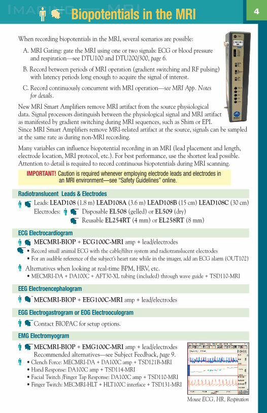
4Imaging — MRIBiopotentials in the MRI
Mouse ECG, HR, Respiration

www.biopac.com
5
Airflow & Gas Analysis Gating Systems Microvascular FlowBiopotentials — page 4 Respiration Stimulation — page 10Blood Pressure
Airflow & Gas AnalysisSetup: TSD137 or TSD237 series + DA100C amp (flow heads from .05 L/sec to 2.6 L/sec)
Transducers consist of a low flow, pneumotach airflow head coupled to a highly sensitive, differential pressure transducer (TSD160A) and connect directly to an airflow cannula and non-rebreathing valve.
TSD137 (heated) or TSD237 (unheated; low thermal inertia)
Important: Contains ferrous material—must be clamped down in the safe MRI operating area.
Gas Analysis Setup: CO2100C amp + 02100C amp + AFT31-MRI gas sampling tubing
Blood PressureArterial Blood Pressure
General arterial pressureSetup: MECMRI-DA + DA100C amp + TSD104A-MRI transducerMicro PressureSetup: MPMS100A control unit + TSD173A/B transducer
Control Unit: MPMS100A-1 one channel, MPMS100A-2 two channelsMRI-compatible Transducers: TSD173A 5 cm fiber/8 m cable or TSD173B 15 cm/8 m
This compact unit is used for a variety of pressure measurements including arterial venous BP,cranial pressure, LVP and RVP. Analog output makes connection with a BIOPAC MP unit easy. Calibration data is stored in the connector.
Small Animal Noninvasive Blood PressureSetup: NIBP200A system + RXCUFSEN9.5/11/13-MRI transducer
The NIBP200A incorporates a built-in pumpthat automatically inflates the blood pressurecuff to occlude the vessel. Once the pumpreaches the inflation point it slowly deflates the cuff, providing a linear drop inpressure. A single pushbutton controls both the inflation and deflation cycles,making the system very operator friendly. MRI-compatible fiber optic cuff/sensor transducers havean 8 m cable and fit 9.5, 11, or 13 mm tail diameters (approx. animal size 100 g -350 g).
Magnetic ResonanceAnimal Physiology in the MRI
The Micro Pressure MeasurementSystem consists of a Control Unit and a Micro Pressure Transducer,purchased separately.
Ultra-miniature fiber opticpressure sensor — no biggerthan a grain of salt on a hair!

www.biopac.com
Gating UnitsThe gating unit is placed in the control room with the MP system and amplifiers.
Dual-Channel GatingSetup: DTU200 Gating system
• Cardio/Respiratory System (DTU200 ) - New dual channel gating unit for small animal cardiac gating. The unit simultaneously monitors two physiological signals—ECG or blood pressure plus respiration—and provides amplification and signal conditioning. A TTL MRI trigger is output for a predetermined number of heart beats after the respiration cycle. The MRI trigger is coincident with each heartbeat and incorporates blanking to remove MRI artifact to prevent false triggering. The MRI is triggered during the animal’s quiet time, which minimizes movement and maximizes image quality. A variety of output signals and conditions can be monitored during the experiment.
Digital TriggerSetup: HLT100C interface module + DTU100 Trigger
• Single channel gating unit (DTU100) - provides a TTL trigger pulse from any physiological signal. The system is usually used with either ECG, blood pressure, or respiration signals. See page 7 for more information.
Microvascular/Laser Doppler FlowSetup: LDF100C amp + TSD147AL probe (1 m) + TSD148 driver (2 m)
For acute preparations inside the MRI, use the LDF100C laser Doppler tissue perfusion monitorto measure microvascular blood flow in tissue. The LDF100C amplifier delivers a low powerbeam of laser light down an optical fiber to the tissue being studied; typically, the volume of tissue sampled by the light is in the order of 1mm3.
RespirationSetup: DA100C amp + TSD110-MRI transducer/sensor/tubing
For high-quality respiration signals, place the anesthetized animal on the sensor pad and runthe tubing through the wave guide to attach to the pressure transducer on the DA100C amp.
TemperatureStand-alone Fiber-Optic Temperature SystemSetup: FOTS100 + TSD180 fiber-optic temperature transducer. See page 10 for details.
Recommended for rectal temperature, due to size.
Temperature AmplifierSetup: MECMRI-TRANS + SKT100C amp + TSD202A or E surface transducer
The SKT100C amplifier module measures surface, core, or air temperature with resolution upto 0.0001°C.
6Imaging — MRIAnimal Physiology in the MRI

Airflow & Gas Analysis Noninvasive Blood Pressure Stimulation — page 10Biopotentials — page 4 Pulse Subject FeedbackElectrodermal Activity — EDA (GSR) Respiration TemperatureGating Units
Airflow & Gas AnalysisAirflow & Lung VolumeSetup: MECMRI-DA + DA100C amp + TSD117-MRI + AFT11A coupler + AFT7-L tubing +
AFT25 mask with valve
Use the Pneumotach Airflow Transducer (TSD117-MRI) to perform a variety of tests relating toairflow and lung volume. Place the TSD117-MRI outside the bore in the MRI Chamber Roomand connect AFT7-L tubing to reach the subject. Medium flow range ± 300 L/min.
Accessories:° Interface TSD117-MRI transducer to AFT7-L tubing: AFT11A coupler° Extend tubing: AFT7-L (3 m) tubing + AFT11D coupler° Facemask with non-rebreathing T-valve: AFT25
Gas Analysis Setup: CO2100C amp + 02100C amp + AFT31-MRI tubing
Electrodermal Activity
Setup: MECMRI-TRANS + EDA100C-MRI amplifier + lead/electrodes
Record EDA inside the MRI. Use AcqKnowledge software filters to improve the quality of theEDA signal, if required, and provide automated analysis.
Electrodes• Disposable Electrodes: BIOPAC recommends EL509 dry disposable
electrodes with GEL101 and LEAD108 (1.8 m) or LEAD108A (3.6 m) for excellent EDA responses.
• Reusable Electrodes: Disposable electrodes are recommended, but reusable TSD203 electrodes will also work for skin conductance.
Gating UnitsTrigger / R-Wave Sync DTU100Setup: HLT100C interface module + DTU100 Trigger
Trigger an MRI System with the occurrence of the R-wave present in ECG, respiratory data orblood pressure for gating purposes. This external hardware unit can accept data from any pulsatileanalog output associated with an MP System and convert that analog signal into a TTL-compatibletrigger to trigger an MRI.
• The timing resolution of the trigger is excellent because it is controlled solely by the real time analog reference signal and is therefore independent of the computer’s operating system and associated communication delays.
Cardio/Respiratory Gating DTU300Setup: DTU300 Gating System
Use the DTU300 to trigger the MRI on the basis of two physiological signals (such as ECG or BPplus respiration). See page 6 for details.
7 Human PhysiologyHuman Physiology in the MRI
EDA data

www.biopac.com
8in the MRIHuman Physiology in the MRINoninvasive Blood Pressure - Wireless Monitoring SystemSetup: HLT100C interface module + NIBP-MRI
The new NIBP-MRI provides real-time, beat-to-beat pressuremeasurement values during magnetic resonance imaging. Thesystem tracks systolic and diastolic blood pressure (using Pulse-Decomposition Analysis technology). The device analyzes thetiming and amplitudes of the primary left ventricular ejectionpulse as well as the arterial pulse reflections, at the middle phalange of the middle finger, at the wrist, or on the upper arm.
PulsePulse - Photo PlethysmographSetup: MECMRI-TRANS + PPG100C-MRI amp + TSD200-MRI transducerThe TSD200-MRI transducer is sensitive to Blood Volume Pulse (BVP) via photo-plethysmo-graphic methods. Use it to record the blood volume pulse pressure waveform. It's primarilydesigned for finger or toe attachment, but can be taped to other body locations with TAPE1.
Pulse - Pressure PadSetup: DA100C amp + AFT30-XL tubing (included) through
wave guide + TSD110-MRI transducer
The TSD110-MRI consists of a differential pressure transducer(TSD160A), sensor (RX110), and tubing (AFT30-XL). Use it to record pulse and pulse rate—it requires no electrical connections between MRI control and chamber rooms andworks on a number of body locations. Affix to finger or majorpulse point with TAPE1.
RespirationRespiratory Effort Transducer — Recommended methodSetup: MECMRI-TRANS + RSP100C amp + TSD201
transducer
The respiratory effort transducer measures changes in thoracicor abdominal circumference that occur as the subject breathes.The transducer comes with an adjustable Velcro strap to fit awide range of subjects.
Pressure Pad / Respiration TransducerSetup: DA100C amp + TSD110-MRI transducer
Place the TSD110-MRI under a strap that is wrapped around the subject’s chest, or tape thetransducer directly to the chest to record respiration. See Pulse above for TSD110-MRI details.
Pulse data
NIBP-MRI data
Respiration data
Subject Feedback
BIOPAC offers a range of subject feedback devices for use inside the MRI.
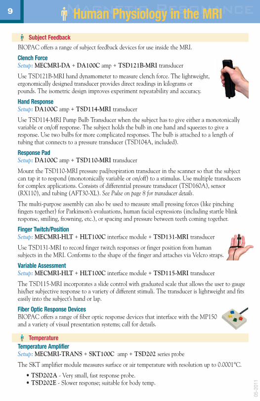
Clench ForceSetup: MECMRI-DA + DA100C amp + TSD121B-MRI transducer
Use TSD121B-MRI hand dynamometer to measure clench force. The lightweight,ergonomically designed transducer provides direct readings in kilograms orpounds. The isometric design improves experiment repeatability and accuracy.
Hand ResponseSetup: DA100C amp + TSD114-MRI transducer
Use TSD114-MRI Pump Bulb Transducer when the subject has to give either a monotonicallyvariable or on/off response. The subject holds the bulb in one hand and squeezes to give aresponse. Use two bulbs for more complicated responses. The bulb is attached to a length of tubing that connects to a pressure transducer (TSD104A, included).
Response PadSetup: DA100C amp + TSD110-MRI transducer
Mount the TSD110-MRI pressure pad/respiration transducer in the scanner so that the subjectcan tap it to respond (monotonically variable or on/off) to a stimulus. Use multiple transducersfor complex applications. Consists of differential pressure transducer (TSD160A), sensor(RX110), and tubing (AFT30-XL). See Pulse on page 8 for transducer details.
The multi-purpose assembly can also be used to measure small pressing forces (like pinching fingers together) for Parkinson’s evaluations, human facial expressions (including startle blinkresponse, smiling, frowning, etc.), or spacing and pressure between teeth coming together.
Finger Twitch/PositionSetup: MECMRI-HLT + HLT100C interface module + TSD131-MRI transducer
Use TSD131-MRI to record finger twitch responses or finger position from human subjects in the MRI. Conforms to the shape of the finger and attaches via Velcro straps.
Variable AssessmentSetup: MECMRI-HLT + HLT100C interface module + TSD115-MRI transducer
The TSD115-MRI incorporates a slide control with graduated scale that allows the user to gaugehis/her subjective response to a variety of different stimuli. The transducer is lightweight and fitseasily into the subject’s hand or lap.
Fiber Optic Response DevicesBIOPAC offers a range of fiber optic response devices that interface with the MP150and a variety of visual presentation systems; call for details.
TemperatureTemperature AmplifierSetup: MECMRI-TRANS + SKT100C amp + TSD202 series probe
The SKT amplifier module measures surface or air temperature with resolution up to 0.0001°C.
• TSD202A - Very small, fast response probe.• TSD202E - Slower response; suitable for body temp.
9 Magnetic ResonanceHuman Physiology in the MRI
05-2
011

0.42 mm
www.biopac.com
Stand-alone Fiber-Optic Temperature System
Setup: FOTS100 + TSD180 or TSD181 FO temperature probe
To interface with UIM100C, add RCA-3.5 mm cable (CBL101, not included).
System uses advanced technology with 62.5 µm core fiber and 50 Hz sampling rate. Excellent system and sensor linearity and accuracy for long-term reliability andrepeatability. Not photonic intensity based. No local heating due to fiber construction.
• FO Rectal temp probe (0.42 mm dia., 8 m): TSD180• FO Surface temp probe (3 mm dia., 8 m): TSD181
STIMULATIONFor Comprehensive Safety Guidelines, see “Safe Use of Electrical Stimulators” online.
Stimulation—Constant Current/Constant VoltageSetup: MECMRI-STMISO + STMISOC/D/E stim isolation
adapter + STM100C stimulator + lead/electrodesUse the stimulator to deliver a variety of electrical stimulation paradigms. AcqKnowledge software provides single pulse, pulsetrains, and arbitrary waveform output options.
Stimulation—Unipolar Wide PulseSetup: MECMRI-STMISO + CBL207 + STM200 stimulator + lead/electrodes
Use the stimulator for any preparation or subject, including pain and stress studies that require lower voltages and wider pulse widths.Trigger the stimulator from the MP150 or a visual presentation system (see below). Use for high-energy nerve or muscle stimulation.
Stimulation ElectrodesUse disposable or reusable electrodes for subject stimulation.
• Disposable: EL509 dry electrodes plus GEL104 + LEAD108 electrode leads• Reusable: EL254RT/258RT plus GEL104 + ADD200 collars• Gel: GEL104 salt-free and chloride-free electrically conductive gel
Visual PresentationSuperLabSetup: STP100W Stimulus Presentation System
The STP100W can present visual stimuli or auditory stimuli, and simultaneously (1ms resolution) send trigger signals to an MP150 for data synchronization and collection purposes.Optional: STIMTRACKER universal marker interface, provides digital trigger info from
SuperLab
E-Prime, DirectRT, MediaLab, Presentation, etc.Setup: STP100C isolated digital interface with CBL110C
Connect to the computer’s parallel printer port to send digital I/O info.
10Imaging — MRIStimulation in the MRI
3 mm

High
-Qua
lity
Phys
iolo
gica
lDa
tafo
rM
RISt
udie
s
•M
RISm
artA
mpl
ifier
s•
Biop
oten
tials
•Ai
rflow
&Ga
sAn
alys
is•
Bloo
dPr
essu
re•
Diffe
rent
ialP
ress
ure
•El
ectro
derm
alAc
tivity
•Ga
ting/
Trig
gerU
nits
•Fo
rce
•La
serD
oppl
erFl
ow•
Mic
roPr
essu
re•
Noni
nvas
ive
Bloo
dPr
essu
re•
Resp
iratio
n•
Stim
ulat
ion
•Su
bjec
tFee
dbac
k•
Tem
pera
ture
•Vi
sual
Pres
enta
tion
Reg
iste
red
toIS
O90
01:2
008
42A
ero
Cam
ino
Gol
eta,
CA
9311
7