foundations ekg i unit 2 summary ischemia mimics · unit 2 summary -ischemia mimics ... bundle or...
TRANSCRIPT

Although STEMI diagnosis is critical not all ST elevation represents STEMI. There are many possible harms
of a false positive STEMI activation or transfer including but certainly not limited to the risks of receiving
anti-platelet agents or even tPA in the setting of a transfer, complications of angiography (bleeding,
dissection, etc), as well as operational issues of the cath lab team being activated overnight or cancelling a
scheduled case.
An EKG with a STEMI pattern must be considered in the context of the patient’s past medical history, HPI,
and exam. This summary will provide an overview of common STEMI mimics including left ventricular
aneurysm hyperkalemia, pericarditis, Brugada syndrome, elevated intracranial pressure, and left ventricular
hypertrophy.
LV Aneurysm is defined by EKG criteria as ST elevation that persists for more than 2 weeks after an MI. It is
important to note that it is considered normal for ST elevation to slowly resolve after MI and it may not
immediately return to normal. Distinguishing LV aneurysm from STEMI can be difficult however typically
there are clear Q waves and a lack of hyperacute T waves. Additionally, the patient should not have pain or
active anginal equivalent symptoms. LITFL Review
Foundations EKG I
Unit 2 Summary - Ischemia Mimics

Brugada syndrome is the result of an inherited or
spontaneous mutation in cardiac sodium channels that
predisposes patients to ventricular tachyarrhythmias
and sudden cardiac death. To be diagnosed with the
syndrome, one must have ECG features and symptoms
consistent with ventricular tachyarrhythmia (syncope,
palpitations, etc). LITFL Review
The image above is an example of Type 1 Brugada
with coved ST-elevation in V1-2 sloping into an
inverted T-wave.
Large and particularly rapid increase in intracranial pressure (a
good example would be a large subarachnoid hemorrhage) can
cause EKG changes that are often confused with ischemia.
Most frequently the changes are deep T wave inversions (aka
cerebral T waves) however sometimes there can be associated
ST elevation and depression. The history and exam are critical
in this situation because normal therapies for ischemia like an-
tiplatelet agents could be devastating if there is a concurrent
intracranial hemorrhage. As an example it is atypical for a
STEMI patient to be altered or obtunded which can be a clue
that there is another or concurrent process. The image above
is an example of cerebral T waves. LITFL Review
Courtesy of Edward Burns of Life in the Fast Lane
Creative Commons License
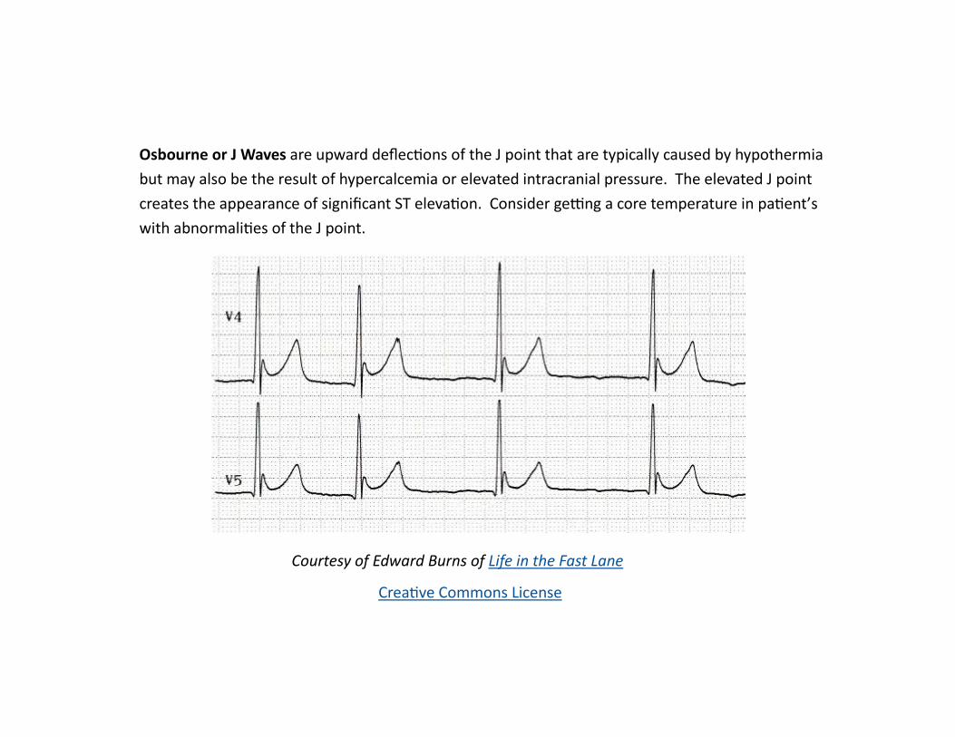
Osbourne or J Waves are upward deflections of the J point that are typically caused by hypothermia
but may also be the result of hypercalcemia or elevated intracranial pressure. The elevated J point
creates the appearance of significant ST elevation. Consider getting a core temperature in patient’s
with abnormalities of the J point.

Courtesy of Edward Burns of Life in the Fast Lane Creative Commons License
Left Ventricular Hypertrophy is frequently the result of untreated hypertension and aortic valve dysfunc-
tion. It causes characteristic EKG abnormalities including massive QRS amplitudes (typically large S waves
in V1-3, III, aVR and large R waves in V4-6, I, and aVL) as well as prolonged QRS duration and ST segment
changes like ST elevation and ST depression. LITFL Review

Courtesy of Edward Burns of Life in the Fast Lane Creative Commons License
Hyperkalemia is a common electrolyte derangement that can cause significant changes to a patient’s
EKG. Classically hyperkalemia progressively leads to peaking T waves, PR prolongation, loss of P waves,
QRS widening, bundle or fascicular block morphology, sine wave, and ventricular dysrhythmia or asysto-
le as the potassium rises. The bizarre appearance of the EKG in hyperkalemia can be easily confused
with STEMI and it is important to keep hyperkalemia in your differential particularly in patients with
known renal dysfunction/failure, patients undergoing chemotherapy, patients using ACE inhibitors or
potassium sparing diuretics, and all patients receiving potassium supplementation. LITFL Review

aVR
Pericarditis frequently causes diffuse ST elevation that can be confused with STEMI however it is im-
portant to recognize that the ST elevation of pericarditis should also be associated with PR depres-
sion (except in aVR which has PR elevation) and NOT be associated with ST depression in leads other
than aVR and V1. Patients with pericarditis often have pain that improves when sitting forward and
is exacerbated by laying back. The clinical history and exam is very important with this diagnosis as
well as it can sometimes be difficult or impossible to distinguish pericarditis and STEMI. The EKG
above is an example of diffuse STE and STD in aVR/V1 with diffuse PR depression and PR elevation in
aVR and V1. LITFL Review
Created by William Burns, MD Edited by Nick Hartman, MD & Kristen Grabow Moore, MD MEd
I
aVR
V1 V4
aVL II V2 V5
aVF III V3 V6