(from the department of genetics, carnegie institutio~ of
TRANSCRIPT

T H E 1NTRACELLULAR GROWTH OF BACTERIOPHAGES
I. LIBERATION OF INTRACELLULAR ]~ACTERIOPHAGE T4 BY PREMATURE LYSIS WITH ANOTHER PHAGE OR WITH CYANIDE
BY A. H. DOERMANN*, $
(From the Department of Genetics, Carnegie Institutio~ of Washington, Cold Spring Harbor)
(Received for publication, August 20, 1951)
Direct studies of bacteriophage reproduction have been handicapped by the fact that the cell wall of the infected bacterium presents a closed door to the investigator in the period between infection and lysis. As a result it was im- possible to demonstrate the presence of intracellular phage particles during this so called latent period, and, much less, to estimate their number or to describe them genetically. This barrier has now been penetrated. I t is the purpose of the first two papers of this series to describe two methods for disrupting infected bacteria in such a way that the intracellular phage particles can be counted and their genetic constitution analyzed.
The first method used to liberate intracellular bacteriophage depends on the induction of premature lysis in infected bacteria by "lysis from without" which occurs when a large excess of phage particles is adsorbed on bacteria (1). I t was found by nephelometric tests that T6 lysates are efficient in disrupting cells when moderately high multiplicities are used (2). The further observation was made that the addition of a large number of T6 particles to bacteria previ- ously infected with T4, would, under some conditions, cause liberation of T4 particles before the expiration of the normal latent period of these cells. I t therefore seemed hopeful that a method of reproducibly disrupting infected bacteria could be developed on the basis of this preliminary knowledge.
* The experiments described here were carried out while the author was a fellow of the Carnegie Institution of Washington. The author is indebted to Dr. M. Demerec and the staff of the Department of Genetics of the Carnegie Institution of Washington for providing facilities for this work. In particular the stimulating discussions with Dr. Barbara McClintock are gratefully acknowledged. The manuscript was prepared while the author held a fellowship in the Department of Biology of the California Institute of Technology. He is grateful to Dr. G. W. Beadle and the staff of that de- partment for their interest in this work, and especially to Dr. Max Delbriick for criticism of the manuscript.
:~ Present address: Biology Division, Oak Ridge National Laboratory, Oak Ridge, Tennessee.
645
The Journal of General Physiology
646 INTRACELLULAR GROWTH OY BACTERIOPHAGES. I
The first experiments in devising a method of this kind were made with phage T5 (3). I t was found that T5 is liberated before the end of the latent period if the infected cells are exposed to a high excess of T6. However, the extremely low rate of adsorption of T5 coupled with difficulties in inactivation of unadsorbed phage by specific antisera indicated that this phage was a poor choice. Hence T4 was chosen because of its fast rate of adsorption and because of the availability of high titer antisera against it. The first experiments with T4, along with the T5 results, showed conclusively that, by itself, lysis from without is not sufficiently rapid for the purpose of this investigation. I t is likely that phage growth continues after the addition of the lysing agent T6. There- fore the a t tempt was made to stop phage growth while T6 was allowed to ac- complish lysis from without. Low temperature could not be used for this purpose
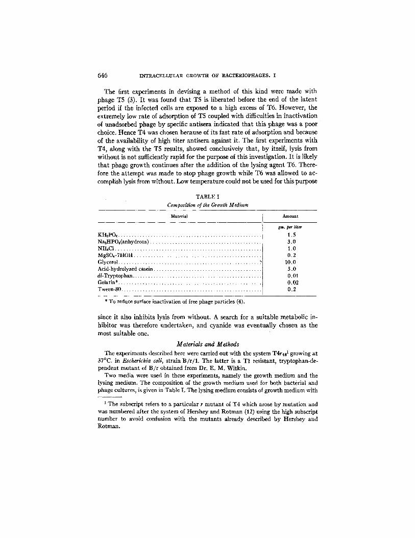
TABLE I Composition of the Growth Medium
Material Amount
gin. p~r l/Ur KH2PO, . . . . . . . . . . . . . . . . . . . . . . . . . . . . . . . . . . . . . . . . . . . . . . . . . . . . . 1.5 N~HPO4(anhydrous) . . . . . . . . . . . . . . . . . . . . . . . . . . . . . . . . . . . . . . . . . 3.0 NHaC1 . . . . . . . . . . . . . . . . . . . . . . . . . . . . . . . . . . . . . . . . . . . . . . . . . . . . . 1.0 MgSO~. 7HOH . . . . . . . . . . . . . . . . . . . . . . . . . . . . . . . . . . . . . . . . . . . . . . 0.2 Glycerol . . . . . . . . . . . . . . . . . . . . . . . . . . . . . . . . . . . . . . . . . . . . . . . . . . . . 10.0 Acid-hydrolyzed casein . . . . . . . . . . . . . . . . . . . . . . . . . . . . . . . . . . . . . . . 5.0 dl-Tryptophan . . . . . . . . . . . . . . . . . . . . . . . . . . . . . . . . . . . . . . . . . . . . . . . . ] O. 01 Gelatin* . . . . . . . . . . . . . . . . . . . . . . . . . . . . . . . . . . . . . . . . . . . . . . . . . . . . . 0.02 Tween-80 . . . . . . . . . . . . . . . . . . . . . . . . . . . . . . . . . . . . . . . . . . . . . . . . . . . . 0.2
* To reduce surface inactivation of free phage particles (4).
since it also inhibits lysis from without. A search for a suitable metabolic in- hibitor was therefore undertaken, and cyanide was eventually chosen as the most suitable one.
Materials and Methods
The experiments described here were carried out with the system T4r4s ~ growing at 37°C. in Escherichia coli, strain B/r/1. The latter is a T1 resistant, tryptophan-de- pendent mutant of B/r obtained from Dr. E. M. Witkin.
Two media were used in these experiments, namely the growth medium and the lysing medium. The composition of the growth medium used for both bacterial and phage cultures, is given in Table I. The lysing medium consists of growth medium with
1 The subscript refers to a particular r mutant of T4 which arose by mutation and was numbered after the system of Hershey and Rotman (12) using the high subscript number to avoid confusion with the mutants already described by Hershey and Rotman.

A. ~. DOEP.MAN~ 647
the addition of one part in ten of a high titer T6 phage filtrate (concentration of T6 in lysing medium was ca. 4 X 10⁹ particles per ml.) and cyanide brought to a final concentration of 0.01 ~. Specially designed experiments showed that at this concentra- tion the cyanide does not inactivate free phage particles, nor does the amount which reaches the plate affect titration by interference with plaque development.
T6 was used as the lysing phage because in several experiments it proved to be a more effective lysing agent than any of the other T phages tested. Since only single stocks of the phages were compared in the early experiments, the superiority of T6 over the other phages may have been due to a difference in the particular stock used, and not to an inherent difference among the phages. In fact, later experiments with different T6 stocks showed marked differences in lysing efficiency, and phage titer proved to be a poor criterion of lysing ability. The experiments described here were made with T6 stocks selected for their ability to induce lysis from without. The se- lections were made on the basis of nephelometric comparisons.
Platings were made in agar layer (0.7 per cent agar) poured over nutrient agar plates (1.3 per cent agar), and in order to assay T4 in the presence of high titer T6, the in- dicator strain, B/6, was used. B/6 is completely resistint to T6 (no host range mutants have so far been found which will lyse the strain used here) and gives full efficiency of plating (compared to B) with T4.
EXPERIMENTAL
Experiments with the Standard Lysing Medium.--The experimental procedure used consisted essentially of a one-step growth experiment (5) with certain modifications. B / r / l cells in the exponential growth phase were concentrated by centrlfugation to about 109 cells per ml. To these concentrated bacteria T4r4s was added and this ad- sorption mixture was incubated for 1 to 2 minutes with aeration, allowing at least 80 per cent of the phage to be adsorbed to the bacteria. Then a 40-fold or larger dilution was made into growth medium containing anti-T4 rabbit Serum. The serum inactivated most of the residual unadsorbed phage. Mter several minutes' incubation in the serum tube, a further dilution was made to reduce the serum concentration to one of relative inactivity, The resulting culture will be referred to as the source culture (SC). The entire experiment was carried out with the infected bacteria from SC. The titer of infected B / r / l in this tube was approximately 10 ~ cells per nil.
Simultaneously with the dilution into the tube containing serum, another dilution from the adsorption tube was made. From the latter an estimation of the unadsorbed phage was made by assaying the supematant after sedimentation of the cells. This step permits calculation of the multiplicity of infection (5).
From SC a further dilution of 1:20 was made at some time before the end of the latent period. The resulting culture, containing approximately 5 X 10³ infected bac- teria per ml., was used for determining the normal end of the latent period and for estimating the average yield of phage per infected cell. I t will be called the control growth tube (GT). In addition, a number of precisely timed 20-fold dilutions were made from SC into lysing medium. These were titrated after they had been incubated in the lysing medium for 30 minutes or longer. Serial platings from the lysing medium cultures over a longer period of time have shown that the phage titer remained constant after 30 minutes' incubation. The titer calculated from these platings, divided by the

648 INTRACELLULAR GROWTH O1 ~" BACTERIOPHAGES. I
titer of infected bacteria given by the preburst control platings, gives the average yield per infected cell. As a working hypothesis, this yield was considered to be the average number of intraceUular phage particles per bacterium at the time of dilution into the lysing medium. Dividing these numbers by the control burst size gives the fraction of the control yield found in the experimental lysing medium tubes.
The results of several typical experiments are shown in Fig. 1 in which the data are plotted on semilogarithmic coordinates. The fraction of the control yield found in a given experimental culture is plotted against the time at which the dilution is made into lysing medium. Curve i shows the results from a single experiment in which the bacteria were infected with an average of 7 phage particles each. Curve 2 is the composite result of four experiments in which the bacteria were infected with single phage particles. Curve 3 is the control one- step growth curve derived from the control growth tube platings in the four experiments of curve 2.
Several striking results can be seen in these experiments. First, it is clearly seen that during the early stages of the latent period the virus-host complex is inactivated by the cyanide-T6 mixture, and that not even the infecting particles are recovered. Even when 7 phage particles were adsorbed on each bacterium, less than two are recovered per cell at the earliest stage tested, and the shape of the curve suggests that if earlier stages had been tested, still fewer would have been recovered. In experiments with singly infected bacteria, the earliest tests indicated that less than one infected bacterium in 80 liberated any phage at all. A second point to be noted is that the multiplicity of infection appears to influence slightly the time at which phage particles can be recovered from the cell, and it continues to affect the fraction found in the bacteria at a given time. That this difference is a real one seems clear from the consistency among the points of curve 2. This result has been observed in each experiment, although the effect appeared to be less pronounced in some experiments made at the lower temperature of 30°C. Attention should also be drawn to the fact that the shape of the curves is clearly not exponential. In fact, it parallels with a delay of several minutes the approximately linear DNA increase observed in this system (6).
In connection with the preceding experiments a test was made to establish whether the cyanide concentration chosen was maximally effective in inhibiting phage synthesis. Using the described technique, but changing the cyanide con- centration of the lysing medium from 0.01 ~ to 0.004 ~ and 0.001 M in parallel aliquots, no difference was detectable in the three lysing media. Thus 0.01 M cyanide is well beyond the minimum concentration necessary and was con- sidered to be adequate for these experiments.
Action of Cyanide in the Absence of the Lysing Agent T6.--Cohen and Anderson (7) reported a loss of infectious centers when infected bacteria were incubated in the presence of the antimetabolite 5-methyltryptophan. Although the details

A. H. DOE~ANN 649
I
I I I I I
I I I I I
<3
\ ",,._ o~< o \ .,,.,, i
% e '~ , . , o <3 n
I I
a ~
~ v
- . . . .
u~ , ~ . ~ ~...=
0D
. ~ . ~
~ 0
~.a ~
~ ~.~ =~.~
e~
~ . ~
~ ~.'~ o - o
3 A ~ l n O HJ.MO~IO - IO~I .LNO0 NI O131) , - IVN I - I .-IO J . N 3 0 ~t3¢J

650 I N T R A C E L L U L A R G R O W T H O F B A C T E R I O P H A G E S . I
of their experiments differed somewhat from those presented here, the loss of infectious centers in their experiments suggested testing whether cyanide could cause a similar loss of infected bacteria in the present procedure. An experi- ment was made which was identical with the standard cyanide lysis experiment
IO0 "
o~ .i-
o
t9
Z O
t.O- z_
w_ >-
°t ~0.1
0.0:
AT 37" / ¢ / , O
/ - - - " -- ~ / ~ ; 7 7 . . . . . . INFECTED BACTERIA
-J O ' - C N - O - - C N - + 1"6
, , ; o . . . . ; . . . . . . . . . . . . I I 20 25 MINUTES OF INCUBATION IN GROWTH MEDIUM AT 57"
FIG. 2. The comparative effect of cyanide alone and cyanide plus T6 on singly infected bacteria at various stages in the latent period.
except that T6 was omitted from one set and included in a parallel set of lysing medium cultures (Fig. 2). As in the case of 5-methyltryptophan, it is seen that cyanide alone caused a loss of infectious centers when added in the early stages of phage growth, although the loss is less than that produced by cyanide and T6 together. Furthermore, in the second half of the latent period, comparison of the two media showed clearly and surprisingly that a definite rise in titer of infective centers occurred even when the lysing agent, T6, was omitted from the

A. ~, DOE~I~NN 651
lysing medium. In fact, during the second half of the latent period, phage liberation is identical in the two media.
In order to see whether lysis is actually occurring and can account for t h e liberation of phage, a nephelometric experiment was made introducing CN- at two points in the latent period. Three cultures of B/r /1 growing exponentially in growth medium were infected with T4r48 (ca. fivefold multiplicity). One culture served as a control for normal lysis. To the second culture cyanide (0.01 M final concentration) was added 7.5 minutes after addition of the virus and to
CN-ADDED TO 2 T4_r41AT 37 e
I20 . ~
CN-ADOED TO I ~ ~GONTROL
" ] 0 J n i I n n I I I , . . . . .
0 I0 20 30 40 50 60 70 80 go 290 MINUTES AFTER ADDITION OF PHAGE TO BACTERIAL CULTURES
Fzo. 3. The turbidity of T4-infected bacterial cultures as affected by addition of cyanide at two stages in the latent period.
iX"
t - u. 40 0
the third tube 17.5 minutes after addition of the T4r48. The turbidities of these three cultures were followed with a nephelometer designed like that described previously by Underwood and Doermann (8), but with four separate units which permit independent readings on the four tubes without removing any of them from the instrument. The results indicate that CN- added to infected bacteria early during the latent period does not induce lysis (Fig. 3). From the plaque count experiment (Fig. 2) it is seen that a loss of infective centers does occur. This loss must therefore be due to some cause other than lysis of these cells. In the later stages of the latent period, the turbidimetric experiment indicates that lysis occurs promptly upon the addition of CN- to the culture

652 INTRACELLULAR GROWTH OF BACTERIOPHAGES. I
4 !
4.= I I I I
I
6 ! I
t
I ~%%\
I ~,
( I I o_ -
3 A ~ N 9 HJ.MO~9 " IO~INOO NI 0"1311 7VNI- I -I0 .LN30 ~1~:1
o
03
0
0 0
=.., ~0
° ~

A. H. DOERM-ANN 653
(Fig. 3). The increase of infective centers in comparable cultures (Fig. 2) at these later stages is probably brought about by liberation of phage particles concurrent with this lysis.
Experiments Using 5-Methyltryptophan as the Metabolic Inhibitor.--In trying to find a suitable metabolic inhibitor for instantaneously stopping phage growth, a large number of experiments was done using the antimetabolite 5-methyltryptophan (5MT) 2 whose bacteriostatic action is blocked by trypto- phan (9). The technique used was similar to the cyanide lysis procedure except that tryptophan was omitted from the lysing medium and 5MT was used in place of cyanide. The results (Fig. 4) are quite similar to the cyanide results in all respects except one. They are similar in failure to recover any phage particles during the early stages of the latent period, in the difference between single and multiple infection, and in the shapes of the curves. They are different, however, in that both the single and the multiple infection curves are moved to the left along the time scale by 3 to 4 minutes. This indicates that more phage is liber- ated per cell if lysis is induced in the presence of 5MT than if it is brought about in the presence of CN-. This difference may be interpreted on the basis of two alternative hypotheses.
First, it might be suspected that CN- penetrates t]ae cell and reaches its site of inhibition more quickly than 5MT. This would allow more phage repro- duction to go on between the time of exposure to the 5MT and the time at which the cell breaks ope.n. In this event, a higher concentration of 5MT would enable penetration of an inhibitory amount in a shorter period of time, thus reducing the amount of phage found. To test this, the concentration of 5MT in the lysing medium was increased fivefold. No difference in the amount of phage liberated was found, suggesting that the rate of penetration of the poison is not limiting its effectiveness.
A second hypothesis is that the reaction blocked by 5MT may be one of the earlier ones involved in the synthesis of phage constituents. At the time of addition of 5MT many individual phages may already have acquired these constituents and thus be able to go on to maturity before lysis disperses the enzyme equipment of the infected cell. Cyanide, on the other hand, may block one of the terminal reactions in phage production, with the result that at a given time fewer individuals will have passed this reaction than will have passed the 5MT-inhibitable step. Consequently, fewer particles will be liberated when using cyanide than when 5MT is used.
DISCUSSION
Earlier experiments (2) and tests made of the lysing efficiency of the T6 stocks used here indicate that rapid tysis occurs when T6 stocks are added in
2 Obtained through the courtesy of Dr. M. L. Tainter, Sterling-Winthrop Research Institute, Rensselaer, New York.

654 I N T R A C E L L U L A R G R O W T t t O~F BACTERIOPHAGES. I
sufficient concentration to bacterial cultures. The very first experiments with bacteria infected with T5 (3) left no doubt that lysis from without by T6 will liberate T5 particles prematurely from infected bacteria. From the evidence contained in the present paper it cannot be definitely established whether the combined action of T6 and cyanide liberates all of the mature phage present in the cells. However, the fact that, during the terminal stages of intracellular development, the cyanide-lysis method yields as much phage as does spontane- ous lysis, suggests that the cyanide method liberates all of the mature phage. Furthermore, during the second half of the latent period, exactly the same amount of phage is liberated by cyanide alone as by cyanide plus T6. This sug- gests that cyanide acts promptly in arresting phage growth. Otherwise one would expect to find a consistently higher number of phage particles in the cyanide medium than in the medium in which cyanide and T6 are combined. The experiments presented here therefore warrant the working hypothesis that mature intracellular phage is effectively liberated by the treatment described, and that the method gives a true picture of the intracellular phage population. The validity of this working hypothesis will be conclusively demonstrated for the phage T3 in the second paper of this series (10).
The bearing of the present experiments on our concept of phage reproduction might be discussed here. The finding that the original infecting particles are not recoverable from the cells during the first stages of the latent period appears at first sight surprising. Nevertheless some indirect evidence indicates that this is to be expected. The discovery that yields from mixedly infected bacteria may con- tain new combinations of the genetic material of the infecting types (11-13) suggests that some alteration of the infecting particles may occur. Furthermore, in mixed infections of bacteria with unrelated phages only one type is repro- duced. The other type, although adsorbedon the cells, it not onlyprevented from multiplying but the infecting particle of that type is lost (5, 14). On the basis of multiple infection experiments with ultraviolet-inactivated phage particles, Luria (15) has proposed that reproduction of phage occurs by reproduction of subunits which are at some later stage assembled into complete virus particles. The failure to find infective phage particles within tho infected cell in the early stages of reproduction agrees with what would have been predicted from these experiments.
The results of our experiments agree quite well with the scheme which Latarjet (16) suggested on the basis of x-ray inactivation studies of phage inside infected bacteria. Latarjet differentiated three segments of the latent period of phage growth. Using T2 he found that during the first segment of 6 to 7 minutes' duration, singly infected bacteria show the same inactivation charac- teristics as do unadsorbed phage particles. In the second period, from time 7 to time 13 minutes, the phage in infected cells became more resistant to x-rays, even during the first 2 minutes of this segment in which the inactivation curves

A. H. DOEP~N 655
still retain a single hit character. During the last 4 minutes of this period the curves take on a multiple hit character. In the final segment, from time 13 minutes to the end of the latent period, tile curves retain the multiple hit charac- ter, but gradually regain the original x-ray sensitivity characteristic of free phage. These x-ray experiments suggest again that a rather drastic alteration occurs to the infecting particle, and that particles with the original character- istics are not found in the cell until the second half of the latent period. This is precisely what is observed in the results presented here. Our experiments were done with T4, but comparison seems legitimate since the two viruses are quite closely related (17).
Results of a similar nature to those discussed here were published by Foster (18). In studying the effect of proflavine on the growth of phage T2, Foster found that the time at which this poison was added influenced the amount of phage liberated by the bacteria. No phage is liberated from T2-infected cells (latent period 21 minutes) if proflavine is added during the first 12 minutes after infection even though lysis of the cells does occur at the normal time. When proflavine is added at later points in the latent period, lysis yields phage particles, the number depending on the time of proflavine addition. When the results of these single infection experiments are compared to the cyanide single infection experiments (Fig. 1) the results are seen to be quite similar. From other experiments Foster concluded that proflavine inhibits one of the final stages in the formation of fully infective phage. These facts, taken together, suggests that proflavine experiments were, in fact, measuring intracellular pha~e.
SUMMARY
A method is described for liberating and estimating intracellular bacterio- phage at any stage during the latent period by arresting phage growth and induc- ing premature lysis of the infected cells. This is brought about by placing the infected bacteria into the growth medium supplemented with 0.01 ~ cyanide and with a high titer T6 lysate. I t was found in some of the later experiments that the T6 lysate is essential only during the first half of the latent period. Cyanide alone will induce lysis during the latter part of the latent period.
Using this method on T4-infected bacteria it is found that during the first half of the latent period no phage particles, not even those originally infecting the bacteria, are recovered. This result is in agreement with the gradually emerging concept that a profound alteration of the infecting phage particle takes place before reproduction ensues. During the second half of the latent period mature phage is found to accumulate within the bacteria at a rate which is parallel to the approximately linear increase of intracellular DNA in this system. However, the phage production lags several minutes behind DNA production.

656 INTRACELLULAR GROWTH OF BACTERIOPHAGES. I
When 5-methyltryptophan replaced cyanide as the metabolic inhibitor, similar results were obtained. The curves were, however, displaced several minutes to the left on the time axis.
The results are compared with Latarjet 's (16) data on x-radiation of infected bacteria and with Foster's data (18) concerning the effect of proflavine on in- fected bacteria. Essential agreement with both is apparent.
BIBLIOGRAPHY
1. Delbriick, M., J. Gen. Physiol., 1940, 23,643. 2. Doermann, A. H., J. Bact., 1948, 55, 257. 3. Doermann, A. H., Ann. Rep. Biol. Lab., Long Island Biol. Assn., 1946, 22. 4. Adams, M. H., J. Gen. Physiol., 1948, 31, 417. 5. Delbriick, M., and Luria, S. E., Arch. Biochem., 1942, 1, 111. 6. Cohen, S. S., Bact. Rev., 1949, 13, 1. 7. Cohen, S, S., and Anderson, T. F., J. Exp. Med., 1946, 84, 525. 8. Underwood, N., and Doermann, A. H., Rev. Scient. Instr., 1947, 18, 665. 9. Anderson, T. F., Science, 1945, 101,565.
10. Anderson, T. F., and Doermann, A. H., J. Gen. Physiol., 1952, 35, 657. 11. Delbfiick, M., and Bailey, W. T., Jr., Cold Spring Harbor Syrup. Quant. Biol.,
1946, 11, 33. 12. Hershey, A. D., and Rotman, R., Proc. Nat. Acad. Sc., 1948, 34, 89. 13. Hershey, A. D., and Rotman, R., Genetics, 1949, 34, 44. 14. Delbrtick, M., J. Bact., 1945, 50, 151. 15. Luria, S. E., Proc. Nat. Acad. So., 1947, 33, 253. 16. Latarjet, R., J. Gen. Physiol., 1948, 31,529. 17. Adams, M. H., in Methods in Medical Research, (J. H. Comroe, editor), Chicago,
The Yearbook Publishers, Inc., 1950, 2, 1. 18. Foster, R. A. C., J. Bact., 1948, 56, 795.