function of arches of the foot : they act as a weight bearing. they act as a locomotive part of...
TRANSCRIPT
Function of Arches of Function of Arches of the Foot : the Foot :
They act as a weight bearing.
They act as a locomotive part of the body in walking & running.
They provide space in the sole of foot to contain and protect the muscles, nerves and blood vessels of the sole.
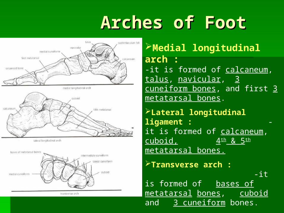
Arches of FootArches of FootMedial longitudinal arch : -it is formed of calcaneum, talus, navicular, 3 cuneiform bones, and first 3 metatarsal bones.
Lateral longitudinal ligament : -it is formed of calcaneum, cuboid, 4th & 5th metatarsal bones.
Transverse arch : -it is formed of bases of metatarsal bones, cuboid and 3 cuneiform bones.

Factors maintaining Factors maintaining arches of Foot :arches of Foot :
Medial longitudinal Medial longitudinal archarch
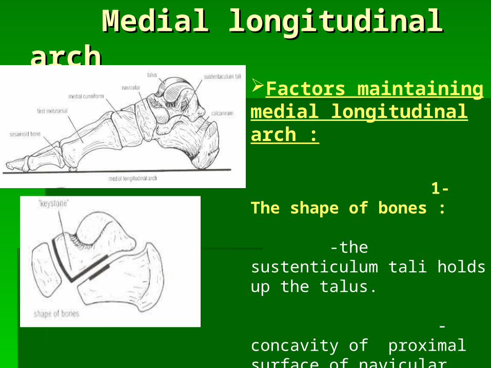
Factors maintaining medial longitudinal arch :
1- The shape of bones : -the sustenticulum tali holds up the talus. -concavity of proximal surface of navicular bone receives the rounded head of talus, which is the keystone in the center of the arch. -concavity of proximal surface of medial cuneiform bone receives the navicular.
Medial longitudinal archMedial longitudinal arch
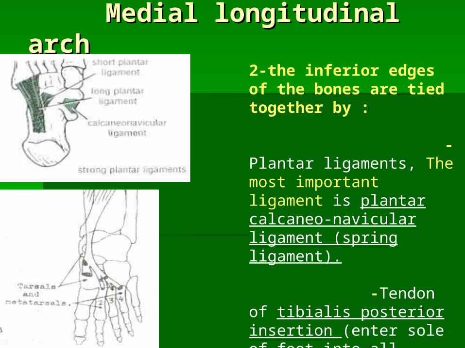
2-the inferior edges of the bones are tied together by : -Plantar ligaments, The most important ligament is plantar calcaneo-navicular ligament (spring ligament). -Tendon of tibialis posterior insertion (enter sole of foot into all tarsal bones except talus + bases of 2,3,4 metatarsal bones).
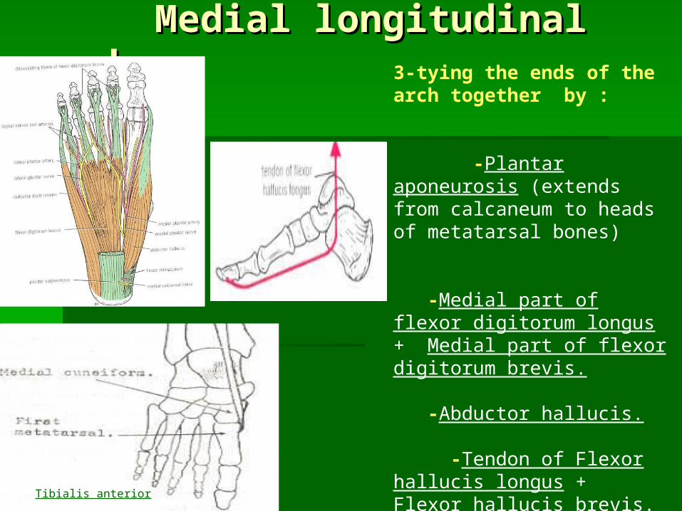
Medial longitudinal archMedial longitudinal arch3-tying the ends of the arch together by : -Plantar aponeurosis (extends from calcaneum to heads of metatarsal bones) -Medial part of flexor digitorum longus + Medial part of flexor digitorum brevis. -Abductor hallucis. -Tendon of Flexor hallucis longus + Flexor hallucis brevis. 4-Suspending the arch from above by : -Tibialis anterior (descends in front of tibia to be inserted into medial sides of medial cuneiform bone + base of 1st metatarsal bone) -Posterior & Medial ligament (deltoid ligament) of ankle joint.
Tibialis anterior
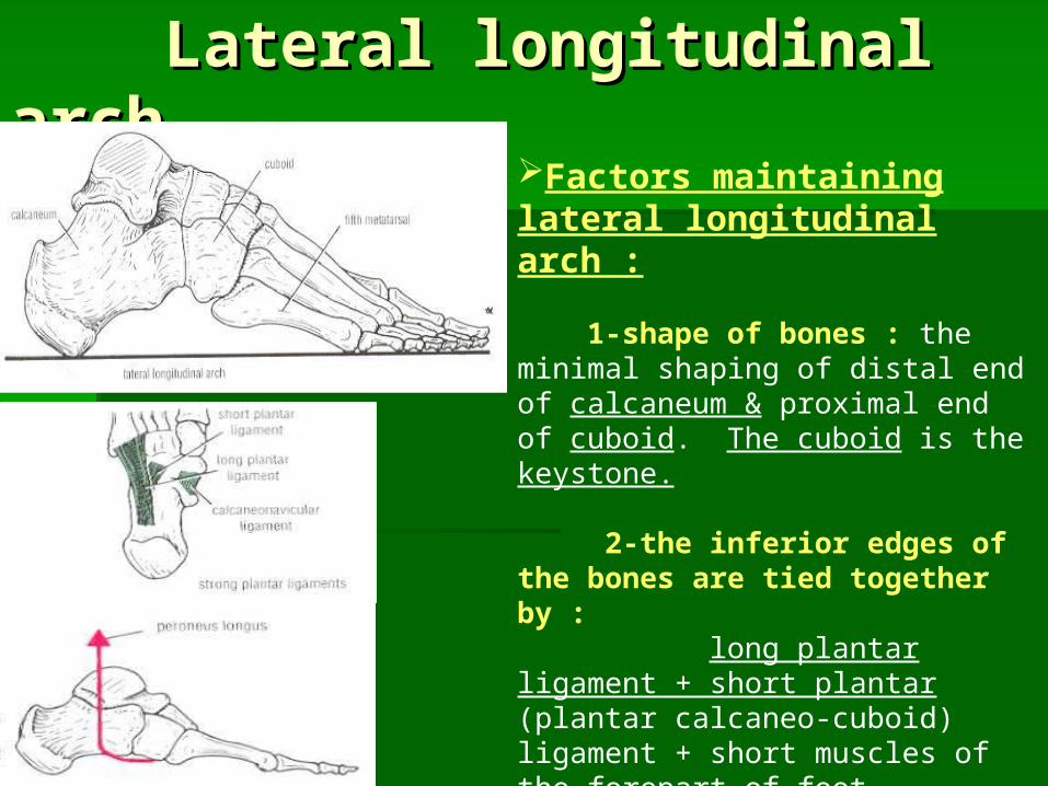
Lateral longitudinal archLateral longitudinal archFactors maintaining lateral longitudinal arch : 1-shape of bones : the minimal shaping of distal end of calcaneum & proximal end of cuboid. The cuboid is the keystone. 2-the inferior edges of the bones are tied together by : long plantar ligament + short plantar (plantar calcaneo-cuboid) ligament + short muscles of the forepart of foot. 3-Tying the ends of the arch together : by plantar aponeurosis + abductor digiti minimi + lateral part of flexor digitorum longus & brevis. 4-Suspending the arch from above : peroneus longus & brevis.

Transverse archTransverse archFactors maintaining transverse arch: 1-Shape of bones : the wedge shape of cuneiform bones + the bases of metatarsal bones. 2-the inferior edges of the bones are tied together by : -Particully, the dorsal interossei + transverse head of adductor hallucis. -Deep interosseus transverse ligaments + long & short plantar ligaments. 3-Tying the ends of the arch together : by peroneus longus tendon. 4-Suspending the arch from above : by the peroneus longus tendon + peroneus brevis.
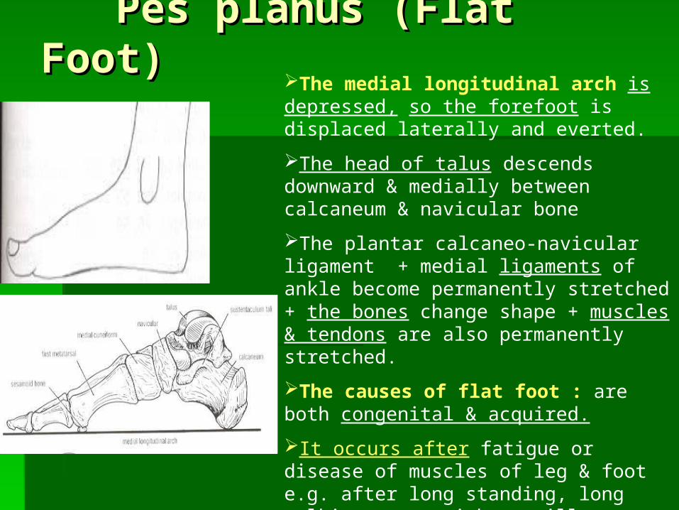
Pes planus (Flat Foot)Pes planus (Flat Foot)The medial longitudinal arch is depressed, so the forefoot is displaced laterally and everted.
The head of talus descends downward & medially between calcaneum & navicular bone
The plantar calcaneo-navicular ligament + medial ligaments of ankle become permanently stretched + the bones change shape + muscles & tendons are also permanently stretched.
The causes of flat foot : are both congenital & acquired.
It occurs after fatigue or disease of muscles of leg & foot e.g. after long standing, long walking, overweight or illness, so the weak muscles & ligaments are stretched and pain is produced after walking for a short distance.
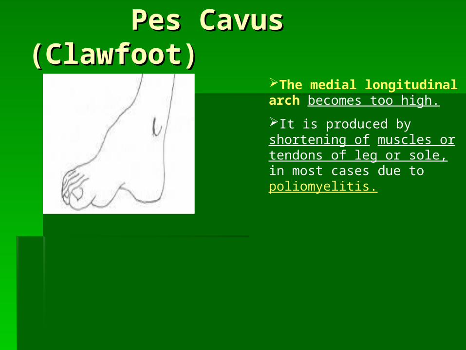
Pes Cavus (Clawfoot)Pes Cavus (Clawfoot)The medial longitudinal arch becomes too high.
It is produced by shortening of muscles or tendons of leg or sole, in most cases due to poliomyelitis.

Veins of the lower limbVeins of the lower limbSuperficial veins : 1-Great & small saphenous veins +their tributaries. -They have numerous valves along its course. -They are situated in superficial fascia, the constant position of great saphenous vein in front of medial malleolus should be remmembered for patients recquiring blood transfusion.
Deep veins : 1-venae comitantes of anterior & posterior tibial arteries. 2-Popliteal vein & Femoral vein + their tributaries. -They have valves to allow blood to pass upwards only .
Perforating veins: -Many of these veins are found in ankle & medial side of lower part of leg. -They connect superficial veins with the deep veins. -They possess valves which allow blood to pass from superficial to deep veins, But prevent passage of blood from deep veins (high blood pressure) to superficial veins (low blood pressure)…. Venous pump
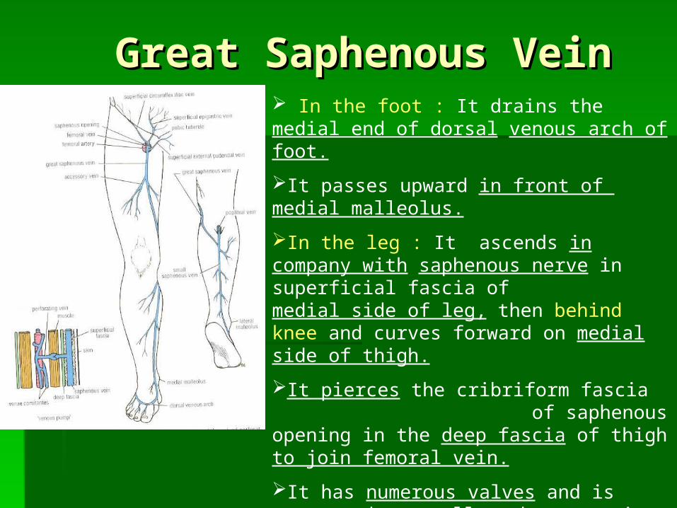
Great Saphenous VeinGreat Saphenous Vein In the foot : It drains the medial end of dorsal venous arch of foot.
It passes upward in front of medial malleolus.
In the leg : It ascends in company with saphenous nerve in superficial fascia of medial side of leg, then behind knee and curves forward on medial side of thigh.
It pierces the cribriform fascia of saphenous opening in the deep fascia of thigh to join femoral vein.
It has numerous valves and is connected to small saphenous vein by anastomotic vein passing behind knee.
It is connected with deep veins via perforating valved veins along medial side of calf.
Varicose VeinsVaricose Veins A varicose vein isA varicose vein is a vein which becomes a vein which becomes
dilated, elongated and tortuous. dilated, elongated and tortuous. It affectsIt affects the the superficial veinssuperficial veins of the lower of the lower
limb.limb. It is producedIt is produced when the when the valves of the valves of the
perforatingperforating veins become incompetentveins become incompetent (so, allow blood to pass from deep veins to (so, allow blood to pass from deep veins to superficial veins).superficial veins).
As a result,As a result, the blood passes from the blood passes from deep veinsdeep veins (high pressure)(high pressure) to to superficial veins (lowsuperficial veins (low pressure),pressure), so the superficial veins become so the superficial veins become dilated, elongated and tortuous.dilated, elongated and tortuous.
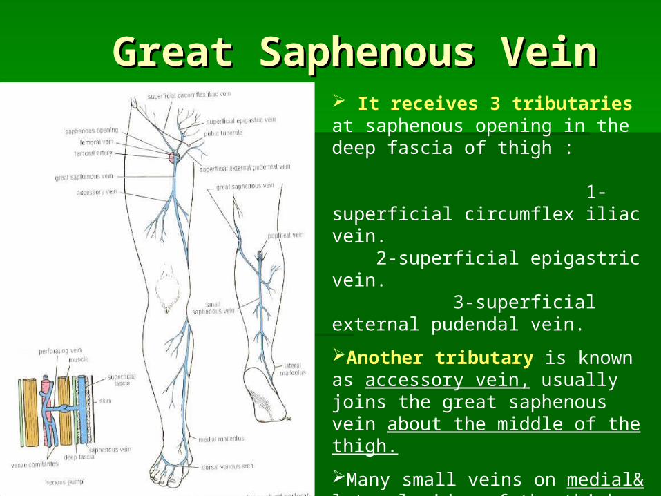
Great Saphenous VeinGreat Saphenous Vein It receives 3 tributaries at saphenous opening in the deep fascia of thigh : 1-superficial circumflex iliac vein. 2-superficial epigastric vein. 3-superficial external pudendal vein.
Another tributary is known as accessory vein, usually joins the great saphenous vein about the middle of the thigh.
Many small veins on medial& lateral sides of the thigh drain into great saphenous vein, but lower part of back of thigh drain into the small saphenous vein in the popliteal fossa.
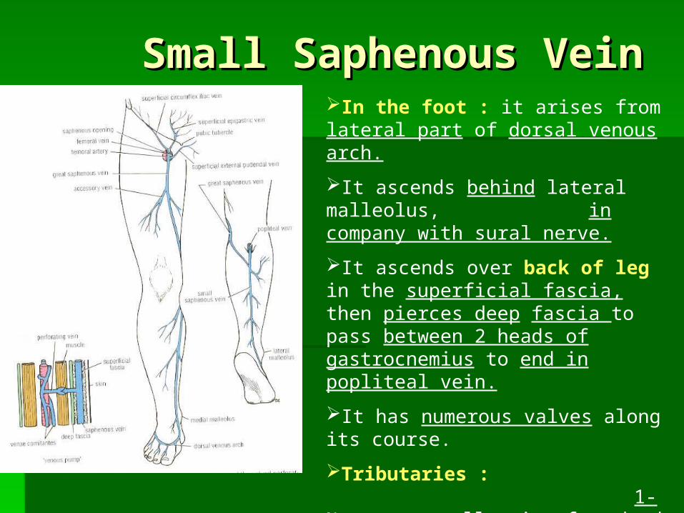
Small Saphenous VeinSmall Saphenous VeinIn the foot : it arises from lateral part of dorsal venous arch.
It ascends behind lateral malleolus, in company with sural nerve.
It ascends over back of leg in the superficial fascia, then pierces deep fascia to pass between 2 heads of gastrocnemius to end in popliteal vein.
It has numerous valves along its course.
Tributaries : 1-Numerous small veins from back of leg. 2-Communicating veins with deep veins of foot. 3-Anastomotic branches that run upward & medially to join great saphenouds vein.
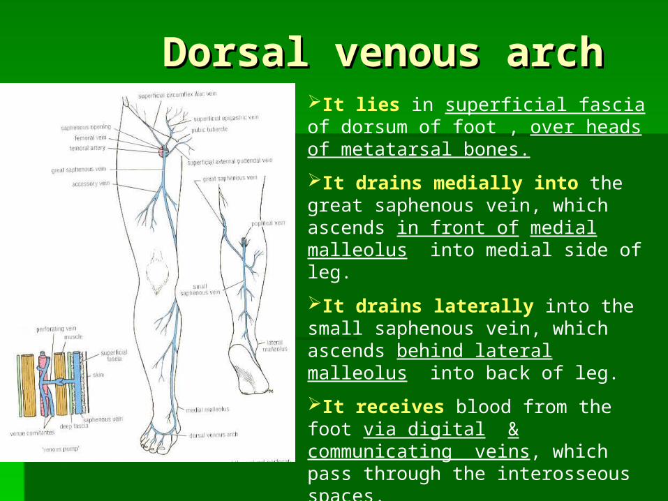
Dorsal venous archDorsal venous archIt lies in superficial fascia of dorsum of foot , over heads of metatarsal bones.
It drains medially into the great saphenous vein, which ascends in front of medial malleolus into medial side of leg.
It drains laterally into the small saphenous vein, which ascends behind lateral malleolus into back of leg.
It receives blood from the foot via digital & communicating veins, which pass through the interosseous spaces.

Popliteal VeinPopliteal VeinIt begins at lower border of popliteus muscle by union of venae comitantes of anterior & posterior tibial arteries.
It passes upwards in the popliteal fossa.
It passes medial to popliteal artery at its lower part, then behind at its middle part, and lateral to the artery at its upper part.
It ends by passing through opening in adductor magnus to become the femoral vein.
Tributaries : 1-Small saphenous vein, which perforates deep fascia to pass between 2 heads of gastrocnemius to end in popliteal vein. 2-Veins that correspond to branches of popliteal artery (5 genicular branches to knee joint + muscular branches)
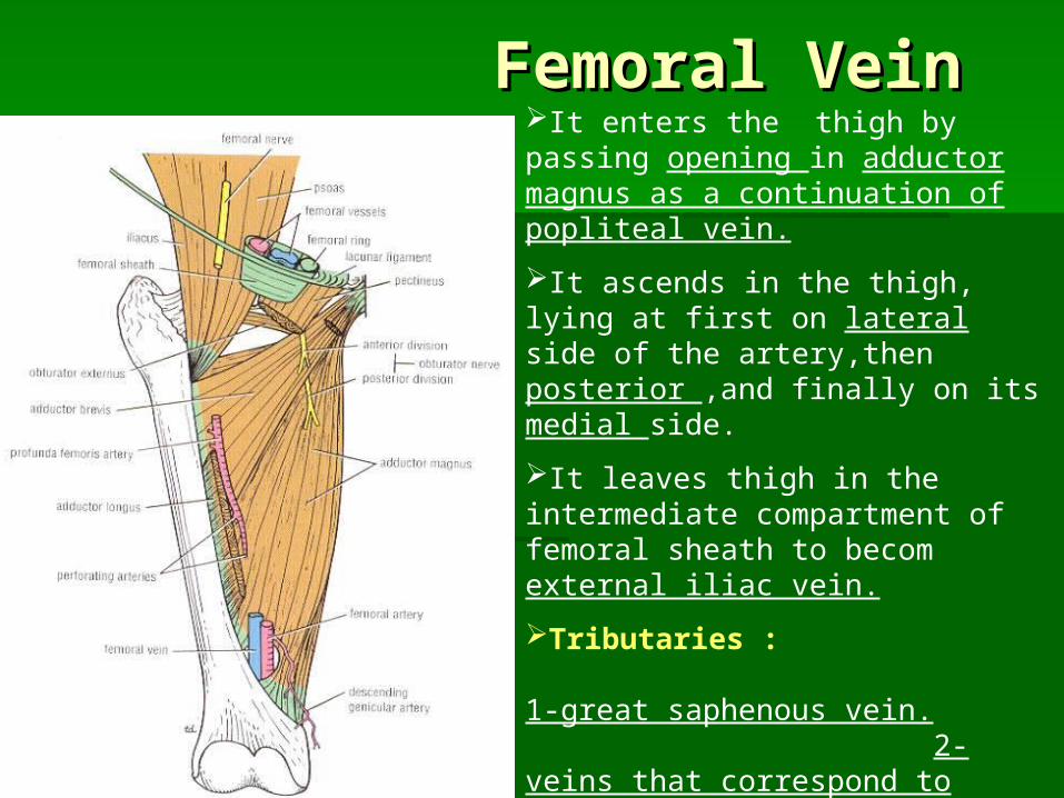
Femoral VeinFemoral VeinIt enters the thigh by passing opening in adductor magnus as a continuation of popliteal vein.
It ascends in the thigh, lying at first on lateral side of the artery,then posterior ,and finally on its medial side.
It leaves thigh in the intermediate compartment of femoral sheath to becom external iliac vein.
Tributaries : 1-great saphenous vein. 2-veins that correspond to branches of artery : -Profunda femoris vein. -Lateral & medial cicumflex femoral veins. -Deep external pudendal vein. -Muscular veins.
Superficial inguinalSuperficial inguinal Lymph Lymph NodesNodes
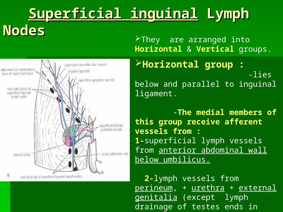
They are arranged into Horizontal & Vertical groups.
Horizontal group : -lies below and parallel to inguinal ligament. -The medial members of this group receive afferent vessels from : 1-superficial lymph vessels from anterior abdominal wall below umbilicus. 2-lymph vessels from perineum, + urethra + external genitalia (except lymph drainage of testes ends in lumbar (para-aortic) L.Ns. at level of L1 vertebra + lower ½ of anal canal. -The lateral members of this group receive afferent superficial lymph vessels from back below level of iliac crest (skin of gluteal region)
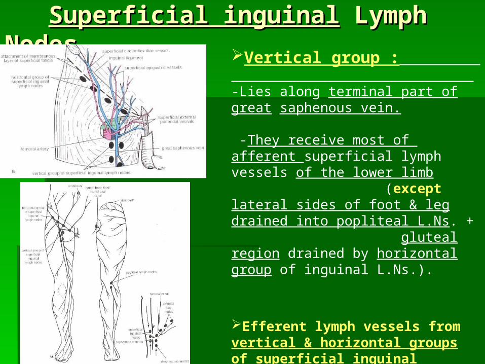
Superficial inguinalSuperficial inguinal Lymph Lymph NodesNodes Vertical group :
-Lies along terminal part of great saphenous vein. -They receive most of afferent superficial lymph vessels of the lower limb (except lateral sides of foot & leg drained into popliteal L.Ns. + gluteal region drained by horizontal group of inguinal L.Ns.).
Efferent lymph vessels from vertical & horizontal groups of superficial inguinal L.Ns. : pass through saphenous opening in the deep fascia to end in deep inguinal L.Ns. (lying along medial side of femoral vein).

PoplitealPopliteal Lymph Nodes : Lymph Nodes :
They are lying in popliteal fossa.
Their afferent lymph vessels from : 1-superficial lymph from skin of lateral side of foot & leg. 2-lymph from knee joint. 3-Deep lymph vessels accompaning anterior & posterior tibial arteries.
Their efferent lymph vessels pass into : deep inguinal lymph nodes (lying along medial side of femoral vein).
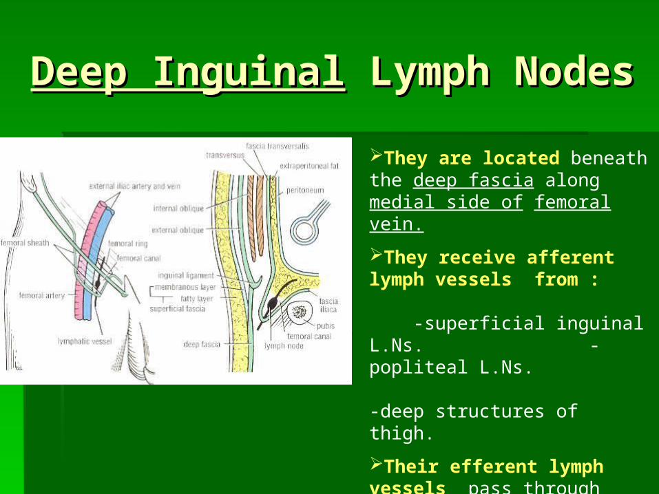
Deep InguinalDeep Inguinal Lymph Lymph NodesNodes
They are located beneath the deep fascia along medial side of femoral vein.
They receive afferent lymph vessels from : -superficial inguinal L.Ns. -popliteal L.Ns. -deep structures of thigh.
Their efferent lymph vessels pass through femoral canal to end into external iliac lymph nodes.


The Propulsive action of The Propulsive action of Foot :Foot : During running :During running :
1-the weight is born 1-the weight is born on the forepart of on the forepart of foot,foot, and the and the heel does not touch the ground.heel does not touch the ground. 2-the forward thrust to the body is 2-the forward thrust to the body is provided by mechanisms described for provided by mechanisms described for walking.walking.
The Propulsive action of The Propulsive action of Foot :Foot :
During Standing immobile :During Standing immobile : the body the body weight is distributed via : weight is distributed via : 1-the heel… 1-the heel… behind.behind. and and 2-the heads of metatarsal 2-the heads of metatarsal bones… bones… in frontin front (including 2 sesamoid (including 2 sesamoid bones under head of 1bones under head of 1stst metatarsal) metatarsal)

The Propulsive action of The Propulsive action of Foot :Foot :
During Walking :During Walking : 1-the body is thrown forward by1-the body is thrown forward by the action of gastrocnemius & soleus (plantarflexion of ankle the action of gastrocnemius & soleus (plantarflexion of ankle joint), so the heel rises from ground, and the joint), so the heel rises from ground, and the body weight is body weight is born onborn on the lateral margin of foot + heads of metatarsal the lateral margin of foot + heads of metatarsal bones. bones. 2-as the heel rises,2-as the heel rises, the the toes are toes are extended at metatarso-phalangeal joints,extended at metatarso-phalangeal joints, and plantar and plantar aponeurosis is streched leading to aponeurosis is streched leading to increasing arches of footincreasing arches of foot. . 3-3-the toes are strongly flexed at inter-phalangeal jointsthe toes are strongly flexed at inter-phalangeal joints by by long & short flexors of foot. long & short flexors of foot. 4-lumbricals & interossei contract4-lumbricals & interossei contract to keep toes to keep toes extended at interphalangeal joints, so that they do not flexing extended at interphalangeal joints, so that they do not flexing during strong action of flexor digitorum longus.during strong action of flexor digitorum longus.

Great Saphenous Vein Cut Down Great Saphenous Vein Cut Down A & B at the ankle. Great saphenous vein is constantly found in front of medial malleolus of tibia.
C & D at the groin. Great saphenous vein drains into femoral vein 2 fingerbreadths below & lateral to pubic tubercle.
Exposure of the vein through a skin incision (a ‘cut down’) is usually performed at ankle, but this site has disadvantages of phlebitis (inflammation of the vein wall) as a complication.
In the groin, phlebitis is rare because the larger diameter of the vein at this site allows the use of large-diameter catheters and rapid infusion of large volumes of fluids.
Great Saphenous Vein in Great Saphenous Vein in Coronary Bypass surgeryCoronary Bypass surgery In occlusive coronary diseaseIn occlusive coronary disease, the diseased , the diseased
arterial segment can be bypassed by inserting a arterial segment can be bypassed by inserting a graft from great saphenous vein.graft from great saphenous vein.
At the donor sit,At the donor sit, the superficial venous bloodthe superficial venous blood ascends the lower limb against gravity by ascends the lower limb against gravity by passing through passing through perforating veinsperforating veins into the into the deep deep veins.veins.
Great saphenous vein Great saphenous vein can also be used tocan also be used to bypass obstructions of brachial or femoralbypass obstructions of brachial or femoral arteries.arteries.
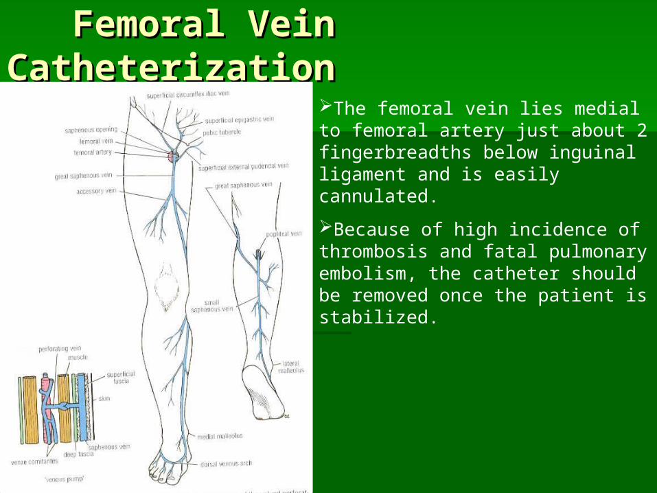
Femoral Vein CatheterizationFemoral Vein Catheterization
The femoral vein lies medial to femoral artery just about 2 fingerbreadths below inguinal ligament and is easily cannulated.
Because of high incidence of thrombosis and fatal pulmonary embolism, the catheter should be removed once the patient is stabilized.
